Ambystoma mexicanum are able to mitigate...
35
Ambystoma mexicanum are able to mitigate teratogenesis in the chondrocranium Chinelo Nnebe
Transcript of Ambystoma mexicanum are able to mitigate...
Ambystoma mexicanum are able to mitigate teratogenesis in the chondrocranium
Chinelo Nnebe
Nnebe
1
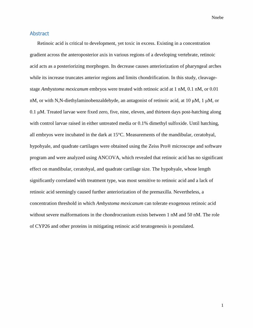
Abstract
Retinoic acid is critical to development, yet toxic in excess. Existing in a concentration
gradient across the anteroposterior axis in various regions of a developing vertebrate, retinoic
acid acts as a posteriorizing morphogen. Its decrease causes anteriorization of pharyngeal arches
while its increase truncates anterior regions and limits chondrification. In this study, cleavage-
stage Ambystoma mexicanum embryos were treated with retinoic acid at 1 nM, 0.1 nM, or 0.01
nM, or with N,N-diethylaminobenzaldehyde, an antagonist of retinoic acid, at 10 μM, 1 μM, or
0.1 μM. Treated larvae were fixed zero, five, nine, eleven, and thirteen days post-hatching along
with control larvae raised in either untreated media or 0.1% dimethyl sulfoxide. Until hatching,
all embryos were incubated in the dark at 15°C. Measurements of the mandibular, ceratohyal,
hypohyale, and quadrate cartilages were obtained using the Zeiss Pro® microscope and software
program and were analyzed using ANCOVA, which revealed that retinoic acid has no significant
effect on mandibular, ceratohyal, and quadrate cartilage size. The hypohyale, whose length
significantly correlated with treatment type, was most sensitive to retinoic acid and a lack of
retinoic acid seemingly caused further anteriorization of the premaxilla. Nevertheless, a
concentration threshold in which Ambystoma mexicanum can tolerate exogenous retinoic acid
without severe malformations in the chondrocranium exists between 1 nM and 50 nM. The role
of CYP26 and other proteins in mitigating retinoic acid teratogenesis is postulated.

Nnebe
2
Introduction
Retinoic acid, more specifically all-trans-retinoic acid, is the most biologically active
derivative of retinol, or vitamin A. Quite remarkably, the body uses this relatively simple, fat-
soluble compound to achieve an overwhelming number of tasks1,2,3,4. It is a key regulator during
chondrocyte differentiation and acts as a morphogen during chordate development5,6,7. However,
exogenous retinoic acid in embryonic compartments requiring low concentrations causes
teratogenesis and mortality8. Disease states such as cleft palate, macrocephaly, and a shortening
of the mandible and maxilla9 encourage an understanding of cellular response to retinoic acid
and its relationship to chondrocranium development. The following details the general pathway
of retinoic acid, its role in chondrification and teratogenesis, and its ability to posteriorize
different regions during development.
General Pathway of Retinoic Acid
Retinoic acid is primarily obtained through ingestion and metabolism of retinol10,11,12, which
can be acquired directly from the diet or synthesized from β-carotene by its oxygenase13,14.
Inside the intestinal lumen, retinol is absorbed and converted into retinyl ester, which acts as a
storage molecule. The ester is then packaged and sent through lymphatic and circulatory vessels
to target cells, especially those of the liver12,15. Inside the liver, enzymes reconvert retinyl ester
into retinol. Through a series of highly regulated pathways, retinol is bound to its plasmatic
carrier protein, RBP, and ejected into the bloodstream16,17.
In embryos, this method of acquisition is different. Viviparous organisms cannot synthesize
retinoids de novo and must obtain retinoic acid and its precursors through the placenta. Two
common modes of delivery are simple diffusion of circulating retinol from the mother’s
bloodstream to the embryonic compartment and transfer of retinol from maternal binding
Nnebe
3
proteins to those of the embryo18,19. In oviparous organisms, retinoic acid is derived from the
metabolism of carotenoids in vegetal cells20.
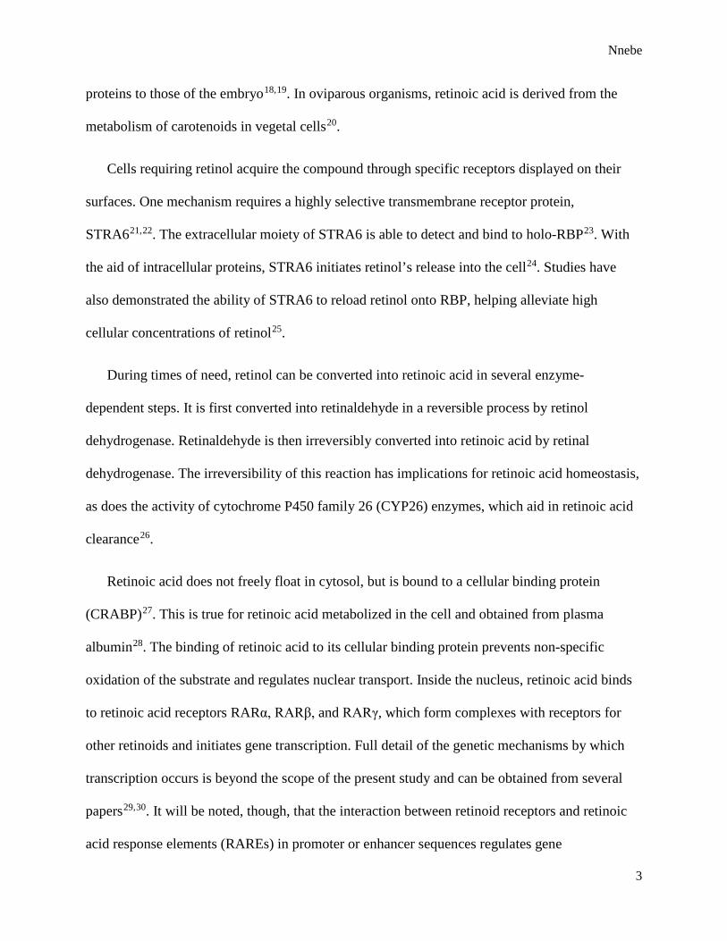
Cells requiring retinol acquire the compound through specific receptors displayed on their
surfaces. One mechanism requires a highly selective transmembrane receptor protein,
STRA621,22. The extracellular moiety of STRA6 is able to detect and bind to holo-RBP23. With
the aid of intracellular proteins, STRA6 initiates retinol’s release into the cell24. Studies have
also demonstrated the ability of STRA6 to reload retinol onto RBP, helping alleviate high
cellular concentrations of retinol25.
During times of need, retinol can be converted into retinoic acid in several enzyme-
dependent steps. It is first converted into retinaldehyde in a reversible process by retinol
dehydrogenase. Retinaldehyde is then irreversibly converted into retinoic acid by retinal
dehydrogenase. The irreversibility of this reaction has implications for retinoic acid homeostasis,
as does the activity of cytochrome P450 family 26 (CYP26) enzymes, which aid in retinoic acid
clearance26.
Retinoic acid does not freely float in cytosol, but is bound to a cellular binding protein
(CRABP)27. This is true for retinoic acid metabolized in the cell and obtained from plasma
albumin28. The binding of retinoic acid to its cellular binding protein prevents non-specific
oxidation of the substrate and regulates nuclear transport. Inside the nucleus, retinoic acid binds
to retinoic acid receptors RARα, RARβ, and RARγ, which form complexes with receptors for
other retinoids and initiates gene transcription. Full detail of the genetic mechanisms by which
transcription occurs is beyond the scope of the present study and can be obtained from several
papers29,30. It will be noted, though, that the interaction between retinoid receptors and retinoic
acid response elements (RAREs) in promoter or enhancer sequences regulates gene
Nnebe
4
expression31,32. A number of genes activated by this general pathway have been documented32.
This study focuses on the relatively few known to have a direct impact on cartilage development.
Retinoic Acid in Chondrification and Teratogenesis
The fact that retinoic acid is involved in the development and modeling of cartilage tissue is
indicated by its ability to modify such cells. Addition of 1 nM or 10 nM retinoic acid to avian
chondrocytes alters their shape, causes the cells to become fibroblastic, drastically reduces their
ability to synthesize proteoglycans, and inhibits the initiation of type X collagen gene
expression33. A decrease in translation and concentration of aggrecan, a protein crucial for proper
functioning of articular cartilage34, is linked to the inhibition of nuclear response to retinoic
acid35. At 50 nM, formation of the second basibranchial cartilage in the Ambystoma mexicanum
chondrocranium does not occur36 and in cranial regions of frog embryos, retinoic acid causes
anterior truncation, posterior or ventral character of mesoderm, and even acephaly37.
Further evidence that suggests retinoic acid is involved in chondrification is the discovery of
RAREs in genes responsible for chondrification, such as the Col11a2 gene, which encodes for a
collagen subunit critical to the extracellular matrix of chondral tissues38. All three retinoic acid
receptors are involved in general cell growth and differentiation41 and RARγ specifically aids in
the formation of healthy cartilage35. What is more, retinoic acid induces the production of
metalloproteinases that are involved in cartilage degradation39,40.
Retinoic Acid as a Posteriorizing Agent
Retinoic acid acts as a posteriorizing agent in various regions of developing vertebrates,
including the pharyngeal arches41. Compared to the anterior mandibular arch, branchial arches
oriented more posteriorly have higher sensitivity to retinoic acid42,43. In frogs, exogenous

Nnebe
5
retinoic acid posteriorizes the neural plate. This causes the anterior neural plate border to
transform into neural crest cells44, which give rise to various cell types, including chondrocytes
and chondroblasts45. In other regions, retinoic acid has been shown to form a concentration
gradient culminating in the posterior domain46; however, it is currently unknown if retinoic acid
forms a similar gradient across the anteroposterior axis of the skull.
Experimental Design and Focus
In light of retinoic acid’s clear effect on chondrification, the present study aimed to document
any cranial teratogenesis observed from exogenous retinoic acid at relatively low concentrations.
Another aim was to determine a relative threshold concentration that cranial chondrocytes of
Ambystoma mexicanum can tolerate before abnormalities are observed.
Because of the lower concentrations of retinoic acid utilized in this study relative to those
previously mentioned43, observation of such dramatic abnormalities such as missing cranial
cartilages and anterior truncation was not expected. Because retinoic acid is crucial to
development, it was expected that embryos treated with higher concentrations of its inhibitor,
N,N-diethylaminobenzaldehyde (DEAB), would have cartilages positioned more anteriorly.

Nnebe
6
Methods
Cleavage-stage axolotl embryos were obtained from the Ambystoma Genetic Stock Center47.
These were raised in 200 mL of either: 1 nM, 0.1 nM, or 0.01 nM retinoic acid; 10 μM, 1 μM, or
0.1 μM DEAB; untreated E3 media48; or 0.1% dimethyl sulfoxide (DMSO). Concentrations of
retinoic acid and DEAB were attained using DMSO. Until hatching, both treated and control
embryos were incubated in the dark at 15°C to prevent light-induced degradation of retinoic
acid49.
Upon their hatching, several treated and control larval axolotls were fixed in 10% formalin.
Clearing was performed with 0.025% potassium hydroxide dissolved in glycerol and water, and
staining with alcian blue (cartilage) and alizarin red (bone) dissolved in the same sort of solution.
Cleared and stained axolotls were stored in 100% glycerol, which was also used for whole-
mount imaging. Live brine shrimp were fed to remaining larvae each day until fixed. These were
fixed five, nine, eleven, and thirteen days after hatching and prepared with the same protocol as
were those fixed upon hatching.
Images of the skull and entire specimen were obtained at 5X magnification using the Zeiss
Pro® microscope and software program, which facilitates the measuring of specimens by using a
tracing tool that computes the total distance of several lines drawn on the captured image. The
total body length of each axolotl was obtained by tracing the notochord from tail to tip, then from
tip to the intermandibular ligament. Cartilages of the chondrocranium were measured using the
procedure outlined below.
Mandibular (Meckel’s) Cartilage
Nnebe
7
The summed length of the two mandibular cartilages was measured using the Zeiss Pro®
software. Each recorded length of the mandible was the length of a single multiple-angled line
drawn through the center of both cartilages from the beginning of the left cartilage to the end of
the right, including the small ligament in between (Figure 1). For mandibles exhibiting
cartilaginous ossification, only measurements of the cartilaginous tissues were taken.
Ceratohyal
Each of the two ceratohyal cartilages was measured and recorded separately. As was the case
for the mandibular cartilage, each ceratohyal was measured from beginning to end, through its
center (Figure 1). The ligament between the ceratohyal and hypohyale was not considered and
was often indicated by a cessation of alcian blue staining or a pinching of the cartilage (Figure
2).
Hypohyale
Each of the two hypohyale cartilages was measured and recorded separately in the same
manner as were the mandibular and ceratohyal cartilages (Figure 1). The ligament between the
two hypohyale cartilages was not considered and was often indicated by the cessation of alcian
blue staining or a thinning of the region (Figure 2).
Quadrate
Both length and width of each of the two quadrate cartilages were recorded. For the length, a
straight line was drawn from the region in which the quadrate contacted the mandible to the
flatter, longer portion of the quadrate (Figure 1). For older aged larvae, the quadrate often
overlapped the mandible. Because focus constraints on the microscope prevented discernment of
the quadrate’s true end, the quadrate in these individuals was measured lengthwise from the
Nnebe
8
region of overlap to the flatter side (Figure 2). The width of the quadrate was measured from the
upper edge of its stouter portion to the point at which its thinner arm overlapped the ethmoid
cartilage (Figures 1 and 2). As an aside, the quadrate likely contacts the more ventrally
positioned trabecular cartilage. However, because this contact was not always observed under the
microscope, the quadrate was measured relative to its proximity to the ethmoid.
These measurements were analyzed using ANCOVA, in which the covariant was body
length and individual ages were disregarded. A simple anatomical diagram of the axolotl
chondrocranium and pharyngeal skeleton was created using literature43,50 and stained individuals
from the present study fixed five days after hatching and showing consensus in the layout and
relative lengths of their cranial cartilages (Figure 2). Axolotls whose skulls considerably
deviated from this diagram were selected for further analysis. Before an individual was classified
as malformed, its stage in development as well as its angle and position on the wet-mount slide
were taken into account, as explained below.
Seemingly deformed axolotls were remounted on glass slides and reimaged. These new
images were compared to those of same-age control axolotls. If the apparent malformation was
observed again in the recaptured image, the embryo was considered malformed and a note of the
malformation was recorded. Posterior or anterior displacement of tissues, misshapen or missing
cartilages, and oddly positioned structures were especially noted, if present.
Nnebe
9
Results
Sample Size
Ninety-one individuals were measured in this study. Of these, twelve were treated with 1 nM
retinoic acid, thirteen with 0.1 nM retinoic acid, eighteen with 0.01 nM retinoic acid, seven with
10 µM DEAB, three with 1 µM DEAB, ten with 0.1 µM DEAB, fourteen with 0.1% DMSO, and
fourteen were not treated at all (Figure 3).
Of the individuals treated with 1 nM retinoic acid, one was fixed upon hatching, two were
fixed five days after hatching, three nine days after hatching, three eleven days after hatching,
and three thirteen days after hatching. Of those treated with 0.1 nM retinoic acid, one was fixed
upon hatching, two five days after hatching, three nine days after hatching, four eleven days after
hatching, and three thirteen days after hatching. Of those treated with 0.01 nM retinoic acid, one
was fixed upon hatching, two five days after hatching, eight nine days after hatching, four eleven
days after hatching, and three thirteen days after hatching. Of those treated with 10 µM DEAB,
two were fixed five days after hatching and five nine days after hatching. The three treated with 1
µM DEAB were all fixed nine days after hatching. Of those treated with 0.1 µM DEAB, two
were fixed five days after hatching and eight nine days after hatching. Of those raised in 0.1%
DMSO, one was fixed upon hatching, two five days after hatching, seven nine days after
hatching, two eleven days after hatching, and two thirteen days after hatching. Of those
untreated, two were fixed upon hatching, two five days after hatching, four nine days after
hatching, three eleven days after hatching, and three thirteen days after hatching (Figure 3).
Malformations Not Observed
Nnebe
10
To summarize, malformed individuals were those who, after reimaging and comparison to
same-age control axolotls, noticeably disaccorded with the skull diagram in shape, position, and
spacing of cartilages (Figure 2). In this study, only individuals whose premaxillae were
positioned anteriorly were considered malformed, as explained below. In all other instances
where cartilage structure and arrangement appeared to vary between treatments, visual
comparisons between individuals of the same age revealed that such variations were due to
cranial growth typical during chondrification. For example, quadrate structure and position
varied with age. In older individuals the quadrate extended from under the mandible to the
trabecular cartilage located just ventral to the ethmoid. On the other hand, in younger individuals
the quadrate was most commonly not overlapped by the mandible (Figure 4). Cranial spacing
was also observed to vary with age. In younger axolotls, cartilages were more closely situated,
giving the skull a more compact shape. In older axolotls, the skull appeared longer (Figure 5).
Premaxillae Potentially Anteriorized in the Absence of Retinoic Acid
The anteroposterior positioning of the premaxilla appears to vary across several individuals
in a treatment-dependent manner. Premaxillae that did not fully cross the mandibular cartilage
were considered anteriorized. Of the ten individuals with a noticeably anterior premaxilla, eight
were treated with DEAB (four at 10 μM, three at 1 μM, and one at 0.1 μM DEAB), one was
treated with 1 nM retinoic acid, and one was raised in 0.1% DMSO (Figure 6). In this group, the
axolotl treated with 1 nM retinoic acid was fixed eleven days after hatching while the other nine
were fixed nine days after hatching.
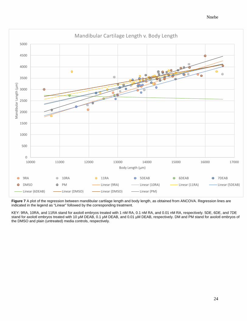
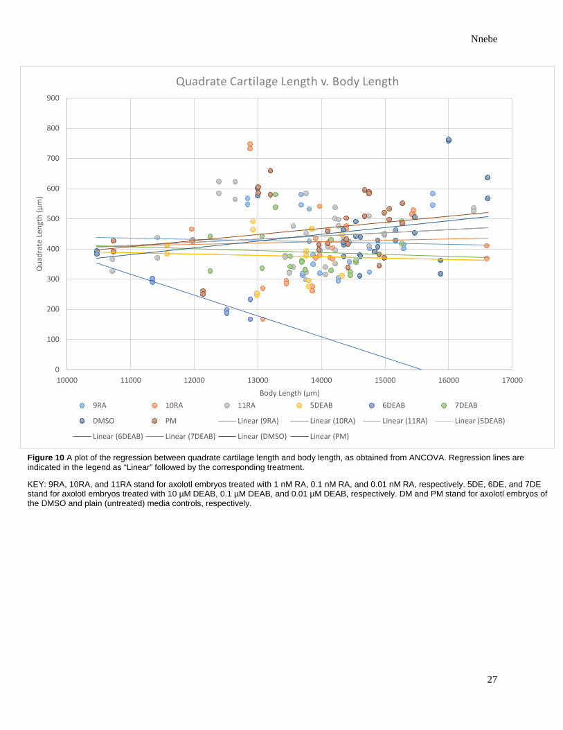
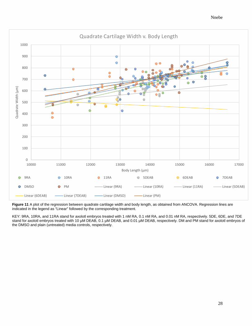
Apart from the Hypohyale, Treatment Has No Significant Effect on Cartilage Size
Nnebe
11
The ANCOVA using body length as a covariant revealed no significant difference between
the type of treatment to which the axolotls were exposed and the measurements of their
mandibular, ceratohyal, or quadrate cartilages (Table 1). With the exclusion of quadrate length,
cartilage size correlated more significantly with body length than it did treatment type (Table 1).
The length of the hypohyale was significantly affected by treatment [p = 0.0007] (Table 1).
Regression lines obtained from ANCOVA demonstrate that the hypohyale was longest in those
raised in 0.1% DMSO and shortest in those treated with 1 µM DEAB or 0.1 µM DEAB (Figures
7 – 11).

Nnebe
12
Discussion
Retinoic Acid Induces Teratogenesis in the Chondrocranium and Modification of the Hypohyale
Previous studies have demonstrated that exogenous retinoic acid causes abnormalities in
cartilage tissues33,34,35. Molar concentrations comparable to those used in this study decrease
cartilage production in vitro33 and at slightly higher concentrations, retinoic acid induces anterior
truncation and acephaly in frog embryos36,37. Studies using Ambystoma mexicanum show that
retinoic acid removes only the second basibranchial cartilage at 50 nM, an ability attributed to
dual embryonic origin and patterning of the pharyngeal skeleton43. Such findings reveal that
cartilages of the same region have varying sensitivities to retinoic acid. This would explain why
the mandibular but not the pharyngeal arches appeared unaffected by retinoic acid in the cited
study and why only the hypohyale was significantly affected by retinoic acid in this study (Table
1). Nevertheless, considerably high concentrations of retinoic acid cause teratogenesis in all
cartilages of the chondrocranium. If axolotls of the present study also were to have been treated
with 100 nM retinoic acid, distortion and compaction of the chondrocranium43 most probably
would have been observed.
Retinoic Acid Potentially Posteriorizes the Axolotl Premaxilla
Studies have highlighted the role of retinoic acid as a posteriorizing agent in various regions
of developing vertebrates41,46. Of specific interest in this study is its ability to posteriorize
pharyngeal arches and the anterior neural plate border. Posteriorization of pharyngeal arches is
suggested by knockout studies of RARα and RARβ in mice41. In frogs, retinoic acid posteriorizes
the neural plate and leads to the production of neural crest cells, which are cartilage tissue cell
lines. Inversely, quail embryos deficient in retinol experience neural crest cell apoptosis42.
Nnebe
13
Although the limited parameters and methods of this study prevent analysis of
posteriorization in areas known to be affected by retinoic acid such as those mentioned above, it
may be conjectured that retinoic acid is able to posteriorize the premaxilla in Ambystoma
mexicanum skulls. Of the relatively few individuals that exhibited anterior positioning of this
structure, the majority were treated with DEAB (Figure 6). Is it possible, then, that inhibition of
retinoic acid was responsible for the apparent anteriorization? To begin answering that question,
it would be useful to know just how much inhibition of retinoic acid actually occurred. This can
be achieved by monitoring cellular concentrations of retinoic acid and retinal dehydrogenase
activity. Does the fact that the majority of individuals with anterior positioning of this structure
were of the same age imply complications in the methods of treatment? Answers to these
questions as well as replication of the study may elucidate underlying mechanisms of retinoic
acid posteriorization.
Teratogenesis is Mitigated in the Ambystoma mexicanum Chondrocranium
Despite the apparent sensitivity of the hypohyale to retinoic acid, Ambystoma mexicanum
overall appear quite effective at mitigating teratogenesis at 1 nM, 0.1 nM, and 0.01 nM retinoic
acid. The present study reports no missing or visibly deformed cartilages, contrary to other
papers43 that linked exogenous retinoic acid to the absence of basibranchial 2 as well as to severe
deformations in the chondrocranium and pharyngeal skeleton. This finding is not startling as the
concentrations used here are orders of magnitude lower than those used in such studies.
Moreover, the amount of retinoic acid actually taken up by CRABP is uncertain and can be at
least partially responsible for the absence of malformations.
Threshold Concentration of Retinoic Acid
Nnebe
14
As gathered from this and the aforementioned study43, it seems that the increase of retinoic
acid from 1 nM to 50 nM is enough to induce observable teratogenesis in Ambystoma mexicanum.
It is not yet clear how specific concentrations within this range affect development in the
chondrocranium. Nevertheless, it can be said that relative to reported malformations caused by 50
nM and 100 nM, abnormalities caused by 0.01 nM to 1 nM retinoic acid are minor if not
nonexistent and the overall structure of the chondrocranium and its tissues appear unaffected.
Therefore, a retinoic acid threshold concentration may exist between 1 nM and 50 nM. Below this
range, Ambystoma mexicanum are able to tolerate exogenous retinoic acid without experiencing
dramatic teratogenesis in their chondrocranium, whereas above this range they cannot.
Retinoic Acid Clearance is Likely Involved in Mitigation of Teratogenesis
The fact that cells of the chondrocranium are able to tolerate certain concentrations of
retinoic acid calls for analysis of retinoic acid cellular clearance. Although retinoic acid
regulation at the cellular level is not fully understood, at least two proteins, CYP26 and CRABP,
have been suggested to be direct influencers. Expression of CRABP is upregulated in regions of
high retinoic acid concentrations and causes subsequent removal of the ligand as well as limited
access to is nuclear receptors51,52,53. However, because CRABP has not yet been found in
cartilage cells54, the role of CYP26 in retinoic acid metabolism is more relevant to this study.
It is generally agreed that the binding of retinoic acid to its nuclear receptor prompts the
production and activity of CYP26 enzymes. One study compared teratogenesis in CYP26-/-
mutant embryos to that associated with excess retinoic acid55, leading to the belief that retinoic
acid is primarily cleared by CYP26. Thus, through activation of CYP26, retinoic acid induces its
own metabolism. Nevertheless, not all cell types respond to retinoic acid by regulation of
CYP26, and the presence and expression of CYP26 in cells of the axolotl chondrocranium have

Nnebe
15
not yet been confirmed. Could its activity, though, be connected to retinoic acid tolerance as
observed in this study? If so, future projects, as later elaborated, may be useful in understanding
the correlation between retinoic acid clearance and teratogenesis.
Of notable mention are two minor contributions to retinoic acid homeostasis— minor
primarily due to limited research in these areas. STRA6 is capable of reloading retinol onto RBP
and is also able to bind to retinoic acid56. Reduced cellular concentrations of retinol leave less of
the substrate available for oxidation to retinoic acid. Perhaps a similar mechanism exists in
which retinoic acid is exported from cells of oviparous embryos? Though this scheme is more
imaginative than evidential, it is probable that cellular retinoic acid concentrations are regulated
by cellular transport due to involvement of STRA6 in regulation. The irreversible conversion of
retinaldehyde to retinoic acid hints at the concentration-dependent activity of retinal
dehydrogenase. Indeed, a feedback loop involving STRA6 and retinal dehydrogenase helps
maintains cellular retinoic acid homeostasis57.
Future Studies
The limited parameters of this study encourage supplementary projects analyzing other
aspects of retinoic acid control. For example, addition of retinoic acid to chondrocytes is known
to cause structural and functional alteration.33 Experiments measuring chondral tissue density,
chondrocyte arrangement and configuration, and alkaline phosphatase activity as well as those
monitoring collagen production after exposure to retinoic acid are thereby useful. Gene
expression profiling may further illustrate cellular sensitivity to retinoic acid and perhaps reveal
other genes important in chondrification. Returning to the role of CYP26 in retinoic acid
clearance, the treatment of CYP26-/- mutants with the same concentrations of retinoic acid and its
inhibitor used here would be an interesting follow-up study.
Nnebe
16
Final Thoughts
In conclusion, noticeable cranial teratogenesis was not produced in Ambystoma mexicanum
by exogenous retinoic acid at concentrations of 1 nM, 0.1 nM, or 0.01 nM in this study. It seems
that a retinoic acid concentration threshold, in which Ambystoma mexicanum can tolerate
exogenous retinoic acid in the chondrocranium, exists between 1 nM and 50 nM.

Nnebe
17
Tables
Cartilage Length Cartilage Width Mandibular Ceratohyal Hypohyale Quadrate Quadrate
Whole Body Length <0.0001 <0.0001 <0.0001 0.7622 <0.0001 Treatment 0.0134 0.0005 0.0235 0.0162 <0.0001 Whole Body Length*Treatment 0.1700 0.3629 0.0007 0.8023 0.1333
Table 1 P-values from ANCOVA highlighting the effect, if any, that treatment type has on cartilage size in axolotl (Ambystoma mexicanum) chondrocrania. The first row gives p-values for the correlation between whole body length and cartilage size. The second gives p-values for the correlation between treatment and cartilage size. The third gives p-values for the correlation between cartilage size and treatment using body length as a covariant. Bolded values are statistically significant (α = 0.05). In-text ANCOVA results are also provided.

Nnebe
18
Figures
i. ii.
iii. iv.
Figure 1 A schematic diagram illustrating the method used to measure cartilages in axolotls (Ambystoma mexicanum). Green and red lines mimic those of the tracing tool used in the Zeiss® microscope and software program, as explained in text. Compare to Figure 2 for relative positions of each cartilage in the chondrocranium.
KEY: i. mandibular cartilage (a. in Figure 2); ii. quadrate cartilage (c. in Figure 2); iii. ceratohyal cartilage (d. in Figure 2); iv. hypohyale cartilage (e. in Figure 2).
Nnebe
19
Figure 2 A simple anatomical diagram labeling known cartilages of the axolotl (Ambystoma mexicanum) chondrocranium and pharyngeal cartilages. Transparency indicates dorsal positioning. The relative positions of these cartilages are based on axolotl embryos fixed five days after hatching and literature, as explained in text. Unknown structures receive a red question mark after their name in the following key.
KEY: a. mandibular/Meckel’s cartilage b. ethmoid cartilage? c. quadrate cartilages d. ceratohyal cartilages e. hypohyale cartilages f. trabecular cartilages? g. basibranchial 1 h. basibranchial 2 i. hypobranchial 1 j. hypobranchial 2 k. ceratobranchial 1 l. ceratobranchial 2 m. ceratobranchial 3 n. ceratobranchial 4 o. dorsal groove p. premaxilla
Nnebe
20
0d
5d
9d
11d 13d 0d 5d 9d
11d
13d
0d 5d
9d
11d
13d
0d
5d
9d
11d 13d 5d 9d
9d
5d
9d
0d
5d
9d
11d
13d
Figure 3 Sample sizes organized by treatment and labeled with age. Each color in the pie chart represents a different treatment, as labeled below each section. Numbers preceded by the letter “d,” as in 0d, 5d, etc. refer to the number of days after hatching the individuals were fixed in each treatment. A total of 91 axolotls were measured.
Nnebe
21
Figure 4 Images of two Ambystoma mexicanum chondrocrania, both treated with 1 nM retinoic acid, as displayed on the images. WB indicates the whole body length of the individual. Left Axolotl was fixed five days after hatching. Right Axolotl was fixed nine days after hatching. Red arrows note the different contact points of the quadrate cartilages.
1 nM RA WB: 13682.55 µm
1 nM RA WB: 3238.026 µm
Fixed five days after hatching. Fixed nine days after hatching.

Nnebe
22
Figure 5 Images of two Ambystoma mexicanum chondrocrania, both untreated as is displayed on images. Left Axolotl was fixed upon hatching. Right Axolotl was fixed nine days after hatching. WB indicates the whole body length of the individual. Note the difference in skull spatial arrangement between the two individuals, as explained in text.
Untreated WB: 14392.931 µm
Untreated WB: 10730.136 µm
Fixed upon hatching Fixed nine days after hatching

Nnebe
23
Figure 6 Images of axolotls (Ambystoma mexicanum) fixed nine days after hatching. Treatments and body lengths are indicated on the bottom left of each individual. Arrowheads indicate the premaxilla. As discussed in text, premaxillae in the untreated axolotl and in the axolotl treated with 0.01 nM retinoic acid are properly positioned in the chondrocranium. However, those raised in 0.1% DMSO, 0.1 µM DEAB, and 10 µM DEAB are positioned more anteriorly. KEY: RA stands for retinoic acid, WB stands for whole body length.
0.1% DMSO WB: 14884.458 µm 10μM DEAB
WB: 14322.609 µm
0.1 μM DEAB WB: 14151.348 µm
Untreated WB: 14417.688 µm
0.01 nM RA WB: 11420.557 µm
Nnebe
24
0
500
1000
1500
2000
2500
3000
3500
4000
4500
5000
10000 11000 12000 13000 14000 15000 16000 17000
Man
dibu
lar L
engt
h (μ
m)
Body Length (μm)
Mandibular Cartilage Length v. Body Length
9RA 10RA 11RA 5DEAB 6DEAB 7DEAB
DMSO PM Linear (9RA) Linear (10RA) Linear (11RA) Linear (5DEAB)
Linear (6DEAB) Linear (DMSO) Linear (DMSO) Linear (PM)
Figure 7 A plot of the regression between mandibular cartilage length and body length, as obtained from ANCOVA. Regression lines are indicated in the legend as “Linear” followed by the corresponding treatment.
KEY: 9RA, 10RA, and 11RA stand for axolotl embryos treated with 1 nM RA, 0.1 nM RA, and 0.01 nM RA, respectively. 5DE, 6DE, and 7DE stand for axolotl embryos treated with 10 µM DEAB, 0.1 µM DEAB, and 0.01 µM DEAB, respectively. DM and PM stand for axolotl embryos of the DMSO and plain (untreated) media controls, respectively.
Nnebe
25
600
800
1000
1200
1400
1600
1800
10000 11000 12000 13000 14000 15000 16000 17000
Cera
tohy
al L
engt
h (μ
m)
Body Length (μm)
Ceratohyal Cartilage Length v. Body Length
9RA 10RA 11RA 5DEAB 6DEAB 7DEAB
DMSO PM Linear (9RA) Linear (10RA) Linear (11RA) Linear (5DEAB)
Linear (6DEAB) Linear (7DEAB) Linear (DMSO) Linear (PM)
Figure 8 A plot of the regression between ceratohyal cartilage length and body length, as obtained from ANCOVA. Regression lines are indicated in the legend as “Linear” followed by the corresponding treatment.
KEY: 9RA, 10RA, and 11RA stand for axolotl embryos treated with 1 nM RA, 0.1 nM RA, and 0.01 nM RA, respectively. 5DE, 6DE, and 7DE stand for axolotl embryos treated with 10 µM DEAB, 0.1 µM DEAB, and 0.01 µM DEAB, respectively. DM and PM stand for axolotl embryos of the DMSO and plain (untreated) media controls, respectively.

Nnebe
26
0
50
100
150
200
250
300
350
400
450
500
10000 11000 12000 13000 14000 15000 16000 17000
Hypo
hyal
e Le
ngth
(μm
)
Body Length (μm)
Hypohyale Cartilage Length v. Body Length
9RA 10RA 11RA 5DEAB 6DEAB 7DEAB
DMSO PM Linear (9RA) Linear (10RA) Linear (11RA) Linear (5DEAB)
Linear (6DEAB) Linear (7DEAB) Linear (DMSO) Linear (PM)
Figure 9 A plot of the regression between hypohyale cartilage length and body length, as obtained from ANCOVA. Regression lines are indicated in the legend as “Linear” followed by the corresponding treatment.
KEY: 9RA, 10RA, and 11RA stand for axolotl embryos treated with 1 nM RA, 0.1 nM RA, and 0.01 nM RA, respectively. 5DE, 6DE, and 7DE stand for axolotl embryos treated with 10 µM DEAB, 0.1 µM DEAB, and 0.01 µM DEAB, respectively. DM and PM stand for axolotl embryos of the DMSO and plain (untreated) media controls, respectively.
Nnebe
27
0
100
200
300
400
500
600
700
800
900
10000 11000 12000 13000 14000 15000 16000 17000
Qua
drat
e Le
ngth
(µm
)
Body Length (µm)
Quadrate Cartilage Length v. Body Length
9RA 10RA 11RA 5DEAB 6DEAB 7DEAB
DMSO PM Linear (9RA) Linear (10RA) Linear (11RA) Linear (5DEAB)
Linear (6DEAB) Linear (7DEAB) Linear (DMSO) Linear (PM)
Figure 10 A plot of the regression between quadrate cartilage length and body length, as obtained from ANCOVA. Regression lines are indicated in the legend as “Linear” followed by the corresponding treatment.
KEY: 9RA, 10RA, and 11RA stand for axolotl embryos treated with 1 nM RA, 0.1 nM RA, and 0.01 nM RA, respectively. 5DE, 6DE, and 7DE stand for axolotl embryos treated with 10 µM DEAB, 0.1 µM DEAB, and 0.01 µM DEAB, respectively. DM and PM stand for axolotl embryos of the DMSO and plain (untreated) media controls, respectively.
Nnebe
28
0
100
200
300
400
500
600
700
800
900
1000
10000 11000 12000 13000 14000 15000 16000 17000
Qua
drat
e W
idth
(µm
)
Body Length (µm)
Quadrate Cartilage Width v. Body Length
9RA 10RA 11RA 5DEAB 6DEAB 7DEAB
DMSO PM Linear (9RA) Linear (10RA) Linear (11RA) Linear (5DEAB)
Linear (6DEAB) Linear (7DEAB) Linear (DMSO) Linear (PM)
Figure 11 A plot of the regression between quadrate cartilage width and body length, as obtained from ANCOVA. Regression lines are indicated in the legend as “Linear” followed by the corresponding treatment.
KEY: 9RA, 10RA, and 11RA stand for axolotl embryos treated with 1 nM RA, 0.1 nM RA, and 0.01 nM RA, respectively. 5DE, 6DE, and 7DE stand for axolotl embryos treated with 10 µM DEAB, 0.1 µM DEAB, and 0.01 µM DEAB, respectively. DM and PM stand for axolotl embryos of the DMSO and plain (untreated) media controls, respectively.

Nnebe
29
References
1 Togari, A., Kondo, M., Arai, M., & Matsumoto, S. (1991). Effects of retinoic acid on bone
formation and resorption in cultured mouse calvaria. General Pharmacology: The Vascular System 22(2),
287-292.
2 Vernet, N., Dennefeld, C., Rochette-Egly, C., Oulad-Abdelghani, M., Chambon, P., Ghyselinck, N.
B., & Mark, M. (2006). Retinoic acid metabolism and signaling pathways in the adult and developing
mouse testis. Endocrinology 147(1), 96-110.
3 Maden, M. (2007). Retinoic acid in the development, regeneration and maintenance of the nervous
system. Nature Reviews Neuroscience 8(10), 755-765.
4 Tang, X. H., & Gudas, L. J. (2011). Retinoids, retinoic acid receptors, and cancer. Annual Review of
Pathology: Mechanisms of Disease 6, 345-364.
5 Vergara, M. N., Arsenijevic, Y., & Rio‐Tsonis, D. (2005). CNS regeneration: a morphogen's
tale. Journal of Neurobiology 64(4), 491-507.
6 Li, X., Schwarz, E. M., Zuscik, M. J., Rosier, R. N., Ionescu, A. M., Puzas, J. E., ... & O’Keefe, R.
J. (2003). Retinoic acid stimulates chondrocyte differentiation and enhances bone morphogenetic protein
effects through induction of Smad1 and Smad5. Endocrinology 144(6), 2514-2523.
7 De Luca, F., Uyeda, J. A., Mericq, V., Mancilla, E. E., Yanovski, J. A., Barnes, K. M., ... & Baron,
J. (2000). Retinoic acid is a potent regulator of growth plate chondrogenesis. Endocrinology 141(1), 346-
353.
8 Napoli, J. L. (1996). Retinoic acid biosynthesis and metabolism. The FASEB Journal 10(9), 993-
1001.
9 Cohlan, S. Q. (1954). Congenital anomalies in the rat produced by excessive intake of vitamin A
during pregnancy. Pediatrics 13(6), 556-567.

Nnebe
30
10 Seegmiller, R. E., Ford, W. H., Carter, M. W., Mitala, J. J., & Powers, W. J. (1997). A
developmental toxicity study of tretinoin administered topically and orally to pregnant Wistar rats.
Journal of the American Academy of Dermatology 36(3), S60-S66.
11 Willhite, C. C., Sharma, R. P., Allen, P. V., & Berry, D. L. (1990). Percutaneous retinoid
absorption and embryotoxicity. Journal of Investigative Dermatology 95(5), 523-529.
12 D’Ambrosio, D. N., Clugston, R. D., & Blaner, W. S. (2011). Vitamin A metabolism: an update.
Nutrients 3(1), 63-103.
13 Olson, J. A., & Hayaishi, O. (1965). The enzymatic cleavage of beta-carotene into vitamin A by
soluble enzymes of rat liver and intestine. Proceedings of the National Academy of Sciences 54(5), 1364-
1370.
14 During, A., Smith, M. K., Piper, J. B., & Smith, J. C. (2001). β-Carotene 15, 15′-Dioxygenase
activity in human tissues and cells: evidence of an iron dependency. The Journal of Nutritional
Biochemistry 12(11), 640-647.
15 Kam, R. K. T., Deng, Y., Chen, Y., & Zhao, H. (2012). Retinoic acid synthesis and functions in
early embryonic development. Cell & Bioscience 2, 11.
16 Smith, J.E., Deen, D.D., Sklan, D. & Goodman, D.S. (1980). Colchicine inhibition of retinol-
binding protein secretion by rat liver, Journal of Lipid Research 21, 229-237.
17 Smith, J. E., Muto, Y., & Goodman, D. S. (1975). Tissue distribution and subcellular localization of
retinol binding protein, in normal and vitamin A-deficient rats, Journal of Lipid Research 16, 318-323.
18 Quadro, L., Hamberger, L., Gottesman, M. E., Colantuoni, V., Ramakrishnan, R., & Blaner, W. S.
(2004). Transplacental delivery of retinoid: the role of retinol-binding protein and lipoprotein retinyl
ester. American Journal of Physiology-Endocrinology and Metabolism 286(5), E844-E851.
19 Marceau, G., Gallot, D., Lemery, D., & Sapin, V. (2007). Metabolism of retinol during mammalian
placental and embryonic development. Vitamins & Hormones 75, 97-115.
20 Niederreither, K., & Dollé, P. (2008). Retinoic acid in development: towards an integrated view.
Nature Reviews Genetics 9(7), 541-553.

Nnebe
31
21 Heller, J. (1975). Interactions of plasma retinol-binding protein with its receptor. Specific binding
of bovine and human retinol-binding protein to pigment epithelium cells from bovine eyes. Journal of
Biological Chemistry 250(10), 3613-3619.
22 Kawaguchi, R., Yu, J., Honda, J., Hu, J., Whitelegge, J., Ping, P., ... & Sun, H. (2007). A
membrane receptor for retinol binding protein mediates cellular uptake of vitamin A. Science 315(5813),
820-825.
23 Barber, T., Esteban-Pretel, G., Marín, M. P., & Timoneda, J. (2014). Vitamin A deficiency and
alterations in the extracellular matrix. Nutrients 6(11), 4984-5017.
24 Kawaguchi, R., Yu, J., Ter-Stepanian, M., Zhong, M., Cheng, G., Yuan, Q., ... & Sun, H. (2011).
Receptor-mediated cellular uptake mechanism that couples to intracellular storage. ACS Chemical
Biology 6(10), 1041-1051.
25 Isken, A., Golczak, M., Oberhauser, V., Hunzelmann, S., Driever, W., Imanishi, Y., ... & von
Lintig, J. (2008). RBP4 disrupts vitamin A uptake homeostasis in a STRA6-deficient animal model for
Matthew-Wood syndrome. Cell Metabolism 7(3), 258-268.
26 Thatcher, J. E., & Isoherranen, N. (2009). The role of CYP26 enzymes in retinoic acid clearance.
Expert Opinion on Drug Metabolism & Toxicology 5(8), 875-886.
27 Ong, D. E., & Chytil, F. (1975). Retinoic acid-binding protein in rat tissue. Partial purification and
comparison to rat tissue retinol-binding protein. Journal of Biological Chemistry 250(15), 6113-6117.
28 Barber, T., Esteban-Pretel, G., Marín, M. P., & Timoneda, J. (2014). Vitamin A deficiency and
alterations in the extracellular matrix. Nutrients 6(11), 4984-5017.
29 Minucci, S., Leid, M., Toyama, R., Saint-Jeannet, J. P., Peterson, V. J., Horn, V., ... & Ozato, K.
(1997). Retinoid X receptor (RXR) within the RXR-retinoic acid receptor heterodimer binds its ligand
and enhances retinoid-dependent gene expression. Molecular and Cellular Biology 17(2), 644-655.
30 Chambon, P. (1996). A decade of molecular biology of retinoic acid receptors. The FASEB Journal
10(9), 940-954.
Nnebe
32
31 Botling, J., Castro, D. S., Öberg, F., Nilsson, K., & Perlmann, T. (1997). Retinoic acid
receptor/retinoid X receptor heterodimers can be activated through both subunits providing a basis for
synergistic transactivation and cellular differentiation. Journal of Biological Chemistry 272(14), 9443-
9449.
32 Balmer, J. E., & Blomhoff, R. (2002). Gene expression regulation by retinoic acid. Journal of Lipid
Research 43(11), 1773-1808.
33 Iwamoto, M., Golden, E. B., Adams, S. L., Noji, S., & Pacifici, M. (1993). Responsiveness to
retinoic acid changes during chondrocyte maturation. Experimental Cell Research 205(2), 213-224.
34 Kiani, C., Liwen, C. H. E. N., Wu, Y. J., Albert, J. Y., & Burton, B. Y. (2002). Structure and
function of aggrecan. Cell Research 12(1), 19-32.
35 Williams, J. A., Kondo, N., Okabe, T., Takeshita, N., Pilchak, D. M., Koyama, E., ... & Napoli, J.
L. (2009). Retinoic acid receptors are required for skeletal growth, matrix homeostasis and growth plate
function in postnatal mouse. Developmental Biology 328(2), 315-327.
36 Altaba, A. R., & Jessell, T. (1991). Retinoic acid modifies mesodermal patterning in early Xenopus
embryos. Genes & Development 5(2), 175-187.
37 Sive, H. L., & Cheng, P. F. (1991). Retinoic acid perturbs the expression of Xhox. lab genes and
alters mesodermal determination in Xenopus laevis. Genes & Development 5(8), 1321-1332.
38 Harris, L. R., Kamarainen, O.-P., Sevakivi, M., Miller, G. C., Clarke, J. W., Potter, J. L., &
Bridgewater, L. C. (2004). A novel retinoic acid-response element requires an enhancer element mediator
for transcriptional activation. Biochemical Journal 383(1), 37-43.
39 Kistler, A., Galli, B., & Kuhn, H. (1991). Retinoic acid-induced cartilage degradation is caused by
cartilage cells. Roux's archives of Developmental Biology 199(7), 377-386.
40 Demircan, K., Hirohata, S., Nishida, K., Hatipoglu, O. F., Oohashi, T., Yonezawa, T., & Ninomiya,
Y. (2005). ADAMTS‐9 is synergistically induced by interleukin‐ 1β and tumor necrosis factor α in
OUMS‐ 27 chondrosarcoma cells and in human chondrocytes. Arthritis & Rheumatism 52(5), 1451-1460.
Nnebe
33
41 Wendling, O., Ghyselinck, N. B., Chambon, P., & Mark, M. (2001). Roles of retinoic acid
receptors in early embryonic morphogenesis and hindbrain patterning. Development 128(11), 2031-2038.
42 Grandel, H., Lun, K., Rauch, G. J., Rhinn, M., Piotrowski, T., Houart, C., & Holder, N. (2002).
Retinoic acid signalling in the zebrafish embryo is necessary during pre-segmentation stages to pattern the
anterior-posterior axis of the CNS and to induce a pectoral fin bud. Development 129(12), 2851-2865.
43 Sefton, E. M., Piekarski, N., & Hanken, J. (2015). Dual embryonic origin and patterning of the
pharyngeal skeleton in the axolotl (Ambystoma mexicanum). Evolution & Development 17(3), 175-184.
44 Villanueva, S., Glavic, A., Ruiz, P., & Mayor, R. (2002). Posteriorization by FGF, Wnt, and
retinoic acid is required for neural crest induction. Developmental Biology 241(2), 289-301.
45 Ericsson, R. (2003). A Comparative Study of Head Development in Mexican Axolotl and
Australian Lungfish: Cell Migration, Cell Fate and Morphogenesis.
46 Thaller, C., & Eichele, G. (1987). Identification and spatial distribution of retinoids in the
developing chick limb bud. Nature 327, 625-628.
47 Ambystoma Genetic Stock Center <http://www.ambystoma.org/genetic-stock-center>
48 Cold Spring Harbor Laboratory Press (2011). E3 medium (for zebrafish embryos). Cold Spring
Harbor Protocols.
49 Tolleson, W. H., Cherng, S.-H., Xia, Q., Boudreau, M., Yin, J. J., Wamer, W. G., … & Fu, P. P.
(2005). Photodecomposition and phototoxicity of natural retinoids. International Journal of
Environmental Research and Public Health 2(1), 147-155.
50 Francis, E.T. (2002). The anatomy of the salamander. Ithaca, NY: Society for the Study of
Amphibians and Reptiles.
51 Leonard, L., Horton, C., Maden, M., & Pizzey, J. A. (1995). Anteriorization of CRABP-I
expression by retinoic acid in the developing mouse central nervous system and its relationship to
teratogenesis. Developmental Biology 168(2), 514-528.
52 Aström, A., Tavakkol, A., Pettersson, U., Cromie, M., Elder, J. T., & Voorhees, J. J. (1991).
Molecular cloning of two human cellular retinoic acid-binding proteins (CRABP). Retinoic acid-induced

Nnebe
34
expression of CRABP-II but not CRABP-I in adult human skin in vivo and in skin fibroblasts in
vitro. Journal of Biological Chemistry 266(26), 17662-17666.
53 Fisher, P.B. (1990). Mechanisms of Differentiation (Vol. 1, pp. 56-57). Boca Raton, FL: CRC Press
54 Oikarinen, A. I., Oikarinen, H., & Uitto, J. (1986). Demonstration of cellular retinoic acid binding
protein (CRABP) in chick embryo tendon cells and effects of retinoids on collagen synthesis in tendon
and sterna. Biochemical Pharmacology 35(19), 3393-3400.
55 Sakai, Y., Meno, C., Fujii, H., Nishino, J., Shiratori, H., Saijoh, Y., ... & Hamada, H. (2001). The
retinoic acid-inactivating enzyme CYP26 is essential for establishing an uneven distribution of retinoic
acid along the anterio-posterior axis within the mouse embryo. Genes & Development 15(2), 213-225.
56 Kawaguchi, R., Zhong, M., Kassai, M., Ter-Stepanian, M., & Sun, H. (2015). Vitamin A Transport
Mechanism of the Multitransmembrane Cell-Surface Receptor STRA6. Membranes 5(3), 425-453.
57 Kam, R. K. T., Deng , Y., Chen, Y., & Zhao, H. (2012). Retinoic acid synthesis and functions in
early embryonic development. Cell & Bioscience 2, 11.