Amaranthus Pollen Allergens: Protein Diversity...
6
Abstract— Allergenic weeds dominate the pollen air flora (> 80%) of Saudi Arabia. Two species viz Amaranthus viridis (Av) and A. lividus (Al) are the most prevalent components. In this study pollen from different Amaranthus species were acquired from three sources Greer, Allergon and Av& Al collected indigenously. To determine IgE mediated sensitization of Av and to observe cross- reactivity patterns with other species, an allergological study was conducted using seven amaranthus species. Allergenic extracts were prepared in buffered saline. Skin prick test (SPT) was conducted on 132 patients. Protein separation of seven Amaranthus species was conducted by SDS-PAGE. The results indicate that the species of Amaranthus vary in their protein profiles with a pattern of cross SPT reactivity between the species. However, as the exposure takes place with prevalent pollen form Av and Al, the commercial extracts using species not present in the region may not be fully relevant to the patients for diagnosis and immunotherapy. Keywords— Allergy, Diagnosis, Pollen, Protein diversity I. INTRODUCTION LLERGY and asthma in both children and adult can be caused by many allergic pollen grains from weeds, trees and grasses [1]. World allergenic pollen flora varies in their nature and quantity from place to place and fluctuates with geography and climate. Bronchial Asthma is a very common allergic disease occurring in all age groups, particularly children, all over the world and the trend of asthma prevalence in both developed and developing countries are increasing over the last 30 years [2]. Environmental factors are known to play an important role in the development and elicitation of asthma in genetically predisposed individuals. Although there has also been an increase in the awareness among Allergists /Physicians to Syed Mohammed Hasnain is with Department of Cell Biology, King Faisal Specialist Hospital and Research Centre, Riyadh, Saudi Arabia. (Tel: +966- 11-5577681; fax: +966-11-4427858; e-mail: [email protected]). Halima Alsini is with Department of Cell Biology, King Faisal Specialist Hospital and Research Centre, Riyadh, Saudi Arabia. (e-mail: [email protected]). Abdulrahman Alfrayh is College of Medicine, King Saud University,Riyadh, SaudiArabia (e-mail: [email protected]). Mohammed Osman Gad-El-Rab is with College of Medicine, King Saud University,Riyadh, SaudiArabia (e-mai: [email protected]). Ayodele A. Alaiya is with Stem Cell & Tissue Engineering Program, King Faisal Specialist Hospital and Research Centre, Riyadh, Saudi Arabia. (e- mail: [email protected]). diagnose asthma, a combination of various other factors may also be involved in the increased prevalence of asthma [3]. A large number of ornamental plants have been introduced to the kingdom in recent years [4]. The genus Amaranthus consist of several species. It is an allergenic weed shedding pollen in the air almost throughout the year in Saudi Arabia with peaks in autumn and spring months.There are a number of Amaranthus species in Saudi Arabia as listed in Table I. Each of them, with some synonym, is known by a common name as well. Both, the common and synonymic names are also presented in this table. However, the dominant species on the ground and frequently encountered pollen in the air belongs to A. viridis [5] (Fig1). Because of non-availability, A. viridis extract is not included in the diagnostic profile in Saudi Arabia by the clinicians and instead, unrelated imported/commercial extract of other Amaranthus spp. With a common name of Pigweeds is included. This is likely to result in false negative reactivity in those patients who are exposed to A. viridis. There are only up to 30% cross‐reactivity within the weeds pollen allergy but no such cross‐reactivity has been documented within all Amaranthus pollen allergens [6]. Apart from the cross reactivity, treatment by immunotherapy may not be successful unless precise molecular relation between offending allergen and desensitizing allergens are established. II. MATERIALS AND METHOD A. Collection of Indigenous Amaranthus Two Amaranthus species (Av, Al) were primarily collected from Riyadh, Jeddah, Taif and Najran regions. Majority of these species were found growing in parklands, home backyard, home gardens, lawns etc. Several lots of flowering Amaranthus were collected at different time intervals from different places. All collections were properly dried. After drying the collected plants, anthers were separated. The separated anthers were further dried, treated and teased with acetone, centrifuged and dried as raw material, stored at 4˚C and used in the preparation of allergen extracts. Pollen samples showing more than 90% purity were included in the investigations. Pollen for commercial sources Amaranthus Pollen Allergens: Protein Diversity and Impact on Allergy Diagnosis Syed Mohammed Hasnain, Halima Alsini, Abdulrahman Al-Frayh, Mohamed Osman Gad-El-Rab, Ayodele A. Alaiya A International Journal of Chemical, Environmental & Biological Sciences (IJCEBS) Volume 4, Issue 1 (2016) ISSN 2320–4087 (Online) 87
Transcript of Amaranthus Pollen Allergens: Protein Diversity...

Abstract— Allergenic weeds dominate the pollen air flora (>
80%) of Saudi Arabia. Two species viz Amaranthus viridis (Av) and
A. lividus (Al) are the most prevalent components. In this study
pollen from different Amaranthus species were acquired from three
sources Greer, Allergon and Av& Al collected indigenously. To
determine IgE mediated sensitization of Av and to observe cross-
reactivity patterns with other species, an allergological study was
conducted using seven amaranthus species. Allergenic extracts were
prepared in buffered saline. Skin prick test (SPT) was conducted on
132 patients. Protein separation of seven Amaranthus species was
conducted by SDS-PAGE. The results indicate that the species of
Amaranthus vary in their protein profiles with a pattern of cross SPT
reactivity between the species. However, as the exposure takes place
with prevalent pollen form Av and Al, the commercial extracts using
species not present in the region may not be fully relevant to the
patients for diagnosis and immunotherapy.
Keywords— Allergy, Diagnosis, Pollen, Protein diversity
I. INTRODUCTION
LLERGY and asthma in both children and adult can be
caused by many allergic pollen grains from weeds, trees
and grasses [1]. World allergenic pollen flora varies in their
nature and quantity from place to place and fluctuates with
geography and climate.
Bronchial Asthma is a very common allergic disease
occurring in all age groups, particularly children, all over the
world and the trend of asthma prevalence in both developed
and developing countries are increasing over the last 30 years
[2].
Environmental factors are known to play an important role
in the development and elicitation of asthma in genetically
predisposed individuals. Although there has also been an
increase in the awareness among Allergists /Physicians to
Syed Mohammed Hasnain is with Department of Cell Biology, King Faisal
Specialist Hospital and Research Centre, Riyadh, Saudi Arabia. (Tel: +966-
11-5577681; fax: +966-11-4427858; e-mail: [email protected]).
Halima Alsini is with Department of Cell Biology, King Faisal Specialist
Hospital and Research Centre, Riyadh, Saudi Arabia. (e-mail:
Abdulrahman Alfrayh is College of Medicine, King Saud
University,Riyadh, SaudiArabia (e-mail: [email protected]).
Mohammed Osman Gad-El-Rab is with College of Medicine, King Saud
University,Riyadh, SaudiArabia (e-mai: [email protected]).
Ayodele A. Alaiya is with Stem Cell & Tissue Engineering Program, King
Faisal Specialist Hospital and Research Centre, Riyadh, Saudi Arabia. (e-
mail: [email protected]).
diagnose asthma, a combination of various other factors may
also be involved in the increased prevalence of asthma [3]. A
large number of ornamental plants have been introduced to the
kingdom in recent years [4].
The genus Amaranthus consist of several species. It is an
allergenic weed shedding pollen in the air almost throughout
the year in Saudi Arabia with peaks in autumn and spring
months.There are a number of Amaranthus species in Saudi
Arabia as listed in Table I. Each of them, with some synonym,
is known by a common name as well. Both, the common and
synonymic names are also presented in this table. However,
the dominant species on the ground and frequently
encountered pollen in the air belongs to A. viridis [5] (Fig1).
Because of non-availability, A. viridis extract is not included
in the diagnostic profile in Saudi Arabia by the clinicians and
instead, unrelated imported/commercial extract of other
Amaranthus spp. With a common name of Pigweeds is
included. This is likely to result in false negative reactivity in
those patients who are exposed to A. viridis. There are only up
to 30% cross‐reactivity within the weeds pollen allergy but no
such cross‐reactivity has been documented within all
Amaranthus pollen allergens [6]. Apart from the cross
reactivity, treatment by immunotherapy may not be successful
unless precise molecular relation between offending allergen
and desensitizing allergens are established.
II. MATERIALS AND METHOD
A. Collection of Indigenous Amaranthus
Two Amaranthus species (Av, Al) were primarily collected
from Riyadh, Jeddah, Taif and Najran regions. Majority of
these species were found growing in parklands, home
backyard, home gardens, lawns etc.
Several lots of flowering Amaranthus were collected at
different time intervals from different places. All collections
were properly dried. After drying the collected plants, anthers
were separated. The separated anthers were further dried,
treated and teased with acetone, centrifuged and dried as raw
material, stored at 4˚C and used in the preparation of allergen
extracts. Pollen samples showing more than 90% purity were
included in the investigations.
Pollen for commercial sources
Amaranthus Pollen Allergens: Protein Diversity
and Impact on Allergy Diagnosis
Syed Mohammed Hasnain, Halima Alsini, Abdulrahman Al-Frayh, Mohamed Osman Gad-El-Rab,
Ayodele A. Alaiya
A
International Journal of Chemical, Environmental & Biological Sciences (IJCEBS) Volume 4, Issue 1 (2016) ISSN 2320–4087 (Online)
87

Based on the international availability, commercial pollen
grains of the following species were purchased from various
commercial suppliers in Europe and USA:
These included: Amaranthus palmeri, Amaranthus
tuberculatus, Amaranthus retroflexus, Amaranthus hybridus
(Greer Laboratory, USA), (Amaranthus retroflexus,
Amaranthus tamariscinus. Allergon Company, Europe).
B. Pollen protein extraction
Both indigenous and commercial pollens were defatted with
excess of diethyl ether / n-butanol to achieve maximum
removal of lipids and pigments. Antigenic protein was
extracted from the defatted pollen with 1:10 weight per
volume (w/v) concentration. The extract was prepared in
Phosphate Buffered Saline [12] (10 mM PBS pH 8 at 4oC for
72 hrs.). After the extraction, it was centrifuged at 4000 rpm
for 15 min and the supernatant was dialyzed (mol. wt. cut
limit: 3500) exhaustively against 85 % PBS, lyophilized by
freeze drying system in small aliquots and stored at –20 oC and
reconstituted, when and as required. Protein content of each
extract was determined by Bradford method [13].The extracts
were sterilized by bacterial filtration by passing through 0.45
mm and 0.22 mm filter using Millipore filter units. For in vivo
SPT, 50% glycerinated extracts were prepared. The purity and
sterility for each extract was tested using Brain Heart Infusion
Agar and Blood Agar for at least 15 days at 37˚ C. The test
was negative indicating no contamination.
C. Sodium Dodecyl Sulphate Polyacrylamide
Electrophoresis (SDS-PAGE)
The procedure outlined by Laemmli [14] was followed.
SDS-PAGE was carried out using 12 % polyacrylamide gel
using Mini Electrophoretic Apparatus (Bio Rad). Extracts with
varying protein concentrations were used in loading. The gels
were calibrated with marker proteins with molecular weights
of 10, 15, 20, 25, 37, 50, 75, 100, 150, 250 kD (Bio-Rad). The
gels were stained using staining solution (10% glacial acetic
acid, 0.25 % Commassie brilliant Blue in 45% methanol), then
destained for varied periods until protein bands appeared clear.
After destaining, the gels were scanned.
D. Skin Prick Test (SPT)
Skin prick tests were performed on 132 allergic patients
attending the Allergy clinic at King Khalid University
Hospital, Riyadh. Phosphate buffered saline and histamine
were also tested as negative and positive control respectively.
The skin response was observed after 15-20 minutes of the test
and graded as per the criteria:
< 3mm negative,
≥3mm low positive,
5-10 mm moderate positive, and
>10 mm strong positive.
E. Skin Prick Test (SPT)
Out of 132 patients’ only seven teen (17) skin test positive
patients agreed to give blood samples. Sera was separated by
centrifugation and stored at -20ºC in small aliquots for further
use. Blood samples from 10 healthy volunteers were also
collected and used as control. An approved Research Advice
Council (RAC) consent form was signed by each patient for
SPT and blood draw.
F. Immunoblot
Electrophoretic transfer of proteins to PVDF membrane
following the method of Towbin et al. [15] Proteins separated
by SDS-PAGE were electrophoretically transferred to a
0.45μm polyvinylidene difluoride (PVDF) membrane for
immunodetection of IgE in serum of sensitized subjects bound
to allergenic proteins. Highly positive sera from hypersensitive
patients were used to determine the IgE binding fractions in
pollen extracts.
PVDF membrane (0.45 μm) of the size of the gel was
soaked in the transfer buffer, Tris glycine buffer (25mM Tris,
200 mM Glycine, 20% methanol, pH8.3) an hour before the
transfer of proteins. Proteins were then blotted to membrane
by electrotransfer using the transfer buffer at 30mA at 4°C
for
overnight.
The un-reacted sights on the membrane were blocked with
5% non-fat milk in 0.05 % Tween20 phosphate buffered saline
(PBST) at room temperature for I hour. Washed by PBST
then, membrane is incubated with pooled sera of positive
individual. Pooled sera from healthy individual showing
negative skin reactivity were used as control.
In all incubations, serum was diluted in the ratio of 1:500
using PBS containing 0.05 %Tween20. Membrane was
washed thoroughly using 0.05% PBST. After washing, the
membrane was blocked by non-fat milk (5%). Membrane was
incubated with antihuman IgE peroxidase conjugate (Sigma) in
the ratio of 1:10000 in 0.05% PBST for I hour at room
temperature. The membrane then washed thoroughly 4 times
by washing buffer 0.05% PBST. After the last wash the
membrane was washed by PBS to remove all the Tween.
Membrane was developed in dark room after ECL super signal
incubation for 5 minutes.
III. RESULTS
A. Protein estimation
Electrophoretic transfer of proteins to PVDF membrane
following the method of Towbin et al. [15] Proteins separated
by SDS-PAGE were electrophoretically transferred to a
0.45μm polyvinylidene difluoride (PVDF) membrane for
immunodetection of IgE in serum of sensitized subjects bound
to allergenic proteins. Highly positive sera from hypersensitive
patients were used to determine the IgE binding fractions in
pollen extracts.
PVDF membrane (0.45 μm) of the size of the gel was
soaked in the transfer buffer, Tris glycine buffer (25mM Tris,
200 mM Glycine, 20% methanol, pH8.3) an hour before the
transfer of proteins. Proteins were then blotted to membrane
by electrotransfer using the transfer buffer at 30mA at 4°C
for
overnight.
The un-reacted sights on the membrane were blocked with
5% non-fat milk in 0.05 % Tween20 phosphate buffered saline
International Journal of Chemical, Environmental & Biological Sciences (IJCEBS) Volume 4, Issue 1 (2016) ISSN 2320–4087 (Online)
88

(PBST) at room temperature for I hour. Washed by PBST
then, membrane is incubated with pooled sera of positive
individual. Pooled sera from healthy individual showing
negative skin reactivity were used as control.
In all incubations, serum was diluted in the ratio of 1:500
using PBS containing 0.05 %Tween20. Membrane was
washed thoroughly using 0.05% PBST. After washing, the
membrane was blocked by non-fat milk (5%). Membrane was
incubated with antihuman IgE peroxidase conjugate (Sigma) in
the ratio of 1:10000 in 0.05% PBST for I hour at room
temperature. The membrane then washed thoroughly 4 times
by washing buffer 0.05% PBST. After the last wash the
membrane was washed by PBS to remove all the Tween.
Membrane was developed in dark room after ECL super signal
incubation for 5 minutes.
B. Skin Prick Test (SPT)
Out of one hundred and thirty two (132) consecutive
patients attending the Allergy clinic at King Khalid University
Hospital, Riyadh (KKUH), sixty five patients (65/47.1%)
reacted positively to Amaranthus extracts. The skin test
reactivity of the 39 patients, who participated in the study are
presented in Table II and Fig 3.
In the whole group (39), 31(76.92%) reacted to A. viridis
(indigenous species), while 26 patients (66.66%) reacted to A.
lividus (indigenous species).
Twenty five patients (64.10%) reacted to both local extracts.
Four patients showed strong reactions to the local Amaranthus
allergens.
Reactions to the other Amaranthus species were as follows:
30(76.92%) to A. retroflexus (Allergon), 37(94.87%) to A.
retroflexus (Greer), 36(92.30%) to A. tuberculatus (Greer),
36(92.30%) to A. hybridus (Greer) and 37(94.87%) to A.
palmeri (Greer).
C. Immunoblot
1. Immunobloting of serum samples (17 patients), we found
that 94% of the Amaranthus sensitized (IgE mediated
positive SPT) individuals have IgE‐binding antibodies to A.
viridis (Indigenous) pollen extract. The major Amaranthus
allergen defined as binding IgE from most subjects is 52,
31Kda. Other IgE‐binding allergens were found at 38, 20,
17and 14Kda. 3.
2. All patients reacted to proteins at 31 Kda and 52 Kda. It was
interesting to note that indigenous extracts contained 31 Kda
& 52 Kda proteins and out of 17 patients, 16 (94%) reacted
to indigenous extract (A. viridis). However, one patient who
did not react to indigenous (A. viridis, possibly an error)
reacted to 31 Kda protein of other indigenous (A. lividus).
Likewise, two patients, who did not react to A. lividus
(patient no. 6&7), reacted to A. viridis. Therefore, it was
100% immune‐reactivity towards two species of indigenous
Amaranthus species (Fig. 4).
Surprisingly, all patients showed allergenicity to sample no.4
(Fig. 5) and 76.47 % showed allergenicity to sample no. 3,
species which are not found in KSA.
IV. DISCUSSION
This study has provided much important information as
regards to Amaranthus allergens that are prevalent in the
Kingdom of Saudi Arabia and those which are imported into
the country for diagnostic and therapeutic reasons.
There are only a handful of companies in the world, mainly
in Europe and North America, producing Amaranthus extract
for diagnostic and therapeutic use. Most imported extract
belong to species which are not found in the Kingdom of Saudi
Arabia. The list of commercial Allergenic Pollen Powder (&
aqueous extracts) revealed that none of them produce extracts
for SPT using A. viridis. Our literature search also indicates
that there are no commercial suppliers of A. viridis pollen
powder and extract. This is an interesting observation because
A. viridis is the dominant species in Saudi Arabia while no
suppliers has access to this pollen in the world market to date.
“Amaranthus extract” means extract from any species or
variety of Amaranthus which may or may not include the
viridis species. Some of the Amaranthus are known as:
Amaranthus lividus (Purple amaranth), Amaranthus palmeri
(Careless weed), Amaranthus retroflexus (Pigweed, Rough
(Redroot)), Amaranthus viridis (Slender amaranth, Green
amaranth) etc.
There may be clinics and hospitals in the Kingdom getting
commercial “Amaranthus extract”, but the question is that they
need to know whether the species they are using is available in
Saudi Arabian environment and how prevalent they are? Are
patient exposed to the same species where they live??
The results has indicated that there are cross‐reactivity
between some species of Amaranthus and that is the reason
that A. retroflexus, a species not found in Saudi Arabia,
reacted in most patients [16]. The A. retroflexus was purchased
from Greer company in the United States was highly purified.
The allergen extract prepared in our Lab using the same
technique, as used for others, gave a high protein content
compared to others. However, when the main allergen in our
environment is identified, it is questionable to use a
cross‐reactive allergens [17].
It has been noted (personal communication with many
Allergists in the Kingdom) that patients undergoing
immunotherapy with “Pollen allergens” are not successfully
treated. The probable reason may be the precise molecular
relationship to desensitize the patient and the causative
allergenic determinants may be different from the determinants
in immunotherapy products. In the present study, we found a
high degree of reactivity to Amaranthus viridis with their IgE
binding allergenic proteins at 31 Kda and 52 Kda Fig. 6. Some
of the commercial extract also contained the same allergenic
proteins [18].
International Journal of Chemical, Environmental & Biological Sciences (IJCEBS) Volume 4, Issue 1 (2016) ISSN 2320–4087 (Online)
89

Our study also revealed that though there are a good number
of individual who are SPT positive but in immunoblot, even a
higher degree of reactivity was recorded.
Fig 1: Amaranthus viridis weed
L1: A.viridis(indigenous), L2: A.lividus(indigenous), L3:
A.retrofluxes(Allergon), L4: A. retrofluxes (Greer), L5: A.tbberculatus(Greer),
L6: A.hybridus(Greer),L7: A.palmeri(Greer),
Fig. 2: 12% SDS-PAGE of different Amaranthus Species
L1: A.viridis(indigenous), L2: A.lividus(indigenous), L3:
A.retrofluxes(Allergon), L4: A. retrofluxes (Greer), L5: A.tbberculatus(Greer),
L6: A.hybridus(Greer),L7: A.palmeri(Greer),
Fig. 3: Mild and Strong SPT reactivities of various Amaranthus
extracts.
L1: A.viridis(indigenous), L2: A.lividus(indigenous), L3:
A.retrofluxes(Allergon), L4: A. retrofluxes (Greer), L5: A.tbberculatus(Greer),
L6: A.hybridus(Greer),L7: A.palmeri(Greer)
Fig. 4: Immunoblot showing Amaranthus species specific IgE
binding fractions of antigenic extracts probed with the sera of SPT
positive patient (p13)
Fig. 5: Individual sensitivity patterns to Amaranthus retroflexus
extract on immunoblotting.
Fig. 6: Immunoblot showing Amaranthus viridis specific IgE binding
fractions of antigenic extracts when probed with the sera of positive
patients (1-9) and control sera (10,11).
International Journal of Chemical, Environmental & Biological Sciences (IJCEBS) Volume 4, Issue 1 (2016) ISSN 2320–4087 (Online)
90
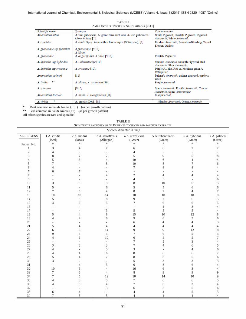
TABLE I
AMARANTHUS SPECIES IN SAUDI ARABIA [7-11]
* Most common in Saudi Arabia (+++) (as per growth pattern)
** Less common in Saudi Arabia (++) (as per growth pattern)
All others species are rare and sporadic.
TABLE II
SKIN TEST REACTIVITY OF 39 PATIENTS TO SEVEN AMARANTHUS EXTRACTS.
*(wheal diameter in mm)
ALLERGENS
Patient No.
1 A. viridis
(local)
*
2 A. lividus
(local)
*
3 A. retroflexus
(Allergon)
*
4 A. retroflexus
(Greer)
*
5 A. tuberculatus
(Greer)
*
6 A. hybridus
(Greer)
*
7 A. palmeri
(Greer)
*
1 3 4 7 6 6 7 7
2 4 - - 4 - - -
3 8 7 7 5 9 5 8
4 5 5 4 10 6 4 4
5 7 - 8 10 8 7 6
6 4 - - 7 4 4 4
7 6 7 - - - - -
8 - - 4 7 4 4 4
9 - - - 4 5 - 6
10 3 3 5 8 10 6 5
11 5 - 6 5 5 6 6
12 7 5 4 8 7 4 7
13 10 10 14 10 10 10 9
14 5 3 8 9 7 6 5
15 4 3 5 7 6 6 5
16 - - - - 4 3 4
17 - - 3 5 5 5 4
18 5 4 8 15 10 12 8
19 4 4 6 9 6 5 6
20 - - - 6 4 4 3
21 5 4 4 4 4 4 4
22 6 6 14 9 9 12 8
23 9 8 5 7 6 5 5
24 4 5 10 6 5 5 7
25 - - - 7 5 3 4
26 3 3 3 7 4 6 5
27 4 - 5 3 - 4 6
28 4 4 6 8 6 6 7
29 5 4 7 8 6 5 6
30 3 - - 5 3 3 3
31 - 4 5 6 8 6 4
32 10 6 4 16 6 3 4
33 7 6 4 8 9 9 9
34 7 5 12 10 14 10 9
35 4 3 5 7 6 6 5
36 4 3 4 7 6 3 4
37 - - 3 6 5 5 6
38 6 4 - 4 4 5 3
39 7 5 5 4 4 4 4
International Journal of Chemical, Environmental & Biological Sciences (IJCEBS) Volume 4, Issue 1 (2016) ISSN 2320–4087 (Online)
91

ACKNOWLEDGMENT
This Research was funded by King Abdul-Aziz City for
Science and Technology (KACST) under a Research Grant
ARP 27-11.The informed consent form was approved by the
Research Advice Council (RAC) of KFSH&RC in Riyadh.
The authors also wished to acknowledge Ms. Cheryl Mijares-
Oblea for her typographical assistance.
REFERENCES
[1] B.S. Anand and K. Pawan, “Aeroallergens in clinical practice of allergy
in India,” Ann Agric Environ Med, vol. 10, pp. 131-136, 2003.
[2] G.W.K. Wong and C.M. Chow, “Childhood Asthma Epidemiology:
Insights from Comparative Studies of Rural and Urban Population,”
Pediatric Pulmonology, vol. 43, pp.107-16, 2008.
[3] M. Moradi-Lakeh, C. El Bcheraoui, F. Daoud, M. Tuffaha, H.
Kravitz, M. Al Saeedi, et al, “Prevalence of asthma in Saudi adults:
findings from a national household survey. 2013,” BMC Pulm Med, vol.
15, pp. 77, Jul 28, 2015. doi: 10.1186/s12890-015-0080-5.
[4] A.R. Al-Frayh, Z. Shakoor, M.O. Gad‐el‐Rab and S.M. Hasnain,
“Increased prevalence of Asthma in Saudi Arabia,” Annals of Allergy,
Asthma and Immunology, vol. 86, pp. 292-96, 2001.
[5] S.M. Hasnain, A.R. Al Frayh, “Prevalence of Allergenic Amaranthus
viridis Pollen in seven Different Regions of Saudi Arabia,” Annals of
Saudi Medicine, vol. 27, issue 4, pp. 259-63, 2007.
[6] R.W. Weber, “Cross-reactivity of pollen allergens: impact on allergen
immunotherapy,” Annals of Allergy, Asthma & Immunology, vol. 99,
no. 3, pp. 203-212, 2007.
[7] United States Department of Agriculture, National Resources
Conservation Service.
[http://plants.usda.gov/core/profile?symbol=AMAL]
[8] Plants for a future.
[http://www.pfaf.org/user/Plant.aspx?LatinName=Amaranthus+caudatu
s].
[9] A.M. Migahid, “Flora of Saudi Arabia,” Riyadh, SA, King Saud
University 1996.
[10] S.A. Chaudhery, “Flora of the Kingdom of Saudi Arabia,” Ministry of
agriculture & water, National Herbarium, Riyadh, 1999.
[11] Ministry of Agriculture, Kingdom of Saudi Arabia
[http://www.moa.gov.sa].
[12] J.E. Slater, RE EschE, R Lockey, “Preparation and standardization of
allergen extracts,” Middleton's allergy principles and practice, 7th ed.,
St. Louis: Mosby, 2009, pp 557-608.
[13] M.M. Bradford, “A rapid and sensitive method for the quantitation of
microgram quantities of protein utilizing the principle of protein-dye
binding,” Anal Biochem, vol. 72, pp. 248-254, 1976.U.K. Laemmli,
“Cleavage of structural proteins during the assembly of the head of
bacteriophage T4,” Nature, vol. 227, pp. 680-685, 1970.
[14] H. Towbin, T. Staehelin and J. Gordon, “Electrophoretic transfer of
proteins from polyacrylamide gel to nitrocellulose sheets,”
Biotechnology, vol. 24, pp. 145-149, 1992.
[15] M. Villalba, R. Barderas, S. Mas, C. Colás and E. Batanero and R.
Rodríguez. “Amaranthaceae pollens: review of an emerging allergy in
the Mediterranean area,” J Investing Allergol Clin Immunol, vol. 24, pp.
371-81, 2014.
[16] M. Tehrani, M. Sankian, M. Assarehzadegan, R. Falak, F. Jabbari and
A. Varasteh, “Immunochemical characterization of Amaranthus
retroflexus pollen extract: extensive cross-reactive allergenic
components among the four species of
Amaranthaceae/Chenopodiaceae,” Iran J Allergy Asthma Immunol, vol.
9, pp. 87-95, 2010.
[17] A.A. Alaiya, H.A. Alsini, M.O. Gad-El-Rab and S.M. Hasnain, “Protein
Profiles of Indigenous and Commercial Extracts of Amaranthus Pollen
for the Diagnosis of Allergy and Asthma Patients,” World Applied
Sciences Journal, vol. 32, pp. 2354-2361, 2014.
International Journal of Chemical, Environmental & Biological Sciences (IJCEBS) Volume 4, Issue 1 (2016) ISSN 2320–4087 (Online)
92