Altered Mental Status - The University of Texas Health...
63
Altered Mental Status CMR Lecture
Transcript of Altered Mental Status - The University of Texas Health...
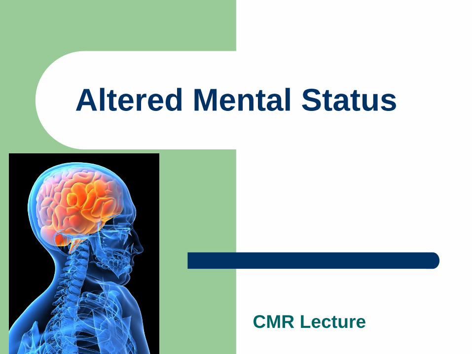
Altered Mental Status
CMR Lecture
Objectives
Overview the definition of “altered mental status”
Develop reasonable differential diagnosis for acute
mental status changes
Explain first steps in diagnosis and management of
common causes of mental status changes
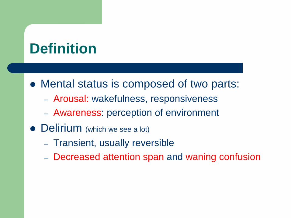
Definition
Mental status is composed of two parts:
– Arousal: wakefulness, responsiveness
– Awareness: perception of environment
Delirium (which we see a lot)
– Transient, usually reversible
– Decreased attention span and waning confusion
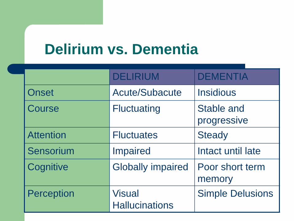
Delirium vs. Dementia
DELIRIUM DEMENTIA
Onset Acute/Subacute Insidious
Course Fluctuating Stable and
progressive
Attention Fluctuates Steady
Sensorium Impaired Intact until late
Cognitive Globally impaired Poor short term
memory
Perception Visual
Hallucinations
Simple Delusions
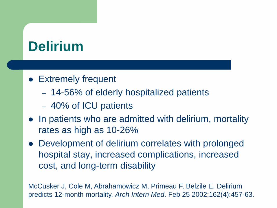
Delirium
Extremely frequent
– 14-56% of elderly hospitalized patients
– 40% of ICU patients
In patients who are admitted with delirium, mortality
rates as high as 10-26%
Development of delirium correlates with prolonged
hospital stay, increased complications, increased
cost, and long-term disability
McCusker J, Cole M, Abrahamowicz M, Primeau F, Belzile E. Delirium
predicts 12-month mortality. Arch Intern Med. Feb 25 2002;162(4):457-63.
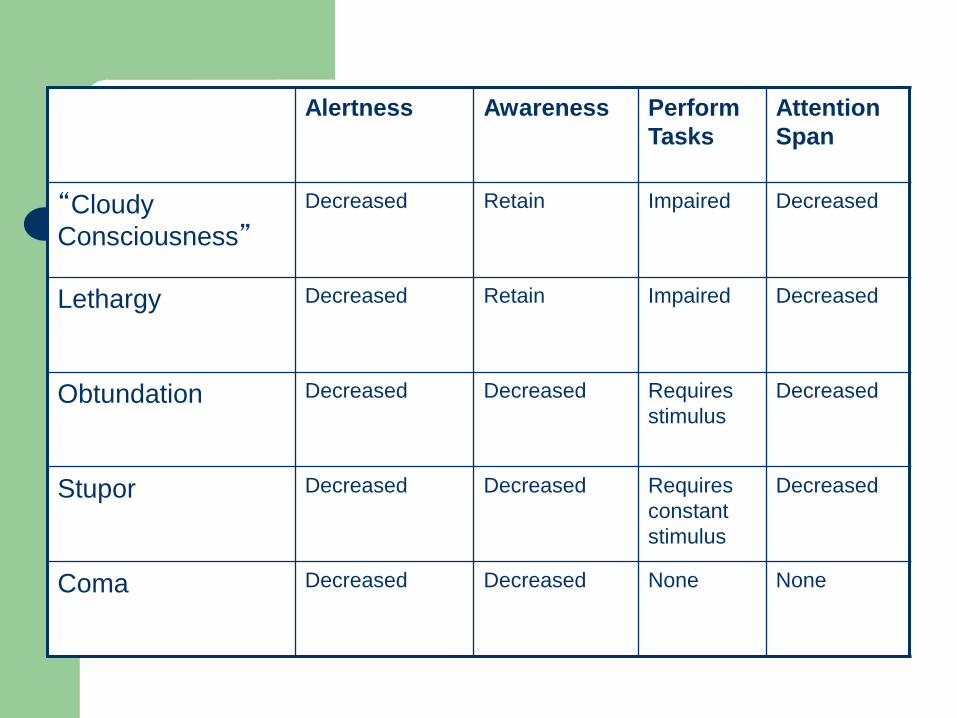
Alertness Awareness Perform
Tasks
Attention
Span
“Cloudy
Consciousness”
Decreased Retain Impaired Decreased
Lethargy Decreased Retain Impaired Decreased
Obtundation Decreased Decreased Requires
stimulus
Decreased
Stupor Decreased Decreased Requires
constant
stimulus
Decreased
Coma Decreased Decreased None None
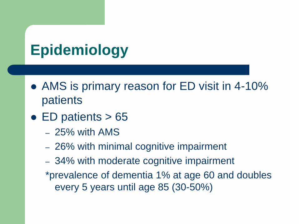
Epidemiology
AMS is primary reason for ED visit in 4-10%
patients
ED patients > 65
– 25% with AMS
– 26% with minimal cognitive impairment
– 34% with moderate cognitive impairment
*prevalence of dementia 1% at age 60 and doubles
every 5 years until age 85 (30-50%)
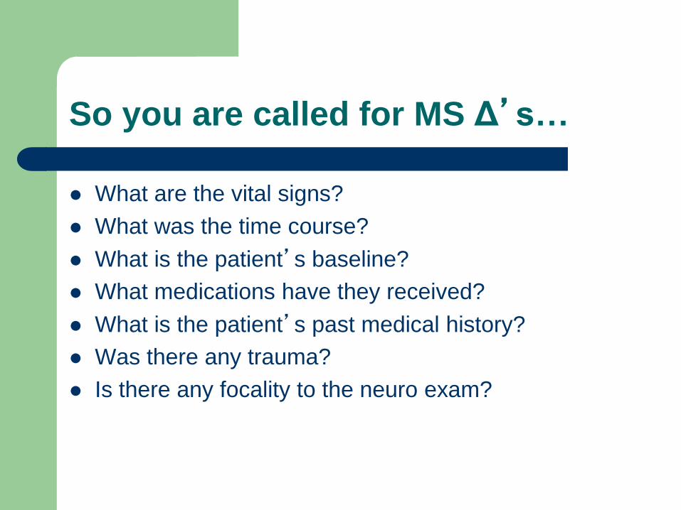
So you are called for MS Δ’s…
What are the vital signs?
What was the time course?
What is the patient’s baseline?
What medications have they received?
What is the patient’s past medical history?
Was there any trauma?
Is there any focality to the neuro exam?
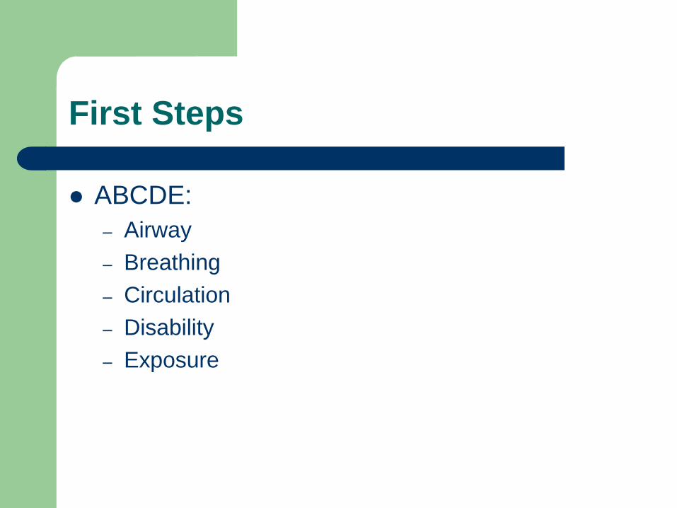
First Steps
ABCDE:
– Airway
– Breathing
– Circulation
– Disability
– Exposure
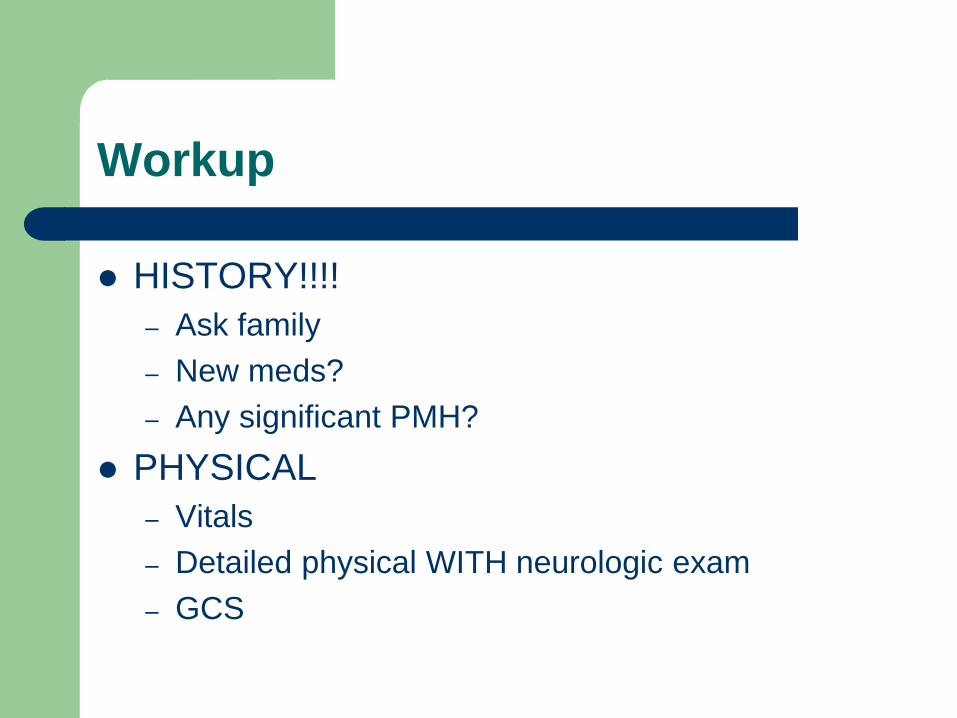
Workup
HISTORY!!!!
– Ask family
– New meds?
– Any significant PMH?
PHYSICAL
– Vitals
– Detailed physical WITH neurologic exam
– GCS
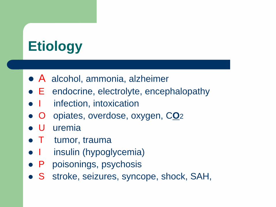
Etiology
A alcohol, ammonia, alzheimer
E endocrine, electrolyte, encephalopathy
I infection, intoxication
O opiates, overdose, oxygen, CO2
U uremia
T tumor, trauma
I insulin (hypoglycemia)
P poisonings, psychosis
S stroke, seizures, syncope, shock, SAH,
Case #1
73 YO WM with h/o HTN and gout admitted for
suspected septic arthritis of left knee. Patient had
arthrocentesis this afternoon, results pending. You
are called at 9pm because patient has had an acute
change in mental status.
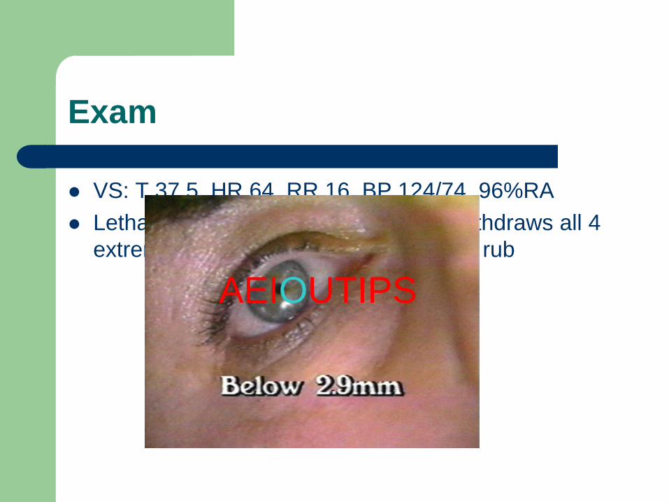
Exam
VS: T 37.5, HR 64, RR 16, BP 124/74, 96%RA
Lethargic, not conversant, moaning, withdraws all 4
extremities to pain, responds to sternal rub
AEIOUTIPS
Drugs
Medications implicated in 30% of cases of delirium
Common causes of mental status changes include
opioids, benzos, any anticholinergics
Clues in the exam
– Opioids: miosis (pinpoint pupils), decreased respirations,
and hypotension
– Anticholinergics: mydriasis, bradycardia, salivation,
lacrimation, and diaphoresis
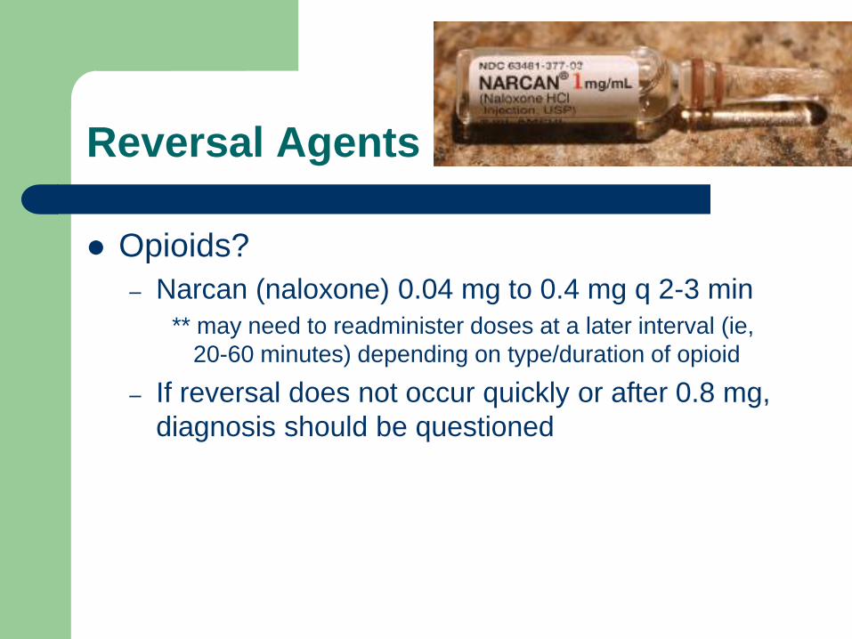
Reversal Agents
Opioids?
– Narcan (naloxone) 0.04 mg to 0.4 mg q 2-3 min
** may need to readminister doses at a later interval (ie,
20-60 minutes) depending on type/duration of opioid
– If reversal does not occur quickly or after 0.8 mg,
diagnosis should be questioned
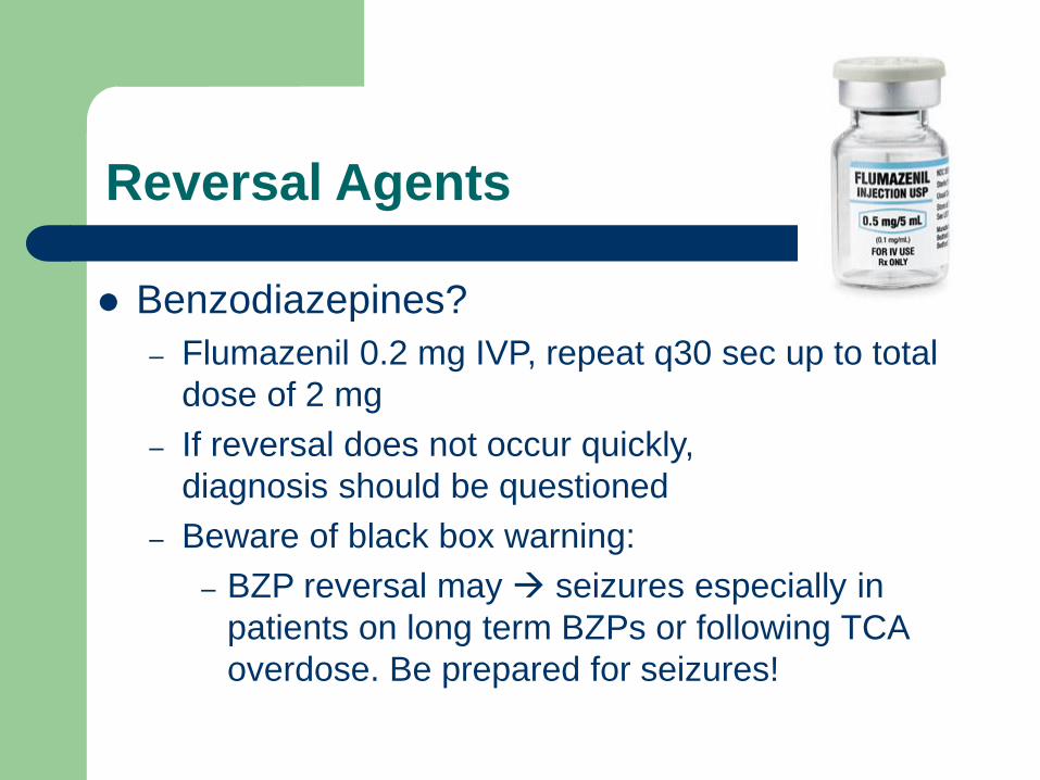
Reversal Agents
Benzodiazepines?
– Flumazenil 0.2 mg IVP, repeat q30 sec up to total
dose of 2 mg
– If reversal does not occur quickly,
diagnosis should be questioned
– Beware of black box warning:
– BZP reversal may seizures especially in
patients on long term BZPs or following TCA
overdose. Be prepared for seizures!
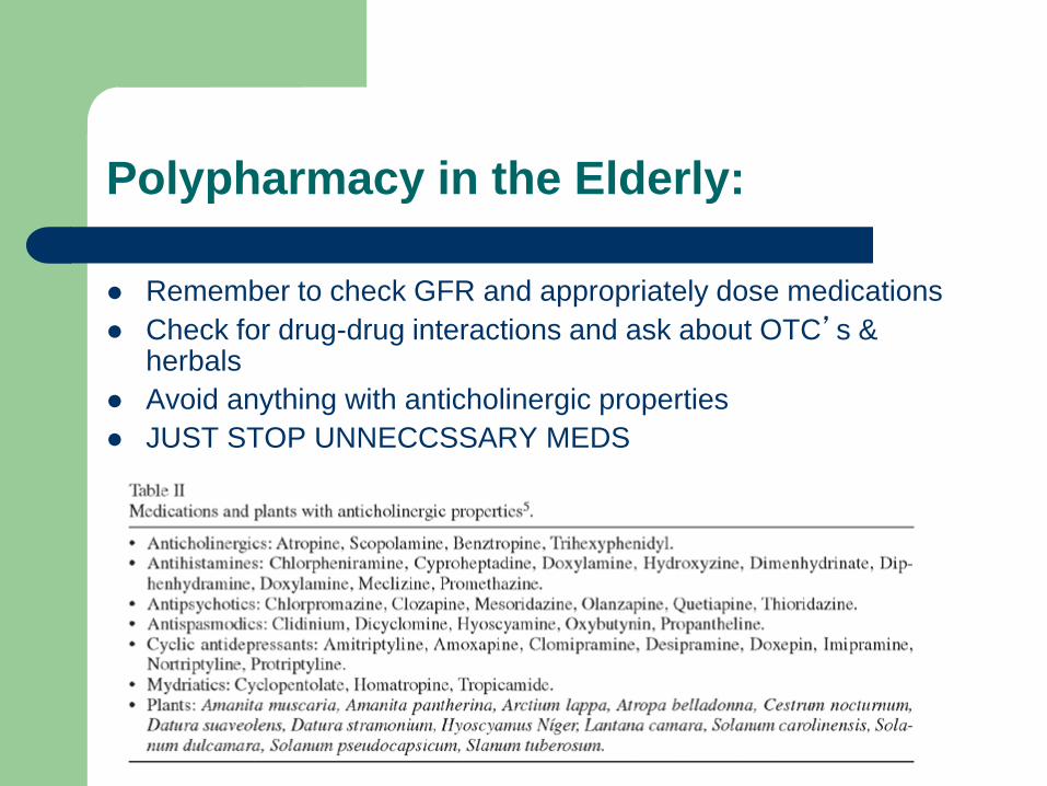
Polypharmacy in the Elderly:
Remember to check GFR and appropriately dose medications
Check for drug-drug interactions and ask about OTC’s & herbals
Avoid anything with anticholinergic properties
JUST STOP UNNECCSSARY MEDS
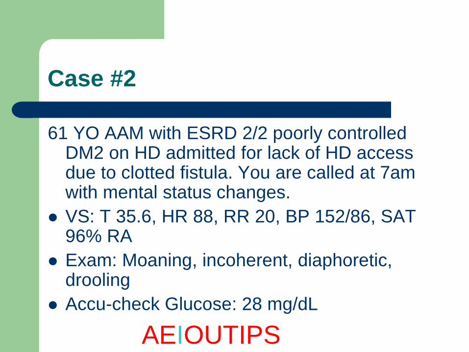
Case #2
61 YO AAM with ESRD 2/2 poorly controlled DM2 on HD admitted for lack of HD access due to clotted fistula. You are called at 7am with mental status changes.
VS: T 35.6, HR 88, RR 20, BP 152/86, SAT 96% RA
Exam: Moaning, incoherent, diaphoretic, drooling
Accu-check Glucose: 28 mg/dL
AEIOUTIPS
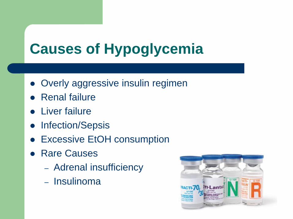
Causes of Hypoglycemia
Overly aggressive insulin regimen
Renal failure
Liver failure
Infection/Sepsis
Excessive EtOH consumption
Rare Causes
– Adrenal insufficiency
– Insulinoma
Hypoglycemia Management
Is patient awake enough to drink some juice, take glucose tabs?
– Three glucose tabs will raise blood sugar by 50 g/dL.
If unable to take PO and has IV access, then give use IV dextrose
– 1 amp D50 = 25 grams of glucose
If patient does not have IV access and unresponsive, give Glucagon 1mg IM/SC.
Always recheck glucose 15-20 minutes later to document return to euglycemia.
Case #3
64 YO obese WF with GOLD class III COPD (on 2L
home O2) admitted for COPD exacerbation. You are
called for mental status changes at 10:55 PM.
VS: T 36.4, HR 88, RR 18, BP 134/66, SAT 99% on
8L O2 via NC
Exam: Lethargic, arouses only to sternal rub, lungs
with poor air exchange
ABG: 7.18 / 103 / 95 / 98% on 8L Via NC
AEIOUTIPS
Hypercapnea because of supplemental Oxygen:
1) V/Q mismatch: if a part of the lung is underventilated it should be underperfused (hypoxic pulmonary vasoconstriction) adding O2 increases perfusion but NOT ventilation
2) Haldane effect: Deoxygenated Hg is able to carry more carbon dioxide than oxygenated Hg
3) Respiratory homeostasis: Chronic elevation of CO2 leads to CO2 being less of a stimulant for respiratory drive, and instead O2 provides stimulus. Hence, supplemental O2 can decrease respiratory drive leading to CO2 retention.

Five Causes of Hypoxia
1. Hypoventilation
2. Shunt
3. Increased Diffusion Gradient
4. Decreased FiO2
5. V-Q Mismatch
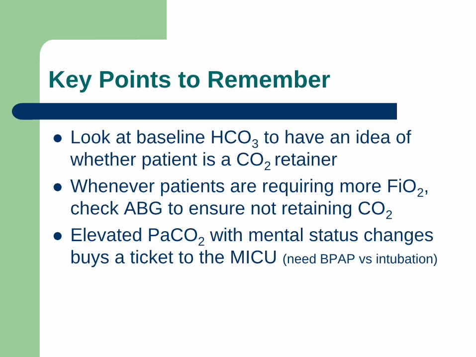
Key Points to Remember
Look at baseline HCO3 to have an idea of
whether patient is a CO2 retainer
Whenever patients are requiring more FiO2,
check ABG to ensure not retaining CO2
Elevated PaCO2 with mental status changes
buys a ticket to the MICU (need BPAP vs intubation)
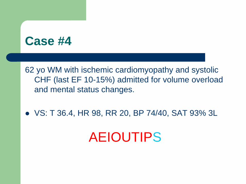
Case #4
62 yo WM with ischemic cardiomyopathy and systolic
CHF (last EF 10-15%) admitted for volume overload
and mental status changes.
VS: T 36.4, HR 98, RR 20, BP 74/40, SAT 93% 3L
AEIOUTIPS
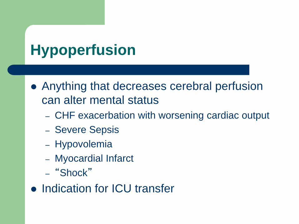
Hypoperfusion
Anything that decreases cerebral perfusion
can alter mental status
– CHF exacerbation with worsening cardiac output
– Severe Sepsis
– Hypovolemia
– Myocardial Infarct
– “Shock”
Indication for ICU transfer
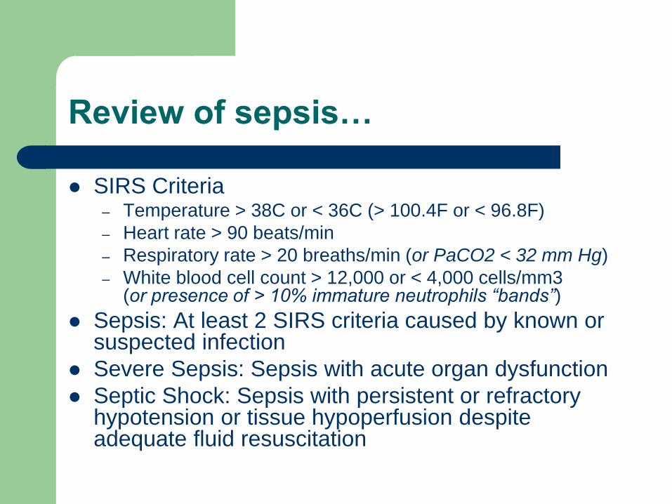
Review of sepsis…
SIRS Criteria – Temperature > 38C or < 36C (> 100.4F or < 96.8F)
– Heart rate > 90 beats/min
– Respiratory rate > 20 breaths/min (or PaCO2 < 32 mm Hg)
– White blood cell count > 12,000 or < 4,000 cells/mm3 (or presence of > 10% immature neutrophils “bands”)
Sepsis: At least 2 SIRS criteria caused by known or suspected infection
Severe Sepsis: Sepsis with acute organ dysfunction
Septic Shock: Sepsis with persistent or refractory hypotension or tissue hypoperfusion despite adequate fluid resuscitation
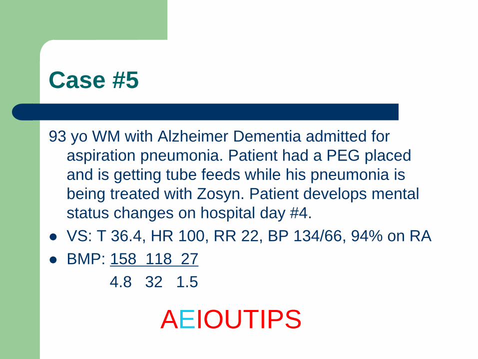
Case #5
93 yo WM with Alzheimer Dementia admitted for
aspiration pneumonia. Patient had a PEG placed
and is getting tube feeds while his pneumonia is
being treated with Zosyn. Patient develops mental
status changes on hospital day #4.
VS: T 36.4, HR 100, RR 22, BP 134/66, 94% on RA
BMP: 158 118 27
4.8 32 1.5
AEIOUTIPS
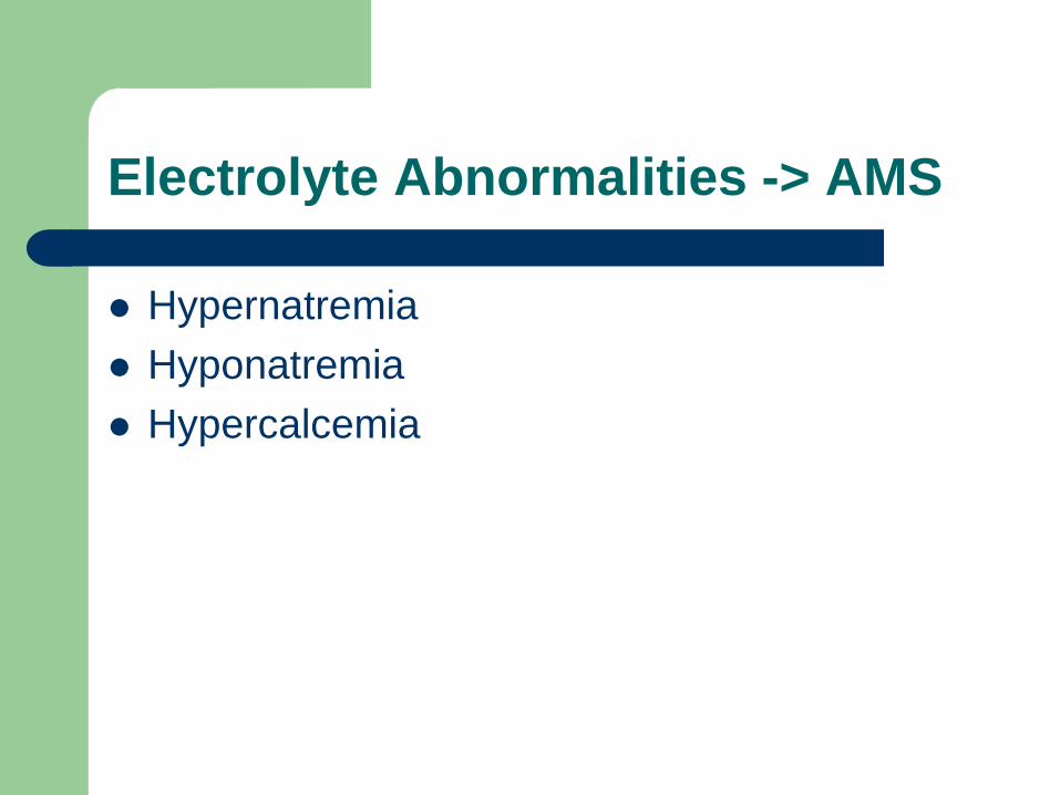
Electrolyte Abnormalities -> AMS
Hypernatremia
Hyponatremia
Hypercalcemia
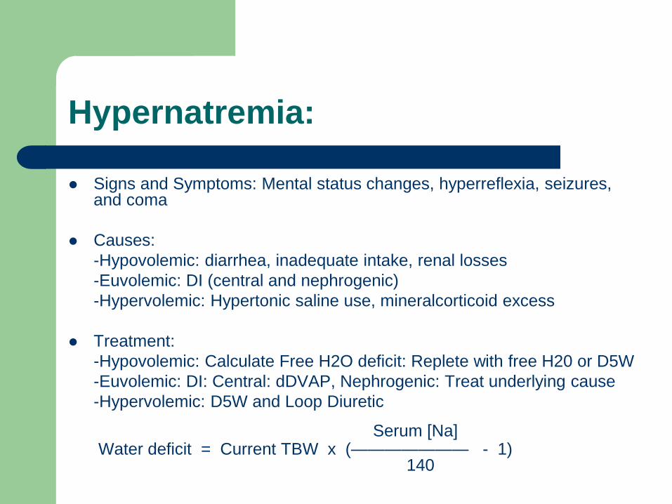
Hypernatremia:
Signs and Symptoms: Mental status changes, hyperreflexia, seizures, and coma
Causes:
-Hypovolemic: diarrhea, inadequate intake, renal losses
-Euvolemic: DI (central and nephrogenic)
-Hypervolemic: Hypertonic saline use, mineralcorticoid excess
Treatment:
-Hypovolemic: Calculate Free H2O deficit: Replete with free H20 or D5W
-Euvolemic: DI: Central: dDVAP, Nephrogenic: Treat underlying cause
-Hypervolemic: D5W and Loop Diuretic
Serum [Na] Water deficit = Current TBW x (——————— - 1) 140
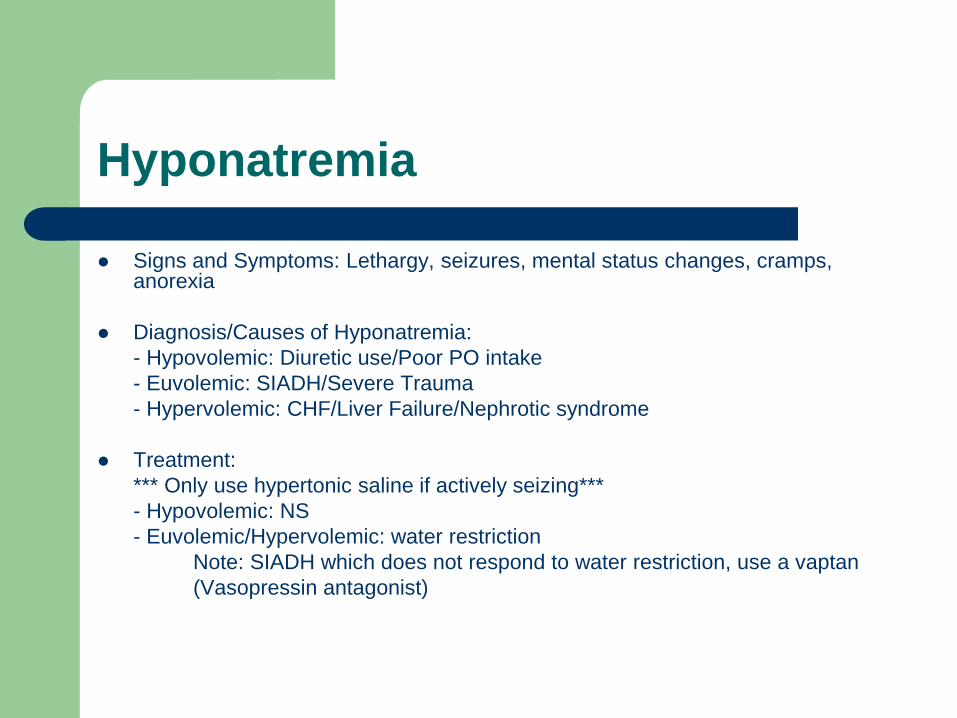
Hyponatremia
Signs and Symptoms: Lethargy, seizures, mental status changes, cramps, anorexia
Diagnosis/Causes of Hyponatremia:
- Hypovolemic: Diuretic use/Poor PO intake
- Euvolemic: SIADH/Severe Trauma
- Hypervolemic: CHF/Liver Failure/Nephrotic syndrome
Treatment:
*** Only use hypertonic saline if actively seizing***
- Hypovolemic: NS
- Euvolemic/Hypervolemic: water restriction
Note: SIADH which does not respond to water restriction, use a vaptan
(Vasopressin antagonist)
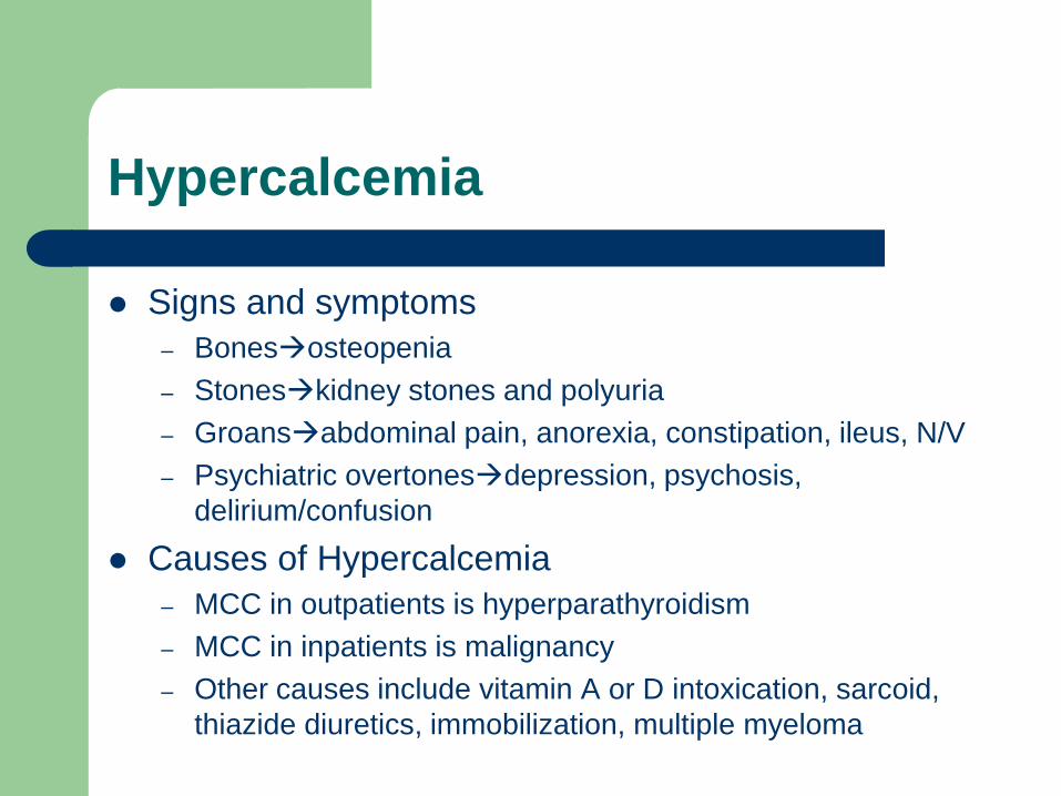
Hypercalcemia
Signs and symptoms
– Bonesosteopenia
– Stoneskidney stones and polyuria
– Groansabdominal pain, anorexia, constipation, ileus, N/V
– Psychiatric overtonesdepression, psychosis,
delirium/confusion
Causes of Hypercalcemia
– MCC in outpatients is hyperparathyroidism
– MCC in inpatients is malignancy
– Other causes include vitamin A or D intoxication, sarcoid,
thiazide diuretics, immobilization, multiple myeloma
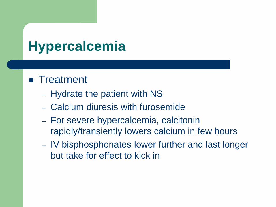
Hypercalcemia
Treatment
– Hydrate the patient with NS
– Calcium diuresis with furosemide
– For severe hypercalcemia, calcitonin
rapidly/transiently lowers calcium in few hours
– IV bisphosphonates lower further and last longer
but take for effect to kick in
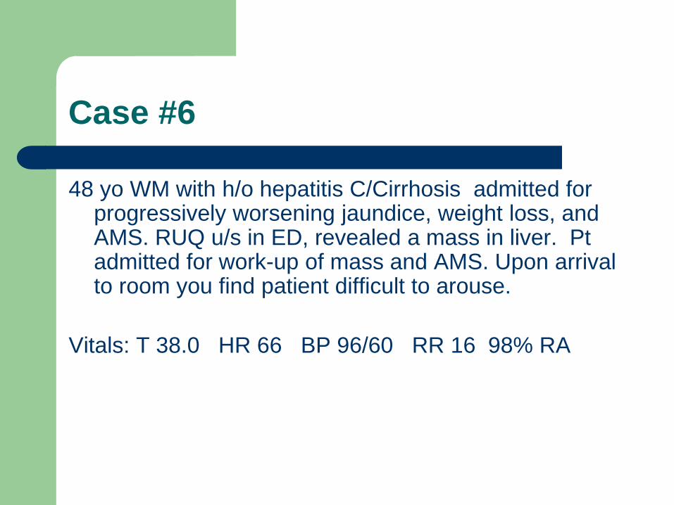
Case #6
48 yo WM with h/o hepatitis C/Cirrhosis admitted for progressively worsening jaundice, weight loss, and AMS. RUQ u/s in ED, revealed a mass in liver. Pt admitted for work-up of mass and AMS. Upon arrival to room you find patient difficult to arouse.
Vitals: T 38.0 HR 66 BP 96/60 RR 16 98% RA
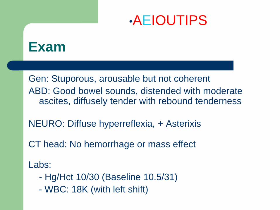
Exam
Gen: Stuporous, arousable but not coherent
ABD: Good bowel sounds, distended with moderate ascites, diffusely tender with rebound tenderness
NEURO: Diffuse hyperreflexia, + Asterixis
CT head: No hemorrhage or mass effect
Labs:
- Hg/Hct 10/30 (Baseline 10.5/31)
- WBC: 18K (with left shift)
•AEIOUTIPS
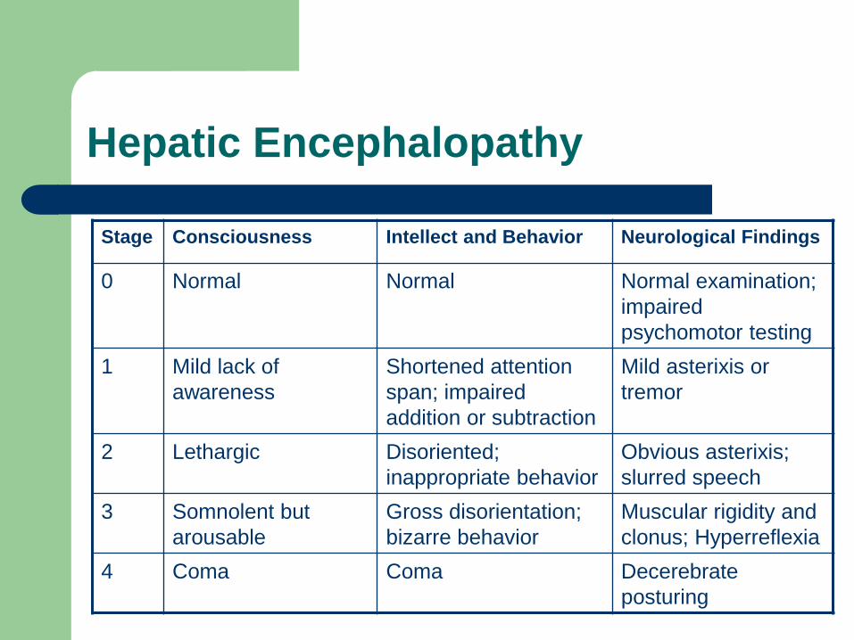
Hepatic Encephalopathy
Stage Consciousness Intellect and Behavior Neurological Findings
0 Normal Normal Normal examination;
impaired
psychomotor testing
1 Mild lack of
awareness
Shortened attention
span; impaired
addition or subtraction
Mild asterixis or
tremor
2 Lethargic Disoriented;
inappropriate behavior
Obvious asterixis;
slurred speech
3 Somnolent but
arousable
Gross disorientation;
bizarre behavior
Muscular rigidity and
clonus; Hyperreflexia
4 Coma Coma Decerebrate
posturing
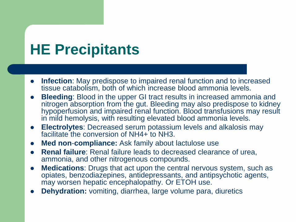
HE Precipitants
Infection: May predispose to impaired renal function and to increased tissue catabolism, both of which increase blood ammonia levels.
Bleeding: Blood in the upper GI tract results in increased ammonia and nitrogen absorption from the gut. Bleeding may also predispose to kidney hypoperfusion and impaired renal function. Blood transfusions may result in mild hemolysis, with resulting elevated blood ammonia levels.
Electrolytes: Decreased serum potassium levels and alkalosis may facilitate the conversion of NH4+ to NH3.
Med non-compliance: Ask family about lactulose use
Renal failure: Renal failure leads to decreased clearance of urea, ammonia, and other nitrogenous compounds.
Medications: Drugs that act upon the central nervous system, such as opiates, benzodiazepines, antidepressants, and antipsychotic agents, may worsen hepatic encephalopathy. Or ETOH use.
Dehydration: vomiting, diarrhea, large volume para, diuretics
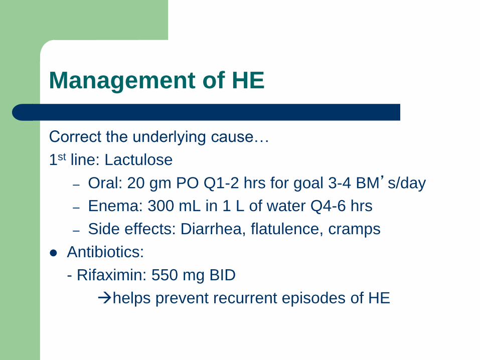
Management of HE
Correct the underlying cause…
1st line: Lactulose
– Oral: 20 gm PO Q1-2 hrs for goal 3-4 BM’s/day
– Enema: 300 mL in 1 L of water Q4-6 hrs
– Side effects: Diarrhea, flatulence, cramps
Antibiotics:
- Rifaximin: 550 mg BID
helps prevent recurrent episodes of HE
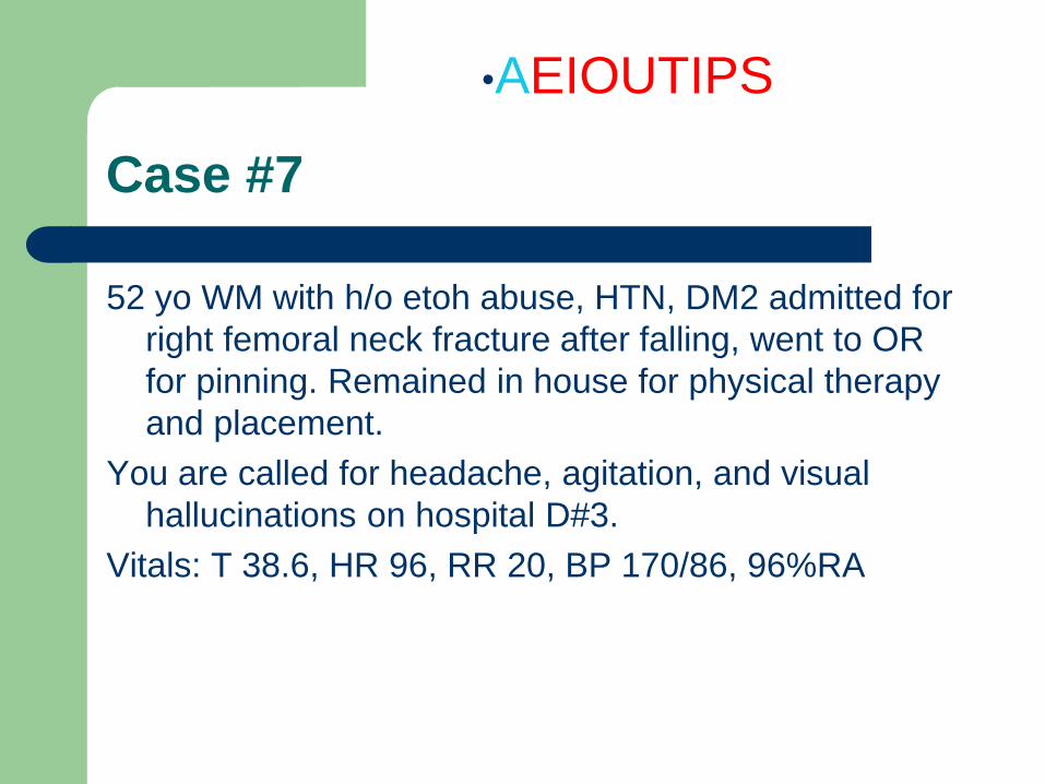
Case #7
52 yo WM with h/o etoh abuse, HTN, DM2 admitted for
right femoral neck fracture after falling, went to OR
for pinning. Remained in house for physical therapy
and placement.
You are called for headache, agitation, and visual
hallucinations on hospital D#3.
Vitals: T 38.6, HR 96, RR 20, BP 170/86, 96%RA
•AEIOUTIPS
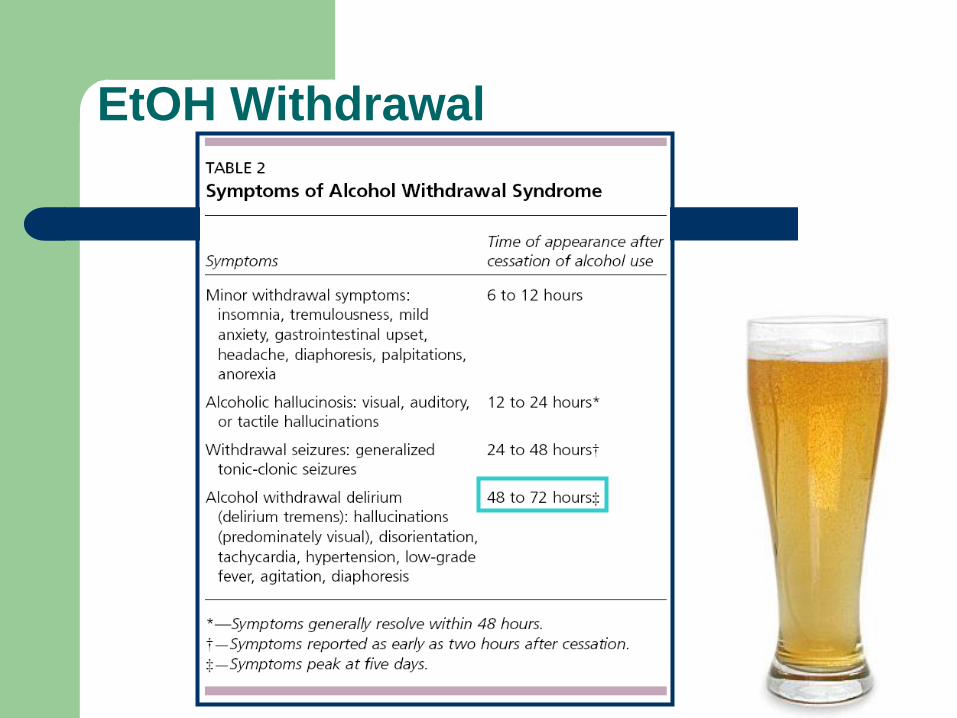
EtOH Withdrawal
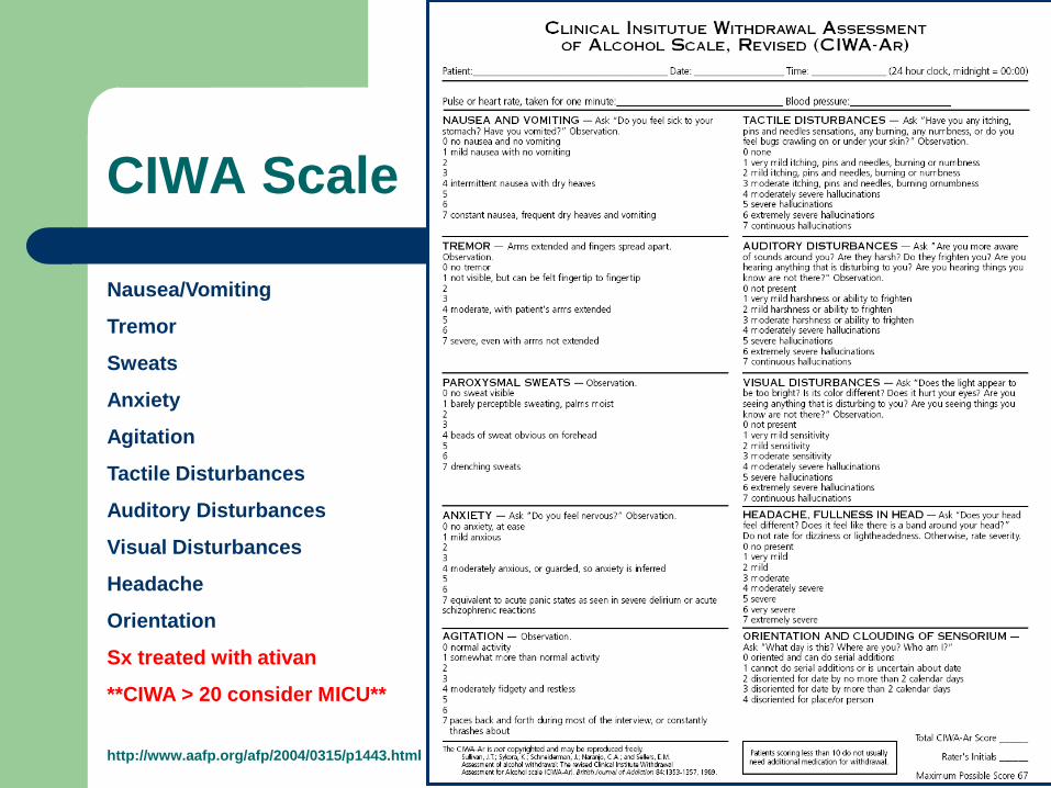
CIWA Scale
Nausea/Vomiting
Tremor
Sweats
Anxiety
Agitation
Tactile Disturbances
Auditory Disturbances
Visual Disturbances
Headache
Orientation
Sx treated with ativan
**CIWA > 20 consider MICU**
http://www.aafp.org/afp/2004/0315/p1443.html
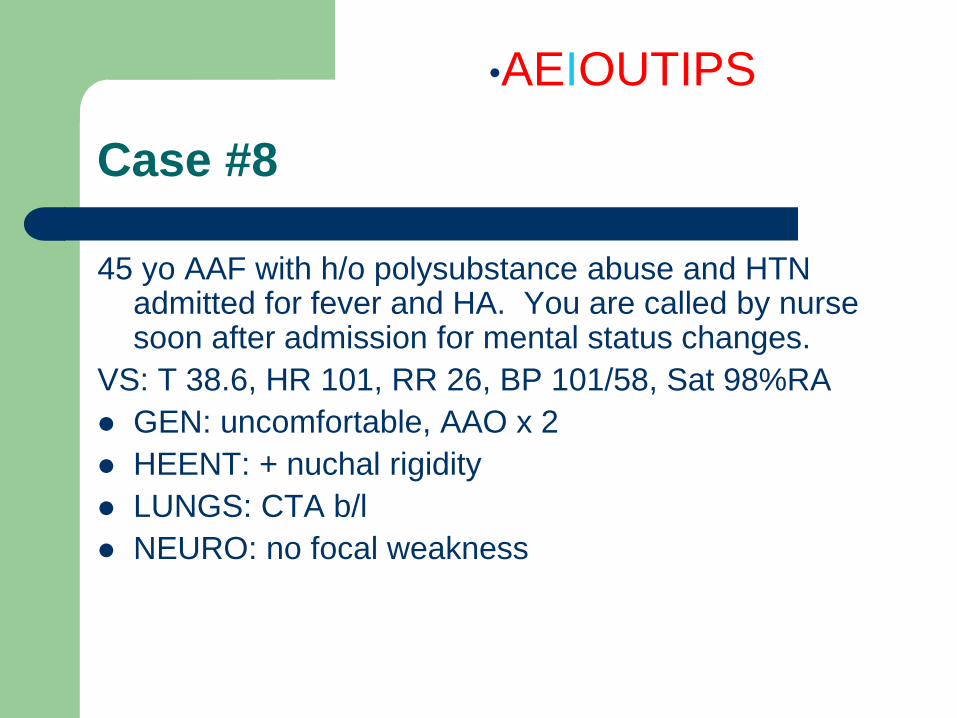
Case #8
45 yo AAF with h/o polysubstance abuse and HTN admitted for fever and HA. You are called by nurse soon after admission for mental status changes.
VS: T 38.6, HR 101, RR 26, BP 101/58, Sat 98%RA
GEN: uncomfortable, AAO x 2
HEENT: + nuchal rigidity
LUNGS: CTA b/l
NEURO: no focal weakness
•AEIOUTIPS
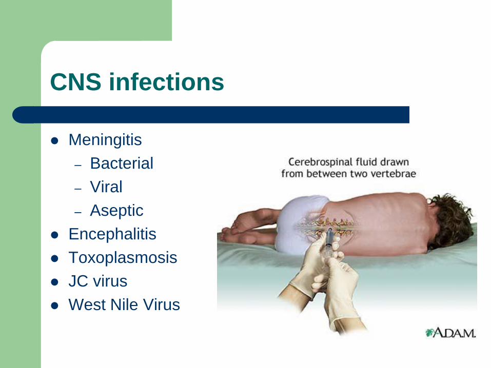
CNS infections
Meningitis
– Bacterial
– Viral
– Aseptic
Encephalitis
Toxoplasmosis
JC virus
West Nile Virus
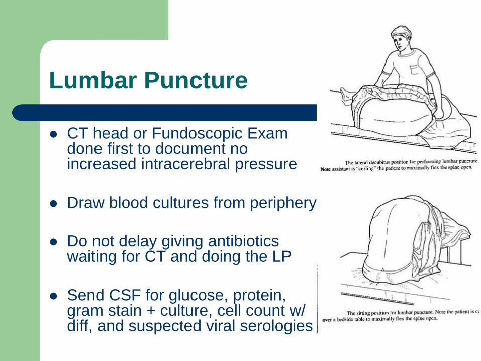
Lumbar Puncture
CT head or Fundoscopic Exam done first to document no increased intracerebral pressure
Draw blood cultures from periphery
Do not delay giving antibiotics waiting for CT and doing the LP
Send CSF for glucose, protein, gram stain + culture, cell count w/ diff, and suspected viral serologies
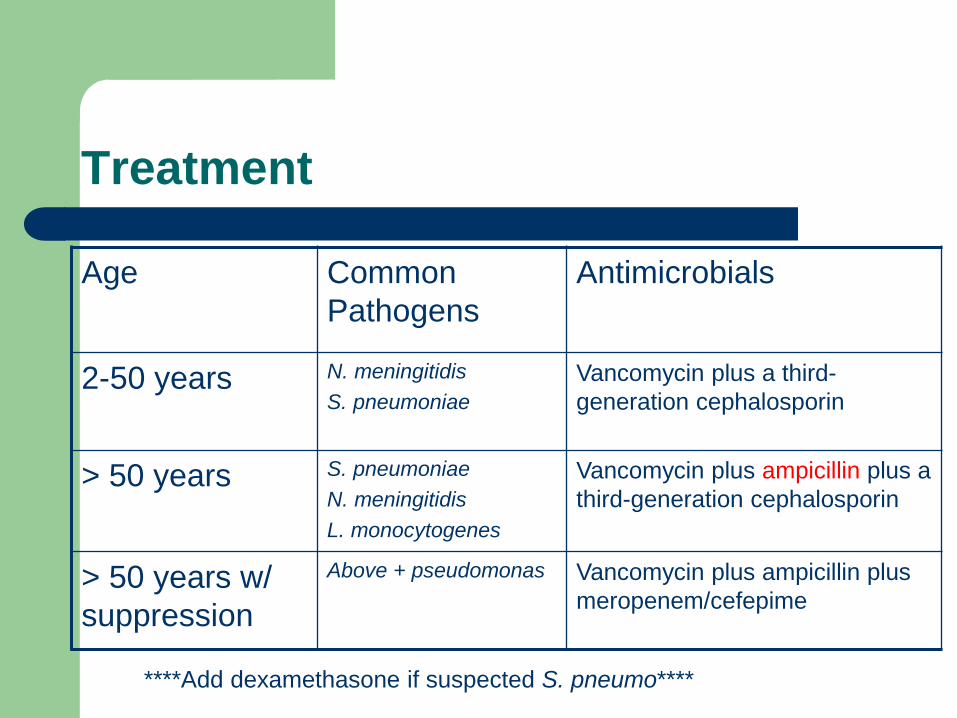
Treatment
Age Common
Pathogens
Antimicrobials
2-50 years N. meningitidis
S. pneumoniae
Vancomycin plus a third-
generation cephalosporin
> 50 years S. pneumoniae
N. meningitidis
L. monocytogenes
Vancomycin plus ampicillin plus a
third-generation cephalosporin
> 50 years w/
suppression
Above + pseudomonas Vancomycin plus ampicillin plus
meropenem/cefepime
****Add dexamethasone if suspected S. pneumo****
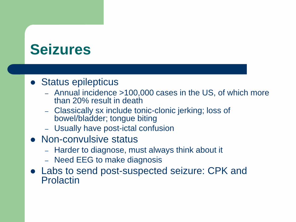
Seizures
Status epilepticus – Annual incidence >100,000 cases in the US, of which more
than 20% result in death
– Classically sx include tonic-clonic jerking; loss of bowel/bladder; tongue biting
– Usually have post-ictal confusion
Non-convulsive status – Harder to diagnose, must always think about it
– Need EEG to make diagnosis
Labs to send post-suspected seizure: CPK and Prolactin
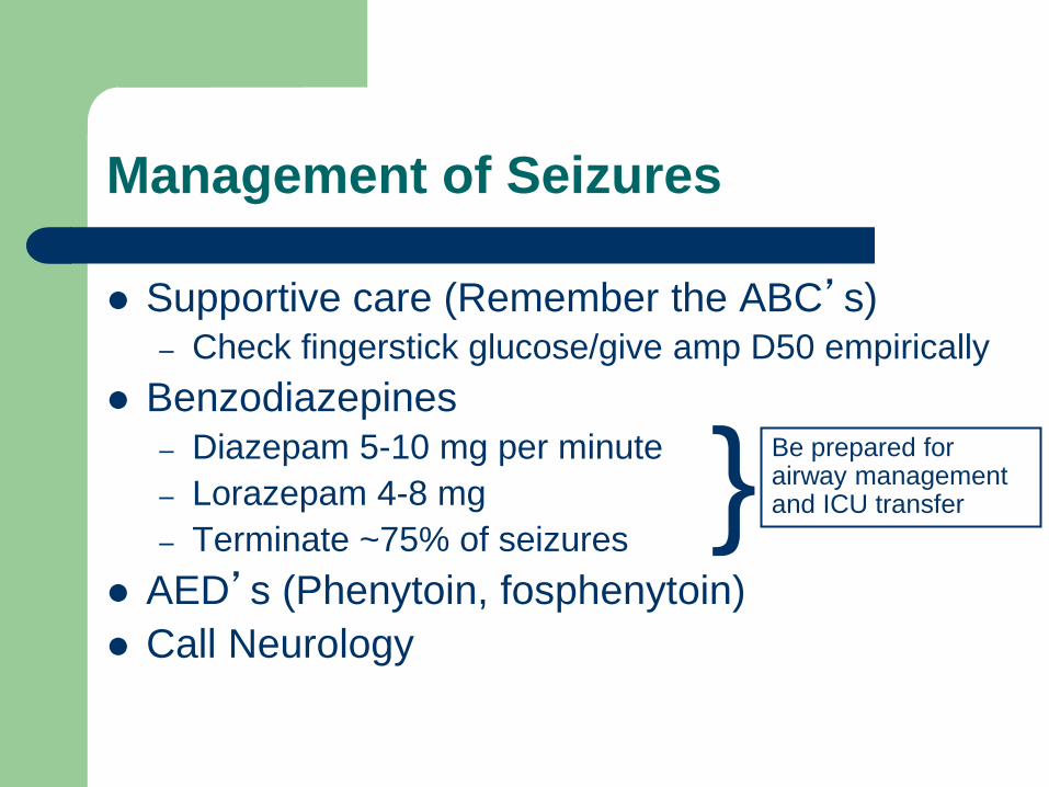
Management of Seizures
Supportive care (Remember the ABC’s) – Check fingerstick glucose/give amp D50 empirically
Benzodiazepines – Diazepam 5-10 mg per minute
– Lorazepam 4-8 mg
– Terminate ~75% of seizures
AED’s (Phenytoin, fosphenytoin)
Call Neurology
} Be prepared for airway management and ICU transfer
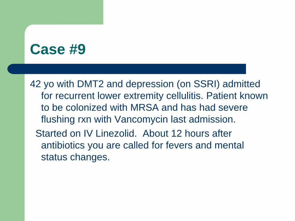
Case #9
42 yo with DMT2 and depression (on SSRI) admitted
for recurrent lower extremity cellulitis. Patient known
to be colonized with MRSA and has had severe
flushing rxn with Vancomycin last admission.
Started on IV Linezolid. About 12 hours after
antibiotics you are called for fevers and mental
status changes.
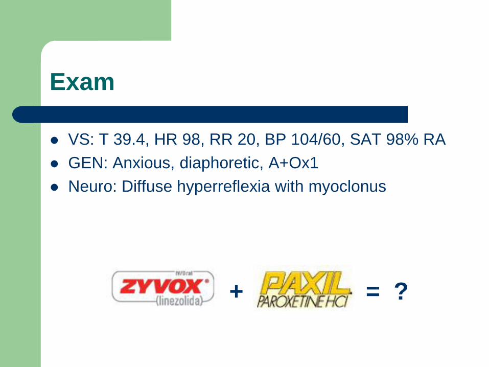
Exam
VS: T 39.4, HR 98, RR 20, BP 104/60, SAT 98% RA
GEN: Anxious, diaphoretic, A+Ox1
Neuro: Diffuse hyperreflexia with myoclonus
+ = ?
Serotonin Syndrome
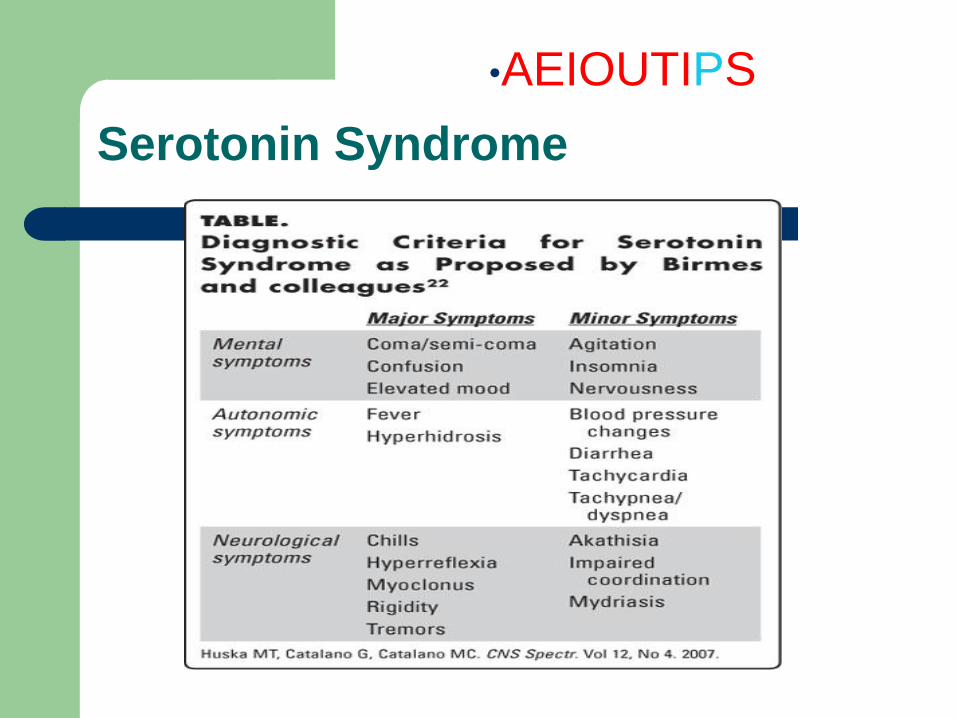
•AEIOUTIPS
Serotonin Syndrome
Treatment
– Discontinuation of all serotonergic agents
– Supportive care aimed at vital signs
– Sedation with benzodiazepines (Ativan 1-2 mg IV)
– If benzos and supportive care fail to improve
agitation and abnormal vital signs, give
cyproheptadine (12 mg orally or by OG/NG)
– Temperature >41.1C (105F) -> immediate
sedation, paralysis, and endotracheal intubation;
avoid antipyretics such as acetaminophen
Case #10
78 yo WM with h/o Stage IIB Colon Cancer admitted
with SOB, found to have a PE. Patient is now on
heparin drip, and he suffers a fall in his room trying
to drag his IV pole to the bathroom. You are called to
assess the patient.
Vitals: T 36.5, HR 52, RR 12, BP 170/88
Exam significant for new LLE weakness
•AEIOUTIPS
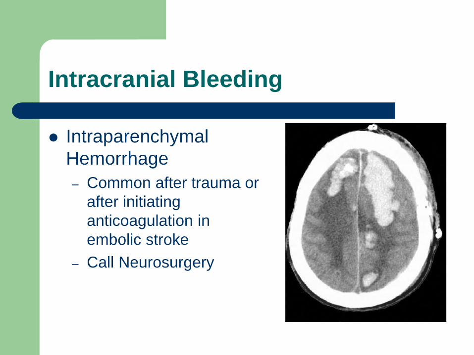
Intracranial Bleeding
Intraparenchymal
Hemorrhage
– Common after trauma or
after initiating
anticoagulation in
embolic stroke
– Call Neurosurgery
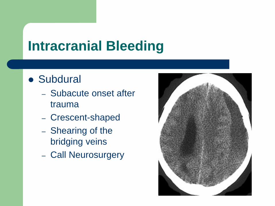
Intracranial Bleeding
Subdural
– Subacute onset after
trauma
– Crescent-shaped
– Shearing of the
bridging veins
– Call Neurosurgery
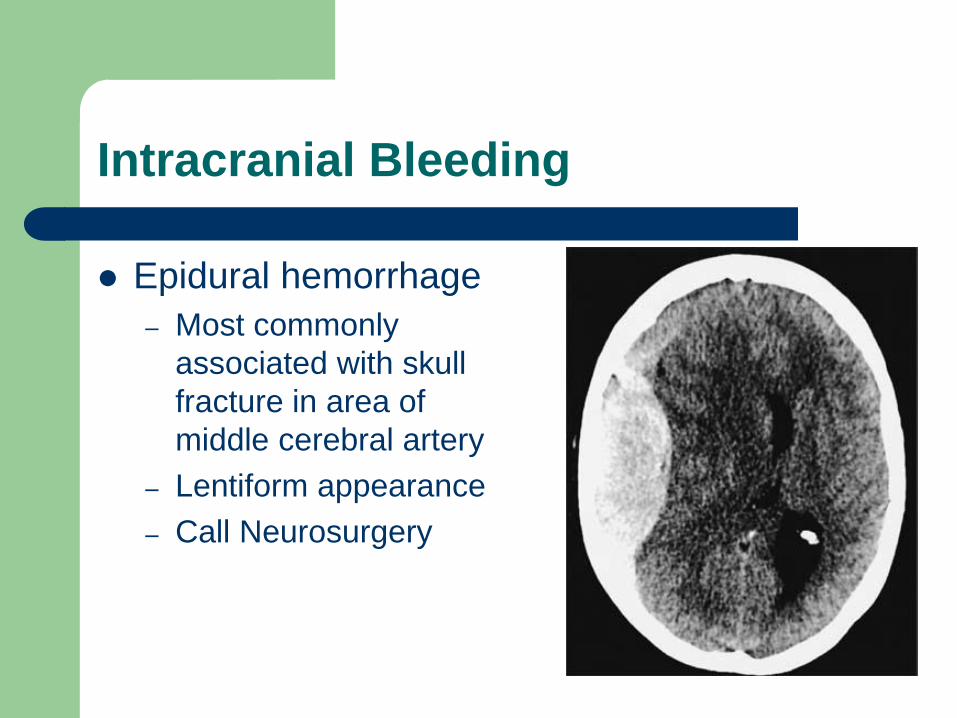
Intracranial Bleeding
Epidural hemorrhage
– Most commonly
associated with skull
fracture in area of
middle cerebral artery
– Lentiform appearance
– Call Neurosurgery
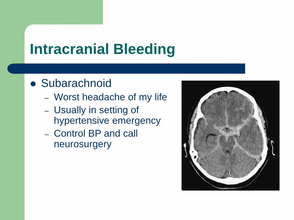
Intracranial Bleeding
Subarachnoid – Worst headache of my life
– Usually in setting of hypertensive emergency
– Control BP and call neurosurgery
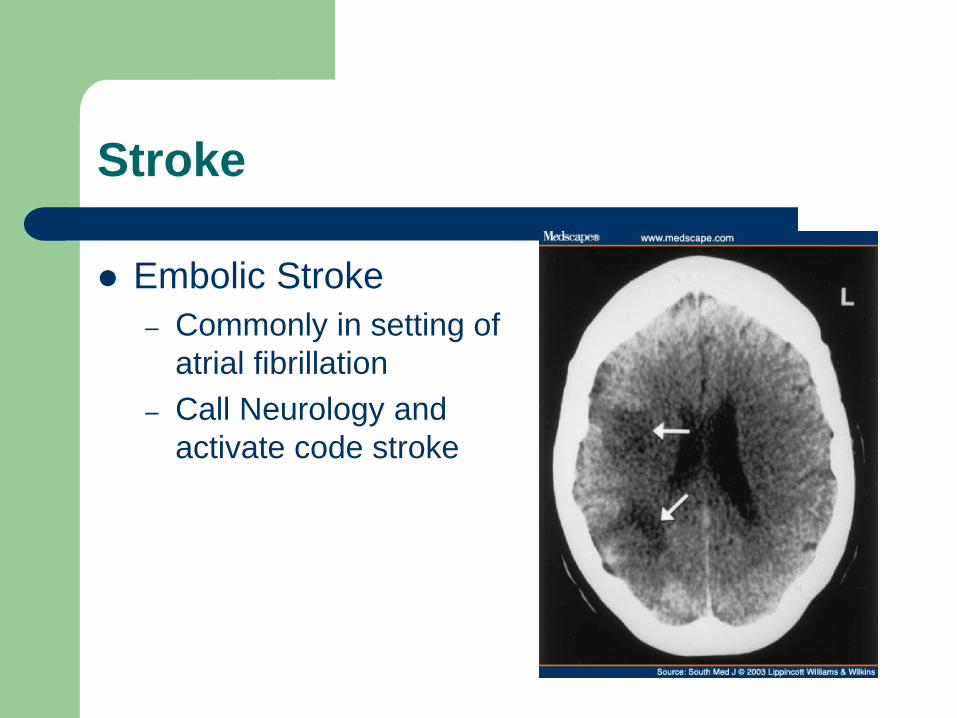
Stroke
Embolic Stroke
– Commonly in setting of
atrial fibrillation
– Call Neurology and
activate code stroke
Case #11
93 yo AAM with HTN and vascular dementia admitted
for UTI. Patient on ceftriaxone IV and awaiting
placement. You are called at 3AM because patient
attempting to climb out of bed, very disoriented, and
trying to pull out Foley.
T-37.7, HR-65, RR-16, BP-120/80
PE: unremarkable
•AEIOUTIPS
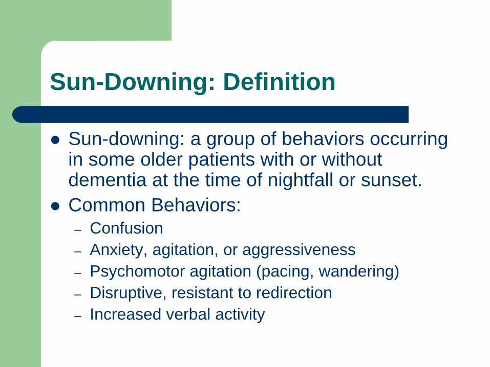
Sun-Downing: Definition
Sun-downing: a group of behaviors occurring in some older patients with or without dementia at the time of nightfall or sunset.
Common Behaviors: – Confusion
– Anxiety, agitation, or aggressiveness
– Psychomotor agitation (pacing, wandering)
– Disruptive, resistant to redirection
– Increased verbal activity
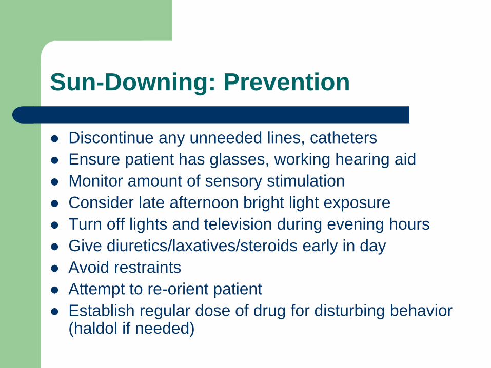
Sun-Downing: Prevention
Discontinue any unneeded lines, catheters
Ensure patient has glasses, working hearing aid
Monitor amount of sensory stimulation
Consider late afternoon bright light exposure
Turn off lights and television during evening hours
Give diuretics/laxatives/steroids early in day
Avoid restraints
Attempt to re-orient patient
Establish regular dose of drug for disturbing behavior (haldol if needed)

Thank you for your attention
Any questions?


![Altered Mental Status · 6/1/2018 2 Overview •Altered mental status: It Could Be [almost] Anything! requires a thorough work-up •What is the differential for altered mental status?](https://static.fdocuments.net/doc/165x107/5e771bc68f2c7b2c9440a58e/altered-mental-status-612018-2-overview-aaltered-mental-status-it-could-be.jpg)