AL/OE-TR-1995-0057 UNITED STATES AIR FORCE …
21
AL/OE-TR-1995-0057 UNITED STATES AIR FORCE ARMSTRONG LABORATORY Dermal Absorption Kinetics of Liquid Chloropentafluorobenzene (CPFB) and 1,2-Dichlorobenzene (DCB) in Rats and Guinea Pigs L. Dong J.H.Grabau Geo-Centers, Inc. 7 Wells Avenue Newton Centre, MA 02159 J.N. McDougal G.W. Jepson D.R.Mattie B.L. Garrity Toxicology Division Wright-Patterson AFB, OH 45433-7400 C. Seckel ManTech Environmental Technology, Inc. P.O. Box 31009 Dayton, OH 45437 March 1995 19961108 058 Approved for public release; distribution is unlimited. Occupational and Environmental Health Directorate Toxicology Division 2856 G Street Wright-Patterson Air Force Base, OH 45433-7400 DTIC QUALITY IMbWSÜiBD 1
Transcript of AL/OE-TR-1995-0057 UNITED STATES AIR FORCE …

AL/OE-TR-1995-0057
UNITED STATES AIR FORCE ARMSTRONG LABORATORY
Dermal Absorption Kinetics of Liquid Chloropentafluorobenzene (CPFB) and 1,2-Dichlorobenzene (DCB) in Rats and
Guinea Pigs
L. Dong J.H.Grabau
Geo-Centers, Inc. 7 Wells Avenue
Newton Centre, MA 02159
J.N. McDougal G.W. Jepson D.R.Mattie B.L. Garrity
Toxicology Division Wright-Patterson AFB, OH 45433-7400
C. Seckel
ManTech Environmental Technology, Inc. P.O. Box 31009
Dayton, OH 45437
March 1995
19961108 058 Approved for public release; distribution is unlimited.
Occupational and Environmental Health Directorate
Toxicology Division 2856 G Street Wright-Patterson Air Force Base, OH 45433-7400
DTIC QUALITY IMbWSÜiBD 1

NOTICES
When US Government drawings, specifications or other data are used for any purpose other than a definitely related Government procurement operation, the Government thereby incurs no responsibility nor any obligation whatsoever, and the fact that the Government may have formulated, furnished, or in any way supplied the said drawings, specifications, or other data is not to be regarded by implication or otherwise, as in any manner licensing the holder or any other person or corporation, or conveying any rights or permission to manufacture, use, or sell any patented invention that may in any way be related thereto.
Please do not request copies of this report from the Armstrong Laboratory. Additional copies may be purchased from:
National Technical Information Service 5285 Port Royal Road Springfield, Virginia 22161
Federal Government agencies and their contractors registered with the Defense Technical Information Center should direct requests for copies of this report to:
Defense Technical Information Center Cameron Station Alexandria, Virginia 22314
DISCLAIMER This Technical Report is published as received and has not
been edited by the Technical Editing Staff of the Armstrong Laboratory.
TECHNICAL REVIEW AND APPROVAL
AL/OE-TR-1995-0057
The experiments reported herein were conducted according to the "Guide for the Care and Use of Laboratory Animals," Institute of Laboratory Animal Resources, National Research Council.
This report has been reviewed by the Office of Public Affairs (PA) and is releasable to the National Technical Information Service (NTIS). At NTIS, it will be available to the general public, including foreign nations.
This technical report has been reviewed and is approved for publication.
FOR THE COMMANDER
TERRY A. CHILDRESS, Lt Col, USAF, BSC Director, Toxicology Division Armstrong Laboratory

•kl
REPORT DOCUMENTATION PAGE Form Apptv OMB No. 070
Public reporting burden for this collection of information is estimated to average 1 hour per response, including the time for reviewing instructions, searching existing data sources, gathe< maintaining the data needed, and completing and reviewing the collection of information. Send comments regarding this burden estimate or any other aspect of this collection of information inc. suggestions for reducing this burden to Washington Headquarters Services, Directorate for Information Operations and Reports, 1215 Jefferson Davis Highway, Suite 1204, Arlington, VA22i. 4302, and to the Office of Management and Budget, Paperwork Reduction Project (0704-0188), Washington, DC 20503
1. AGENCY USE ONLY (Leave Blank) REPORT DATE March 1995
3. REPORT TYPE AND DATES COVERED
Interim - July 1993 - March 1995 TITLE AND SUBTITLE
Dermal Absorption Kinetics of Liquid Chloropentafluorobenzene (CPFB) and 1,2-Dichlorobenzene (DCB) in Rats and Guinea Pigs
AUTHOR(S)
L. Dong, C. Seckel, B.L. Garrity, G.W. Jepson, D.R. Mattie, J.H. Grabau, and J.N. McDougal,
5. FUNDING NUMBERS Contract N00014-95-D-0048 Contract F33615-90-C-0532 PE 61102F PR 2312 TA 2312A2 WU 2312A201
PERFORMING ORGANIZATION NAME(S) AND ADDRESS(ES)
Geo-Centers, Inc. ManTech Environmental Technology, Inc. 7 Wells Avenue P.O. Box 31009 Newton Centre, MA 02159 Dayton, OH 45437
PERFORMING ORGANIZATION REPORT NUMBER
SPONSORING/MONITORING AGENCY NAME(S) AND ADDRESS(ES) Armstrong Laboratory, Occupational and Environmental Health Directorate Toxicology Division, Human Systems Center Air Force Materiel Command Wright-Patterson AFB OH 45433-7400
10. SPONSORING/MONITORING AGENCY REPORT NUMBER
AL/OE-TR-1995-0057
11. SUPPLEMENTARY NOTES
12a. DISTRIBUTION/AVAILABILITY STATEMENT
Approved for public release; distribution is unlimited. 12b. DISTRIBUTION CODE
13. ABSTRACT (Maximum 200 words)
The potential for occupational or accidental skin exposure to chemicals requires a full understanding of chemical absorption through the skin. To determine whether species differences in dermal absorption are due to physical and physiological dissimilarities, male F-344 rats and Hartley guinea pigs were chosen to provide different characteristics in the skin. Two model chemicals Chloropentafluorobenzene (CPFB) and 1,2-Dichlorobenzene (DCB) were chosen to provide a range of volatilities and water and lipid solubilities for in vivo study. Twenty four hours prior to exposure each animal was fitted with a jugular cannula and the glass cell was attached to the animal's back. All the animals were exposed to pure liquid CPFB and DCB for 2 to 4 hours in a 3.14 cm , septum sealed glass cell. Blood was serially drawn and analyzed for these chemicals. The blood concentration of the absorbed chemicals peaked during the first 1/2 to 2 hours of exposure and stayed constant or declined up to 4 hours. The decline in blood concentrations suggests that the rate of penetration decreases during the exposure, especially with DCB and the guinea pig. Additionally, in each species with each chemical, the histological/pathological changes in the exposure area of the skin were examined. There was a correlation between the onset of minimal skin damage and the time when the chemical peak value was reached in the blood. These studies suggest that prolonged contact of chemicals with the skin may reduce penetration of some chemicals. 14. SUBJECT TERMS
Skin penetration Skin damage Blood concentration
Species difference Pharmacokinetics
Chloropentafluorobenzene (CPFB) 1,2-Dichlorobenzene (DCB)
15. NUMBER OF PAGES
24
16. PRICE CODE
17. SECURITY CLASSIFICATION OF REPORT UNCLASSIFIED
18. SECURITY CLASSIFICATION OF THIS PAGE UNCLASSIFIED
19. SECURITY CLASSIFICATION OF ABSTRACT UNCLASSIFIED
20. LIMITATION OF ABSTRACT
UL
NSN 7540-01-280-5500 Standard Form 298 (Rev. 2-89) Prescribed by ANSI Std. Z39-18 298-102

THIS PAGE INTENTIONALLY LEFT BLANK
it
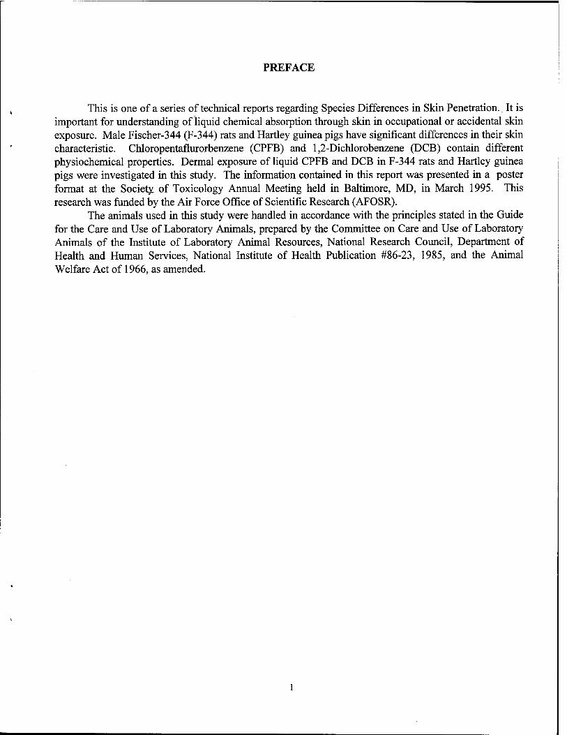
PREFACE
This is one of a series of technical reports regarding Species Differences in Skin Penetration. It is important for understanding of liquid chemical absorption through skin in occupational or accidental skin exposure. Male Fischer-344 (F-344) rats and Hartley guinea pigs have significant differences in their skin characteristic. Chloropentaflurorbenzene (CPFB) and 1,2-Dichlorobenzene (DCB) contain different physiochemical properties. Dermal exposure of liquid CPFB and DCB in F-344 rats and Hartley guinea pigs were investigated in this study. The information contained in this report was presented in a poster format at the Society of Toxicology Annual Meeting held in Baltimore, MD, in March 1995. This research was funded by the Air Force Office of Scientific Research (AFOSR).
The animals used in this study were handled in accordance with the principles stated in the Guide for the Care and Use of Laboratory Animals, prepared by the Committee on Care and Use of Laboratory Animals of the Institute of Laboratory Animal Resources, National Research Council, Department of Health and Human Services, National Institute of Health Publication #86-23, 1985, and the Animal Welfare Act of 1966, as amended.

TABLE OF CONTENTS
SECTION PAGE
PREFACE • 1
LIST OF FIGURES 3
LIST OF TABLES 4
ABBREVIATIONS 5
INTRODUCTION 6
MATERIALS AND METHODS 7
Experimental Animals 7
Chemicals '
Test Agent Quality Control • 7
Surgery Preparation °
Dermal Exposures °
Pathological Analysis • °
Analytical Methods For CPFB And DCB In The Blood 8
RESULTS • 9
DISCUSSION 15
CONCLUSIONS 16
REFERENCES 17
i

LIST OF FIGURES
FIGURE TITLE PAGE
1. Dermal Exposure Surgical Preparation For F-344 Rats 10
2. Average Concentration Of CPFB In The Venous Blood Of 12 F-344 Rats "In a Dermal Exposure Lasting 4 Hours 11
3. Average Concentration Of CPFB In The Venous Blood Of 8 Guinea Pigs In A Dermal Exposure Lasting 2 Hours 11
4. Average Concentration Of DCB In The Venous Blood Of 7 F-344 Rats In a Dermal Exposure Lasting 4 Hours 12
5. Average Concentration Of DCB In the Venous Blood Of 6 Guinea Pigs In Three Combined Dermal Exposure Experiments Lasting 4 Hours 12

LIST OF TABLES
TABLE TITLE PAGE
1. Physical Properties Of CPFB And DCB 7
2. Histological/Pathological Changes In The Skin 13

ABBREVIATIONS
B.W.
cm
cone
CPFB
DCB
ECD
g
GC
i.d.
Inflam.
i.p.
Kg
mg
min
ml
Necro.
PMN
S.D.
u
ug
Body weight
Centimeter
Concentration
Chloropentafluorobenzene
1,2-Dichlorobenzene
Electron capture detector
Gram
Gas Chromatograph
Inside diameter
Dermal inflammation
Intraperitoneally
kilogram
Milligram
Minutes
Milliliter
Epidermal necrosis
Polymorphonuclear cell margination
Standard deviation
Unit
Microgram

INTRODUCTION
Skin is only a partial barrier to chemicals that may contact the skin in occupational and environmental settings. These dermal exposures may result from contact with chemical vapors, liquid immersions or transient splashes. In order to define the human health risk associated with dermal exposure to a particular chemical, quantitative information about the chemical movement through and interaction with the skin is required.
The structure and composition of skin vary among species. Density and size of hair follicles, density of sebaceous, eccrine and apocrine glands, capillary density and distance fromtiie surface, as well as thickness of the various layers of the epidermis and dermis are skin features of interest. A significant failing of skin toxicology research is the marginal ability of the data from dermal absorption research in laboratory animals to be predictive of the human situation. The toxicology literature regarding skin absorption primarily describes chemical absorption after dermal application with only a limited understanding of the basic physiologic or anatomic principles involved. As a result, unless these laboratory experiments were designed specifically to mimic a human exposure scenario, it is very hard to apply the results of animal studies to human exposure situations. It has been widely accepted that anatomical and physiological properties of skin impact the movement of chemicals across the dermal barrier. These properties are especially important to quantitatively describe species differences in dermal absorption of chemicals. To study whether species differences in dermal absorption are due to these skin dissimilarities, an in vivo method has the advantage of intact skin of the whole animal which has blood flow, normal metabolism and immune responses. An in vivo method in rats was developed in this laboratory for dermal vapor exposures [1] and was compared with available human vapor exposures [2]. We applied the previous in vivo method used for vapors (i.e. pharmacokinetic approach) to dermal penetration of liquids in two different species.
Male F-344 rats and Hartley guinea pigs were chosen based on significant differences in their skin [3]. In this particular study of "Comparison of Anatomical Characteristics of the Skin for Several Laboratory Animals" (abbreviated as "Comparison" in the rest of this report), the skin anatomical difference in 7 species was investigated from this laboratory. The Hartley guinea pig had thicker stratum corneum as well as a thicker total epidermis, stratum granulosum. The F-344 rat present a greater average arteriole fractional area as well as vascular depth for capillaries, venules and arterioles. The thickness of the dermis was greater in the Hartley guinea pig than in the F-344 rat with approximately a two-fold difference.
Different chemicals with different characteristics penetrate the skin differently [4,5]. Two model chemicals chloropentafluorobenzene (CPFB) and 1,2-dichlorobenzene (DCB) were chosen to provide a range of water and lipid solubilities.
The previous study from this laboratory reveals that skin damage resulted from extended exposure of organic chemicals. This will have an important impact on dermal absorption kinetics.
Our study investigated dermal penetration of CPFB and DCB of 99.9% concentration to F-344 rat and Hartley guinea pig. The blood concentrations of CPFB and DCB in these two species resulting from the dermal exposure were quantified. The objective of this study was to compare the two species versus the two chemicals. Meanwhile the effect of continuous contact of these two chemicals on the skin of the two species was also investigated. This work is an initial step in an overall project to describe species differences in dermal penetration.

MATERIALS AND METHODS
Experimental Animals. Male Fischer 344 rats, (Charles River Breeding Laboratory, Raleigh, NC), 200-250g and Male
Hartley guinea pigs (Charles River Breeding Laboratory, Raleigh NC or Portage, MI), 300-375g were used in this studies. Prior to the study the animals were randomized and group housed (two to three per cage) in clear plastic cages with wood chip bedding and were kept in a portable laminar air flow enclosure with a light/dark cycle set at 12-h intervals. Water and feed (Purina Lab Chow) were available ad libitum?
Chemicals. The CPFB (99.9%) and DCB (99.9%) used in this study were purchased from Aldrich Chemical
Co. (Milwaukee, WI). The physical properties of CPFB and DCB are shown in Table 1.
Table 1. Physical Properties of CPFB and DCB
Chemical Molecular Weight
Volatility (mmHg)
25°C
Water Solubility
*Rat Fat/Air Partition
Coefficient
chloropentafluorobenzene 202 14.1 medium 766
1,2-dichlorobenzene 147 0.96 low 26,606
* The fat/air partition coefficients were determined from this laboratory.
n-Hexane was used as an extraction solvent for CPFB and was obtained from either Fisher Scientific or Baxter.
Isooctane was used as an extraction solvent for DCB and was obtained from Sigma-Aldrich.
Test Agent Quality Control. The purity of the chemicals was determined by capillary gas chromatography. A Hewlett-Packard
5890 gas Chromatograph equipped with an electron capture detector was used in conjunction with a Hewlett-Packard 3392A computing integrator (Hewlett-Packard Co., Palo Alto, CA) to measure peak area and record chromatograms. CPFB and DCB were diluted in hexane to provide peak areas within the detection limits of the instrument.
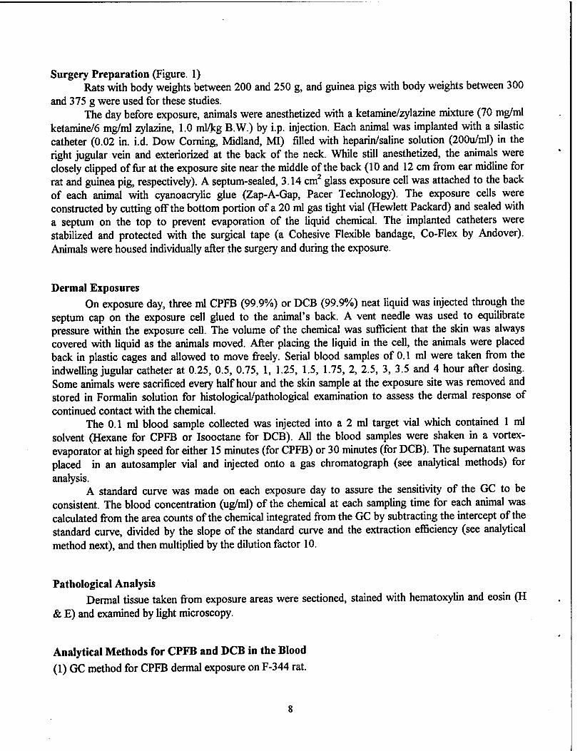
Surgery Preparation (Figure. 1) Rats with body weights between 200 and 250 g, and guinea pigs with body weights between 300
and 375 g were used for these studies. The day before exposure, animals were anesthetized with a ketamine/zylazine mixture (70 mg/ml
ketamine/6 mg/ml zylazine, 1.0 ml/kg B.W.) by i.p. injection. Each animal was implanted with a silastic catheter (0.02 in. i.d. Dow Corning, Midland, MI) filled with heparin/saline solution (200u/ml) in the right jugular vein and exteriorized at the back of the neck. While still anesthetized, the animals were closely clipped of fur at the exposure site near the middle of the back (10 and 12 cm from ear midline for rat and guinea pig, respectively). A septum-sealed, 3.14 cm2 glass exposure cell was attached to the back of each animal with cyanoacrylic glue (Zap-A-Gap, Pacer Technology). The exposure cells were constructed by cutting off the bottom portion of a 20 ml gas tight vial (Hewlett Packard) and sealed with a septum on the top to prevent evaporation of the liquid chemical. The implanted catheters were stabilized and protected with the surgical tape (a Cohesive Flexible bandage, Co-Flex by Andover). Animals were housed individually after the surgery and during the exposure.
Dermal Exposures On exposure day, three ml CPFB (99.9%) or DCB (99.9%) neat liquid was injected through the
septum cap on the exposure cell glued to the animal's back. A vent needle was used to equilibrate pressure within the exposure cell. The volume of the chemical was sufficient that the skin was always covered with liquid as the animals moved. After placing the liquid in the cell, the animals were placed back in plastic cages and allowed to move freely. Serial blood samples of 0.1 ml were taken from the indwelling jugular catheter at 0.25, 0.5, 0.75, 1, 1.25, 1.5, 1.75, 2, 2.5, 3, 3.5 and 4 hour after dosing. Some animals were sacrificed every half hour and the skin sample at the exposure site was removed and stored in Formalin solution for histological/pathological examination to assess the dermal response of continued contact with the chemical.
The 0.1 ml blood sample collected was injected into a 2 ml target vial which contained 1 ml solvent (Hexane for CPFB or Isooctane for DCB). All the blood samples were shaken in a vortex- evaporator at high speed for either 15 minutes (for CPFB) or 30 minutes (for DCB). The supernatant was placed in an autosampler vial and injected onto a gas Chromatograph (see analytical methods) for analysis.
A standard curve was made on each exposure day to assure the sensitivity of the GC to be consistent. The blood concentration (ug/ml) of the chemical at each sampling time for each animal was calculated from the area counts of the chemical integrated from the GC by subtracting the intercept of the standard curve, divided by the slope of the standard curve and the extraction efficiency (see analytical method next), and then multiplied by the dilution factor 10.
Pathological Analysis Dermal tissue taken from exposure areas were sectioned, stained with hematoxylin and eosin (H
& E) and examined by light microscopy.
Analytical Methods for CPFB and DCB in the Blood
(1) GC method for CPFB dermal exposure on F-344 rat.

Chemical analysis was accomplished using a Hewlett Packard 5880 gas Chromatograph equipped with an electron capture detector (ECD) and a HP 7673 A auto sampler. The chemical was separated with Hexane (Fisher) solvent on a ECONO CAP, phase SE-30 column (Cat # 19650, Alltech) using a splitless injection mode of sample deposition on the column. The carrier gas was an ultrapure 95% argon/5% methane mixture with a column flow of 3.34 ml/minute and a total flow of 30.6 ml/minute. The oven temperature was 105°C, the injector temperature was 125°C and the detector temperature was 300°C. The detection limit for CPFB was 0.01 ug/ml. The linear range of the standard curve was between 0.025 to 5 ug/ml. Retention time was 1.45 min. The extraction efficiency was 0.93.
(2) GC method for CPFB dermal exposure on Hartley guinea pig. Chemical analysis was accomplished using a Varian 3500 capillary gas Chromatograph equipped
with an ECD because of the GC used for CPFB in F-344 rats was unavailable at the time. The chemical was separated with Hexane solvent (Baxter) on a SE-30 column (Cat #19650, Alltech). The carrier gas was nitrogen with a column flow 5 ml/minute and a total flow of 23 ml/minute. The oven temperature was 40°C, the injector temperature was 175°C and the detector temperature was 300°C. The detection limit for CPFB was 0.01 ug/ml. The linear range of the standard curve was between 0.025 to 3 ug/ml. Retention time was 2.3 min. The extraction efficiency was 0.93.
(3) GC method for DCB dermal exposure on both guinea pig and rat. A Hewlett Packard 5880 gas Chromatograph equipped with an ECD and a HP 7673A auto
sampler was used. The isooctane (Sigma-Aldrich) was used as the separation solvent. DCB was separated with a ECONO CAP, phase CARBOWAX column (Cat # 19653, Alltech). The carrier gas was 95% argon/5% methane mixture with a column flow of 3.5 ml/minute and a total flow of 30.2 ml/minute. The oven temperature was 125°C, the injector temperature was 150°C and the detector temperature was 300°C. The detection limit for DCB was 0.025 ug/ml. The linear range of the standard curve was between 0.025 to 5 ug/ml. Retention time was 4.4 min. The extraction efficiency was 0.93 for F-344 rat and 0.97 for Hartley guinea pig respectively.
RESULTS
Mean blood concentrations for CPFB exposures in 12 rats slowly increased to a peak of 8.5 ug/ml at 3 hours post dosing (Figure 2). Mean blood concentrations for CPFB in 8 guinea pigs rose quickly to above 1 ug/ml at 0.5 hour and stayed constant up to 2 hours (Figure 3). Mean blood concentrations in 7 rats for DCB exposures increased rapidly during the first hour showing an average of just over 5 ug/ml at 1 hour (Figure 4) and slightly declined out to 4 hours. In a group of 6 guinea pigs, mean blood concentrations of DCB quickly increased to about 1.5 ug/ml at 45 minutes (Figure 5) and then decreased out to 4 hours.
Histopathological analysis (Table 2) showed that rat skin exposed to CPFB was comparable to controls until minor changes in polymorphonuclear cell margination occurred at 2.5 hours. Inflammation was not present until 3 hours. With guinea pigs, CPFB caused minimal inflammation at 1.5 hours. Contact with DCB caused minimal inflammation at 4 hours in rats, but caused minimal inflammation much earlier (1 hour) in guinea pigs.

■<3- o tt. __;
L_ u cH u c o 3
•4-» O
u a. cd v* n. V V u •a a. c ^_ cä es u cd
00 3 C
3 e en CO U u i— 1— « en O 3) V. 00
c E £
Q o
"to
• W4
V k 9 es
10

RAT/CPFB
1.5 2 2.5
Time (hours)
-mean mean+S.D. mean-S.D.
3.5
Figure 2. Average concentration of CPFB in the venous blood of 12 F-344 rats in a dermal exposure lasting 4 hours.
GUINEA PIG/CPFB
-m m-
< 1 H I I
0.4 0.6 0.8 1 1.2 1.4 1.6 1.8
Time (hours)
-mean mean+S.D. mean-S.D.
Figure 3. Average concentration of CPFB in the venous blood of 8 guinea pigs in a dermal exposure lasting 2 hours.
11

RAT/DCB
1.5 2 2.5
Time (hours)
mean+S.D.
mean-S.D.
Figure 4. Average concentration of DCB in the venous blood of 7 F-344 rats in a dermal exposure lasting 4 hours.
GUINEA PIG/DCB
0.5 1.5 2 2.5
Time (hours)
-mean
mean+S.D.
mean-S.D.
Figure 5. Average concentration of DCB in the venous blood of 6 guinea pigs in three combined dermal exposure experiments lasting 4 hours.
12

es w WÜ o o e
2 « (/>
es 4>
J3 u M e
u in
5 e es
JS • is U u ja es H
© 00
e
2*. o
W
«s
m
ER
"es u ES
v u m
s V N e
© &. o s C es
■*->
S a 2 e
+ + + N « Ifi
+ + IS IS
+. + © IS »H ^
+. + © IS *4 ^
© © ©
© © ©
* 3 fc
4
+ + + IS ■* VJ
+ + + Tt ^ "V
+ + + N N H
+. + © IS *H ö
Ml * 5 *
■fcu
o
"1
25 3 "> o w cn o in
w
m ju "es u ca u V «B
e s e v
2 ja S u Q ■ is
+ + IS ^
+ +
© © ©
MS * NS &
2^
+ + + N « «
+ + + IS <*> f>
+ + + N (O N
+ + IS IS
+. + © IS »H ö
© ©
5 I 2 Ml
SB w
13

«s
H
•a H 0 4J
09 01 U
4J 0
£
O u 8 CA
I U 2
"rt .«2
•II 0) c
en O u u c en
- c £ en O
u u e
"c3
<» .2 O D, U 4>
i
C en S 5 "3 «3
03
•a ii
u 03
£.S £ * en en t2 O !- Ö 2 o> '2 c c
11S Is a, g o> fcs
'•a 8 —- 03 eg C « s
§2
gl
II E "O II II
N m t "l O
03
o C/3
14

DISCUSSION
The shape of CPFB blood concentration curves in rats (Figure 2) and guinea pigs (Figure 3) were similar, i.e. they both rose and plateaued. Guinea pig blood concentrations plateaued much earlier than rats but the actual blood concentrations was lower by a factor of about 8. The shape of the DCB blood concentration curves was also similar for both species, i.e. they rose to a peak and then decreased over time. Peak DCB blood concentrations in the guinea pig were nearly 4 times lower than those of the rat. The skin surface area exposed in both species was the same but the guinea pigs were twice the size of the rats. Although the lower chemical concentration per body weight appears in the heavier animal, the anatomical and physiological differences between the two species may have contributed to it. The literature establishes that the outermost layer of skin, the stratum corneum is typically the major barrier to transdermal ingress [5]. As stated in "Comparison" study from this laboratory, the stratum corneum in Hartley guinea pig was significantly thicker than F-344 rat by a factor of 3. This seems to support the theory of stratum corneum as a major barrier in the skin, since lower blood concentration of CPFB and DCB in Hartley guinea pig in our study coincides with the thicker dermal barrier found in the guinea pig skin. In the following, a serial correlations between our results and "Comparison" studies are being discussed.
The hair follicle of the skin does not contain the stratum corneum layer, therefore the chemical absorption in this area is easier than the area where stratum corneum exists. The average number of follicle ostia was 21.9 for F-344 rat and 15.4 for Hartley guinea pig respectively in "Comparison", but it was not significantly different in statistics. An average vascular depth and fractional area for capillaries, venules and arterioles may also be important factors for skin penetration. The greater vascular fractional area, the more capillaries, venules and arterioles involved per dermis area, therefore the chemical penetration process may be facilitated in these regions other than those where less vasculatures exist. The fractional area for arteriole in F-344 rat was significantly greater than Hartley guinea pig. This appears in agreement with the higher blood concentration of CPFB and DCB achieved in rats. On the other hand, the deeper the vascular depth, the further away the vasculature is from the surface area of the skin, then the skin penetration is expected to be less favorable. The F-344 rat was found a greater average vascular depth than guinea pig; its impact on skin penetration was not clear. The blood concentration of the chemical depends on several factors besides the amount of chemical absorbed. Some of these factors are blood flow, hepatic metabolism, partition coefficient in different tissues, etc. Based on the complex physiology present in the mammalian system, a physiologically-based pharmacokinetic model might be a useful way to study species differences in skin penetration. This has been our focus in the past [6,7] and will be the next step in this project.
Table 2 shows that, guinea pig skin damage with both chemicals presented earlier than in the rat, indicating the guinea pig may be more sensitive to the skin damage than the rat. In both species tested, dermal exposure of DCB caused minimal PMNs marginating/migrating and minimal supperative dermatitis a half hour earlier than CPFB, suggesting DCB may cause inflammation and PMNs more rapidly than CPFB.
There was some interesting correlation between the onset of minimal skin damage and the time when the chemical peak value was reached in the venous blood. F-344 rats exposed to CPFB presented minimal supperative dermatitis at 3 hours post dosing when a 8.5 mg/L CPFB peak was reached at the same time in the blood. Hartley guinea pigs exposed to CPFB presented minimal PMNs marginating/migrating per post-capillary venule at 1 hour when the CPFB peak value was reached closely at 45 minutes (the skin samples were collected every half hour only). Dermal exposure with DCB in
15

Hartley guinea pig started dermal inflammation at 1 hour while the peak DCB blood concentration was present 15 minutes earlier. In contrast, the peak value of DCB in rat was present at 1 hour post dosing, but the minimal skin damage did not appear until 2 hours. It is worthwhile to mention that once the chemical peak was reached in the blood it started to either decline out or maintain about the same from 2 to 4 hours, indicating a steady state being reached. Currently an immunochemical methodology is being developed in this laboratory which will surely enable us to detect skin histopatholögical changes more precisely in the near future.
CONCLUSIONS
There were differences in skin penetration of CPFB and DCB between F-344 rat and Hartley guinea pig. This difference might be due to the different stratum corneum found in these two species. Prolonged contact of CPFB and DCB neat liquid with the skin causes histological/pathological changes in the skin of F-344 rat and Hartley guinea pig. Dermal damage due to prolonged contact of CPFB and DCB may impact the skin penetration of these chemicals.
16

REFERENCES
1. McDougal, J.N.; Jepson, G.W.; Clewell III, HJ.; MacNaughton, M.G.; and Andersen, M.E. "A Physiological Pharmacokinetic Model for Dermal Absorption of Vapors in the Rat." Toxicology and Applied Pharmacology. 85:286-294 (1986).
2. McDougal, J.N.; Jepson, G.W.; Clewell III, HJ.; Gargas, M.L..; and Andersen, M.E. "Dermal Absorption of Organic Chemical Vapors in Rats and Humans." Fundamental and Applied Toxicology. 14:299-308(1990).
3. Grabau, J.H.; Mattie, D.R.; Dong, L.; Jepson, G.W.; and McDougal, J.N. "Comparison of Anatomical Characteristics of the Skin for Several Laboratory Animals." Air Force Technical Report AL/OE-TR-1995-0066. Armstrong Laboratory, Wright-Patterson Air Force Base, OH.
4. Grandjean, P.; Berlin, A.; Gilbert, M.; and Penning, W. "Preventing Percutaneous Absorption of Industrial Chemicals: The "Skin" Denotation. American Journal of Industrial Medicine. 14:97- 107 (1988).
5. Surber, C; Wilhelm, K.; Maibach, HJ.; Hall, L.L.; and Guy, R.H. "Partitioning of Chemicals into Human Stratum Corneum: Implications for Risk Assessment Following Dermal Exposure." Fundamental and Applied Toxicology. 15:99-107 (1990).
6. Jepson, G.W.; Clewell III, H.J.; and Andersen, M.E. "A Rapid, Physiological Based Method for Evaluating Candidate Chemical Warfare Agent Uptake Simulants." Air Force Technical Report AAMRL-TR-8-045. Wright-Patterson Air Force Base, OH.
7. McDougal, J.N. "Physiologically-Based Pharmacokinetic Modeling in Dermatotoxicology." F.N. Marzulli and H.I. Maibach (Eds.), Hemisphere Publishing Corporation, Washington. 37-60 (1991).
17


![094-0057-20 [Unlocked by Www.freemypdf.com]](https://static.fdocuments.net/doc/165x107/577c83031a28abe054b32f1f/094-0057-20-unlocked-by-wwwfreemypdfcom.jpg)

![57 F1 742, Federal Reporter - Public.Resource.Org › pub › us › case › reporter › F › 0057 › 0057.f1.0742.pdf742:']. nOER.u. QPQRTElt, vol. 57.i.T})js ·.~~~~nt]dQesnot](https://static.fdocuments.net/doc/165x107/60dd651e2c0b615a0667b025/57-f1-742-federal-reporter-a-pub-a-us-a-case-a-reporter-a-f-a-0057.jpg)