Allograft Anterior Cruciate Ligament Reconstruction ... VYAS, MD, PhD1 • STEPHEN J. RABUCK, MD 1...
12
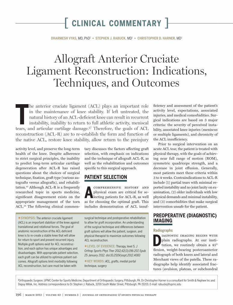
196 | march 2012 | volume 42 | number 3 | journal of orthopaedic & sports physical therapy [ CLINICAL COMMENTARY ] T he anterior cruciate ligament (ACL) plays an important role in the maintenance of knee stability. If left untreated, the natural history of an ACL-deficient knee can result in recurrent instability, inability to return to full athletic activity, meniscal tears, and articular cartilage damage. 27 Therefore, the goals of ACL reconstruction (ACL-R) are to re-establish the form and function of the native ACL, restore knee stability, allow return to the preinjury activity level, and preserve the long-term health of the knee. Despite adherence to strict surgical principles, the inability to predict long-term articular cartilage degeneration after ACL-R has raised questions about the choices of surgical technique, fixation, graft type (various au- tografts versus allografts), and rehabili- tation. 10 Although ACL-R is a frequently researched topic in sports medicine, significant disagreement exists on the appropriate management of the torn ACL. 28 The following clinical commen- T T SYNOPSIS: The anterior cruciate ligament (ACL) is an important stabilizer of the knee against translational and rotational forces. The goal of anatomic reconstruction of the ACL-deficient knee is to re-create a stable knee that will allow for return to sport and prevent recurrent injury. Multiple graft options exist for ACL reconstruc- tion, and each option has unique advantages and disadvantages. With appropriate patient selection, each graft can be utilized to optimize patient out- comes. Allograft options limit morbidity following ACL reconstruction, but care must be taken with surgical technique and postoperative rehabilitation to allow for graft incorporation. An understanding of the surgical technique and differences between graft options will allow the patient, surgeon, and physical therapist to maximize outcomes following ACL reconstruction. T T LEVEL OF EVIDENCE: Therapy, level 5. J Orthop Sports Phys Ther 2012;42(3):196-207, Epub 25 January 2012. doi:10.2519/jospt.2012.4083 T T KEY WORDS: ACL, grafts, medial portal technique, surgery 1 Orthopaedic Surgeon, UPMC Center for Sports Medicine, Department of Orthopaedic Surgery, Pittsburgh, PA. Dr Christopher Harner is a consultant for Smith & Nephew Inc and Depuy Mitek, Inc. Address correspondence to Dr Stephen J. Rabuck, 3200 South Water Street, Pittsburgh, PA 15203. E-mail: [email protected] DHARMESH VYAS, MD, PhD 1 • STEPHEN J. RABUCK, MD 1 • CHRISTOPHER D. HARNER, MD 1 Allograft Anterior Cruciate Ligament Reconstruction: Indications, Techniques, and Outcomes tary discusses the factors affecting graft selection, with emphasis on indications and the technique of allograft ACL-R, as well as the rehabilitation and outcomes specific to this surgical approach. PATIENT SELECTION A comprehensive history and physical exam are critical for se- lecting patients for ACL-R, as well as for choosing the optimal graft. This includes demonstration of ACL insuf- ficiency and assessment of the patient’s activity level, expectations, associated injuries, and medical comorbidities. Sur- gical indications are based on 3 major criteria: the severity of perceived insta- bility, associated knee injuries (meniscus or multiple ligaments), and chronicity of the ACL insufficiency. Prior to surgical intervention on an acute ACL tear, the patient is treated with physical therapy, with the goals of achiev- ing near full range of motion (ROM), symmetric quadriceps strength, and a decrease in joint effusion. Generally, most patients meet these criteria within 3 to 4 weeks. Contraindications to ACL-R include (1) partial tears with minimal re- ported instability and no joint laxity on ex- amination, (2) older individuals with low physical demands and minimal instability, and (3) comorbidities that make surgical intervention unsafe for the patient. PREOPERATIVE (DIAGNOSTIC) IMAGING Radiographs D iagnostic imaging begins with plain radiographs. At our insti- tution, we routinely obtain a 45° flexion, weight-bearing posteroanterior radiograph of both knees and lateral and Merchant views of the patella. These ra- diographs help identify associated frac- tures (avulsion, plateau, or subchondral
Transcript of Allograft Anterior Cruciate Ligament Reconstruction ... VYAS, MD, PhD1 • STEPHEN J. RABUCK, MD 1...
196 | march 2012 | volume 42 | number 3 | journal of orthopaedic & sports physical therapy
[ clinical commentary ]
The anterior cruciate ligament (ACL) plays an important role in the maintenance of knee stability. If left untreated, the natural history of an ACL-deficient knee can result in recurrent instability, inability to return to full athletic activity, meniscal
tears, and articular cartilage damage.27 Therefore, the goals of ACL reconstruction (ACL-R) are to re-establish the form and function of the native ACL, restore knee stability, allow return to the preinjury
activity level, and preserve the long-term health of the knee. Despite adherence to strict surgical principles, the inability to predict long-term articular cartilage degeneration after ACL-R has raised questions about the choices of surgical technique, fixation, graft type (various au-tografts versus allografts), and rehabili-tation.10 Although ACL-R is a frequently researched topic in sports medicine, significant disagreement exists on the appropriate management of the torn ACL.28 The following clinical commen-
TT SYNOPSIS: The anterior cruciate ligament (ACL) is an important stabilizer of the knee against translational and rotational forces. The goal of anatomic reconstruction of the ACL-deficient knee is to re-create a stable knee that will allow for return to sport and prevent recurrent injury. Multiple graft options exist for ACL reconstruc-tion, and each option has unique advantages and disadvantages. With appropriate patient selection, each graft can be utilized to optimize patient out-comes. Allograft options limit morbidity following ACL reconstruction, but care must be taken with
surgical technique and postoperative rehabilitation to allow for graft incorporation. An understanding of the surgical technique and differences between graft options will allow the patient, surgeon, and physical therapist to maximize outcomes following ACL reconstruction.
TT LEVEL OF EVIDENCE: Therapy, level 5. J Orthop Sports Phys Ther 2012;42(3):196-207, Epub 25 January 2012. doi:10.2519/jospt.2012.4083
TT KEY WORDS: ACL, grafts, medial portal technique, surgery
1Orthopaedic Surgeon, UPMC Center for Sports Medicine, Department of Orthopaedic Surgery, Pittsburgh, PA. Dr Christopher Harner is a consultant for Smith & Nephew Inc and Depuy Mitek, Inc. Address correspondence to Dr Stephen J. Rabuck, 3200 South Water Street, Pittsburgh, PA 15203. E-mail: [email protected]
DHARMESH VYAS, MD, PhD1 • STEPHEN J. RABUCK, MD1 • CHRISTOPHER D. HARNER, MD1
Allograft Anterior Cruciate Ligament Reconstruction: Indications,
Techniques, and Outcomes
tary discusses the factors affecting graft selection, with emphasis on indications and the technique of allograft ACL-R, as well as the rehabilitation and outcomes specific to this surgical approach.
PATIENT SELECTION
A comprehensive history and physical exam are critical for se-lecting patients for ACL-R, as well
as for choosing the optimal graft. This includes demonstration of ACL insuf-
ficiency and assessment of the patient’s activity level, expectations, associated injuries, and medical comorbidities. Sur-gical indications are based on 3 major criteria: the severity of perceived insta-bility, associated knee injuries (meniscus or multiple ligaments), and chronicity of the ACL insufficiency.
Prior to surgical intervention on an acute ACL tear, the patient is treated with physical therapy, with the goals of achiev-ing near full range of motion (ROM), symmetric quadriceps strength, and a decrease in joint effusion. Generally, most patients meet these criteria within 3 to 4 weeks. Contraindications to ACL-R include (1) partial tears with minimal re-ported instability and no joint laxity on ex-amination, (2) older individuals with low physical demands and minimal instability, and (3) comorbidities that make surgical intervention unsafe for the patient.
PREOPERATIVE (DIAGNOSTIC) IMAGING
Radiographs
Diagnostic imaging begins with plain radiographs. At our insti-tution, we routinely obtain a 45°
flexion, weight-bearing posteroanterior radiograph of both knees and lateral and Merchant views of the patella. These ra-diographs help identify associated frac-tures (avulsion, plateau, or subchondral
42-03 Vyas.indd 196 2/22/2012 6:16:23 PM

journal of orthopaedic & sports physical therapy | volume 42 | number 3 | march 2012 | 197
impaction), gauge the amount of joint space narrowing in the 3 compartments, and assess patellar height (lateral view), tilt, and subluxation (Merchant view). A long-cassette anteroposterior view of the bilateral lower extremity is obtained to determine overall limb alignment. Im-portantly, radiographs are a prerequisite to assess the status of the growth plate in pediatric patients.
Magnetic Resonance ImagingIf a patient’s history and physical exam suggest an ACL tear, then noncontrast magnetic resonance imaging of the knee is obtained (FIGURE 1). Discontinuity of the ACL in the coronal and sagittal planes is a reliable indication of an ACL tear. In addition, magnetic resonance imaging helps to identify associated injuries such as meniscal tears, chondral damage (in-cluding bone bruises), and concomitant ligament injuries (posterior cruciate ligament, medial and lateral collateral ligaments, and posterolateral corner). Allograft reconstructions may be ben-efi cial for multiligament injuries, as they decrease operative time and minimize the associated morbidity of graft harvesting.
PREOPERATIVE REHABILITATION
Rupture of the ACL results in signifi cant hemarthrosis, which may a� ect outcomes following ACL-R.
Large e� usions can result in quadriceps inhibition. In patients with an ACL-defi -cient knee, the role of the quadriceps as a dynamic stabilizer of the knee should not be underestimated. The return of quadriceps function and a reduction in e� usion are among the primary goals of preoperative rehabilitation. Inadequate quadriceps strength has been shown to produce altered gait patterns following ACL-R26 and an increase in the transfer of forces across the reconstructed ACL.
One of the early pitfalls of arthroscop-ically assisted ACL-R was the develop-ment of postoperative sti� ness. This complication was largely attributed to poor preoperative ROM and early sur-gical intervention during the infl amma-tory phase of healing.18,44,45 More recent studies have shown early surgical inter-vention to be safe,3,9,20 with the best in-dicator of postoperative ROM loss being the patient’s preoperative ROM.30 Pa-tients should be carefully evaluated for preoperative ROM defi cits and aggres-sively treated to prevent postoperative complications. The patient is ready for surgery once the infl ammatory period
has resolved. This period may last 2 to 3 weeks and corresponds to a decrease in e� usion and a resultant increase in ROM. Flexion must be adequate enough to al-low for knee hyperfl exion during ACL re-construction. Depending on concomitant pathology, surgery may be performed early (eg, displaced bucket-handle menis-cus tears) or may be delayed (eg, medial collateral ligament disruption).
GRAFT SELECTION
Principles of Graft Selection
Successful ACL-R depends on several factors, including stable fi x-ation, biological graft-bone integra-
tion, adequate graft strength, and, most importantly, anatomic positioning. Ac-cordingly, graft selection is a very impor-tant part of preoperative planning and depends on several factors: the patient’s preference, age, activity level, and physi-cal requirements; expected outcomes; the time line for return to play; associated ligamentous injuries; medical comorbidi-ties; previous surgery; tissue availability; and surgeon preference and experience.
TABLE 1Comparison of the Advantages
and Disadvantages of Graft Options
Graft Type Advantages Disadvantages
Allograft, bone-tendon • Bone-to-bone healing
• Decreased surgical time
• Predictable graft size
• Availability
• Cost
• Infectious disease transmission
• Risk of bridging physis
• Higher failure rate
• Delayed incorporation
Allograft, soft tissue • Decreased surgical time
• Predictable graft size
• Availability
• Less risk with bridging physis
• Cost
• Infectious disease transmission
• Higher failure rate
• Delayed incorporation
Autograft, bone-tendon • Faster incorporation and healing
• Better outcomes in young, active patients
• Prolonged surgical time
• Donor site morbidity (anterior knee pain)
• Risk of bridging physis
• Risk of fracture
Autograft, soft tissue • Faster incorporation and healing
• Better outcomes in young, active patients
• Less risk with bridging physis
• Prolonged surgical time
• Donor site morbidity (knee fl exion
weakness)
• Unpredictable graft size
• Compromise of medial structures
FIGURE 1. Sagittal magnetic resonance imaging demonstrating anterior cruciate ligament rupture.
42-03 Vyas.indd 197 2/22/2012 6:16:24 PM

198 | march 2012 | volume 42 | number 3 | journal of orthopaedic & sports physical therapy
[ clinical commentary ]Whereas significant debate still exists regarding the best graft choice, what is widely accepted is that the ideal graft should reflect the anatomy and biome-chanics of the native ACL, minimize har-vest site morbidity, be amenable to stable fixation, and result in rapid remodeling and incorporation into the reconstructed knee. As such, graft selection is a trade-off between the benefits and potential morbidity of each graft (TABLE 1).
Graft TypesGraft sources for ACL-R fall into 3 major categories: synthetic/artificial ligaments, autografts, and allografts. Synthetic grafts such as scaffolds (carbon fiber) and stents (Kennedy ligament augmentation device), and prostheses such as polytetra-fluoroethylene Gore-Tex (W.L. Gore & As-sociates, Inc, Elkton, MD), polyethylene terephthalate (Leeds-Keio artificial liga-ment), and Dacron (Invista, Wichita, KS), have demonstrated poor clinical results secondary to loosening, fatigue failure, and a strong host immune response.29,43,58 Commonly used autografts include the central third of the patellar tendon, hamstring tendons (semitendinosus and gracilis), and the quadriceps tendon. Several allograft options are available as well. These can be divided into grafts providing bone-to-bone healing and grafts consisting solely of soft tissue. The patellar-tendon allograft is the only op-tion for proximal and distal osseous inte-gration. Achilles tendon and quadriceps tendon allografts contain a single osseous attachment. Soft tissue allografts include the hamstring, tibialis anterior, tibialis posterior, and peroneus longus tendons, as well as the tensor fascia lata.Allograft Advantages/Disadvantages All graft types offer distinct advantages and disadvantages to the patient and the surgeon. The benefits of allograft use in-clude absence of donor site morbidity, shortened operating time, availability for complex cases (multiligament knee and revision ACL-R), greater availability and more predictable graft sizes, and compa-rable strength and stiffness to autograft
tissue at the time of reconstruction.4,35,38,52 Significant disadvantages of allograft tis-sue are potentially higher failure rates, increased time to incorporation, vari-ability in mechanical strength due to sec-ondary sterilization techniques, risk of disease transmission, immunogenic reac-tion, lack of long-term outcome data (es-pecially for young patients under the age of 25), and higher cost.31,46,58 The senior author’s (C.D.H.) preferred technique is anatomic single-bundle ACL-R using a bone-patellar tendon-bone autograft.
INDICATIONS FOR ALLOGRAFT ACL RECONSTRUCTION
In the senior author’s (C.D.H.) prac-tice, the graft choice for each ACL-R is tailored to suit the individual pa-
tient. We routinely utilize multiple graft choices, with approximately 80% of them being autografts (70% bone-patellar ten-don-bone and 30% hamstring) and 20% allografts (100% bone-patellar tendon-bone). Generally, allograft tissue is re-served for patients older than 40 years. In this population, the autograft benefits of more rapid incorporation and healing do not appear to warrant the increased morbidity from harvesting a graft from the patient. Allograft tissue is only used in special circumstances for patients who are 12 to 30 years old, and autografts are also strongly recommended for patients who are 30 to 40 years old. The ideal can-didate for an allograft at our center is a
mildly to moderately active patient older than 40 years who experiences symptom-atic instability during activities of daily living and whose clinical presentation is consistent with ACL rupture. Other in-dications for use of allograft tissue are reconstruction of multiligament knee in-juries, revision ACL-R, cases where au-tograft tissue is inadequate, and patient preference (TABLE 2).1,41,42
TECHNIQUE FOR MEDIAL PORTAL ACL-R
We provide a brief description of our technique for anatomic ACL-R using the medial portal
for femoral tunnel drilling. We aim for anatomic placement of the femoral and tibial tunnels. Femoral tunnel placement is done via the medial portal, allow-ing placement independent of the tibial tunnel (the transtibial technique). This technique may be utilized in all cases of primary (single-bundle, double-bundle, or augmentation) or revision ACL-R, and is not dependent on the choice of graft, instrumentation, or final fixation.
Portals and IncisionsDuring the procedure, we utilize 3 portals (FIGURE 2): anterolateral (viewing), an-teromedial (working), and superolateral (outflow). The medial portal is made un-der direct arthroscopic visualization us-ing a spinal needle. This is done not only to avoid damaging the medial meniscus
TABLE 2A Summary of the Relative
Indications for Allograft Anterior Cruciate Ligament Reconstruction
Abbreviation: ACL-R, anterior cruciate ligament reconstruction.*It is the author’s preference to use an autograft in revision cases if that graft has not been harvested in previous surgery and if the patient meets criteria for autograft use (ie, age, activity level).
Most Common Indications for Allograft ACL-R
• Patients older than 40 y
• Multiple ligament knee injuries
• Prior harvest from donor sites
• Patient preference
• Revision ACL-R*
42-03 Vyas.indd 198 2/22/2012 6:16:25 PM

journal of orthopaedic & sports physical therapy | volume 42 | number 3 | march 2012 | 199
but also to allow adequate clearance from the medial femoral condyle. When using allograft tissue, a 3-cm vertical incision is made on the anteromedial aspect of the tibia for drilling the tibial tunnel later in the procedure. The location of this incision is estimated by provisional placement of the tibial tunnel ACL guide midway between the anterior and poste-rior borders of the tibia.
Allograft PreparationBone-Patellar Tendon-Bone Our prefer-ence is for the bone-patellar tendon-bone allograft (FIGURE 3). The central 10 mm of the patellar tendon is utilized, with bone plugs measuring 20 mm in length both proximally and distally. The plugs
are designed to be trapezoidal in shape, and the leading plug is tapered to facili-tate graft passage. Two 1.5-mm holes are drilled in the tibial bone plug and a num-ber 5 Ethibond (braided, nonabsorbable; Ethicon, Inc, Somerville, NJ) is threaded through the holes. These will be used to secure fi nal plug fi xation over a post. The femoral bone plug is secured using the EndoButton CL (Smith & Nephew Inc, Memphis, TN) device to provide suspen-sory fi xation (FIGURE 4).Soft Tissue In certain cases, a soft tissue allograft is used. This graft is generally doubled over to increase its diameter. It is important to pay careful attention to the necessary diameter of the graft because di-mensions may not be consistent through-out the entire length of the graft. The goal is to prepare an appropriately sized graft
with consistent dimensions and adequate length for ACL-R. We use an EndoButton CL device, over which the graft is doubled. A suture is tied within the proximal por-tion of the graft, and a second nonabsorb-able suture is secured within the distal portion of the graft to be tied around a post for tibial fi xation (FIGURE 4).
Arthroscopic ACL-RThe fat pad is left intact to prevent post-operative scarring, patellar entrapment, and pain. Assessment of any associated intra-articular pathology is performed before preparation of the femoral and tibial insertion sites. On the femoral side, using the location of the torn ACL remnant as a guide (FIGURE 5), we mark the center of the anatomic ACL insertion site with a 30° Steadman awl (FIGURES 6A
and 6B). On the tibial side, a signifi cant portion of the ACL stump is preserved to enhance proprioceptive and vascular properties.25 We do not routinely perform a notchplasty unless it is needed for bet-ter visualization (1-2 mm) or to alleviate graft impingement.Femoral Tunnel Placement The native ACL footprint, although variable in each individual, is generally 4 to 6 mm ante-rior to the posterior femoral cortex with the knee at 90° of fl exion. Appropriate tunnel position is further confi rmed via intraoperative fl uoroscopy by taking a lateral image of the knee (90° of fl exion and overlapping condyles) with the awl still in position (FIGURE 6C). With the knee hyperfl exed, a guide pin is placed in the anatomic footprint (FIGURE 7A) and an acorn reamer is carefully advanced over the guide wire to avoid damaging the cartilage of the medial femoral condyle (FIGURE 7B). Finally, a 3.2-mm EndoBut-ton drill is used to breach the lateral femoral cortex.Tibial Tunnel Placement Anatomic tibial tunnel position is also accomplished us-ing a combination of visual arthroscopic landmarks (FIGURE 8) and fl uoroscopic im-aging (FIGURE 9). An ACL elbow-tip guide is set at 50° to 55° and placed at the inter-section between the posterior edge of the
FIGURE 2. Incisions for allograft anterior cruciate ligament reconstruction. Anterolateral viewing portal, anteromedial working portal, and superolateral outfl ow portal. A medial tibial incision is used for tibial tunnel placement and graft passage.
FIGURE 3. Bone-patellar tendon-bone allograft prior to (A) and following (B) graft preparation.
FIGURE 4. Posterior tibialis allograft prior to (A) and following (B) graft preparation.
FIGURE 5. Arthroscopic image of anterior cruciate ligament rupture.
42-03 Vyas.indd 199 2/22/2012 6:16:27 PM

200 | march 2012 | volume 42 | number 3 | journal of orthopaedic & sports physical therapy
[ CLINICAL COMMENTARY ]
anterior horn of the lateral meniscus and the midline of the tibial spines. Verifi ca-tion of the Kirschner wire placement is done with arthroscopy and fl uoroscopy. After correct placement of the guide pin, a cannulated compaction reamer is used over the Kirschner wire.Graft Passage Using passing sutures, the graft is advanced up the tibial tunnel and the tendinous portion of the bone-patel-lar tendon-bone allograft is maintained in
the posterior aspect of both tunnels (FIG-
URE 10). After clearing the lateral femoral cortex with the EndoButton, the device is toggled to engage the cortex and prevent passage back into the tunnel. Tension is applied to the tibial sutures, and the knee is cycled to minimize graft creep. Graft isometry and impingement are checked.
Graft FixationMultiple options exist for graft fi xation. These options include tying over a post, suspensory fi xation, and interference screws, and are dependent to some extent on the type of graft chosen (ie, bone block or soft tissue alone). Our choice for tibial fi xation is tying over a post (4.5-mm AO fully threaded cortical screw over a washer, bicortical purchase). After the far cortex is engaged but before fi nal seating, the tibial sutures are individually tied around the post. The screw-and-washer construct is then fully tightened and the Lachman and pivot shift tests are performed for fi -
nal verifi cation of graft tension. We prefer suspensory and suture/post fi xation with bone plugs to allow for maximal healing between the graft and host bone (FIGURE 11).
There are many benefi ts to perform-ing fi xation in this manner. The tunnels in the above technique are dilated to ex-actly fi t the graft, providing circumfer-ential interaction between the graft and apposed bone within the tunnel. There is no fi xation device within the tunnel to interfere with graft healing or the interaction of the graft with potentially benefi cial growth factors. Lastly, should revision surgery be required, the retained hardware will not limit surgical options.
POSTOPERATIVE REHABILITATION AND RETURN TO PLAY
ROM and Strengthening
Rehabilitation following ACL-R should follow a logical progression that allows for progressive strength-
FIGURE 6. The anatomic femoral insertion is identifi ed during arthroscopy (A and B). The planned tunnel placement is confi rmed with fl uoroscopy (C).
FIGURE 7. Femoral tunnel placement. With the knee hyperfl exed, a guide pin is placed in the anatomic footprint of the anterior cruciate ligament on the medial wall of the lateral femoral condyle (A). Arthroscopic view of guide wire placement in anatomic footprint (B).
FIGURE 8. The tibial guide is placed in the anatomic footprint for passage of the tibial guide pin.
42-03 Vyas.indd 200 2/22/2012 6:16:29 PM

journal of orthopaedic & sports physical therapy | volume 42 | number 3 | march 2012 | 201
ening and protection of the reconstructed ACL. Following ACL-R, the overall goals of rehabilitation are to minimize in-fl ammation, to restore knee motion and quadriceps strength, to enhance pro-prioception, neuromuscular control, and dynamic joint stability, and to ensure a return to sport-specifi c activities.
During the initial postoperative pe-riod, guarded ROM exercises help to initiate the processes of healing and strengthening. For the fi rst week after surgery, the patients are asked to bear weight with crutches and a knee brace locked in full extension. Basic home ex-ercises include quadriceps sets, straight leg raises, calf pumps, and heel slides. The goal during this phase is to protect the graft while regaining quadriceps strength and full passive and active knee extension symmetrical to the uninvolved side. At the fi rst postoperative visit (1
week), we initiate heel-to-toe gait train-ing with the brace unlocked and enroll the patient in formal physical therapy. Important milestones for ROM are full passive extension within 1 week and full active extension within 2 weeks. Goals for knee fl exion are 90° by 2 weeks and full symmetrical fl exion by 8 weeks after reconstruction. The patient is allowed to wean himself or herself from crutches after 6 weeks.
As rehabilitation progresses, sport-specifi c activities are initiated. There should be a logical, supervised progres-sion from protected, simple exercises to complex, sport-specifi c drills aimed at regaining neuromuscular control and returning the patient to full participa-tion. Some protocols follow a time line for transition through specifi c phases, assuming that graft incorporation will occur over time and that protecting the
FIGURE 9. The tibial guide pin placement is verifi ed via fl uoroscopy on anteroposterior (A) and lateral (B) views.
FIGURE 10. Arthroscopic view of graft passage before (A) and after (B) the bone plug has been advanced into the femoral tunnel. Fluoroscopic imaging to confi rm EndoButton placement (C).
FIGURE 11. Postoperative radiographs following allograft anterior cruciate ligament reconstruction.
42-03 Vyas.indd 201 2/22/2012 6:16:31 PM

202 | march 2012 | volume 42 | number 3 | journal of orthopaedic & sports physical therapy
[ clinical commentary ]graft will allow for incorporation and pro-gression of activities once adequate time has passed. Other protocols follow objec-tive measurements of muscle strength in the involved and uninvolved sides. Once the patient has regained adequate strength and proprioception to minimize the forces transmitted across the graft and adequate healing/maturation has taken place, the patient is allowed to re-turn to sport.
A combination of the previous ap-proaches is used to best rehabilitate pa-tients; however, good measures of patient rehabilitation and adequate assessment prior to return to sport are lacking in the literature.6 Suggested objective mea-sures to determine a patient’s readiness to return to sport include muscle strength testing, thigh circumference, ROM, laxity testing, validated questionnaires, and hop testing.6,57 Specifically, the importance of testing the involved extremity in isola-tion (eg, unilateral hop tests) is increas-ing because unilateral deficits may not be apparent during bipedal tasks.11,36 Devel-oping and adhering to these protocols is even more important for individuals with an allograft. Compared to autografts, al-lografts are at greater risk of rupture, and they undergo delayed incorporation and healing. Additionally, with allografts, pa-tients experience less morbidity associ-ated with graft harvesting, particularly in the early phases of rehabilitation, which further increases the risks of being overly aggressive during rehabilitation. Progres-sion through the rehabilitation process is individualized, based on an evaluation of objective measures during each phase of rehabilitation. Guidelines can be helpful to allow for adequate graft incorporation and healing (APPENDIX).
Return to PlayThe expected return to full activity is typically 9 to 12 months after surgery (APPENDIX). However, accelerated rehabil-itation programs that allow early return to sport have been described.14 These programs have drawn the attention of athletes and coaches who have new ex-
pectations and pressures for surgeons to meet those expectations. These protocols should be utilized with caution. Biologic healing and incorporation of ACL grafts require time.12 Rehabilitation protocols have not been shown to change the time to graft healing and maturation. Mount-ing data have shown that the reconstruct-ed ACL is at greatest risk of failure during the initial 9 months following reconstruc-tion, especially in patients undergoing al-lograft reconstruction.56
More systematic protocols examining objective measures for return to sport are being developed. The specific measures proposed include the visual analog scale for pain, thigh circumference, ROM, the International Knee Documentation Committee Subjective Knee Form, hop tests, and isokinetic testing.57 Other po-tential measures include an assessment comparing the involved and contralateral sides, fatigue resistance, and measures of neuromuscular control. By develop-ing these measures, we may be able to better assess the protective capabilities of dynamic knee stabilizers prior to re-turn to play. These tests are even more important for patients with an allograft-reconstructed knee, where healing and cellular repopulation are delayed.12 As stated earlier, the risks associated with an accelerated program in this popula-tion are compounded by the fact that these patients clinically appear ready to progress more rapidly than patients who have the morbidity associated with auto-graft harvesting.
Bracing and ACL-RPerceived benefits of bracing following ACL-R are decreased swelling, improved ROM, protection of the graft, improved proprioception, and improved walking kinematics to regain a normal gait pat-tern.8 Knee immobilizers, functional braces, and hyperextension bracing have all been described as options in the im-mediate postoperative period.34 The ben-efit of bracing in the early postoperative period has been demonstrated primarily for swelling and ROM, because bracing
does appear to help achieve full knee extension.33,34
The use of functional bracing follow-ing ACL-R has not had a clear role in the late phases of rehabilitation. Routine bracing to achieve satisfactory objective outcome measures of stability, strength, single-leg hop performance, and ROM has been under scrutiny, as the literature has not demonstrated that braces are ef-fective at achieving those goals.32,59 How-ever, functional bracing appears to help prevent reinjury in skiers.53
Effect of Allograft Tissue on RehabilitationThe use of allograft tissue allows for reha-bilitation without the postoperative mor-bidity of autograft harvesting. Allografts are associated with less anterior knee pain and hamstring weakness than pa-tellar tendon and hamstring autografts, respectively.23,54 As a result, patients with allograft ACL-R may be better prepared for early physical therapy, which may al-low for easier progression of and adher-ence to exercises than for those who have undergone autograft ACL-R. Even so, al-lograft ACL-R should follow a delayed re-habilitation protocol to allow for allograft incorporation and healing, as discussed earlier.12
It has been demonstrated that the strength of nonirradiated allograft tissue is comparable to that of autograft tissue. However, allograft remodeling (ligamen-tization) and incorporation are slower and presumably more susceptible to early failure.7,17 This hypothesis was tested via a model that compared patellar-tendon autografts to allografts in goats that un-derwent ACL-R. The authors showed that after 6 months, the autograft-recon-structed knees had less anterior/poste-rior displacement, twice the force to ACL failure, a greater cross-sectional area, and a greater number of small-diameter col-lagen fibrils.21 In our practice, we delay the return-to-play time line for patients who undergo allograft ACL-R (6 to 9 months for autograft ACL-R and 9 to 12 months for allograft reconstruction). In this study, compared to autograft tissue,
42-03 Vyas.indd 202 2/22/2012 6:16:32 PM

journal of orthopaedic & sports physical therapy | volume 42 | number 3 | march 2012 | 203
allograft tissue demonstrated more simi-lar cellular repopulation and reorganiza-tion of collagen fibrils to the native ACL, albeit at a delayed rate.21 Greater laxity at 6 months in the allograft group, as mea-sured by anterior translation, was noted as well, leading the authors to suggest prolonged protection in patients under-going allograft ACL-R.21
GRAFT PROCESSING
To minimize the risk of disease transmission from the allograft, careful processing of the tissue is
paramount. Contamination can occur from pathogens originating within the donor’s blood/organs or during tissue processing/packaging. This process be-gins with a careful screening of all donors for risk factors of communicable disease. To minimize the risk of harvesting a con-taminated graft, standards for the timing and methods of procurement have been set by the American Association of Tis-sue Banks.
Terminal sterilization is the last step before the graft can be stored and even-tually utilized for ACL-R. Historically, ethylene oxide and gamma irradiation have been effective in terminal steriliza-tion; however, ethylene oxide has been shown to result in chronic synovitis and has largely been abandoned in favor of gamma irradiation.22 Relatively low doses (1.5-2.0 mrad) can effectively kill bacte-ria, fungi, and spores. To deactivate HIV, doses of 2.5 mrad are required.16,49 A bio-mechanical trade-off exists at doses above 2.0 mrad: studies have demonstrated a reduction in the biomechanical proper-ties of the graft.13,15 Newer methods of sterilization have been developed and patented by various companies. These methods include patented washes that use a combination of detergents, antibi-otics, alcohol, and peroxide to safely dis-infect the tissue and minimize the risk of disease transmission while limiting the detrimental effect on the biomechanical properties of the graft. As these newer methods are refined, future studies will
be needed to define their effects on the biomechanical properties of the graft.
OUTCOMES
Allografts
Allograft options include grafts with bone-to-bone healing (patellar tendon, quadriceps ten-
don, Achilles tendon) and those with soft tissue alone (hamstring, anterior tibialis, posterior tibialis, and peroneus longus tendons). In the long term, al-lograft tissue has been shown to undergo ligamentization and resemble the native ACL both grossly and microscopically.21,37 Clinically, the reported failure rates of al-lograft tissue vary. When looking at tibi-alis anterior allografts, the literature is inconsistent, with good outcomes and low failure rates (5.5%) reported in some studies50 and early graft slippage and failure rates as high as 23% in young patients in other studies.47,48 Interest-ingly, the age-dependent variation in outcomes has not been established in bone-patellar tendon-bone allografts. Barber et al5 showed no difference in outcomes between patients older than 40 years and patients younger than 40 years when bone-patellar tendon-bone allografts were used. One theory is that the variation in healing between younger and older patients may be negated by the process of bone-to-bone healing in bone-patellar tendon-bone allografts.Comparison to Autografts Historically, outcome research has reported compa-rable results between allograft and au-tograft ACL-R. Initial outcome studies found similar stability between groups, with less morbidity for the allograft group.2,19 Some outcomes, such as post-operative ROM, appeared to favor al-lograft reconstruction.19 These studies were generally smaller cohorts, and the results were not always reproducible.
Recently, several meta-analyses have reviewed the recent literature and compared the outcomes of allograft re-construction to those of autograft recon-struction, with mixed results. Carey et
al10 reviewed laxity data in addition to subjective outcomes after a minimum follow-up of 2 years. This study evalu-ated 191 autograft and 266 allograft reconstructions and found no signifi-cant difference between groups. In this review, patients treated with bone plug and soft tissue reconstructions were included in both groups. Rerupture of the reconstructed ACL was not specifi-cally examined but was included with all other reasons for failure of the re-construction, with the data favoring the use of autografts. A significant rerupture rate was identified in the allograft group in a meta-analysis by Krych et al24 that compared bone-patellar tendon-bone allografts to autografts. In that study, 256 autograft reconstructions and 278 allograft reconstructions were evaluated after a minimum follow-up of 2 years. Return to sport was allowed between 6 and 12 months after reconstruction. Results on the Lachman and pivot shift tests and rates of return to the preinjury level of activity were similar between groups.
In one of the largest meta-analyses, Prodromos et al40 compared the stability of allograft and autograft reconstructions after a minimum of 2 years of follow-up. The authors found that the use of auto-grafts resulted in significantly better knee stability compared to allografts. They then compared soft tissue allografts to soft tissue autografts and bone-tendon allografts to bone-patellar tendon-bone autografts. The finding of improved sta-bility with autograft reconstruction was still present when soft tissue grafts were used. A similar but less pronounced trend was present in the bone-tendon recon-structions. In summary, short-term data have demonstrated subtle differences in stability and graft rerupture rates be-tween allograft and autograft ACL-R.
Longer follow-up has helped to dem-onstrate differences in graft choices between populations. Spindler et al51 re-ported on a cohort of 446 patients, 84% of whom were still being followed after 6 years. Their findings suggest greater im-
42-03 Vyas.indd 203 2/22/2012 6:16:33 PM

204 | march 2012 | volume 42 | number 3 | journal of orthopaedic & sports physical therapy
[ clinical commentary ]
REFERENCES
1. Allen CR, Giffin JR, Harner CD. Revision anterior cruciate ligament reconstruction. Orthop Clin North Am. 2003;34:79-98.
2. Bach BR, Jr., Aadalen KJ, Dennis MG, et al. Primary anterior cruciate ligament reconstruc-tion using fresh-frozen, nonirradiated patellar tendon allograft: minimum 2-year follow-up. Am J Sports Med. 2005;33:284-292.
3. Bach BR, Jr., Jones GT, Sweet FA, Hager CA. Arthroscopy-assisted anterior cruciate ligament reconstruction using patellar tendon substitu-tion. Two- to four-year follow-up results. Am J Sports Med. 1994;22:758-767.
4. Baer GS, Harner CD. Clinical outcomes of al-lograft versus autograft in anterior cruciate ligament reconstruction. Clin Sports Med. 2007;26:661-681. http://dx.doi.org/10.1016/j.csm.2007.06.010
5. Barber FA, Aziz-Jacobo J, Oro FB. Anterior cruciate ligament reconstruction using patellar tendon allograft: an age-dependent outcome evaluation. Arthroscopy. 2010;26:488-493. http://dx.doi.org/10.1016/j.arthro.2009.08.022
6. Barber-Westin SD, Noyes FR. Factors used to de-termine return to unrestricted sports activities after anterior cruciate ligament reconstruction. Arthroscopy. 2011;27:1697-1705. http://dx.doi.org/10.1016/j.arthro.2011.09.009
7. Barbour SA, King W. The safe and effective use of allograft tissue--an update. Am J Sports Med. 2003;31:791-797.
8. Beynnon BD, Johnson RJ, Fleming BC, et al. The effect of functional knee bracing on the anterior cruciate ligament in the weightbearing and nonweightbearing knee. Am J Sports Med. 1997;25:353-359.
9. Bottoni CR, Liddell TR, Trainor TJ, Freccero DM, Lindell KK. Postoperative range of mo-tion following anterior cruciate ligament reconstruction using autograft hamstrings:
a prospective, randomized clinical trial of early versus delayed reconstructions. Am J Sports Med. 2008;36:656-662. http://dx.doi.org/10.1177/0363546507312164
10. Carey JL, Dunn WR, Dahm DL, Zeger SL, Spindler KP. A systematic review of anterior cruciate ligament reconstruction with autograft compared with allograft. J Bone Joint Surg Am. 2009;91:2242-2250. http://dx.doi.org/10.2106/JBJS.I.00610
11. Chmielewski TL. Asymmetrical lower extremity loading after ACL reconstruction: more than meets the eye. J Orthop Sports Phys Ther. 2011;41:374-376. http://dx.doi.org/10.2519/jospt.2011.0104
12. Corsetti JR, Jackson DW. Failure of anterior cru-ciate ligament reconstruction: the biologic basis. Clin Orthop Relat Res. 1996;325:42-49.
13. Curran AR, Adams DJ, Gill JL, Steiner ME, Scheller AD. The biomechanical effects of low-dose irradiation on bone-patellar tendon-bone allografts. Am J Sports Med. 2004;32:1131-1135. http://dx.doi.org/10.1177/0363546503260060
14. Decarlo MS, Shelbourne KD, McCarroll JR, Rettig AC. Traditional versus accelerated rehabilitation following ACL reconstruction: a one-year follow-up. J Orthop Sports Phys Ther. 1992;15:309-316.
15. Fideler BM, Vangsness CT, Jr., Lu B, Orlando C, Moore T. Gamma irradiation: effects on bio-mechanical properties of human bone-patellar tendon-bone allografts. Am J Sports Med. 1995;23:643-646.
16. Fideler BM, Vangsness CT, Jr., Moore T, Li Z, Rasheed S. Effects of gamma irradiation on the human immunodeficiency virus. A study in fro-zen human bone-patellar ligament-bone grafts obtained from infected cadavera. J Bone Joint Surg Am. 1994;76:1032-1035.
17. Gulotta LV, Rodeo SA. Biology of autograft and allograft healing in anterior cruciate ligament reconstruction. Clin Sports Med. 2007;26:509-524. http://dx.doi.org/10.1016/j.csm.2007.06.007
18. Harner CD, Irrgang JJ, Paul J, Dearwater S, Fu FH. Loss of motion after anterior cruciate ligament reconstruction. Am J Sports Med. 1992;20:499-506.
19. Harner CD, Olson E, Irrgang JJ, Silverstein S, Fu FH, Silbey M. Allograft versus autograft anterior cruciate ligament reconstruction: 3- to 5-year outcome. Clin Orthop Relat Res. 1996;324:134-144.
20. Hunter RE, Mastrangelo J, Freeman JR, Purnell ML, Jones RH. The impact of surgical timing on postoperative motion and stability following anterior cruciate ligament reconstruction. Ar-throscopy. 1996;12:667-674.
21. Jackson DW, Grood ES, Goldstein JD, et al. A comparison of patellar tendon autograft and allograft used for anterior cruciate ligament reconstruction in the goat model. Am J Sports Med. 1993;21:176-185.
22. Jackson DW, Windler GE, Simon TM. Intraarticu-lar reaction associated with the use of freeze-dried, ethylene oxide-sterilized bone-patella
provements in validated knee scores and return-to-sport function in patients who underwent autograft reconstruction. In addition, a recent study on a cohort of military cadets demonstrated the ACL rerupture rate to be as much as 3 times higher after allograft reconstruction than after autograft reconstruction.39 As a result, there has been a trend toward favoring autograft reconstruction in higher-demand, younger athletes.
COMPLICATIONS
Complications specific to the me-dial portal technique for ACL-R can occur secondary to incorrect place-
ment of the medial portal and resultant damage to the medial femoral condyle. This is usually the result of inadequate clearance from the medial femoral con-dyle for safe passage of the guide pins and drill bits. Other complications generally associated with arthroscopic ACL-R us-ing either autograft or allograft tissue include iatrogenic injury to the menisci, articular cartilage, and tibial spines, postoperative infection, arthrofibro-sis, deep vein thrombosis, or failure of graft healing despite a properly executed reconstruction.
Complications related specifically to allograft tissue primarily relate to the risk of disease transmission and the im-mune response to allograft tissue by the host. The potential for infection is low, with few reported cases in the literature of either viral (HIV and hepatitis) or bac-terial transmission.55 The HIV transmis-sion rate is approximately 1 in 1.5 million. The risk of disease transmission can be reduced by polymerase chain reaction testing for viral transmission and close adherence to the recommendations for screening, harvesting, and storage out-lined by the American Association of Tissue Banks. Complications associated with autograft harvesting, which can be avoided by utilizing allograft tissue, in-clude fracture of the donor site that can result from quadriceps and bone-patellar tendon-bone harvesting.
SUMMARY
When deciding between al-lograft and autograft ACL-R, it is important to consider the
advantages and disadvantages of each technique and which graft option will best address the patient’s needs. These considerations will extend into the post-operative period. Extra caution is neces-sary when allograft tissue has been used for ACL-R, because patients tend to clini-cally improve faster than their graft can incorporate itself. As a result, they may desire a return to activities that the graft is not yet prepared to handle. t
42-03 Vyas.indd 204 2/22/2012 6:16:34 PM

journal of orthopaedic & sports physical therapy | volume 42 | number 3 | march 2012 | 205
tendon-bone allografts in the reconstruction of the anterior cruciate ligament. Am J Sports Med. 1990;18:1-10; discussion 10-11.
23. Kartus J, Movin T, Karlsson J. Donor-site mor-bidity and anterior knee problems after anterior cruciate ligament reconstruction using auto-grafts. Arthroscopy. 2001;17:971-980. http://dx.doi.org/10.1053/jars.2001.28979
24. Krych AJ, Jackson JD, Hoskin TL, Dahm DL. A meta-analysis of patellar tendon autograft versus patellar tendon allograft in anterior cruciate ligament reconstruction. Arthroscopy. 2008;24:292-298. http://dx.doi.org/10.1016/j.arthro.2007.08.029
25. Lee BI, Min KD, Choi HS, Kim JB, Kim ST. Arthroscopic anterior cruciate ligament recon-struction with the tibial-remnant preserving technique using a hamstring graft. Arthros-copy. 2006;22:340.e1-340.e7. http://dx.doi.org/10.1016/j.arthro.2005.11.010
26. Lewek M, Rudolph K, Axe M, Snyder-Mackler L. The effect of insufficient quadriceps strength on gait after anterior cruciate ligament re-construction. Clin Biomech (Bristol, Avon). 2002;17:56-63.
27. Maletius W, Messner K. Eighteen- to twenty-four-year follow-up after complete rupture of the anterior cruciate ligament. Am J Sports Med. 1999;27:711-717.
28. Marx RG, Jones EC, Angel M, Wickiewicz TL, Warren RF. Beliefs and attitudes of members of the American Academy of Orthopaedic Surgeons regarding the treatment of ante-rior cruciate ligament injury. Arthroscopy. 2003;19:762-770.
29. Mascarenhas R, MacDonald PB. Anterior cruci-ate ligament reconstruction: a look at prosthet-ics--past, present and possible future. McGill J Med. 2008;11:29-37.
30. Mauro CS, Irrgang JJ, Williams BA, Harner CD. Loss of extension following anterior cruciate ligament reconstruction: analysis of incidence and etiology using IKDC criteria. Arthroscopy. 2008;24:146-153. http://dx.doi.org/10.1016/j.arthro.2007.08.026
31. McAllister DR, Joyce MJ, Mann BJ, Vang-sness CT, Jr. Allograft update: the current status of tissue regulation, procurement, processing, and sterilization. Am J Sports Med. 2007;35:2148-2158. http://dx.doi.org/10.1177/0363546507308936
32. McDevitt ER, Taylor DC, Miller MD, et al. Functional bracing after anterior cruciate liga-ment reconstruction: a prospective, random-ized, multicenter study. Am J Sports Med. 2004;32:1887-1892.
33. Melegati G, Tornese D, Bandi M, Volpi P, Schon-huber H, Denti M. The role of the rehabilitation brace in restoring knee extension after anterior cruciate ligament reconstruction: a prospective controlled study. Knee Surg Sports Traumatol Arthrosc. 2003;11:322-326. http://dx.doi.org/10.1007/s00167-003-0386-3
34. Mikkelsen C, Cerulli G, Lorenzini M, Bergstrand G, Werner S. Can a post-operative brace in
slight hyperextension prevent extension deficit after anterior cruciate ligament reconstruction? A prospective randomised study. Knee Surg Sports Traumatol Arthrosc. 2003;11:318-321. http://dx.doi.org/10.1007/s00167-003-0406-3
35. Miller MD, Harner CD. The use of allograft. Techniques and results. Clin Sports Med. 1993;12:757-770.
36. Myer GD, Schmitt LC, Brent JL, et al. Utilization of modified NFL combine testing to identify functional deficits in athletes following ACL reconstruction. J Orthop Sports Phys Ther. 2011;41:377-387. http://dx.doi.org/10.2519/jospt.2011.3547
37. Nin JR, Leyes M, Schweitzer D. Anterior cruciate ligament reconstruction with fresh-frozen patel-lar tendon allografts: sixty cases with 2 years’ minimum follow-up. Knee Surg Sports Trauma-tol Arthrosc. 1996;4:137-142.
38. Olson EJ, Harner CD, Fu FH, Silbey MB. Clinical use of fresh, frozen soft tissue allografts. Ortho-pedics. 1992;15:1225-1232.
39. Pallis MP, Svoboda SJ, Cameron KL, Owens BD. Survival comparison of allograft and autograft ACL reconstruction at US military academy. American Orthopaedic Society for Sports Medi-cine Annual Meeting. San Diego, CA: 2011.
40. Prodromos C, Joyce B, Shi K. A meta-analysis of stability of autografts compared to allografts after anterior cruciate ligament reconstruc-tion. Knee Surg Sports Traumatol Arthrosc. 2007;15:851-856. http://dx.doi.org/10.1007/s00167-007-0328-6
41. Safran MR, Harner CD. Revision ACL surgery: technique and results utilizing allografts. Instr Course Lect. 1995;44:407-415.
42. Safran MR, Harner CD. Technical considerations of revision anterior cruciate ligament surgery. Clin Orthop Relat Res. 1996;325:50-64.
43. Seroyer ST, Bach BR, Jr. Graft choices in ACL reconstruction. In: Bach BR, Jr., Provencher MT, eds. ACL Surgery: How to Get It Right the First Time and What to Do if It Fails. Thorofare, NJ: SLACK Incorporated; 2010:71-77.
44. Shelbourne KD, Johnson GE. Outpatient surgi-cal management of arthrofibrosis after anterior cruciate ligament surgery. Am J Sports Med. 1994;22:192-197.
45. Shelbourne KD, Patel DV. Timing of surgery in anterior cruciate ligament-injured knees. Knee Surg Sports Traumatol Arthrosc. 1995;3:148-156.
46. Sikka RS, Narvy SJ, Vangsness CT, Jr. Anterior cruciate ligament allograft surgery: underreport-ing of graft source, graft processing, and donor age. Am J Sports Med. 2011;39:649-655. http://dx.doi.org/10.1177/0363546510382222
47. Singhal MC, Gardiner JR, Johnson DL. Failure of primary anterior cruciate ligament surgery using anterior tibialis allograft. Arthroscopy. 2007;23:469-475. http://dx.doi.org/10.1016/j.arthro.2006.12.010
48. Smith CK, Howell SM, Hull ML. Anterior lax-ity, slippage, and recovery of function in the first year after tibialis allograft ante-rior cruciate ligament reconstruction. Am J
Sports Med. 2011;39:78-88. http://dx.doi.org/10.1177/0363546510378652
49. Smith RA, Ingels J, Lochemes JJ, Dutkowsky JP, Pifer LL. Gamma irradiation of HIV-1. J Orthop Res. 2001;19:815-819. http://dx.doi.org/10.1016/S0736-0266(01)00018-3
50. Snow M, Campbell G, Adlington J, Stanish WD. Two to five year results of primary ACL reconstruction using doubled tibialis anterior allograft. Knee Surg Sports Traumatol Arthrosc. 2010;18:1374-1378. http://dx.doi.org/10.1007/s00167-009-0997-4
51. Spindler KP, Huston LJ, Wright RW, et al. The prognosis and predictors of sports function and activity at minimum 6 years after anterior cruci-ate ligament reconstruction: a population cohort study. Am J Sports Med. 2011;39:348-359. http://dx.doi.org/10.1177/0363546510383481
52. Spindler KP, Wright RW. Clinical practice. Anterior cruciate ligament tear. N Engl J Med. 2008;359:2135-2142. http://dx.doi.org/10.1056/NEJMcp0804745
53. Sterett WI, Briggs KK, Farley T, Steadman JR. Effect of functional bracing on knee injury in skiers with anterior cruciate ligament recon-struction: a prospective cohort study. Am J Sports Med. 2006;34:1581-1585. http://dx.doi.org/10.1177/0363546506289883
54. Tashiro T, Kurosawa H, Kawakami A, Hikita A, Fukui N. Influence of medial hamstring tendon harvest on knee flexor strength after anterior cruciate ligament reconstruction. A detailed evaluation with comparison of single- and double-tendon harvest. Am J Sports Med. 2003;31:522-529.
55. Tomford WW. Transmission of disease through transplantation of musculoskeletal allografts. J Bone Joint Surg Am. 1995;77:1742-1754.
56. Van Eck CF, Schkrohowsky JG, Ramirez C, Work-ing Z, Irrgang JJ, Fu FH. Failure rate and predic-tors of failure after anatomic ACL reconstruction with allograft. International Society of Arthros-copy, Knee Surgery & Orthopaedic Sports Medi-cine Congress. Rio de Janeiro, Brazil: 2011.
57. van Grinsven S, van Cingel RE, Holla CJ, van Loon CJ. Evidence-based rehabilitation fol-lowing anterior cruciate ligament reconstruc-tion. Knee Surg Sports Traumatol Arthrosc. 2010;18:1128-1144. http://dx.doi.org/10.1007/s00167-009-1027-2
58. West RV, Harner CD. Graft selection in anterior cruciate ligament reconstruction. J Am Acad Orthop Surg. 2005;13:197-207.
59. Wright RW, Fetzer GB. Bracing after ACL re-construction: a systematic review. Clin Orthop Relat Res. 2007;455:162-168. http://dx.doi.org/10.1097/BLO.0b013e31802c9360
@ MOREINFORMATIONWWW.JOSPT.ORG
42-03 Vyas.indd 205 2/22/2012 6:16:35 PM

206 | march 2012 | volume 42 | number 3 | journal of orthopaedic & sports physical therapy
[ clinical commentary ]
Phase 1: 0 to 6 WeeksGoals• Protect graft fixation• Minimize effects of immobilization• Control inflammation• Full extension range of motion• Educate patient on rehabilitationBrace:• 0 to 1 week, locked in full extension for ambulation and sleeping• 1 to 6 weeks, unlocked for ambulation, remove for sleepingWeight-bearing status:• 0 to 6 weeks, weight bearing as tolerated with 2 crutchesTherapeutic exercises:• Heel slides• Quadriceps sets, hamstring sets• Patellar mobilization• Non–weight-bearing gastrocnemius/soleus, hamstring stretches• Straight leg raise (all planes) with brace locked in full extension until
quadriceps strength is sufficient to prevent extension lag• Quadriceps isometrics at 60° and 90° of knee flexion
Phase 2: 6 to 8 WeeksCriteria for Advancement to Phase 21. Good quadriceps set, straight leg raise without knee extension lag2. Approximately 90° of knee flexion3. Full extension4. No signs of active inflammationGoals• Initiate weight-bearing (closed kinetic chain) exercises• Restore normal gait• Protect graft fixationBrace/weight-bearing status:• Discontinue use of brace and crutches as allowed by physician when
the patient has full extension and can perform a straight leg raise with-out extension lag
• Patient must exhibit nonantalgic gait pattern. Consider using single crutch or cane until gait is normalized
Therapeutic exercises:• Wall slides, 0° to 45°, progressing to minisquats• 4-way hip• Stationary bike (begin with high seat and low tension to promote range
of motion. Progress to single leg)• Weight-bearing terminal knee extension with resistive tubing or weight
machine• Toe raises• Balance exercises (eg, single-leg balance)• Hamstring curls• Aquatic therapy with emphasis on normalization of gait
• Continue hamstring stretches. Progress to weight-bearing gastrocnemius/soleus stretches
Phase 3: 2 to 6 MonthsBegins at approximately 8 weeks and extends through approximately
6 monthsGoals• Full range of motion• Improve strength, endurance, and proprioception of the lower extremity
to prepare for functional activities• Avoid overstressing the graft• Protect the patellofemoral jointTherapeutic exercises:• Continue and progress previous flexibility and strengthening activities• Seated knee extensions, 90° to 45°, and progress to eccentrics• Advance weight-bearing activities (leg press, single-leg minisquats
0° to 45° of flexion, step-ups beginning at 5 cm and progressing to 20 cm, etc)
• Progress proprioceptive activities (slide board, use of ball, etc)• Progress aquatic program to include pool running, swimming
Phase 4: 6 to 9 MonthsBegins at approximately 6 months and extends through approximately
9 monthsCriteria for Advancement to Phase 41. Full, pain-free range of motion2. No evidence of patellofemoral joint irritation3. Strength and proprioception of approximately 70% of the uninvolved
side4. Physician clearance to initiate advanced weight-bearing exercises and
functional progressionGoals• Continue and progress previous flexibility and strengthening activities• Functional progression, including:
- Walk/job progression- Forward/backward running at half, three-quarters, and full speed
Phase 5: After 9 MonthsCriteria for Advancement to Phase 51. No patellofemoral or soft tissue complaint2. Necessary joint range of motion, strength, endurance, and propriocep-
tion to safely return to work or athletics3. Physician clearance to resume partial or full activityGoals• Initiate cutting and jumping activities• Completion of appropriate functional progression• Maintenance of strength, endurance, and proprioception• Patient education with regard to any possible limitations
APPENDIX
REHABILITATION FOLLOWING ALLOGRAFT ACL RECONSTRUCTION
42-03 Vyas.indd 206 2/22/2012 6:16:36 PM

journal of orthopaedic & sports physical therapy | volume 42 | number 3 | march 2012 | 207
Therapeutic exercises:• Functional progression, including but not limited to the following:
- Walk/job progression- Forward/backward running at half, three-quarters,
and full speed- Cutting- Plyometric activities appropriate to patient’s goals
- Sport-specific drills
• Safe, gradual return to sport after successful completion of functional progression
• Maintenance program for strength and endurance
Brace:
• Functional brace may be recommended by the physician for use during sports for the first 1 to 2 years after surgery
APPENDIX
EARN CEUs With JOSPT’s Read for Credit Program
JOSPT’s Read for Credit (RFC) program invites Journal readers to study and analyze selected JOSPT articles and successfully complete online quizzes about them for continuing education credit. To participate in the program:
1. Go to www.jospt.org and click on “Read for Credit” in the left-hand navigation column that runs throughout the site or on the link in the “Read for Credit” box in the right-hand column of the home page. 2. Choose an article to study and when ready, click “Take Exam” for that article. 3. Login and pay for the quiz by credit card. 4. Take the quiz. 5. Evaluate the RFC experience and receive a personalized certificate of continuing education credits.
The RFC program o�ers you 2 opportunities to pass the quiz. You may review all of your answers—including the questions you missed. You receive 0.2 CEUs, or 2 contact hours, for each quiz passed. The Journal website maintains a history of the quizzes you have taken and the credits and certificates you have been awarded in the “My CEUs” section of your “My JOSPT” account.
42-03 Vyas.indd 207 2/22/2012 6:16:36 PM