All-in-one mitochondria-targeted NIR-II fluorophores for ...
8
All-in-one mitochondria-targeted NIR-II fluorophores for cancer therapy and imaging† Yujia Zheng,‡ ab Qianqian Li,‡ ab Jing Wu, a Ziyi Luo, a Wenyi Zhou, a Anguo Li, a Yanling Chen, a Tuerxunayi Rouzi, a Tian Tian, b Hui Zhou, ad Xiaodong Zeng, ad Yang Li, ad Xiaoding Cheng, ad Yongchang Wei, c Zixin Deng, a Fuling Zhou * a and Xuechuan Hong * abd Small-molecule subcellular organelle-targeting theranostic probes are crucial for early disease diagnosis and treatment. The imaging window of these molecules is mainly focused on the visible and near- infrared region (below 900 nm) which limits the tissue penetration depth and therapeutic effects. Herein, a novel NIR-II small-molecule probe H4–PEG-Glu with a thiopyrylium cation was synthesized. H4–PEG-Glu not only can quickly and effectively image mitochondria in acute myeloid leukemia (AML) cells, and induce G 0 /G 1 phase arrest by the intrinsic mitochondrial apoptosis pathway w/o irradiation, but also exhibit moderate cytotoxicity against AML cancer cells in a dose dependent-manner without laser irradiation. The THP-1 cells treated with H4–PEG-Glu upon NIR laser irradiation showed enhanced chemo- and photothermal therapy (CPTT) with 93.07% 6.43 apoptosis by Annexin V staining. Meanwhile, H4–PEG-Glu displayed high synergistic CPTT effects in vivo, as well as specific NIR-II tumor imaging in AML patient derived PDX mouse models for the first time. Our work lays down a solid foundation for designing small-molecule NIR-II mitochondria-selective theranostic probes. Introduction Mitochondria, as an important type of subcellular organelles, plays an essential role in energy production and cell survival. 1 Mitochondria regulates many cellular behaviors such as cellular signaling, differentiation, growth, apoptosis, metabolism and death. 2 Because of the importance of mitochondria in cellular regulation and death, a number of small-molecule mitochondria-targeted theranostic probes, including rhoda- mine, triphenyl phosphonium (TPP), F16, dequalinium (DQA), and guanidine have been developed. 3 Furthermore, when introducing 2-deoxy-D-glucose into theranostic probes, it is possible to enhance their potency against drug-resistant tumors in one delivery system. 4 It is worth noting that an F16 derivative 5BMF was rstly accomplished with an IC 50 value of 50 nM against H2228 cells and in vivo uorescence imaging at 550 nm. 5 However, small-molecule subcellular organelle- targeting theranostic probes are still in their infancy. The imaging window of these molecules is mainly focused on the visible and near-infrared region (below 900 nm) which still limits the tissue penetration depth and therapeutic effects. Acute Myeloid Leukemia (AML) is a heterogeneous and complex hematological malignancy that affects 1 million people each year globally. 6 Various treatments such as molecularly targeted chemotherapy and allogeneic hematopoietic stem cell trans- plantation (HSCT) have been extensively applied in clinic treatment. 7 However, the 5 year survival rate for patients 20 years older with AML is still quite gloomy (25%), and the treatment of AML remains one of the most formidable chal- lenges in clinic research. Recent developments in cancer therapy were mostly focused on solid tumor models, and very few small-molecule probes have been exploited for the diag- nosis and treatment of AML owing to the circulation of cancer cells in the immune system. 8 Thus, it is imperative to pursue a novel therapeutic strategy for AML. In recent years, uorescence imaging in the second near- infrared window (NIR-II, 1000–1700 nm) has emerged as a novel technique, and been widely used in tumor imaging, image-guided surgery and therapy with deep tissue penetration and subcellular resolution. 9 To date, several types of NIR-II inorganic materials, 10 macromolecules 11 and small-molecule uorophores, 12 such as quantum dots, 13 ultrane gold nano- clusters, 14 single-walled carbon nanotubes, 15 rare earth a Department of Hematology, Zhongnan Hospital of Wuhan University, State Key Laboratory of Virology, Wuhan University School of Pharmaceutical Sciences, Wuhan 430071, China. E-mail: [email protected]; [email protected] b College of Science, Innovation Center for Traditional Tibetan Medicine Modernization and Quality Control, Tibet University, Lhasa, 850000, China c Department of Radiation Oncology, Zhongnan Hospital of Wuhan University, Wuhan, 430071, China d Shenzhen Institute of Wuhan University, Shenzhen 518057, China † Electronic supplementary information (ESI) available. See DOI: 10.1039/d0sc04727a ‡ These authors contributed equally to this work. Cite this: Chem. Sci. , 2021, 12, 1843 All publication charges for this article have been paid for by the Royal Society of Chemistry Received 27th August 2020 Accepted 26th November 2020 DOI: 10.1039/d0sc04727a rsc.li/chemical-science © 2021 The Author(s). Published by the Royal Society of Chemistry Chem. Sci. , 2021, 12, 1843–1850 | 1843 Chemical Science EDGE ARTICLE Open Access Article. Published on 27 November 2020. Downloaded on 10/22/2021 3:57:41 AM. This article is licensed under a Creative Commons Attribution-NonCommercial 3.0 Unported Licence. View Article Online View Journal | View Issue
Transcript of All-in-one mitochondria-targeted NIR-II fluorophores for ...

ChemicalScience
EDGE ARTICLE
Ope
n A
cces
s A
rtic
le. P
ublis
hed
on 2
7 N
ovem
ber
2020
. Dow
nloa
ded
on 1
0/22
/202
1 3:
57:4
1 A
M.
Thi
s ar
ticle
is li
cens
ed u
nder
a C
reat
ive
Com
mon
s A
ttrib
utio
n-N
onC
omm
erci
al 3
.0 U
npor
ted
Lic
ence
.
View Article OnlineView Journal | View Issue
All-in-one mitoc
aDepartment of Hematology, Zhongnan H
Laboratory of Virology, Wuhan Universi
Wuhan 430071, China. E-mail: Xhy78@whubCollege of Science, Innovation Center for Tra
and Quality Control, Tibet University, LhasacDepartment of Radiation Oncology, Zhongn
430071, ChinadShenzhen Institute of Wuhan University, Sh
† Electronic supplementary informa10.1039/d0sc04727a
‡ These authors contributed equally to th
Cite this: Chem. Sci., 2021, 12, 1843
All publication charges for this articlehave been paid for by the Royal Societyof Chemistry
Received 27th August 2020Accepted 26th November 2020
DOI: 10.1039/d0sc04727a
rsc.li/chemical-science
© 2021 The Author(s). Published by
hondria-targeted NIR-IIfluorophores for cancer therapy and imaging†
Yujia Zheng,‡ab Qianqian Li,‡ab Jing Wu,a Ziyi Luo,a Wenyi Zhou,a Anguo Li,a
Yanling Chen,a Tuerxunayi Rouzi,a Tian Tian,b Hui Zhou, ad Xiaodong Zeng, ad
Yang Li,ad Xiaoding Cheng,ad Yongchang Wei,c Zixin Deng,a Fuling Zhou*a
and Xuechuan Hong *abd
Small-molecule subcellular organelle-targeting theranostic probes are crucial for early disease diagnosis
and treatment. The imaging window of these molecules is mainly focused on the visible and near-
infrared region (below �900 nm) which limits the tissue penetration depth and therapeutic effects.
Herein, a novel NIR-II small-molecule probe H4–PEG-Glu with a thiopyrylium cation was synthesized.
H4–PEG-Glu not only can quickly and effectively image mitochondria in acute myeloid leukemia (AML)
cells, and induce G0/G1 phase arrest by the intrinsic mitochondrial apoptosis pathway w/o irradiation, but
also exhibit moderate cytotoxicity against AML cancer cells in a dose dependent-manner without laser
irradiation. The THP-1 cells treated with H4–PEG-Glu upon NIR laser irradiation showed enhanced
chemo- and photothermal therapy (CPTT) with 93.07% � 6.43 apoptosis by Annexin V staining.
Meanwhile, H4–PEG-Glu displayed high synergistic CPTT effects in vivo, as well as specific NIR-II tumor
imaging in AML patient derived PDX mouse models for the first time. Our work lays down a solid
foundation for designing small-molecule NIR-II mitochondria-selective theranostic probes.
Introduction
Mitochondria, as an important type of subcellular organelles,plays an essential role in energy production and cell survival.1
Mitochondria regulates many cellular behaviors such as cellularsignaling, differentiation, growth, apoptosis, metabolism anddeath.2 Because of the importance of mitochondria in cellularregulation and death, a number of small-moleculemitochondria-targeted theranostic probes, including rhoda-mine, triphenyl phosphonium (TPP), F16, dequalinium (DQA),and guanidine have been developed.3 Furthermore, whenintroducing 2-deoxy-D-glucose into theranostic probes, it ispossible to enhance their potency against drug-resistant tumorsin one delivery system.4 It is worth noting that an F16 derivative5BMF was rstly accomplished with an IC50 value of �50 nMagainst H2228 cells and in vivo uorescence imaging at
ospital of Wuhan University, State Key
ty School of Pharmaceutical Sciences,
.edu.cn; [email protected]
ditional Tibetan Medicine Modernization
, 850000, China
an Hospital of Wuhan University, Wuhan,
enzhen 518057, China
tion (ESI) available. See DOI:
is work.
the Royal Society of Chemistry
�550 nm.5 However, small-molecule subcellular organelle-targeting theranostic probes are still in their infancy. Theimaging window of these molecules is mainly focused on thevisible and near-infrared region (below �900 nm) which stilllimits the tissue penetration depth and therapeutic effects.Acute Myeloid Leukemia (AML) is a heterogeneous and complexhematological malignancy that affects �1 million people eachyear globally.6 Various treatments such as molecularly targetedchemotherapy and allogeneic hematopoietic stem cell trans-plantation (HSCT) have been extensively applied in clinictreatment.7 However, the 5 year survival rate for patients 20years older with AML is still quite gloomy (�25%), and thetreatment of AML remains one of the most formidable chal-lenges in clinic research. Recent developments in cancertherapy were mostly focused on solid tumor models, and veryfew small-molecule probes have been exploited for the diag-nosis and treatment of AML owing to the circulation of cancercells in the immune system.8 Thus, it is imperative to pursuea novel therapeutic strategy for AML.
In recent years, uorescence imaging in the second near-infrared window (NIR-II, 1000–1700 nm) has emerged asa novel technique, and been widely used in tumor imaging,image-guided surgery and therapy with deep tissue penetrationand subcellular resolution.9 To date, several types of NIR-IIinorganic materials,10 macromolecules11 and small-moleculeuorophores,12 such as quantum dots,13 ultrane gold nano-clusters,14 single-walled carbon nanotubes,15 rare earth
Chem. Sci., 2021, 12, 1843–1850 | 1843
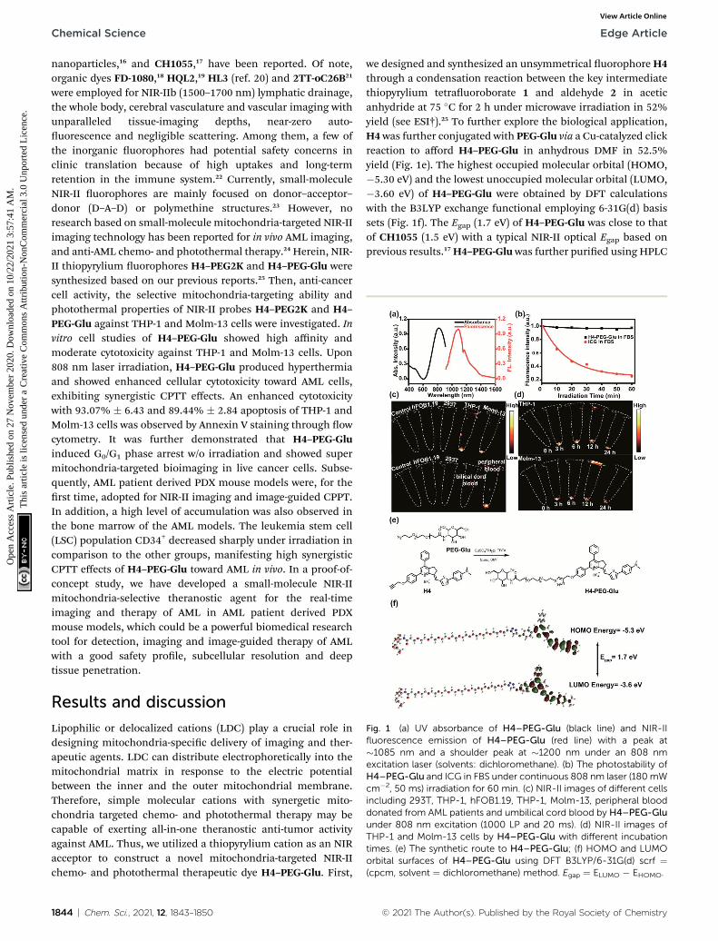
Fig. 1 (a) UV absorbance of H4–PEG-Glu (black line) and NIR-IIfluorescence emission of H4–PEG-Glu (red line) with a peak at�1085 nm and a shoulder peak at �1200 nm under an 808 nmexcitation laser (solvents: dichloromethane). (b) The photostability ofH4–PEG-Glu and ICG in FBS under continuous 808 nm laser (180mWcm�2, 50 ms) irradiation for 60 min. (c) NIR-II images of different cellsincluding 293T, THP-1, hFOB1.19, THP-1, Molm-13, peripheral blooddonated from AML patients and umbilical cord blood by H4–PEG-Gluunder 808 nm excitation (1000 LP and 20 ms). (d) NIR-II images ofTHP-1 and Molm-13 cells by H4–PEG-Glu with different incubationtimes. (e) The synthetic route to H4–PEG-Glu; (f) HOMO and LUMOorbital surfaces of H4–PEG-Glu using DFT B3LYP/6-31G(d) scrf ¼(cpcm, solvent ¼ dichloromethane) method. Egap ¼ ELUMO � EHOMO.
Chemical Science Edge Article
Ope
n A
cces
s A
rtic
le. P
ublis
hed
on 2
7 N
ovem
ber
2020
. Dow
nloa
ded
on 1
0/22
/202
1 3:
57:4
1 A
M.
Thi
s ar
ticle
is li
cens
ed u
nder
a C
reat
ive
Com
mon
s A
ttrib
utio
n-N
onC
omm
erci
al 3
.0 U
npor
ted
Lic
ence
.View Article Online
nanoparticles,16 and CH1055,17 have been reported. Of note,organic dyes FD-1080,18 HQL2,19 HL3 (ref. 20) and 2TT-oC26B21
were employed for NIR-IIb (1500–1700 nm) lymphatic drainage,the whole body, cerebral vasculature and vascular imaging withunparalleled tissue-imaging depths, near-zero auto-uorescence and negligible scattering. Among them, a few ofthe inorganic uorophores had potential safety concerns inclinic translation because of high uptakes and long-termretention in the immune system.22 Currently, small-moleculeNIR-II uorophores are mainly focused on donor–acceptor–donor (D–A–D) or polymethine structures.23 However, noresearch based on small-molecule mitochondria-targeted NIR-IIimaging technology has been reported for in vivo AML imaging,and anti-AML chemo- and photothermal therapy.24 Herein, NIR-II thiopyrylium uorophores H4–PEG2K and H4–PEG-Glu weresynthesized based on our previous reports.25 Then, anti-cancercell activity, the selective mitochondria-targeting ability andphotothermal properties of NIR-II probes H4–PEG2K and H4–PEG-Glu against THP-1 and Molm-13 cells were investigated. Invitro cell studies of H4–PEG-Glu showed high affinity andmoderate cytotoxicity against THP-1 and Molm-13 cells. Upon808 nm laser irradiation, H4–PEG-Glu produced hyperthermiaand showed enhanced cellular cytotoxicity toward AML cells,exhibiting synergistic CPTT effects. An enhanced cytotoxicitywith 93.07% � 6.43 and 89.44% � 2.84 apoptosis of THP-1 andMolm-13 cells was observed by Annexin V staining through owcytometry. It was further demonstrated that H4–PEG-Gluinduced G0/G1 phase arrest w/o irradiation and showed supermitochondria-targeted bioimaging in live cancer cells. Subse-quently, AML patient derived PDX mouse models were, for therst time, adopted for NIR-II imaging and image-guided CPPT.In addition, a high level of accumulation was also observed inthe bone marrow of the AML models. The leukemia stem cell(LSC) population CD34+ decreased sharply under irradiation incomparison to the other groups, manifesting high synergisticCPTT effects of H4–PEG-Glu toward AML in vivo. In a proof-of-concept study, we have developed a small-molecule NIR-IImitochondria-selective theranostic agent for the real-timeimaging and therapy of AML in AML patient derived PDXmouse models, which could be a powerful biomedical researchtool for detection, imaging and image-guided therapy of AMLwith a good safety prole, subcellular resolution and deeptissue penetration.
Results and discussion
Lipophilic or delocalized cations (LDC) play a crucial role indesigning mitochondria-specic delivery of imaging and ther-apeutic agents. LDC can distribute electrophoretically into themitochondrial matrix in response to the electric potentialbetween the inner and the outer mitochondrial membrane.Therefore, simple molecular cations with synergetic mito-chondria targeted chemo- and photothermal therapy may becapable of exerting all-in-one theranostic anti-tumor activityagainst AML. Thus, we utilized a thiopyrylium cation as an NIRacceptor to construct a novel mitochondria-targeted NIR-IIchemo- and photothermal therapeutic dye H4–PEG-Glu. First,
1844 | Chem. Sci., 2021, 12, 1843–1850
we designed and synthesized an unsymmetrical uorophoreH4through a condensation reaction between the key intermediatethiopyrylium tetrauoroborate 1 and aldehyde 2 in aceticanhydride at 75 �C for 2 h under microwave irradiation in 52%yield (see ESI†).25 To further explore the biological application,H4 was further conjugated with PEG-Glu via a Cu-catalyzed clickreaction to afford H4–PEG-Glu in anhydrous DMF in 52.5%yield (Fig. 1e). The highest occupied molecular orbital (HOMO,�5.30 eV) and the lowest unoccupied molecular orbital (LUMO,�3.60 eV) of H4–PEG-Glu were obtained by DFT calculationswith the B3LYP exchange functional employing 6-31G(d) basissets (Fig. 1f). The Egap (1.7 eV) of H4–PEG-Glu was close to thatof CH1055 (1.5 eV) with a typical NIR-II optical Egap based onprevious results.17 H4–PEG-Gluwas further puried using HPLC
© 2021 The Author(s). Published by the Royal Society of Chemistry

Fig. 2 (a) The cell viability of 293T, THP-1, hFOB1.19, and Molm-13after incubation with different concentrations ofH4–PEG-Glu for 24 hwithout laser irradiation (n ¼ 6). (b) Temperature variation of H4–PEG-Glu in serum at various concentrations under irradiation by an 808 nmlaser at a power density of 1.2 W cm�2 for 5 min. (c) Temperaturevariation of H4–PEG-Glu in serum (60 mM) under 808 nm laser irra-diation at different power densities. (d) Temperature elevation of H4–PEG-Glu (60 mM) in serum over several ON/OFF cycles involvingirradiation with an 808 nm laser (1.4 W cm�2) for 5 min followed bypassive cooling. (e) Photothermal conversion ability of H4–PEG-Glu(60 mM) under 808 nm laser irradiation (1.2 W cm�2) for 5.5 min andnaturally cooled to ambient temperature. (f) Liner time data versus�ln(q) during the cooling period.
Edge Article Chemical Science
Ope
n A
cces
s A
rtic
le. P
ublis
hed
on 2
7 N
ovem
ber
2020
. Dow
nloa
ded
on 1
0/22
/202
1 3:
57:4
1 A
M.
Thi
s ar
ticle
is li
cens
ed u
nder
a C
reat
ive
Com
mon
s A
ttrib
utio
n-N
onC
omm
erci
al 3
.0 U
npor
ted
Lic
ence
.View Article Online
and all compounds were characterized by 1H-NMR, 13C-NMR,HRMS and MALDI-TOF-MS (Fig. S1–S6†).
H4–PEG-Glu exhibited excellent solubility in commonorganic solvents and high aqueous solubility. The UV-vis-NIRabsorption and NIR-II uorescence emission spectra of H4–PEG-Glu in CH2Cl2 were investigated under 808 nm excitation.As shown in Fig. 1a, the absorption peak was at �810 nm, whilethe uorescence emission peak was at �1085 nm anda shoulder peak was at �1200 nm. The NIR-II quantum yield(QY) of H4–PEG-Glu in fetal bovine serum (FBS) was measuredto be �1.3% under 808 nm laser excitation using IR-26 dye asa reference (QY ¼ 0.5%) (Fig. S7†). H4–PEG-Glu showedspherical shapes with an average particle size of �96 nm andhydrodynamic radius of �120 nm, as determined from trans-mission electron microscopy (TEM) and dynamic light scat-tering (DLS), respectively (Fig. S8†). H4–PEG-Glu displayednegligible decay in FBS under continuous 808 nm laser irradi-ation at a power density of 180 mW cm�2 for 60 min (50 msexposure time), while indocyanine green (ICG) exhibited a sharpdecrease in uorescence intensity under the same conditions,indicating its high resistance to photo-bleaching for NIR-IIbiomedical application (Fig. 1b). Additionally, the cellularuptake behavior and imaging ability of ALM cancer cells werealso investigated. As depicted in Fig. 1c and d, the humanmyeloid leukemia mononuclear cells THP-1 and Molm-13exhibited high affinity and strong intracellular NIR-II uores-cence whereas negligible binding was observed in 293T andhFOB1.19 cells treated with H4–PEG-Glu (20 mM) (Fig. 1c). TheTHP-1 and Molm-13 cell lines can be clearly visualized aer 3 hof incubation, and uorescence signals reach a maximum at6 h, while H4–PEG2K has demonstrated almost no targetingability without the glucose moiety (Fig. 1d, Fig. S12a and b†). Tofurther illustrate the specicity toward AML cancer cells, wefurther studied the cellular uptake behavior in peripheral blooddonated from AML patients and umbilical cord blood collectedfrom healthy people (Fig. 1c). We were delighted to nd out thatthe peripheral blood samples had bright NIR-II uorescenceinstead of umbilical cord blood, indicating that H4–PEG-Gluhas an AML targeting and accumulation capability and isa promising candidate for targeted NIR-II imaging.
The cell viabilities of H4–PEG-Glu were investigated by CCK-8 assays with 293T, hFOB1.19, THP-1 and Molm-13 cell lineswith different concentrations within 24 h without laser irradi-ation. As shown in Fig. 2a, no obvious cytotoxicity of H4–PEG-Glu was observed even at higher concentrations up to 60 mM,suggesting H4–PEG-Glu had excellent biocompatibility toward293T and hFOB1.19 normal cells (Fig. 2a). In contrast, H4–PEG-Glu suppressed proliferation of AML cells (THP-1 and Molm-13)in a concentration-dependent manner with IC50 values of 23.97� 3.24 and 29.66 � 1.09 mM, respectively, indicating a speciccytotoxic effect against AML cells without laser irradiation(Fig. 2a). Thereaer, we evaluated the photothermal perfor-mance of H4–PEG-Glu. The temperature of H4–PEG-Glu inserum at various concentrations (0 mM, 10 mM, 20 mM, 40 mM, 60mM, 80 mM) was recorded under 808 nm laser irradiation ata power density of 1.2 W cm�2 for 5 min. Rapid photothermaleffects occurred on laser irradiation even at the dose of 40 mM
© 2021 The Author(s). Published by the Royal Society of Chemistry
(Fig. 2b). The temperature increased signicantly (up to 63 �C)at the dose of 60 mM by varying the laser power density (1.2 and1.4 W cm�2) (Fig. 2c). The photothermal stability of H4–PEG-Glu (60 mM) in serum was further assessed by continuouslymonitoring the temperature variation under 808 nm laserirradiation (1.4 W cm�2) for 5 min, and it was naturally cooledto room temperature for ve heating/cooling cycles. As shown inFig. 2d, no obvious decline of temperature was observed in itsphotothermal conversion performance. The photothermalconversion efficiency of H4–PEG-Glu was �11.6% according tothe reported methods (Fig. 2e and f).26 No signicant amount ofROS in vitro was observed in any group of H4–PEG-Glu under808 nm laser irradiation (Fig. S9†). As a consequence, the goodphotostability and excellent photothermal performance of H4–PEG-Glu make it an ideal NIR-II probe for in vivo uorescenceimage-guided photothermal therapy.
Subsequently, the photothermal ablation of THP-1 andMolm-13 cells induced by H4–PEG-Glu was conducted under808 nm NIR laser irradiation (1.2 W cm�2). Aer NIR lasertreatment, the cells were observed with a bright-eld micro-scope. As shown in Fig. 3a, H4–PEG-Glu (20 mM) treated THP-1or Molm-13 cells had an enhanced chemo- and photothermalcytotoxicity under NIR irradiation compared to that under PBS,
Chem. Sci., 2021, 12, 1843–1850 | 1845

Fig. 3 (a) The bright-field microscopy images of THP-1 and Molm-13cells treated with PBS, 808 nm laser, H4–PEG-Glu (20 mM), and H4–PEG-Glu (20 mM) under 808 nm laser irradiation (1.2 W cm�2, 5 min).(b) The cell viability of THP-1 and Molm-13 by the Trypan BlueExclusion test according to the bright-field microscopy images. (c)Proportions of Annexin V+ THP-1 cells and Annexin V+ Molm-13 cellscalculated from the cell apoptosis in Fig. 3d. (d) Flow cytometryanalysis of cell apoptosis of THP-1 and Molm-13 cells treated with PBS,808 nm laser, H4–PEG-Glu (20 mM), and H4–PEG-Glu (20 mM) under808 nm laser irradiation (1.2W cm�2, 5min). *p < 0.05; **p < 0.01; ***p< 0.001; ns, not significant.
Fig. 4 (a) Flow cytometry analysis of the cell cycle of THP-1 andMolm-13 cells treated with PBS, H4–PEG-Glu (20 mM), and H4–PEG-Glu (20 mM) under 808 nm laser irradiation (1.2 W cm�2, 5 min). (b)Proportions of G0/G1, S, and G2/M phase cells in THP-1 and Molm-13.*p < 0.05; **p < 0.01; ***p < 0.001; ns, not significant.
Chemical Science Edge Article
Ope
n A
cces
s A
rtic
le. P
ublis
hed
on 2
7 N
ovem
ber
2020
. Dow
nloa
ded
on 1
0/22
/202
1 3:
57:4
1 A
M.
Thi
s ar
ticle
is li
cens
ed u
nder
a C
reat
ive
Com
mon
s A
ttrib
utio
n-N
onC
omm
erci
al 3
.0 U
npor
ted
Lic
ence
.View Article Online
808 nm laser or H4–PEG-Glu (20 mM) alone. The cell viability ofTHP-1 and Molm-13 cells was also quantitatively analyzed bythe Trypan Blue dye exclusion test. As shown in Fig. 3b,approximately 39.57%� 5.87 of THP-1 cells and 42.9%� 5.58 ofMolm-13 cells were reduced by H4–PEG-Glu (20 mM) inducedsynergistic therapy. The apoptosis of THP-1 and Molm-13 cellswas also studied by Annexin V staining through ow cytometry.THP-1 and Molm-13 cells treated with H4–PEG-Glu under808 nm laser irradiation showed an enhanced cytotoxicity with93.07% � 6.43 and 89.44% � 2.84 apoptosis, respectively. Theresults were in good agreement with the aforementioned cellviability (Fig. 3c and d). All these results demonstrated that theNIR-II probe H4–PEG-Glu exhibited effective chemo- and pho-tothermal synergistic treatment (CPTT) toward ALM cancer cells(THP-1 and Molm-13) in vitro.
To further explore the mechanism of toxicity and apoptosisof H4–PEG-Glu, the cell cycle progression of the above cancercells was analyzed by uorescence-activated cell sorting (FACS)analysis. THP-1 or Molm-13 cells in the G0/G1 phase and G2/Mphase have unreplicated diploid (2n) DNA content and repli-cated ploid (4n) DNA, implying the different procedures of DNA
1846 | Chem. Sci., 2021, 12, 1843–1850
replication. As shown in Fig. 4a and b, THP-1 cells treated withH4–PEG-Glu under 808 nm laser irradiation exhibited an obvi-ously increased percentage in the G0/G1 phase (53.64%� 7.46 to66.68% � 0.78) and a decreased percentage in the S phase andG2/M phase (42.24% � 9.40 to 28.61% � 3.55), demonstratingthatH4–PEG-Glu inhibited DNA replication due to inducing cellcycle arrest in the G0/G1 phase. A similar apoptosis pathway wasalso observed in Molm-13 cells.
Mitochondria is critically important in energy productionand apoptosis.27 To further assess the targeting ability of H4–PEG-Glu to mitochondria and its efficiency in mitochondria-targeted cancer therapy, THP-1 and Molm-13 cells were incu-bated with H4–PEG-Glu (20 mM) for 24 h followed by stainingwith Mito-tracker Green (5 mM), a commercial available kit usedto label the mitochondria. The intrinsic uorescence of H4–PEG-Glu (red color) was observed to be completely overlappedwith Mito-tracker (Green) (Fig. 5a, Fig. S10†), implying itsexcellent targeting ability to mitochondria. The feedback ofmitochondrial membrane potential (MMP) reected differentcellular status and has been used for assessing the therapeuticeffects.28 Thus, MMP levels of THP-1 and Molm-13 cells in eachgroup were detected using a commercial uorescent probe JC-1.The uorescence images of each group have been shown inFig. 5b. According to Fig. 5b, the red uorescence signal fromTHP-1 cancer cells was bright while the green uorescencesignal was weak before H4–PEG-Glu or laser treatment,demonstrating that the MMP level was normal. With the prog-ress of the treatment process, the green uorescence signalintensied and red uorescence signal gradually vanished,illustrating the decrease of the MMP level, and the trans-formation of JC-1 from J-aggregates into monomers. Upon
© 2021 The Author(s). Published by the Royal Society of Chemistry

Fig. 5 (a) Confocal fluorescence images of THP-1 and Molm-13 cellstreated with H4–PEG-Glu (20 mM) for 24 h without laser irradiation.The cell mitochondria stained with Mito-Tracker Green showeda green fluorescence signal, 40,6-diamidino-2-phenylindole (DAPI)labeled nucleus showed a blue fluorescence signal and H4–PEG-Glushowed a red fluorescence signal. (b) Immunofluorescence mito-chondrial membrane potential staining images of AML cells withdifferent treatments. The cell nucleus stained with DAPI showed a bluefluorescence signal, JC-1 monomers showed a green fluorescencesignal, and JC-1 aggregates showed a red fluorescence signal. *p <0.05; **p < 0.01; ***p < 0.001; ns, not significant.
Fig. 6 (a) The representative NIR-II in vivo images of AML patientderived PDX mouse models (n ¼ 3), the blue arrow points to thelocation of tumor sites in bone marrow. (b) The schematic diagram ofAML patient derived PDX mouse models and treatment. (c) Theproportion of CD34+ cells in peripheral blood mononuclear cells(PBMC) and bone marrow (BM) after treatment. (d) The immunohis-tochemical staining images of bone marrow (up left to right: AMLmicewithout treatment, mice treated with H4–PEG-Glu, and mice treatedwith H4–PEG-Glu under 808 nm laser irradiation, down left to right:immunohistochemical staining images were the enlargement of thered square in left images). *p < 0.05; **p < 0.01; ***p < 0.001; ns, notsignificant.
Edge Article Chemical Science
Ope
n A
cces
s A
rtic
le. P
ublis
hed
on 2
7 N
ovem
ber
2020
. Dow
nloa
ded
on 1
0/22
/202
1 3:
57:4
1 A
M.
Thi
s ar
ticle
is li
cens
ed u
nder
a C
reat
ive
Com
mon
s A
ttrib
utio
n-N
onC
omm
erci
al 3
.0 U
npor
ted
Lic
ence
.View Article Online
treatment withH4–PEG-Glu under 808 nm laser irradiation, thered uorescence signals of THP-1 cells vanished, suggestingthat the therapeutic effects of THP-1 cancer cells were greatlyenhanced. The same phenomenon was also detected in Molm-13 cancer cells. All these results demonstrated thatH4–PEG-Gluis a novel therapeutic probe with NIR-II uorescence andmitochondrial-targeting properties.
Inspired by the intriguing chemo- and photothermalperformance of H4–PEG-Glu in vitro, subsequently, we envi-sioned that H4–PEG-Glu can achieve great NIR-II image-guidedtherapeutic effects in vivo. Thus, the NIR-II uorescenceimaging performance of H4–PEG-Glu was rstly evaluated invivo using AML patient derived PDX mouse models. H4–PEG-Glu (200 mL, 1 mg mL�1 in PBS) was rst administered into AMLpatient derived PDXmouse models (n¼ 3) through the tail vein.As shown in Fig. 6a, the uorescence signal was graduallyenhanced at the bone marrow (blue arrow in Fig. 6a) of AMLpatient derived PDX mouse models, and reached a maximum at
© 2021 The Author(s). Published by the Royal Society of Chemistry
6 h post injection (808 nm excitation, 90 mW cm�2, 1000 LP and100 ms). In contrast, no obvious signals were detected in bonemarrow at all-time points i.v. post-injection ofH4–PEG2K in theAML patient derived PDX mouse models or in the normal B-NDG mice (n ¼ 3), demonstrating that H4–PEG-Glu exhibitedspecic targeting ability to AML cancer cells in bone marrow(Fig. S12c†). Thereaer, AML patient derived PDX mousemodels (n ¼ 4 group, �20 g each) were treated with PBS, H4–PEG-Glu (200 mg), and H4–PEG-Glu (200 mg) under 808 nm laserirradiation (1.2 W cm�2, 5 min), respectively. No obvious body
Chem. Sci., 2021, 12, 1843–1850 | 1847

Chemical Science Edge Article
Ope
n A
cces
s A
rtic
le. P
ublis
hed
on 2
7 N
ovem
ber
2020
. Dow
nloa
ded
on 1
0/22
/202
1 3:
57:4
1 A
M.
Thi
s ar
ticle
is li
cens
ed u
nder
a C
reat
ive
Com
mon
s A
ttrib
utio
n-N
onC
omm
erci
al 3
.0 U
npor
ted
Lic
ence
.View Article Online
weight loss was observed for all groups during the CPPT processin a week. Then they were all sacriced to collect the peripheralblood mononuclear cells (PBMC) and bone marrow (BM) toanalyze the expression of AML stem cell marker CD34. Theimmunohistochemical analysis of bone marrow (BM) wasfurther conducted. The results showed that CD34 expressed inH4–PEG-Glu under the 808 nm laser irradiation group (the rightimages in Fig. 6d) was obviously lower than that in the controlgroup (le images in Fig. 6d) and in the H4–PEG-Glu group (themiddle images in Fig. 6d). The CD34+ cells in all three groupswere also quantied by ow cytometry. The percentage of CD34+
cells in PBMC and bone marrow with H4–PEG-Glu under808 nm laser irradiation was signicantly lower than that of theother two groups (Fig. 6c). To assess the hepatotoxicity of H4–PEG-Glu, H4–PEG-Glu (200 mg) and PBS were injected in ICRfemale mice (n ¼ 3 per group, �20 g each), respectively. Theblood was collected at 7 days post-injection to analyze thebiochemistry index (ALT, AST, CREA, BUN). The concentrationin the experiment group was similar to that in the PBS group,indicating that H4–PEG-Glu has no obvious toxicity to thekidneys and liver (Fig. S11†). All the above results demonstratedthat H4–PEG-Glu is a potential small-molecule NIR-II probe forclinical translation in AML cancer theranostics.
Experimental section
Methods and materials are provided in the ESI. All animalexperiments were approved by the Chinese Regulations for theAdministration of Affairs Concerning Experimental Animalsand Institutional Animal Care and Use Committee (IACUC) ofWuhan University. The study has been approved by the Insti-tutional Ethical Committee of Zhongnan Hospital of WuhanUniversity, China (No. 2018278), and performed in accordancewith the ethical standards as laid down in the 1964 Helsinkideclaration and its later amendments or comparable ethicalstandards.
Conclusions
In conclusion, we have successfully designed and synthesizeda novel mitochondria-targeted NIR-II uorophore H4–PEG-Gluwith a new thiopyrylium skeleton for AML CPTT. H4–PEG-Glushowed excellent water solubility, high photo-stability andbiocompatibility, and superior targeting affinity toward AMLcancer cells (THP-1 and Molm-13) and peripheral blooddonated from AML patients. H4–PEG-Glu not only can quicklyand effectively image mitochondria in acute myeloid leukemia(AML) cells, and induce G0/G1 phase arrest by the intrinsicmitochondrial apoptosis pathway w/o irradiation, but alsoexhibited moderate cytotoxicity against AML cancer cells ina dose dependent-manner without laser irradiation. THP-1 andMolm-13 cells treated with H4–PEG-Glu upon NIR laser irradi-ation showed enhanced CPTT with 93.07% � 6.43 and 89.44%� 2.84 apoptosis by Annexin V staining. AML patient derivedPDXmouse models were nally used for the rst time for in vivomitochondria-targeted NIR-II Imaging of acute myeloidleukemia and image-guided CPTT. The percentage of CD34+
1848 | Chem. Sci., 2021, 12, 1843–1850
cells in PBMC and bone marrow was signicantly reduced aerH4–PEG-Glu treatment and laser irradiation. To the best of ourknowledge, this is the rst NIR-II small-molecule probe formitochondrial-targeted chemo- and photothermal synergistictherapy for AML cancer. It is hoped that this novel NIR-II cationuorophore may provide a practical strategy to develop small-molecule NIR-II uorophores for non-solid tumor therapywith a deeper penetration depth and higher resolution.
Conflicts of interest
There are no conicts to declare.
Acknowledgements
The work was supported by the National Key R&D Program ofChina (2020YFA0908800), NSFC (81773674, 81770179), Shenz-hen Science and Technology Research Grant(JCYJ20190808152019182), Hubei Province Scientic andTechnical Innovation Key Project (2020BAB058), the AppliedBasic Research Program of Wuhan Municipal Bureau of Scienceand Technology (2019020701011429), the Local DevelopmentFunds of Science and Technology Department of Tibet(XZ202001YD0028C), Health Commission of Hubei ProvinceScientic Research Project (WJ2019M177, WJ2019M178,WJ2019H008), the Fundamental Research Funds for the CentralUniversities (ZNJC201931).
Notes and references
1 (a) D. Jaque, L. Martınez Maestro, B. Del Rosal, P. Haro-Gonzalez, A. Benayas, J. L. Plaza, E. Martın Rodrıguez andJ. Garcıa Sole, Nanoscale, 2014, 6, 9494–9530; (b) H. He,J. Wang, H. Wang, N. Zhou, D. Yang, D. R. Green andB. Xu, J. Am. Chem. Soc., 2018, 140, 1215–1218; (c) Y. Liu,J. Zhou, L. Wang, X. Hu, X. Liu, M. Liu, Z. Cao,D. Shangguan and W. Tan, J. Am. Chem. Soc., 2016, 138,12368–12374; (d) R. M. Shallis, R. Wang, A. Davidoff, X. Maand A. M. Zeidan, Blood Rev., 2019, 36, 70–87.
2 H. S. Jung, J.-H. Lee, K. Kim, S. Koo, P. Verwilst, J. L. Sessler,C. Kang and J. S. Kim, J. Am. Chem. Soc., 2017, 139, 9972–9978.
3 (a) R. Guo, J. Yin, Y. Ma, Q. Wang and W. Lin, J. Mater. Chem.B, 2018, 6, 2894–2900; (b) Y. Koide, Y. Urano, S. Kenmoku,H. Kojima and T. Nagano, J. Am. Chem. Soc., 2007, 129,10324–10325; (c) M. Sibrian- Vazquez, I. V. Nesterova,T. J. Jensen and M. G. H. Vicente, Bioconjugate Chem.,2008, 19, 705–713; (d) J. Zielonka, J. Joseph, A. Sikora,M. Hardy, O. Ouari, J. Vasquez-Vivar, G. Cheng, M. Lopezand B. Kalyanaraman, Chem. Rev., 2017, 117, 10043–10120.
4 (a) G. Cheng, J. Zielonka, B. P. Dranka, D. McAllister,A. C. Mackinnon, J. Joseph and B. Kalyanaraman, CancerRes., 2012, 72, 2634–2644; (b) J. C. Wang, Z. Jiang,L. P. Xiang, Y. F. Li, M. R. Ou, X. Yang, J. W. Shao, Y. S. Lu,L. F. Lin, J. Z. Chen, Y. Dai and L. Jia, Sci. Rep., 2014, 4,5006; (c) B. Kalyanaraman, G. Cheng, M. Hardy, O. Ouari,
© 2021 The Author(s). Published by the Royal Society of Chemistry
Edge Article Chemical Science
Ope
n A
cces
s A
rtic
le. P
ublis
hed
on 2
7 N
ovem
ber
2020
. Dow
nloa
ded
on 1
0/22
/202
1 3:
57:4
1 A
M.
Thi
s ar
ticle
is li
cens
ed u
nder
a C
reat
ive
Com
mon
s A
ttrib
utio
n-N
onC
omm
erci
al 3
.0 U
npor
ted
Lic
ence
.View Article Online
M. Lopez, J. Joseph, J. Zielonka and M. B. Dwinell, RedoxBiol., 2018, 14, 316–327.
5 H. Chen, J. Wang, X. Feng, M. Zhu, S. Hoffmann, A. Hsu,K. Qian, D. Huang, F. Zhao, W. Liu, H. Zhang andZ. Cheng, Chem. Sci., 2019, 10, 7946–7951.
6 (a) H. Dohner, D. J. Weisdorf and C. D. Bloomeld, N. Engl. J.Med., 2015, 373, 1136–1152; (b) J. N. Saultz and R. Garzon, J.Clin. Med., 2016, 5, 33; (c) B. M. Barth, E. I. Altinoglu,S. S. Shanmugavelandy, J. M. Kaiser, D. Crespo-Gonzalez,N. A. Divittore, C. Mcgovern, T. M. Goff, N. R. Keasey,J. H. Adair, T. P. Loughran, D. F. Claxton and M. Kester,ACS Nano, 2011, 5, 5325–5337.
7 (a) P. Bose, P. Vachhani and J. E. Cortes, Curr. Treat. OptionsOncol., 2017, 18, 17; (b) K. D. Miller, L. Nogueira,A. B. Mariotto, J. H. Rowland, K. R. Yabroff, C. M. Alfano,A. Jemal, J. L. Kramer and R. L. Siegel, Ca-Cancer J. Clin.,2019, 69, 363–385.
8 (a) W. Wang, G. Liang, W. Zhang, D. Xing and X. Hu, Chem.Mater., 2018, 30, 3486–3498; (b) Y. Chen, Z. Li, H. Wang,Y. Wang, H. Han, Q. Jin and J. Ji, ACS Appl. Mater.Interfaces, 2016, 8, 6852–6858; (c) Y. Zhang, C. Y. Ang,M. Li, S. Y. Tan, Q. Qu and Y. Zhao, ACS Appl. Mater.Interfaces, 2016, 8, 6869–6879.
9 (a) S. Kunjachan, J. Ehling, G. Storm, F. Kiessling andT. Lammers, Chem. Rev., 2015, 115, 10907–10937; (b)G. Hong, J. C. Lee, J. T. Robinson, U. Raaz, L. Xie,N. F. Huang, J. P. Cooke and H. Dai, Nat. Med., 2012, 18,1841–1846; (c) G. Hong, S. Diao, J. Chang, A. L. Antaris,C. Chen, B. Zhang, S. Zhao, D. N. Atochin, P. L. Huang,K. I. Andreasson, C. J. Kuo and H. Dai, Nat. Photonics,2014, 8, 723; (d) B. Li, L. Lu, M. Zhao, Z. Lei and F. Zhang,Angew. Chem., Int. Ed., 2018, 57, 7483; (e) C. Qu, Y. Xiao,H. Zhou, B. Ding, A. Li, J. Lin, X. Zeng, H. Chen, K. Qian,X. Zhang, W. Fang, J. Wu, Z. Deng, Z. Cheng and X. Hong,Adv. Opt. Mater., 2019, 7, 1900229.
10 (a) D. Kim, N. Lee, Y. I. Park and T. Hyeon, BioconjugateChem., 2017, 28, 115–123; (b) G. Hong, S. Diao,A. L. Antaris and H. Dai, Chem. Rev., 2015, 115, 10816–10906; (c) H. Zhang, Z.-H. Chen, X. Liu and F. Zhang, NanoRes., 2020, 13, 1795–1809.
11 (a) K. Li and B. Liu, Chem. Soc. Rev., 2014, 43, 6570–6597; (b)Y. Liu, J. Liu, D. Chen, X. Wang, Z. Liu, H. Liu, L. Jiang, C. Wuand Y. Zou, Macromolecules, 2019, 52, 5735–5740; (c) B. Guo,J. Chen, N. Chen, M. Wu, K. Li, C. Liu and B. Liu, Adv. Mater.,2019, 31, 1808355; (d) Z. Zhang, X. Fang, Z. Liu, H. Liu,D. Chen, S. He, X. Zhang and C. Wu, Angew. Chem., Int.Ed., 2020, 59, 3691–3698; (e) H. Zhou, S. Li, X. Zeng,M. Zhang, L. Tang, Q. Li, D. Chen, X. Meng and X. Hong,Chin. Chem. Lett., 2020, 31, 1382–1386.
12 (a) S. He, J. Song, J. Qu and Z. Cheng, Chem. Soc. Rev., 2018,47, 4258–4278; (b) J. Li, Y. Liu, Y. Xu, L. Li, Y. Sun andW. Huang, Coord. Chem. Rev., 2020, 415, 213318; (c) B. Li,M. Zhao and F. Zhang, ACS Mater. Lett., 2020, 2, 905–917;(d) C. Sun, B. Li, M. Zhao, S. Wang, Z. Lei, L. Lu, H. Zhang,L. Feng, C. Dou, D. Yin, H. Xu, Y. Cheng and F. Zhang, J.Am. Chem. Soc., 2019, 141, 19221–19225; (e) Q. Wang,
© 2021 The Author(s). Published by the Royal Society of Chemistry
Y. Dai, J. Xu, J. Cai, X. Niu, L. Zhang, R. Chen, Q. Shen,W. Huang and Q. Fan, Adv. Funct. Mater., 2019, 29, 1901480.
13 (a) Y. Du, B. Xu, T. Fu, M. Cai, F. Li, Y. Zhang and Q. Wang, J.Am. Chem. Soc., 2010, 132, 1470–1471; (b) J. Lin, Q. Li,X. Hong and Y. Xiao, Sci. China: Chem., 2020, 63, 766–770;(c) C. Li, Y. Zhang, M. Wang, Y. Zhang, G. Chen, L. Li,D. Wu and Q. Wang, Biomaterials, 2014, 35, 393–400; (d)C. Li, W. Li, H. Liu, Y. Zhang, G. Chen, Z. Li and Q. Wang,Angew. Chem., Int. Ed., 2020, 132, 253–258; (e) M. Zhang,J. Yue, R. Cui, Z. Ma, H. Wan, Y. Zhou, Y. Kuang,Y. Zhong, D. W. Pang and H. Dai, Proc. Natl. Acad. Sci. U.S. A., 2018, 115, 6590.
14 H. Liu, G. Hong, Z. Luo, J. Chen, J. Chang, M. Gong, H. He,J. Yang, X. Yuan, L. Li, X. Mu, J. Wang, W. Mi, J. Luo, J. Xieand X.-D. Zhang, Adv. Mater., 2019, 31, 1901015.
15 (a) K. Welsher, Z. Liu, S. P. Sherlock, J. T. Robinson, Z. Chen,D. Daranciang and H. Dai, Nat. Nanotechnol., 2009, 4, 773–780; (b) S. Diao, G. Hong, J. T. Robinson, L. Jiao,A. L. Antaris, J. Z. Wu, C. L. Choi and H. Dai, J. Am. Chem.Soc., 2012, 134, 16971; (c) C. Liang, S. Diao, C. Wang,H. Gong, T. Liu, G. Hong, X. Shi, H. Dai and Z. Liu, Adv.Mater., 2014, 26, 5646; (d) J. D. Harvey, P. V. Jena,H. A. Baker, G. H. Zerze, R. M. Williams, T. V. Galassi,D. Roxbury, J. Mittal and D. A. Heller, Nat. Biomed. Eng.,2017, 1, 0041.
16 (a) Y. Zhong, Z. Ma, S. Zhu, J. Yue, M. Zhang, A. L. Antaris,J. Yuan, R. Cui, H. Wan, Y. Zhou, W. Wang, N. F. Huang,J. Luo, Z. Hu and H. Dai, Nat. Commun., 2017, 8, 737; (b)P. Wang, Y. Fan, L. Lu, L. Liu, L. Fan, M. Zhao, Y. Xie,C. Xu and F. Zhang, Nat. Commun., 2018, 9, 2898; (c)Y. Fan and F. Zhang, Adv. Opt. Mater., 2019, 7, 1801417; (d)H. Zhou, Y. Xiao and X. Hong, Chin. Chem. Lett., 2018, 29,1425–1428.
17 (a) A. L. Antaris, H. Chen, K. Cheng, Y. Sun, G. Hong, C. Qu,S. Diao, Z. Deng, X. Hu, B. Zhang, X. Zhang, O. K. Yaghi,Z. R. Alamparambil, X. Hong, Z. Cheng and H. Dai, Nat.Mater., 2016, 15, 235–242; (b) A. L. Antaris, H. Chen,S. Diao, Z. Ma, Z. Zhang, S. Zhu, J. Wang, A. X. Lozano,Q. Fan, L. Chew, M. Zhu, K. Cheng, X. Hong, H. Dai andZ. Cheng, Nat. Commun., 2017, 8, 15269; (c) H. Zhou,X. Zeng, A. Li, W. Zhou, L. Tang, W. Hu, Q. Fan, X. Meng,H. Deng, L. Duan, Y. Li, Z. Deng, X. Hong and Y. Xiao, Nat.Commun., 2020, 11, 6183.
18 (a) B. Li, Z. Lei and F. Zhang, Angew. Chem., Int. Ed., 2018, 57,7483–7487; (b) J. Yang and X. Hong, Sci. China: Chem., 2019,62, 7–8.
19 Q. Li, Q. Ding, Y. Li, X. Zeng, Y. Liu, S. Lu, H. Zhou, X. Wang,J. Wu, X. Meng, Z. Deng and Y. Xiao, Chem. Commun., 2020,56, 3289.
20 Y. Li, Y. Liu, Q. Li, X. Zeng, T. Tian, W. Zhou, Y. Cui, X. Wang,X. Cheng, Q. Ding, X. Wang, J. Wu, H. Deng, Y. Li, X. Meng,Z. Deng, X. Hong and Y. Xiao, Chem. Sci., 2020, 11, 2621–2626.
21 Y. Li, Z. Cai, S. Liu, H. Zhang, S. T. H. Wong, J. W. Y. Lam,R. T. K. Kwok, J. Qian and B. Z. Tang, Nat. Commun., 2020,11, 1255.
22 L. Szablewski, Biochim. Biophys. Acta, 2013, 1835, 164–169.
Chem. Sci., 2021, 12, 1843–1850 | 1849

Chemical Science Edge Article
Ope
n A
cces
s A
rtic
le. P
ublis
hed
on 2
7 N
ovem
ber
2020
. Dow
nloa
ded
on 1
0/22
/202
1 3:
57:4
1 A
M.
Thi
s ar
ticle
is li
cens
ed u
nder
a C
reat
ive
Com
mon
s A
ttrib
utio
n-N
onC
omm
erci
al 3
.0 U
npor
ted
Lic
ence
.View Article Online
23 (a) H. Zhang, Y. Fan, P. Pei, C. Sun, L. Lu and F. Zhang,Angew. Chem., Int. Ed., 2019, 58, 10153–10157; (b) B. Li,M. Zhao, L. Feng, C. Dou, S. Ding, G. Zhou, L. Lu,H. Zhang, F. Chen, X. Li, G. Li, S. Zhao, C. Jiang, Y. Wang,D. Zhao, Y. Cheng and F. Zhang, Nat. Commun., 2020, 11,3102; (c) H. Ma, C. Liu, Z. Hu, P. Yu, X. Zhu, R. Ma, Z. Sun,C.-H. Zhang, H. Sun, S. Zhu and Y. Liang, Chem. Mater.,2020, 32, 2061–2069; (d) Q. Yang, Z. Hu, S. Zhu, R. Ma,H. Ma, Z. Ma, H. Wan, T. Zhu, Z. Jiang, W. Liu, L. Jiao,H. Sun, Y. Liang and H. Dai, J. Am. Chem. Soc., 2018, 140,1715–1724.
24 (a) Y.-X. Zhu, H.-R. Jia, G. Gao, G.-Y. Pan, Y.-W. Jiang, P. Li,N. Li, C. Zhou, C. She, N. W. Ulrich, Z. Chen and F.-G. Wu,Biomaterials, 2020, 232, 119668; (b) G.-Y. Pan, H.-R. Jia,
1850 | Chem. Sci., 2021, 12, 1843–1850
Y.-X. Zhu, R.-H. Wang, F.-G. Wu and Z. Chen, ACSBiomater. Sci. Eng., 2017, 3, 3596–3606.
25 (a) B. Ding, Y. Xiao, H. Zhou, X. Zhang, C. Qu, F. Xu, Z. Deng,Z. Cheng and X. Hong, J. Med. Chem., 2019, 62, 2049–2059;(b) X. Hong, H. Zhou, Y. Xiao, B. Ding, J. Duan, CN Pat.,109369633B, 2018.
26 X. Yang, D. Wang, Y. Shi, J. Zou, Q. Zhao, Q. Zhang,W. Huang, J. Shao, X. Xie and X. Dong, ACS Appl. Mater.Interfaces, 2018, 10, 12431–12440.
27 X.-S. Hou, H.-S. Wang, B. P. Mugaka, G.-J. Yang and Y. Ding,Biomater. Sci., 2018, 6, 2786–2797.
28 X. Li, M. Tian, G. Zhang, R. Zhang, R. Feng, L. Guo, X. Yu,N. Zhao and X. He, Anal. Chem., 2017, 89, 3335–3344.
© 2021 The Author(s). Published by the Royal Society of Chemistry