All About Scopes. Labeling A Microscope Body Tube Revolving Nosepiece Objective Stage Clips...
21
All About Scopes
-
Upload
earl-davis -
Category
Documents
-
view
223 -
download
0
Transcript of All About Scopes. Labeling A Microscope Body Tube Revolving Nosepiece Objective Stage Clips...
All About Scopes

Labeling A Microscope
Body Tube
Revolving Nosepiece
Objective
Stage Clips
Diaphragm
Light Source
Ocular Lens
Arm
Stage
Course Adjustment KnobFine Adjustment Knob
Base
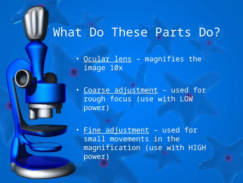
What Do These Parts Do?
• Ocular lens – magnifies the image 10x
• Coarse adjustment – used for rough focus (use with LOW power)
• Fine adjustment – used for small movements in the magnification (use with HIGH power)
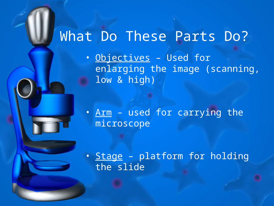
What Do These Parts Do?• Objectives – Used for enlarging the
image (scanning, low & high)
• Arm – used for carrying the microscope
• Stage – platform for holding the slide
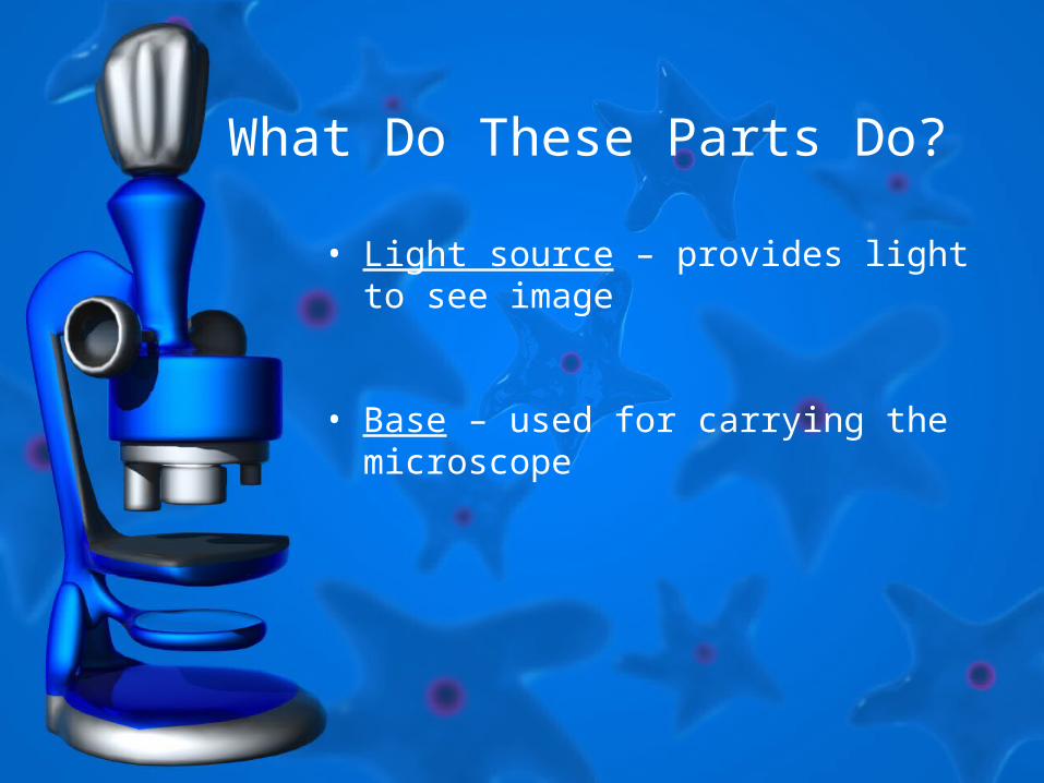
What Do These Parts Do?
• Light source – provides light to see image
• Base – used for carrying the microscope
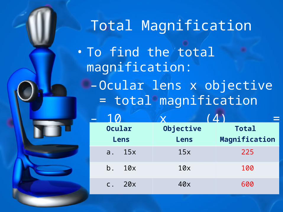
Total Magnification
• To find the total magnification:– Ocular lens x objective =
total magnification– 10 x (4) = 40
Ocular
Lens
Objective
Lens
Total
Magnification
a. 15x 15x 225
b. 10x 10x 100
c. 20x 40x 600

Microscope Image
• What you see on the stage is different from what you view when you look in a microscope– The image is reversed and inverted– This means it’s upside down and flipped
On the Stage Through The Scope
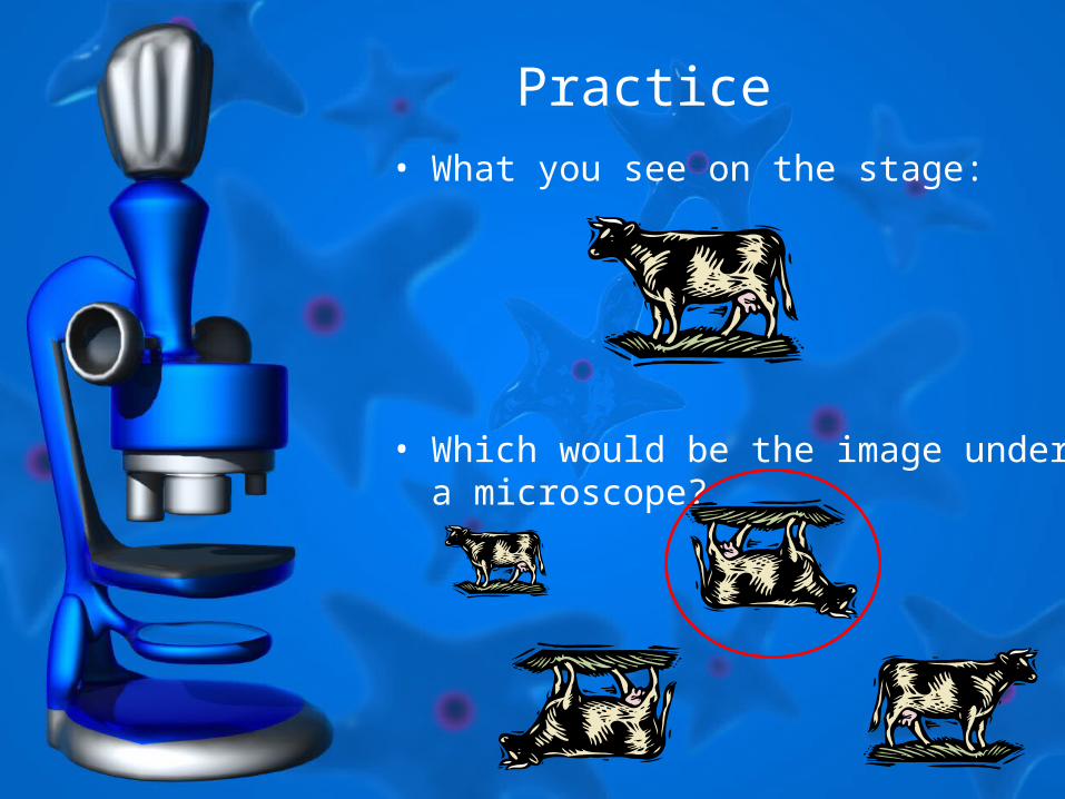
Practice• What you see on the stage:
• Which would be the image under a microscope?
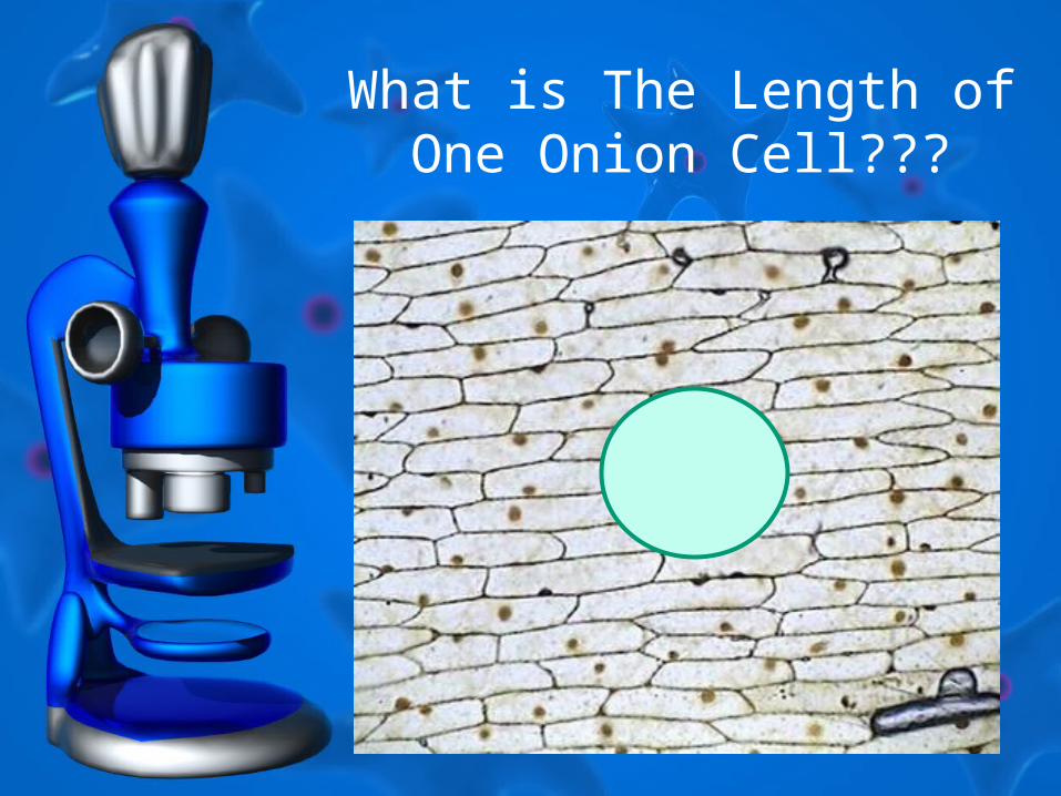
What is The Length of One Onion Cell???
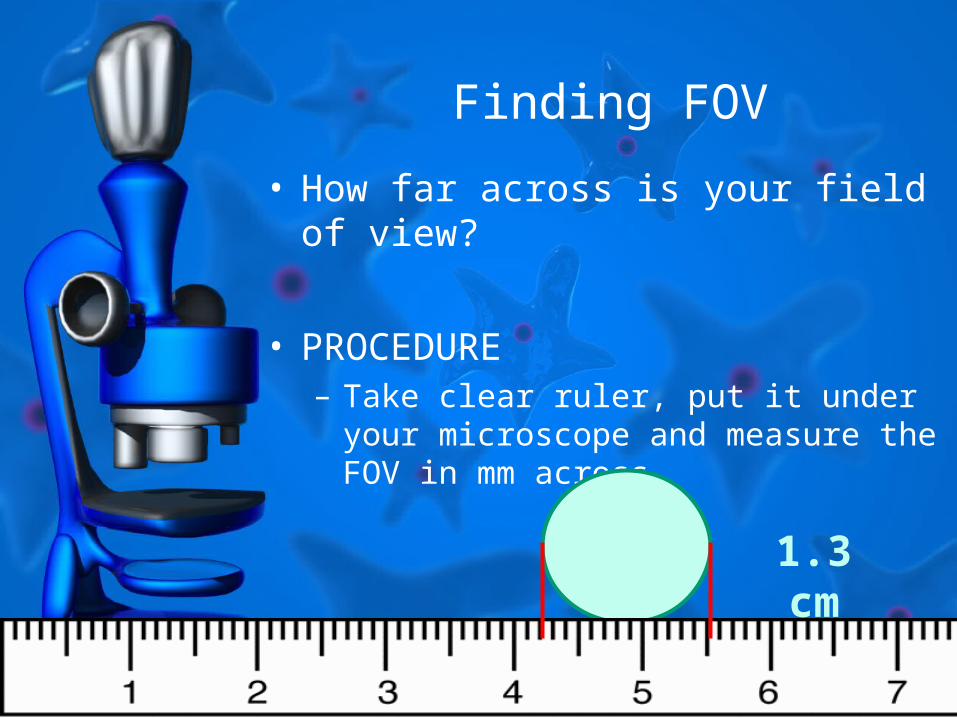
Finding FOV
• How far across is your field of view?
• PROCEDURE– Take clear ruler, put it under your
microscope and measure the FOV in mm across
1.3 cm
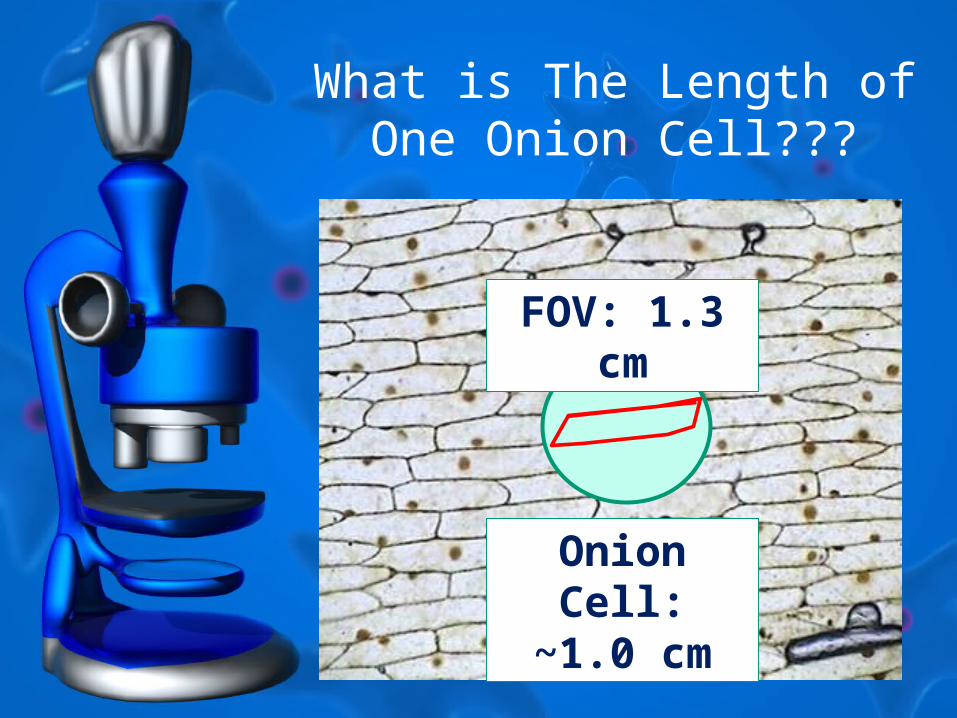
What is The Length of One Onion Cell???
FOV: 1.3 cm
Onion Cell: ~1.0 cm
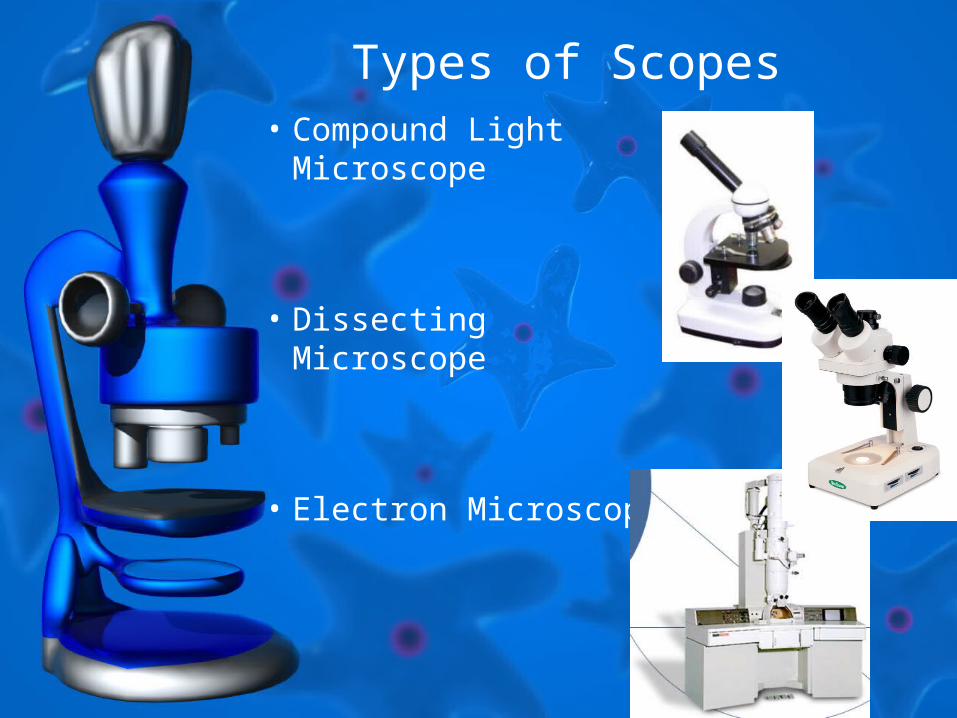
Types of Scopes• Compound Light
Microscope
• Dissecting Microscope
• Electron Microscope

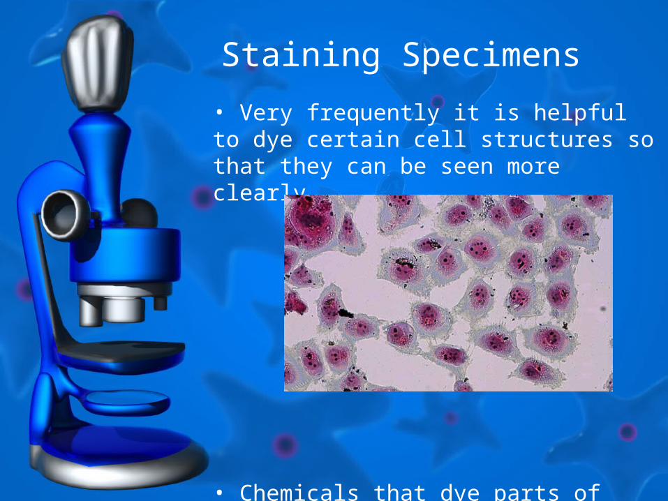
Staining Specimens
• Very frequently it is helpful to dye certain cell structures so that they can be seen more clearly.
• Chemicals that dye parts of cells for this purpose are called stains.
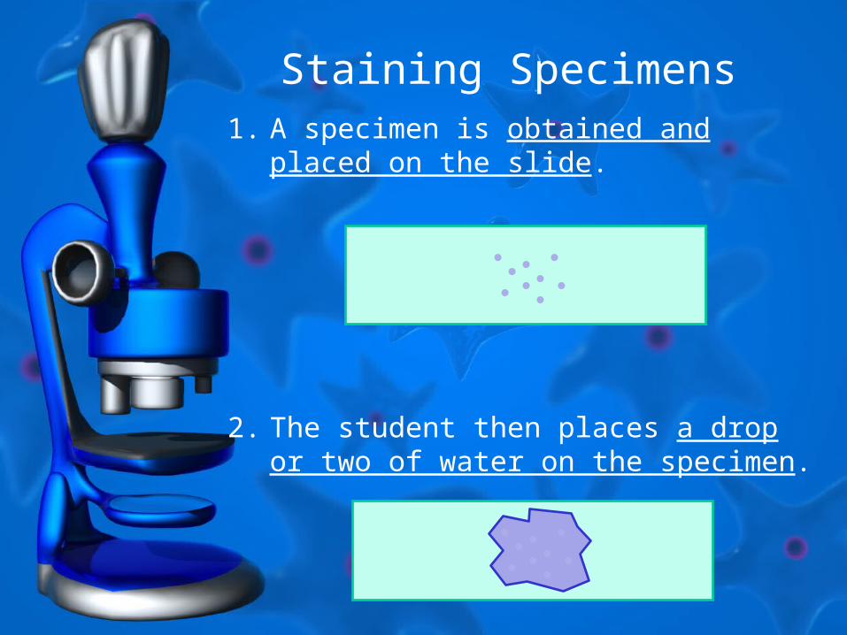
Staining Specimens1. A specimen is obtained and placed on
the slide.
2. The student then places a drop or two of water on the specimen.
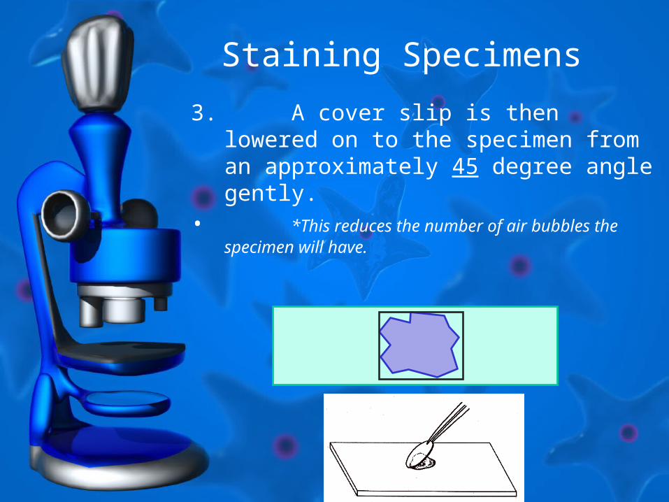
3. A cover slip is then lowered on to the specimen from an approximately 45 degree angle gently.
• *This reduces the number of air bubbles the specimen will have.
Staining Specimens
Staining Specimens
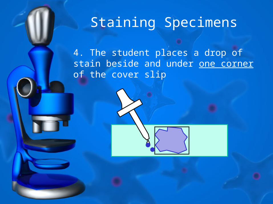
4. The student places a drop of stain beside and under one corner of the cover slip
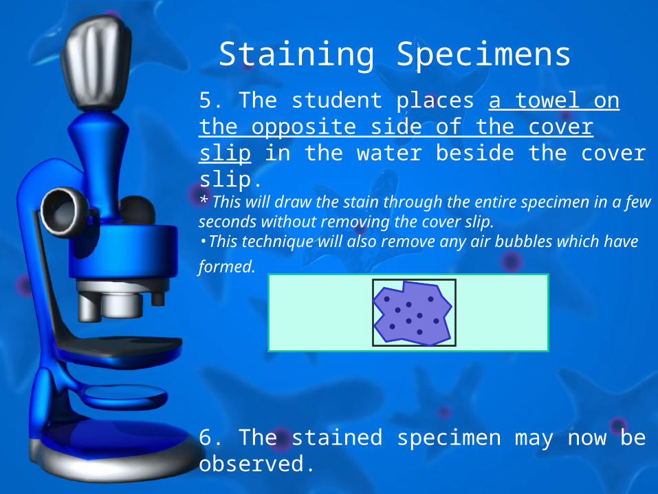
Staining Specimens5. The student places a towel on the opposite side of the cover slip in the water beside the cover slip. * This will draw the stain through the entire specimen in a few seconds without removing the cover slip. •This technique will also remove any air bubbles which
have formed.
6. The stained specimen may now be observed.
Staining Specimens
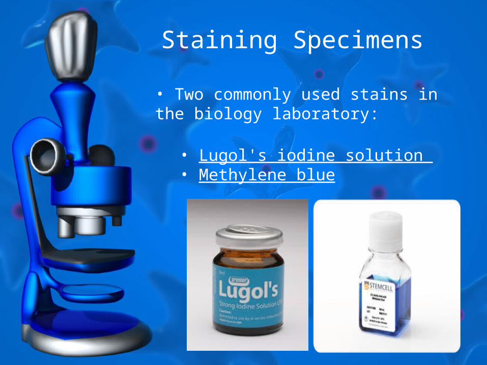
• Two commonly used stains in the biology laboratory:
• Lugol's iodine solution • Methylene blue
Staining Specimens
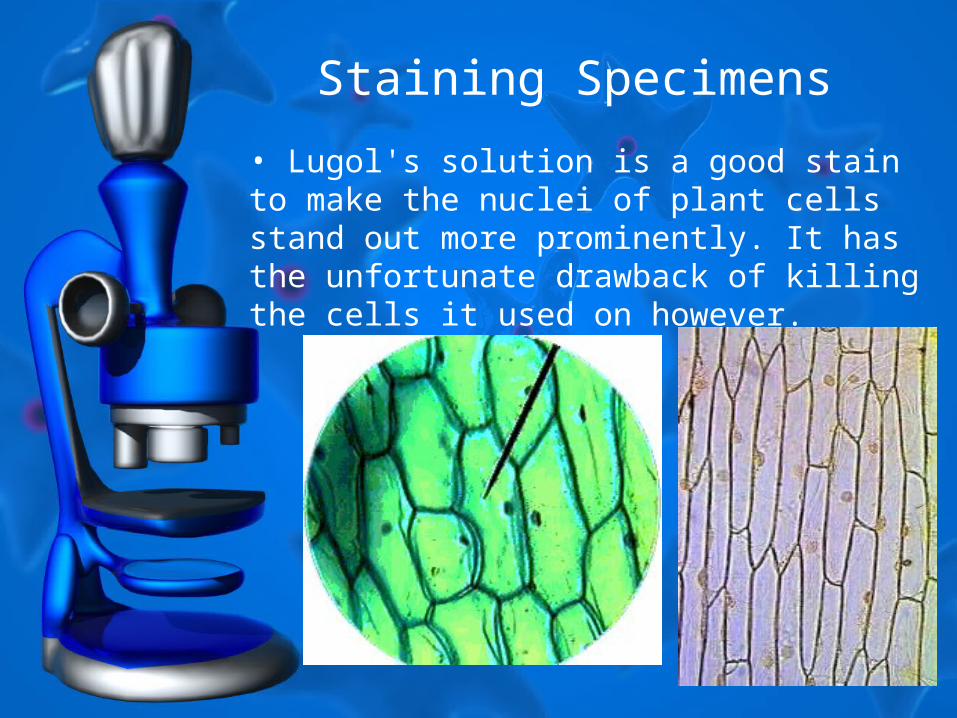
• Lugol's solution is a good stain to make the nuclei of plant cells stand out more prominently. It has the unfortunate drawback of killing the cells it used on however.
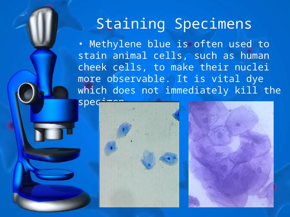
Staining Specimens• Methylene blue is often used to stain animal cells, such as human cheek cells, to make their nuclei more observable. It is vital dye which does not immediately kill the specimen