AidC, a Novel N-Acylhomoserine Lactonase from the …aem.asm.org/content/78/22/7985.full.pdf ·...
8
AidC, a Novel N-Acylhomoserine Lactonase from the Potato Root- Associated Cytophaga-Flavobacteria-Bacteroides (CFB) Group Bacterium Chryseobacterium sp. Strain StRB126 Wen-Zhao Wang, a Tomohiro Morohoshi, a Nobutaka Someya, b and Tsukasa Ikeda a Department of Material and Environmental Chemistry, Graduate School of Engineering, Utsunomiya University, Utsunomiya, Tochigi, Japan, a and Hokkaido Agricultural Research Center (HARC), National Agriculture and Food Research Organization (NARO), Memuro-cho, Kasai-gun, Hokkaido, Japan b N-Acylhomoserine lactones (AHLs) are used as quorum-sensing (QS) signal molecules by many Gram-negative bacteria. We have reported that Chryseobacterium sp. strain StRB126, which was isolated from the root surface of potato, has AHL-degrading activity. In this study, we cloned and characterized the aidC gene from the genomic library of StRB126. AidC has AHL-degrading activity and shows homology to several metallo--lactamase proteins from Bacteroidetes, although not to any known AHL-de- grading enzymes. Purified AidC, as a maltose-binding fusion protein, showed high degrading activity against all tested AHLs, whether short- or long-chain forms, with or without substitution at carbon 3. High-performance liquid chromatography (HPLC) analysis revealed that AidC functions as an AHL lactonase catalyzing AHL ring opening by hydrolyzing lactones. An as- say to determine the effects of covalent and ionic bonding showed that Zn 2 is important to AidC activity both in vitro and in vivo. In addition, the aidC gene could also be PCR amplified from several other Chryseobacterium strains. In conclusion, this study indicated that the aidC gene, encoding a novel AHL lactonase, may be widespread throughout the genus Chryseobac- terium. Our results extend the diversity and known bacterial hosts of AHL-degrading enzymes. Q uorum sensing (QS) is a cell-cell communication mechanism that allows bacterial populations to coordinate gene expres- sion in response to cell density. It is believed that QS constitutes a central element in the social life of bacteria, conferring to the members of a bacterial community the ability to behave as an organized multicellular organism (2, 3). The majority of the QS systems thus far described in Gram-negative bacteria rely on N- acyl-L-homoserine lactones (AHLs) as signaling molecules; these QS systems have been found in more than 100 bacterial species, including many pathogens (10). AHL-mediated QS regulates the expression of many genes and is responsible for bioluminescence, production of pigments and antibiotics, biofilm formation, and many other factors that are important for pathogenicity (6, 24). In general, AHL-negative mutants show defects in pathogenicity; hence, it is expected that disrupting or manipulating the QS sig- nals could inhibit the expression of virulence and infection of host cells (26). Many AHL-degrading genes have been cloned and character- ized from bacteria, fungi, mammalian cells, and metagenomic li- braries constructed from environmental soil samples (20, 22). AHL-degrading enzymes have been divided into two functional groups: AHL lactonases, which catalyze AHL ring opening by hy- drolyzing lactones, and AHL acylases, which hydrolyze the amide bond of AHL (22). The first demonstrated and best-studied AHL- degrading enzyme is AiiA lactonase, which belongs to the metallo- -lactamase superfamily and was identified from Bacillus sp. strain 240B1 (8). Expression of aiiA in the plant pathogen Pecto- bacterium carotovorum subsp. carotovorum significantly attenu- ates pathogenicity on some crops (7). Transgenic plants express- ing AHL lactonase exhibited significantly enhanced resistance to the infection of P. carotovorum subsp. carotovorum (7). The AHL lactonases, which also belong to the metallo-lactamase superfam- ily, were called “AiiA-type lactonases,” with respect to being first identified as AiiAs. AiiA-type lactonases have been identified from a vast range of bacterial original hosts, including Firmicutes, Aci- dobacteria, and Proteobacteria (22, 28). Several AiiA-type lacto- nases have been studied in depth and hold potential for applica- tion in the biocontrol of plant diseases (22, 28). In previous studies, we have reported the putative AHL lacto- nase activities in the potato root-associated strains of Chryseobac- terium sp., which is a member of the Cytophaga-Flavobacterium- Bacteroides (CFB) group (16). Recently, it was reported that the fish pathogen Tenacibaculum maritimum, also a member of the CFB group, produces and degrades AHLs (18). However, to our knowledge, there are no reports of the isolation and characteriza- tion of any AHL-degrading gene from the CFB group bacteria. In this study, we report the cloning of a gene that encodes a novel AiiA-type lactonase from the potato root-associated Chryseobac- terium sp. strain StRB126 and characterization of the enzymatic kinetics of the novel AHL lactonase. MATERIALS AND METHODS Bacterial strains, plasmids, compounds, and growth conditions. Se- lected bacterial strains and plasmids used in this study are listed in Table 1. Escherichia coli was grown at 37°C in Luria-Bertani (LB) medium (19). All other bacteria were grown at 30°C in tryptic soy broth (TSB; Nippon Becton, Dickinson, Tokyo, Japan). Solid bacterial media were made by adding agar at a final concentration of 1.5% to the liquid media. Antibi- otics were added at final concentrations of 100 g/ml ampicillin, 100 g/ml chloramphenicol, 50 g/ml kanamycin, and 10 g/ml gentamicin Received 10 July 2012 Accepted 29 August 2012 Published ahead of print 31 August 2012 Address correspondence to Tomohiro Morohoshi, [email protected] -u.ac.jp. Copyright © 2012, American Society for Microbiology. All Rights Reserved. doi:10.1128/AEM.02188-12 November 2012 Volume 78 Number 22 Applied and Environmental Microbiology p. 7985–7992 aem.asm.org 7985 on October 12, 2018 by guest http://aem.asm.org/ Downloaded from
Transcript of AidC, a Novel N-Acylhomoserine Lactonase from the …aem.asm.org/content/78/22/7985.full.pdf ·...

AidC, a Novel N-Acylhomoserine Lactonase from the Potato Root-Associated Cytophaga-Flavobacteria-Bacteroides (CFB) GroupBacterium Chryseobacterium sp. Strain StRB126
Wen-Zhao Wang,a Tomohiro Morohoshi,a Nobutaka Someya,b and Tsukasa Ikedaa
Department of Material and Environmental Chemistry, Graduate School of Engineering, Utsunomiya University, Utsunomiya, Tochigi, Japan,a and Hokkaido AgriculturalResearch Center (HARC), National Agriculture and Food Research Organization (NARO), Memuro-cho, Kasai-gun, Hokkaido, Japanb
N-Acylhomoserine lactones (AHLs) are used as quorum-sensing (QS) signal molecules by many Gram-negative bacteria. Wehave reported that Chryseobacterium sp. strain StRB126, which was isolated from the root surface of potato, has AHL-degradingactivity. In this study, we cloned and characterized the aidC gene from the genomic library of StRB126. AidC has AHL-degradingactivity and shows homology to several metallo-�-lactamase proteins from Bacteroidetes, although not to any known AHL-de-grading enzymes. Purified AidC, as a maltose-binding fusion protein, showed high degrading activity against all tested AHLs,whether short- or long-chain forms, with or without substitution at carbon 3. High-performance liquid chromatography(HPLC) analysis revealed that AidC functions as an AHL lactonase catalyzing AHL ring opening by hydrolyzing lactones. An as-say to determine the effects of covalent and ionic bonding showed that Zn2� is important to AidC activity both in vitro and invivo. In addition, the aidC gene could also be PCR amplified from several other Chryseobacterium strains. In conclusion, thisstudy indicated that the aidC gene, encoding a novel AHL lactonase, may be widespread throughout the genus Chryseobac-terium. Our results extend the diversity and known bacterial hosts of AHL-degrading enzymes.
Quorum sensing (QS) is a cell-cell communication mechanismthat allows bacterial populations to coordinate gene expres-
sion in response to cell density. It is believed that QS constitutes acentral element in the social life of bacteria, conferring to themembers of a bacterial community the ability to behave as anorganized multicellular organism (2, 3). The majority of the QSsystems thus far described in Gram-negative bacteria rely on N-acyl-L-homoserine lactones (AHLs) as signaling molecules; theseQS systems have been found in more than 100 bacterial species,including many pathogens (10). AHL-mediated QS regulates theexpression of many genes and is responsible for bioluminescence,production of pigments and antibiotics, biofilm formation, andmany other factors that are important for pathogenicity (6, 24). Ingeneral, AHL-negative mutants show defects in pathogenicity;hence, it is expected that disrupting or manipulating the QS sig-nals could inhibit the expression of virulence and infection of hostcells (26).
Many AHL-degrading genes have been cloned and character-ized from bacteria, fungi, mammalian cells, and metagenomic li-braries constructed from environmental soil samples (20, 22).AHL-degrading enzymes have been divided into two functionalgroups: AHL lactonases, which catalyze AHL ring opening by hy-drolyzing lactones, and AHL acylases, which hydrolyze the amidebond of AHL (22). The first demonstrated and best-studied AHL-degrading enzyme is AiiA lactonase, which belongs to the metallo-�-lactamase superfamily and was identified from Bacillus sp.strain 240B1 (8). Expression of aiiA in the plant pathogen Pecto-bacterium carotovorum subsp. carotovorum significantly attenu-ates pathogenicity on some crops (7). Transgenic plants express-ing AHL lactonase exhibited significantly enhanced resistance tothe infection of P. carotovorum subsp. carotovorum (7). The AHLlactonases, which also belong to the metallo-lactamase superfam-ily, were called “AiiA-type lactonases,” with respect to being firstidentified as AiiAs. AiiA-type lactonases have been identified from
a vast range of bacterial original hosts, including Firmicutes, Aci-dobacteria, and Proteobacteria (22, 28). Several AiiA-type lacto-nases have been studied in depth and hold potential for applica-tion in the biocontrol of plant diseases (22, 28).
In previous studies, we have reported the putative AHL lacto-nase activities in the potato root-associated strains of Chryseobac-terium sp., which is a member of the Cytophaga-Flavobacterium-Bacteroides (CFB) group (16). Recently, it was reported that thefish pathogen Tenacibaculum maritimum, also a member of theCFB group, produces and degrades AHLs (18). However, to ourknowledge, there are no reports of the isolation and characteriza-tion of any AHL-degrading gene from the CFB group bacteria. Inthis study, we report the cloning of a gene that encodes a novelAiiA-type lactonase from the potato root-associated Chryseobac-terium sp. strain StRB126 and characterization of the enzymatickinetics of the novel AHL lactonase.
MATERIALS AND METHODSBacterial strains, plasmids, compounds, and growth conditions. Se-lected bacterial strains and plasmids used in this study are listed in Table 1.Escherichia coli was grown at 37°C in Luria-Bertani (LB) medium (19). Allother bacteria were grown at 30°C in tryptic soy broth (TSB; NipponBecton, Dickinson, Tokyo, Japan). Solid bacterial media were made byadding agar at a final concentration of 1.5% to the liquid media. Antibi-otics were added at final concentrations of 100 �g/ml ampicillin, 100�g/ml chloramphenicol, 50 �g/ml kanamycin, and 10 �g/ml gentamicin
Received 10 July 2012 Accepted 29 August 2012
Published ahead of print 31 August 2012
Address correspondence to Tomohiro Morohoshi, [email protected].
Copyright © 2012, American Society for Microbiology. All Rights Reserved.
doi:10.1128/AEM.02188-12
November 2012 Volume 78 Number 22 Applied and Environmental Microbiology p. 7985–7992 aem.asm.org 7985
on October 12, 2018 by guest
http://aem.asm
.org/D
ownloaded from

as required. AHLs used in this study, N-hexanoyl-L-homoserine lactone(C6-HSL), N-octanoyl-L-homoserine lactone (C8-HSL), N-decanoyl-L-homoserine lactone (C10-HSL), N-dodecanoyl-L-homoserine lactone(C12-HSL), N-(3-oxohexanoyl)-L-homoserine lactone (3OC6-HSL),N-(3-oxooctanoyl)-L-homoserine lactone (3OC8-HSL), N-(3-oxode-canoyl)-L-homoserine lactone (3OC10-HSL), and N-(3-oxododecanoyl)-L-homoserine lactone (3OC12-HSL), were synthesized by a previously de-scribed method (4).
Cloning of an AHL-degrading gene from StRB126. A standard pro-tocol for genetic manipulation was used as described previously (19). TheAHL-degrading gene of StRB126 was cloned by a previously describedmethod (26). Briefly, both the genomic library of StRB126 and the AHL-responsive plasmid pLux28 were transformed into E. coli DH5�. Thetransformants were grown on LB agar plates containing ampicillin andchloramphenicol. The resulting colonies were inoculated into 200 �l offresh LB medium containing ampicillin, chloramphenicol, and 0.1 mMisopropyl-�-D-thiogalactopyranoside (IPTG) in 96-well plates. After in-cubation at 30°C for 20 h with gentle shaking, the cell cultures in each wellwere evaluated for luminescence activities using a Luminescenser JNR-II(Atto, Tokyo, Japan). Positive clones were sequenced using a BigDye Ter-minator version 3.1 sequencing kit and an ABI Prism 3100 genetic ana-lyzer (Applied Biosystems, Tokyo, Japan).
Cloning of the aidC gene and its homolog. The aidC-coding regionon the positive clone pST126-E1 was excised by EcoRV and PstI digestionand inserted into the SmaI-PstI sites of the pHSG398 cloning vector forconstruction of pHSG-aidC126. The aidC gene homologs from the root-associated Chryseobacterium sp. strains StRB340, StRB341, and StRB342were amplified with GoTaq DNA polymerase (Promega, Tokyo, Japan)and the following primer set: 5=-CTC AGC TGG CAT TAG CAT GGGTCT TGA ATC-3= and 5=-GCA GAC AGC TAT TCT GTT AGT TTT CAGCAG C-3=. The aidC gene homolog from Chryseobacterium gleum strain
NBRC 15054 was also amplified with the same cycling parameters and thefollowing primer set: 5=-AGC TTG CGC TAG CTT GGG TAT TGA ACCAGG-3= and 5=-CTA CCT GTT ACC TAT CAT CTG CCT CTG TC-3=.PCR was performed using the following cycling parameters: 95°C for 30 s,58°C for 30 s, and 72°C for 1 min for 30 cycles. The PCR products werecloned into the pGEM-T easy cloning vector (Promega), excised by SphIand PstI digestion, and inserted into the SphI-PstI sites of pUC118 forconstruction of pUC118-aidC340, pUC118-aidC341, pUC118-aidC342,and pUC118-aidC15054.
Purification of AidC as an MBP fusion. The aidC-coding region inthe genome of StRB126 was amplified with GoTaq DNA polymerase andthe following primers, containing BamHI and PstI restriction sites (un-derlined), respectively: 5=-GGA TCC ATG AAT AGA AAG CGG GTTATT GGC-3= and 5=-CTG CAG AGC TAT TCT GTT AGT TTC AGCAGC-3=. PCR was performed using the following cycling parameters:95°C for 30 s, 57°C for 30 s, and 72°C for 3 min for 30 cycles. The PCRproducts were cloned into the pGEM-T easy cloning vector, excised byBamHI and PstI digestion, and inserted into the BamHI-PstI sites ofpMAL-c2X for construction of pMAL-aidC. For expression and purifica-tion of the maltose-binding protein (MBP)-AidC fusion protein, thecomplete preculture of E. coli DH5� harboring pMAL-aidC was inocu-lated into 300 ml of fresh LB medium and incubated for 2 h at 37°C, withshaking. Expression of the recombinant MBP-AidC fusion was inducedupon the addition of 0.1 mM IPTG after 2 h, and expression was contin-ued for an additional 8 h at 37°C. After incubation, cells were harvested bycentrifugation and resuspended with 3 ml of BugBuster protein extractionreagent (Novagen, Inc., Madison, WI) and then incubated for 10 min atroom temperature, with gentle shaking. Next, the suspension was soni-cated, centrifuged at 10,000 � g for 5 min to remove the cell debris, andfiltrated. Protein purification was performed using the same method de-scribed previously (26). As a negative control, we also purified an MBP-
TABLE 1 Bacterial strains and plasmids
Strain or plasmid Description Source
StrainsE. coli
DH5� F� supE44 �lacU169 (�80 lacZ�M15) hsdR17 recA1 endA1 gyrA96 thi-1 relA1 Nippon Gene
C. violaceumCV026 ATCC 31532 derivative, cviI::Tn5xylE Kmr, Smr 11VIR07 ATCC 12472 derivative, cviI::Kmr, Apr 13
Chryseobacterium sp.StRB126 AHL-degrading strain isolated from potato root surface 16StRB340 AHL-degrading strain isolated from potato root surface 16StRB341 AHL-degrading strain isolated from potato root surface 16StRB342 AHL-degrading strain isolated from potato root surface 16
C. gleumNBRC 15054 Clinical isolate, type strain NBRC
PlasmidspUC118 Cloning vector; Apr TaKaRa BiopST126-E1 8.8-kb Sau3AI fragment from StRB126 genomic DNA in pUC118 This studypHSG398 Cloning vector; Cmr TaKaRa BiopHSG-aidC126 pHSG398 containing aidC gene from StRB126 This studypLux28 8.8-kb SalI fragment from pHV200 in pSTV28 vector; Cmr 26pUC118-aidC340 pUC118 containing aidC gene from StRB340 This studypUC118-aidC341 pUC118 containing aidC gene from StRB341 This studypUC118-aidC342 pUC118 containing aidC gene from StRB342 This studypUC118-aidC15054 pUC118 containing aidC gene from NBRC15054 This studypGEM-T easy Cloning vector; Apr PromegapMAL-c2X Cloning vector to make MBP fusions; Apr New England BiolabspMAL-aidC pMAL-c2X containing aidC gene from StRB126 This study
Wang et al.
7986 aem.asm.org Applied and Environmental Microbiology
on October 12, 2018 by guest
http://aem.asm
.org/D
ownloaded from

LacZ� fusion from E. coli DH5� harboring pMAL-c2X. Expression andpurification of recombinant MBP-AidC and MBP-LacZ� were checkedby SDS-PAGE analysis.
Detection of AHL-degrading activity of AidC. The residual AHLswere detected using AHL biosensors Chromobacterium violaceum CV026and VIR07, which respond to exogenous AHLs by producing the purplepigment violacein (11, 13). For detecting the AHL-degrading activity ofAidC in vivo and in vitro, we used a previously described method (26). Todetermine the chemical structures of products from the reaction betweenAidC and AHLs, 50 �l of purified protein solution was mixed with 49 �l ofcolumn buffer [20 mM Tris-HCl buffer and 200 mM NaCl (pH 7.4)] and1 �l of 200 mM C10-HSL stock solution (in methanol). After incubationat 30°C for 10 min, reactions were stopped with an equal volume of ace-tonitrile, and the mixture was vortexed and centrifuged to pellet the pre-cipitated protein. The hydrolyzed C10-HSL, as a control, was made byincubating C10-HSL in 10 mM NaOH at room temperature for 30 min.Samples (25 �l) were analyzed by high-performance liquid chromatogra-phy (HPLC) according to a previously described method (26).
Characterization of enzymatic activity of AidC. To determine theenzyme kinetics of AidC, purified MBP-AidC (final concentration, 1 �M)was added to a substrate solution in the column buffer, with a final volumeof 40 �l �-butyrolactone (�-BL). L-Homoserine lactone (HSL), C6-HSL,C8-HSL, C10-HSL, C12-HSL, 3OC6-HSL, 3OC8-HSL, and 3OC12-HSLwere selected as the substrates for an enzyme specificity analysis. Thereaction mixtures were incubated at 30°C, stopped by adding 40 �l ace-tonitrile, and subjected to HPLC analysis. The residual AHL and its hy-drolysis product were quantified by HPLC. All experiments were per-formed in triplicate, and all velocities were determined at time points atwhich no more than 20% of the substrate had been consumed. The kcat
and Km values were calculated based on Michaelis-Menten equation. Theeffects of various metal ions and a metal-chelating reagent (EDTA) onAidC activity were examined both in vitro and in vivo. For the in vitroassay, 1 mM EDTA and 1 mM metal ions comprising Cu2, Ca2, Fe2,Mn2, Mg2, Zn2, and Co2 were mixed with 1 �M purified AidCprotein in column buffer (pH 7.4). After incubation at 30°C for 10 min,the remaining activity was measured under the standard conditions de-scribed above. For the in vivo assay, the full preculture of E. coli harboringthe plasmid pMAL-aidC was diluted into fresh LB medium with 1 mMEDTA and 1 mM metal ions. IPTG was then added to induce AidC over-expression after 4 h cultivation. After an additional 8-h cultivation, crudecell extracts were prepared and adjusted to the same concentration. TheAHL-degrading activities of crude cell extracts were examined by the sameprocedure as used in the in vitro assay.
Nucleotide sequence accession number. The nucleotide sequences ofaidC and its flanking open reading frames (ORFs) from Chryseobacteriumsp. StRB126 have been deposited in the DDBJ/EMBL/GenBank databasesunder accession no. AB733663.
RESULTS AND DISCUSSIONIdentification of AHL-degrading gene from the Chryseobac-terium sp. StRB126. For cloning the AHL degradation gene, apUC118-based StRB126 genomic library was constructed andused to transform E. coli DH5� harboring pLux28 (26). We thenmeasured the luminescence activities of the resulting colonies onfresh LB medium. When approximately 2,300 transformants werescreened, three clones expressed a low level of luminescence. Toelucidate whether the reduction in luminescence resulted fromdegradation of AHL, E. coli DH5� cells harboring the plasmidsfrom the positive clones were inoculated into LB medium con-taining 10 �M C6-HSL or C10-HSL. After incubation for 6 h, theresidual AHL in the culture supernatant was detected by Chromo-bacterium violaceum reporters, which produced the purple pig-ment violacein in response to the presence of AHLs. Conse-quently, E. coli DH5� harboring a single plasmid, designated
pST126-E1, showed clear AHL-degrading activity against bothC6-HSL and C10-HSL (Fig. 1A).
The sequence of the genomic DNA fragment (8,784 bp), whichwas inserted into pST126-E1, contained five complete open read-ing frames (ORFs) (Fig. 1B). The third ORF (orf3) encoded aputative metallo-�-lactamase of 330 amino acids. The Orf3 aminoacid sequence showed low-level similarity (13% identity) to theknown AHL lactonases, including AiiA from Bacillus sp. 240B1 (7,8). To test whether orf3 encodes an AHL-degrading enzyme, thecomplete orf3 was amplified by PCR and subcloned into thepHSG398 easy vector. E. coli DH5� harboring the orf3-expressingplasmid showed clear AHL-degrading activity at the same level asE. coli DH5� harboring pST126-E1 (Fig. 1A). These results sug-gested that orf3 encodes an AHL-degrading enzyme. Therefore,we named orf3 the autoinducer degrading gene from Chryseobac-terium sp. (aidC) as being the first AHL-degrading gene identifiedfrom the genus Chryseobacterium.
AidC encodes an AHL lactonase. We purified AidC as an MBPfusion in an in vitro AHL-degrading assay. The MBP-AidC fusionprotein was overproduced in E. coli DH5� and purified by maltoseaffinity chromatography, and the purified proteins were analyzedby 10% SDS-PAGE. The results revealed that the overexpressedprotein was approximately 80 kDa in size (Fig. 2A), which wascongruent with the predicted molecular weight of MBP-AidC,based on its amino acid sequence. When the AHL-degrading ac-tivity of the purified protein was examined, purified MBP-AidCcompletely degraded 100 �M C6-HSL and C10-HSL within 2 h,whereas MBP-LacZ� did not (Fig. 2B). In our previous study,putative AHL lactonase activities were detected from StRB126(16). Therefore, to determine whether AidC functions as an AHLlactonase, the structure of C10-HSL treated with MBP-AidC wasanalyzed by HPLC. Fractionation of the C10-HSL standard re-vealed a single major HPLC peak, with a retention time of approx-imately 6.3 min (Fig. 3A). To prepare the lactone ring-openedC10-HSL, C10-HSL was hydrolyzed by 10 mM NaOH. Fraction-ation of the hydrolyzed C10-HSL revealed one major HPLC peakwith a retention time of approximately 3.9 min (Fig. 3B). To ex-amine the enzymatic properties of AidC, solutions of MBP-LacZ�and MBP-AidC were mixed with C10-HSL and incubated at 30°C
FIG 1 (A) AHL-degrading activity of E. coli DH5� harboring pUC118,pST126-E1, and pHSG-aidC. Subcultures of E. coli DH5� harboring theseplasmids were mixed with 10 �M C6-HSL or C10-HSL and incubated at 37°Cfor 2 h. The residual AHL was detected using C. violaceum strain CV026 orVIR07. (B) Arrangement of the predicted ORFs on the original genomic clonepST126-E1. The scale represents a 1-kb length of nucleotides. The filled arrowindicates the aidC gene, and the open arrows indicate the other ORFs. EV,EcoRV; P, PstI.
AHL Lactonase from Chryseobacterium
November 2012 Volume 78 Number 22 aem.asm.org 7987
on October 12, 2018 by guest
http://aem.asm
.org/D
ownloaded from
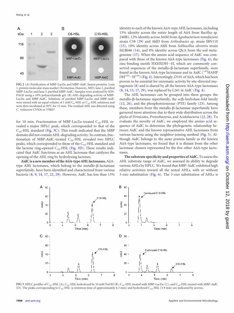
for 10 min. Fractionation of MBP-LacZ�-treated C10-HSL re-vealed a major HPLC peak, which corresponded to that of theC10-HSL standard (Fig. 3C). This result indicated that the MBPdomain did not contain AHL-degrading activity. In contrast, frac-tionation of MBP-AidC-treated C10-HSL revealed two HPLCpeaks, which corresponded to those of the C10-HSL standard andthe lactone ring-opened C10-HSL (Fig. 3D). These results indi-cated that AidC functions as an AHL lactonase that catalyzes theopening of the AHL ring by hydrolyzing lactones.
AidC is a new member of the AiiA-type AHL lactonases. AiiA-type AHL lactonases, which belong to the metallo-�-lactamasesuperfamily, have been identified and characterized from variousbacteria (8, 9, 14, 17, 22, 29). However, AidC has less than 13%
identity to each of the known AiiA-type AHL lactonases, including13% identity across the entire length of AiiA from Bacillus sp.240B1, 12% identity across AttM from Agrobacterium tumefaciensstrain C58 (29) and AhlD from Arthrobacter sp. strain IBN110(15), 10% identity across AhlS from Solibacillus silvestris strainStLB046 (14), and 8% identity across QlcA from the soil meta-genome (17). When the amino acid sequence of AidC was com-pared with those of the known AiiA-type lactonases (Fig. 4), thezinc-binding motifs HXHXDHH, which are commonly con-served sequences of the metallo-�-lactamase superfamily, werefound in the known AiiA-type lactonases and in AidC (145HAHPDH150H239) (Fig. 4). Interestingly, D191 of AiiA, which has beenproven to be essential for enzymatic activity by site-directed mu-tagenesis (8) and is shared by all the known AiiA-type lactonases(8, 14, 15, 17, 29), was replaced by L261 in AidC (Fig. 4).
The AHL lactonases can be grouped into three groups: themetallo-�-lactamase superfamily, the �/�-hydrolase-fold family(12, 26), and the phosphotriesterase (PTE) family (23). Amongthese, members from the metallo-�-lactamase superfamily havegarnered more attention due to their wide distribution across thephyla of Firmicutes, Proteobacteria, and Acidobacteria (22, 28). Toevaluate the novelty of AidC, we employed the amino acid se-quence of AidC to determine the phylogenetic relationship be-tween AidC and the known representative AHL lactonases fromvarious bacteria using the neighbor-joining method (Fig. 5). Al-though AidC belongs to the same protein family as the knownAiiA-type lactonases, we found that it is distant from the otherlactonase clusters represented by the five other AiiA-type lacto-nases.
The substrate specificity and properties of AidC. To assess theAHL substrate range of AidC, we assessed its ability to degradevarious AHLs by HPLC. We found that MBP-AidC exhibited highrelative activities toward all the tested AHLs, with or without3-oxo substitution (Fig. 6). The 3-oxo substitution of AHLs is
FIG 2 (A) Purification of MBP-LacZ� and MBP-AidC fusion proteins. Lane1, protein molecular mass marker (Fermentas, Hanover, MD); lane 2, purifiedMBP-LacZ�; and lane 3, purified MBP-AidC. Samples were analyzed by SDS-PAGE using a 10% polyacrylamide gel. (B) AHL-degrading activity of MBP-LacZ� and MBP-AidC. Solutions of purified MBP-LacZ� and MBP-AidCwere mixed with an equal volume of 1 mM C6-HSL or C10-HSL solutions andwere then incubated at 30°C for 15 min. The residual AHL was detected usingC. violaceum CV026 or VIR07.
FIG 3 HPLC profiles of C10-HSL (A), C10-HSL hydrolyzed by 10 mM NaOH (B), C10-HSL treated with MBP-LacZ� (C), and C10-HSL treated with MBP-AidC(D). The peaks corresponding to C10-HSL (a retention time of approximately 6.3 min) and hydrolyzed C10-HSL (3.9 min) are indicated by arrows.
Wang et al.
7988 aem.asm.org Applied and Environmental Microbiology
on October 12, 2018 by guest
http://aem.asm
.org/D
ownloaded from

known to negatively affect the enzymatic activity of AiiA fromBacillus sp. 240B1 (25, 26). AidC, on the other hand, showedslightly higher degrading activity against 3-oxo-substituted AHLsthan against unsubstituted AHLs (Fig. 6). In addition, althoughdifferences in acyl acid chain length did not significantly affect theenzymatic activity of AiiA (25), AidC was somewhat more effec-tive at degrading C6-HSL and C8-HSL than C10-HSL and C12-HSL(Fig. 6). AidC did not show any degrading activity toward L-ho-moserine lactone or �-butyrolactone, similar to AiiA (25).
The optimal pH for AHL-degrading activity of MBP-AidC wasexamined using C10-HSL as the substrate at 30°C. AHL-degradingactivity was enhanced as pH increased and reached a maximum atpH 8, but no or little activity was detected when pH was adjusted
to pH 5 or below (Fig. 7A). The optimal temperature for the AHL-degrading activity of MBP-AidC was also examined using C10-HSL as the substrate. MBP-AidC displayed more than 80% of itsmaximum activity at 20°C to 50°C, but the relative activity wasgreatly reduced when temperature exceeded 50°C (Fig. 7B). Cor-respondingly, strain StRB126 could grow at temperatures rangingfrom 20°C to 50°C, reaching its optimum temperature at 30°C(data not shown). To determine the thermostability of AidC, pu-rified MBP-AidC was preincubated at various temperatures for2 h and the residual enzymatic activity was then determined. More
FIG 4 Comparison of amino acid sequences of AidC and five known AiiA-type lactonases. The amino acid sequence of AidC was compared with those of AiiAfrom Bacillus sp. 240B1, AhlD from Arthrobacter sp. strain IBN110, AttM from A. tumefaciens strain A6, AhlS from S. silvestris, and QlcA from the soilmetagenome. Sequences were aligned using the ClustalW program (21) and shaded using the Genedoc program (http://www.nrbsc.org/gfx/genedoc/). The twozinc-binding motifs are boxed with rectangles. The amino acid residues that are essential for enzymatic activity of the known AHL lactonases are indicated bysolid triangles.
FIG 5 Phylogenetic tree based on amino acid sequences of AidC, AiiA, AhlD,AttM, AhlS, QlcA, and AiiM from Microbacterium testaceum strain StLB037,AidH from Ochrobactrum sp. strain T63, and QsdA from Rhodococcus eryth-ropolis strain W2. The dendrogram was constructed by the neighbor-joiningmethod using the ClustalW program that is included with NJplot software.The scale bar represents 0.1 substitutions per amino acid position.
FIG 6 Substrate specificity of purified MBP-AidC. Purified MBP-AidC wasmixed with substrate solutions in the column buffer (pH 7.4). After incubationat 30°C for 10 min, the residual AHL and its hydrolysis products were quanti-fied by HPLC. We defined 100% relative activity as the activity toward C6-HSL.The data were reproduced at least three times, and error bars indicate standarddeviations.
AHL Lactonase from Chryseobacterium
November 2012 Volume 78 Number 22 aem.asm.org 7989
on October 12, 2018 by guest
http://aem.asm
.org/D
ownloaded from

than 90% of the activity remained after incubation at tempera-tures of 50°C or less, but the enzymatic activity was markedlyreduced after incubation at temperatures over 60°C (data notshown).
Kinetic analysis of AHL lactonase activity of AidC. AHL hy-drolysis kinetics of AidC was determined by a plot of the substrateconcentration and velocity. The kcat and Km values were calculatedby fitting the data to the Michaelis-Menten equation (Table 2).The AHL lactonase activity of AidC showed comparable catalyticactivities against a range of structurally different AHLs, with kcat
and Km values in the range of 1.97 to 2.35 s�1 and 0.046 to 0.072mM, respectively. Within these narrow ranges, the enzymeshowed higher affinities (Km), faster hydrolysis rates (kcat), andstronger catalytic efficiencies (kcat/Km) toward the AHLs withshort acyl side chains than toward those with the longer deriva-tives. Additionally, AidC displayed somewhat lower kcat andkcat/Km values against the unsubstituted AHLs than against the3-oxo-substituted AHLs (Table 2). Hydrolysis kinetics of the AiiAfrom Bacillus sp. 240B1 have been determined by plotting velocityversus substrate concentration (25). Compared with AiiA fromBacillus sp. 240B1, AidC showed lower Km and kcat values buthigher catalytic efficiencies (kcat/Km) toward the AHLs (data notshown). In addition, Km values of AidC against AHLs are at levelsof tens of micromolar and even higher for several other AHL lac-tonases (1, 5, 25), but AHLs can function as signals at multiple
orders of magnitude below that level. Thus, in natural environ-ments, the reaction rate should depend on the AidC concentrationand the AHL concentration. The AidC/AHL reaction resembles abimolecular reaction with a corresponding catalytic efficiency(kcat/Km). These results suggested that AidC may function muchmore effectively at quenching the bacterial QS systems which em-ploy higher concentrations of AHLs than those employing lowerconcentrations of AHLs.
Zinc is essential for AHL-degrading activity of AidC. Se-quence alignment of AidC and the known AiiA-type lactonasesrevealed that they share the zinc-binding motifs HXHXDHH inthe central region (Fig. 4). To verify whether AidC requires anycofactor for its enzymatic activity, the effects of several divalentmetal ions on the AHL-degrading activity of AidC were evaluated.For the in vitro assay, purified MBP-AidC was mixed with C10-HSL in column buffer containing EDTA and divalent metal ions.When 1 mM EDTA was added to this reaction mixture, the AHL-degrading activity of AidC was decreased by approximately 50%(Fig. 8A). However, AidC activity was successfully restored to 82%by the addition of Zn2, but not by any other of the metal ionstested (Fig. 8A). For in vivo assays, E. coli harboring pMAL-aidCwas cultivated in the LB medium containing EDTA and divalentmetal ions, and the AHL-degrading activity of AidC in the crudecell extracts was then determined. The AHL-degrading activity ofAidC was abolished by the addition of 1 mM EDTA but was re-stored by the addition of Zn2 (Fig. 8B). These results suggestedthat Zn2 is essential for the AHL-degrading activity of AiiA andthe known AiiA-type lactonases. In addition, the AHL-degradingactivity of AidC activity was markedly inhibited by the addition ofCu2 (data not shown). This feature of AidC also corresponded tothat of the known AiiA-type lactonases (25).
aidC is widely conserved in the genus Chryseobacterium. In aprevious study, we isolated 34 AHL-degrading Chryseobacteriumstrains from the roots of potato plants and divided them into eightrelated species groups (16). We now attempted to amplify by PCRprocedures the aidC-homologous genes from the three AHL-de-grading Chryseobacterium strains which belong to group VI, aswell as StRB126 (16). The group VI strains, which were StLB126,StLB340, StLB341, and StLB342, had higher AHL-degrading ac-tivity than the other groups (16). Specific primers were designed
FIG 7 (A) Optimal pH of AHL-degrading activity of purified MBP-AidC. Purified MBP-AiiM was mixed with C10-HSL in reaction buffer with a pH rangingfrom pH 3 to pH 8 and was then incubated at 30°C. The data were reproduced at least three times, and error bars indicate standard deviations. (B) Optimaltemperature of AHL-degrading activity of purified MBP-AidC. Purified MBP-AiiM was mixed with C10-HSL in the reaction buffer (pH 7.4) and incubated attemperatures ranging from 10 to 80°C. After incubation for 10 min, the residual substrate was quantified by HPLC. We defined 100% relative activity as theactivity in the column buffer (pH 7.4) at 30°C. Control samples, which had been prepared under the same conditions but without AidC, were used to exclude theinfluence of autodegradation at high pH or high temperature. The data were reproduced at least three times, and error bars indicate standard deviations.
TABLE 2 Kinetic parameters of AidC against AHLsa
AHL kcat (s�1) Km (mM)kcat/Km
(mM�1 · s�1)
C6-HSL 2.31 � 0.16 0.055 � 0.0043 42.03OC6-HSL 2.35 � 0.29 0.046 � 0.0021 51.1C8-HSL 2.01 � 0.31* 0.064 � 0.0049* 31.43OC8-HSL 2.12 � 0.24* 0.064 � 0.0037* 33.1C10-HSL ND ND ND3OC10-HSL 1.97 � 0.11* 0.072 � 0.0053* 27.4C12-HSL ND ND ND3OC12-HSL ND ND NDa The data are the means from triplicate experiments. The significance was determinedby Student’s t test (*, P � 0.05). ND, not determined due to poor solubility of thesubstrate in the column buffer.
Wang et al.
7990 aem.asm.org Applied and Environmental Microbiology
on October 12, 2018 by guest
http://aem.asm
.org/D
ownloaded from

based on the DNA sequences of aidC from StRB126. FollowingPCR amplification and DNA sequencing, aidC gene homologswere successfully cloned from all group VI strains. AidC fromStLB126, StLB340, StLB341, and StLB342 had highly similar DNAsequences (more than 99% identity). In addition, E. coli DH5�harboring these aidC homologs showed significant AHL-degrad-ing activity against various AHLs (data not shown). These resultssuggested that aidC sequences are highly conserved among groupVI strains.
Based on BLAST search results, an aidC homologous gene wasfound in the whole-genome shotgun sequence of C. gleum NBRC15054 (accession no. ACKQ00000000). However, we have previ-ously reported that C. gleum NBRC 15054 did not show any AHL-degrading activity (16). To assess whether the AidC homolog(aidC15054) from NBRC 15054 shows AHL lactonase activity,aidC15054 was amplified from the chromosome of NBRC 15054and cloned into the pUC118 cloning vector. E. coli DH5� harbor-ing pUC118-aidC15054 showed a clear AHL-degrading activitylevel similar to AidC from StRB126. These results may suggest thatthe aidC gene homolog was not expressed in NBRC 15054 cells.NBRC 15054 appears distant from StRB126 in the phylogeneticanalysis. Therefore, these results suggested that the aidC gene ho-molog may be widespread throughout Chryseobacterium strains,with or without AHL-degrading activity. In addition, in our pre-vious study, aiiM homologous genes (encoding a group of AHLlactonases, AiiM) were only found from the potato leaf-associatedMicrobacterium isolates, but not found in strains from other re-sources (27). Correspondingly, although the aiiM homologousgenes were highly similar to each other, their neighboring genesshowed high diversity (27). When comparing the neighboringgenes of aidC and aidC15054, we found it interesting that, althoughtheir downstream genes showed no identity, the nearest three up-stream genes of aidC shared high identities with the correspond-ing genes located upstream of aidC15054 (data not shown). Theseresults also suggested that the aidC gene may be widespread in thegenus Chryseobacterium.
In summary, in this study, a novel AHL lactonase, AidC, wasidentified and characterized from a CFB group. Although AidCshowed conserved zinc-binding motif sequences, as did theknown AiiA-type lactonases, it showed little identity to knownAHL lactonases; thus, we classified it into a novel group. In our
previous study, AHL-degrading Chryseobacterium strains couldbe isolated from potato roots, which were collected from variousareas (16). Our results suggested that CFB bacteria may also retainfunctional AHL lactonases, extending the diversity of AiiA-typelactonases and again highlighting the relationship between AHLlactonase and metallo-�-lactamases.
ACKNOWLEDGMENT
This work was supported by Grants-in-Aid from the Bio-oriented Tech-nology Research Advancement Institution (BRAIN), Japan.
REFERENCES1. Afriat-Jurnou L, Jackson CJ, Tawfik DS. 2012. Reconstructing a missing
link in the evolution of a recently diverged phosphotriesterase by active-site loop remodeling. Biochemistry 51:6047– 6055.
2. Antunes LC, Ferreira RB. 2009. Intercellular communication in bacteria.Critic. Rev. Microbiol. 35:69 – 80.
3. Atkinson S, Williams P. 2009. Quorum sensing and social networking inthe microbial world. J. R. Soc. Interface 6:959 –978.
4. Chhabra SR, et al. 2003. Synthetic analogues of the bacterial signal (quo-rum sensing) molecule N-(3-oxododecanoyl)-L-homoserine lactone asimmune modulators. J. Med. Chem. 46:97–104.
5. Chow JY, et al. 2010. Directed evolution of a thermostable quorum-quenching lactonase from the amidohydrolase superfamily. J. Biol. Chem.285:40911– 40920.
6. de Kievit TR, Iglewski BH. 2000. Bacterial quorum sensing in pathogenicrelationships. Infect. Immun. 68:4839 – 4849.
7. Dong YH, et al. 2001. Quenching quorum-sensing-dependent bacterialinfection by an N-acyl homoserine lactonase. Nature 411:813– 817.
8. Dong YH, Xu JL, Li XZ, Zhang LH. 2000. AiiA, an enzyme that inacti-vates the acylhomoserine lactone quorum-sensing signal and attenuatesthe virulence of Erwinia carotovora. Proc. Natl. Acad. Sci. U. S. A. 97:3526 –3531.
9. Flagan S, Ching WK, Leadbetter JR. 2003. Arthrobacter strain VAI-Autilizes acyl-homoserine lactone inactivation products and stimulatesquorum signal biodegradation by Variovorax paradoxus. Appl. Environ.Microbiol. 69:909 –916.
10. Galloway WR, Hodgkinson JT, Bowden SD, Welch M, Spring DR. 2011.Quorum sensing in Gram-negative bacteria: small-molecule modulationof AHL and AI-2 quorum sensing pathways. Chem. Rev. 111:28 – 67.
11. McClean KH, et al. 1997. Quorum sensing and Chromobacterium viola-ceum: exploitation of violacein production and inhibition for the detec-tion of N-acylhomoserine lactones. Microbiology 143:3703–3711.
12. Mei GY, Yan XX, Turak A, Luo ZQ, Zhang LQ. 2010. AidH, analpha/beta-hydrolase fold family member from an Ochrobactrum sp.strain, is a novel N-acylhomoserine lactonase. Appl. Environ. Microbiol.76:4933– 4942.
FIG 8 The effects of EDTA and metal ions on AHL-degrading activity of AidC by in vitro (A) and in vivo (B) assays. For the in vitro assay, purified MBP-AidCwas incubated in reaction buffer containing substrate and 1 mM each metal ion. After incubation at 30°C for 10 min, the residual activity of AidC was analyzedby HPLC. For the in vivo assay, E. coli harboring pMAL-aidC was cultivated with 1 mM EDTA and 1 mM each metal ion. Crude cell extracts were prepared andused for the AHL-degrading assay. We defined 100% relative activity as the activity in the absence of EDTA or metal ions; this was represented as PC (positivecontrol). The data were reproduced at least three times, and error bars indicate standard deviations.
AHL Lactonase from Chryseobacterium
November 2012 Volume 78 Number 22 aem.asm.org 7991
on October 12, 2018 by guest
http://aem.asm
.org/D
ownloaded from

13. Morohoshi T, Kato M, Fukamachi K, Kato N, Ikeda T. 2008. N-acylhomoserine lactone regulates violacein production in Chromobac-terium violaceum type strain ATCC 12472. FEMS Microbiol. Lett. 279:124 –130.
14. Morohoshi T, Tominaga Y, Someya N, Ikeda T. 2012. Complete genomesequence and characterization of the N-acylhomoserine lactone-degrading gene of the potato leaf-associated Solibacillus silvestris. J. Biosci.Bioeng. 113:20 –25.
15. Park SY, et al. 2003. AhlD, an N-acylhomoserine lactonase in Arthrobac-ter sp., and predicted homologues in other bacteria. Microbiology 149:1541–1550.
16. Rashid R, Morohoshi T, Someya N, Ikeda T. 2011. Degradation ofN-acylhomoserine lactone quorum sensing signaling molecules by potatoroot surface-associated Chryseobacterium strains. Microb. Environ. 26:144 –148.
17. Riaz K, et al. 2008. A metagenomic analysis of soil bacteria extends thediversity of quorum-quenching lactonases. Environ. Microbiol. 10:560 –570.
18. Romero M, Avendano-Herrera R, Magarinos B, Camara M, Otero A.2010. Acylhomoserine lactone production and degradation by the fishpathogen Tenacibaculum maritimum, a member of the Cytophaga-Flavobacterium-Bacteroides (CFB) group. FEMS Microbiol. Lett. 304:131–139.
19. Sambrook J, Fritsch EF, Maniatis T. 1989. Molecular cloning: a labora-tory manual, 2nd ed. Cold Spring Harbor Laboratory Press, Cold SpringHarbor, NY.
20. Schipper C, et al. 2009. Metagenome-derived clones encoding two novellactonase family proteins involved in biofilm inhibition in Pseudomonasaeruginosa. Appl. Environ. Microbiol. 75:224 –233.
21. Thompson JD, Higgins DG, Gibson TJ. 1994. CLUSTAL W: improving
the sensitivity of progressive multiple sequence alignment through se-quence weighting, position-specific gap penalties and weight matrixchoice. Nucleic Acids Res. 22:4673– 4680.
22. Uroz S, Dessaux Y, Oger P. 2009. Quorum sensing and quorum quench-ing: the yin and yang of bacterial communication. Chembiochem 10:205–216.
23. Uroz S, et al. 2008. A Rhodococcus qsdA-encoded enzyme defines a novelclass of large-spectrum quorum-quenching lactonases. Appl. Environ.Microbiol. 74:1357–1366.
24. Visick KL, Fuqua C. 2005. Decoding microbial chatter: cell-cell commu-nication in bacteria. J. Bacteriol. 187:5507–5519.
25. Wang LH, Weng LX, Dong YH, Zhang LH. 2004. Specificity and enzymekinetics of the quorum-quenching N-acyl homoserine lactone lactonase(AHL-lactonase). J. Biol. Chem. 279:13645–13651.
26. Wang WZ, Morohoshi T, Ikenoya M, Someya N, Ikeda T. 2010. AiiM,a novel class of N-acylhomoserine lactonase from the leaf-associated bac-terium Microbacterium testaceum. Appl. Environ. Microbiol. 76:2524 –2530.
27. Wang WZ, Morohoshi T, Someya N, Ikeda T. 2012. Diversity anddistribution of N-acylhomoserine lactone (AHL)-degrading activity andAHL-lactonase (AiiM) in genus Microbacterium. Microbes Environ. 27:330 –333.
28. Zhang HB, Wang LH, Zhang LH. 2007. Detection and analysis of quo-rum-quenching enzymes against acyl homoserine lactone quorum-sensing signals. Curr. Protoc. Microbiol. 1:1C.3. doi:10.1002/9780471729259.mc01c03s05.
29. Zhang HB, Wang LH, Zhang LH. 2002. Genetic control of quorum-sensing signal turnover in Agrobacterium tumefaciens. Proc. Natl. Acad.Sci. U. S. A. 99:4638 – 4643.
Wang et al.
7992 aem.asm.org Applied and Environmental Microbiology
on October 12, 2018 by guest
http://aem.asm
.org/D
ownloaded from