AHA/ASA Guideline - SISA LombardiaAHA/ASA Guideline Downloaded from stroke.ahajournals.org by on...
24
ISSN: 1524-4628 Copyright © 2007 American Heart Association. All rights reserved. Print ISSN: 0039-2499. Online Stroke is published by the American Heart Association. 7272 Greenville Avenue, Dallas, TX 72514 DOI: 10.1161/STROKEAHA.107.183689 2007;38;2001-2023; originally published online May 3, 2007; Stroke and Mario Zuccarello Derk Krieger, Marc Mayberg, Lewis Morgenstern, Christopher S. Ogilvy, Paul Vespa Joseph Broderick, Sander Connolly, Edward Feldmann, Daniel Hanley, Carlos Kase, the value of this guideline as an educational tool for neurologists. Interdisciplinary Working Group: The American Academy of Neurology affirms Research Council, and the Quality of Care and Outcomes in Research Association/American Stroke Association Stroke Council, High Blood Pressure Adults: 2007 Update: A Guideline From the American Heart Guidelines for the Management of Spontaneous Intracerebral Hemorrhage in http://stroke.ahajournals.org/cgi/content/full/38/6/2001 located on the World Wide Web at: The online version of this article, along with updated information and services, is http://www.lww.com/reprints Reprints: Information about reprints can be found online at [email protected] 410-528-8550. E-mail: Fax: Kluwer Health, 351 West Camden Street, Baltimore, MD 21202-2436. Phone: 410-528-4050. Permissions: Permissions & Rights Desk, Lippincott Williams & Wilkins, a division of Wolters http://stroke.ahajournals.org/subscriptions/ Subscriptions: Information about subscribing to Stroke is online at by on June 26, 2007 stroke.ahajournals.org Downloaded from
Transcript of AHA/ASA Guideline - SISA LombardiaAHA/ASA Guideline Downloaded from stroke.ahajournals.org by on...

ISSN: 1524-4628 Copyright © 2007 American Heart Association. All rights reserved. Print ISSN: 0039-2499. OnlineStroke is published by the American Heart Association. 7272 Greenville Avenue, Dallas, TX 72514
DOI: 10.1161/STROKEAHA.107.183689 2007;38;2001-2023; originally published online May 3, 2007; Stroke
and Mario Zuccarello Derk Krieger, Marc Mayberg, Lewis Morgenstern, Christopher S. Ogilvy, Paul Vespa Joseph Broderick, Sander Connolly, Edward Feldmann, Daniel Hanley, Carlos Kase,
the value of this guideline as an educational tool for neurologists.Interdisciplinary Working Group: The American Academy of Neurology affirms
Research Council, and the Quality of Care and Outcomes in Research Association/American Stroke Association Stroke Council, High Blood Pressure
Adults: 2007 Update: A Guideline From the American Heart Guidelines for the Management of Spontaneous Intracerebral Hemorrhage in
http://stroke.ahajournals.org/cgi/content/full/38/6/2001located on the World Wide Web at:
The online version of this article, along with updated information and services, is
http://www.lww.com/reprintsReprints: Information about reprints can be found online at
[email protected]. E-mail:
Fax:Kluwer Health, 351 West Camden Street, Baltimore, MD 21202-2436. Phone: 410-528-4050. Permissions: Permissions & Rights Desk, Lippincott Williams & Wilkins, a division of Wolters
http://stroke.ahajournals.org/subscriptions/Subscriptions: Information about subscribing to Stroke is online at
by on June 26, 2007 stroke.ahajournals.orgDownloaded from

Guidelines for the Management of SpontaneousIntracerebral Hemorrhage in Adults
2007 UpdateA Guideline From the American Heart Association/American StrokeAssociation Stroke Council, High Blood Pressure Research Council,and the Quality of Care and Outcomes in Research Interdisciplinary
Working GroupThe American Academy of Neurology affirms the value of this guideline
as an educational tool for neurologists.
Joseph Broderick, MD, FAHA, Chair; Sander Connolly, MD, FAHA, Vice-Chair;Edward Feldmann, MD, FAHA; Daniel Hanley, MD, FAHA; Carlos Kase, MD, FAHA;
Derk Krieger, MD; Marc Mayberg, MD, FAHA; Lewis Morgenstern, MD, FAHA;Christopher S. Ogilvy, MD; Paul Vespa, MD; Mario Zuccarello, MD
Purpose—The aim of this statement is to present current and comprehensive recommendations for the diagnosis andtreatment of acute spontaneous intracerebral hemorrhage.
Methods—A formal literature search of Medline was performed through the end date of August 2006. The results of thissearch were complemented by additional articles on related issues known to the writing committee. Data weresynthesized with the use of evidence tables. The American Heart Association Stroke Council’s Levels of Evidencegrading algorithm was used to grade each recommendation. Prerelease review of the draft guideline was performed by5 expert peer reviewers and by the members of the Stroke Council Leadership Committee. It is intended that thisguideline be fully updated in 3 years’ time.
Results—Evidence-based guidelines are presented for the diagnosis of intracerebral hemorrhage, the management ofincreased arterial blood pressure and intracranial pressure, the treatment of medical complications of intracerebralhemorrhage, and the prevention of recurrent intracerebral hemorrhage. Recent trials of recombinant factor VII to slowinitial bleeding are discussed. Recommendations for various surgical approaches for treatment of spontaneousintracerebral hemorrhage are presented. Finally, withdrawal-of-care and end-of-life issues in patients with intracerebralhemorrhage are examined. (Stroke. 2007;38:2001-2023.)
Key Words: AHA Scientific Statement � intracerebral hemorrhage � treatment
Intracerebral hemorrhage (ICH) causes 10% to 15% offirst-ever strokes, with a 30-day mortality rate of 35% to
52%; half of the deaths occur in the first 2 days.1–3 In onepopulation study of 1041 ICHs, 50% were deep in location, 35%were lobar, 10% were cerebellar, and 6% were in the brain
stem.4 Death at 1 year for ICH varies by location of ICH: 51%for deep hemorrhage, 57% for lobar, 42% for cerebellar, and65% for brain stem.5 Of the estimated 67 000 patients who hadan ICH in the United States during 2002, only 20% are expectedto be functionally independent at 6 months.3
The American Heart Association makes every effort to avoid any actual or potential conflicts of interest that may arise as a result of an outsiderelationship or a personal, professional, or business interest of a member of the writing panel. Specifically, all members of the writing group are requiredto complete and submit a Disclosure Questionnaire showing all such relationships that might be perceived as real or potential conflicts of interest.
This guideline was approved by the American Heart Association Science Advisory and Coordinating Committee on April 4, 2007. A single reprint isavailable by calling 800-242-8721 (US only) or by writing the American Heart Association, Public Information, 7272 Greenville Ave, Dallas, TX75231-4596. Ask for reprint No. 71-0411. To purchase additional reprints, call 843-216-2533 or e-mail [email protected].
This guideline has been copublished in Circulation.Expert peer review of AHA Scientific Statements and Guidelines is conducted at the AHA National Center. For more on AHA statements and
guidelines development, visit http://www.americanheart.org/presenter.jhtml?identifier�3023366.Permissions: Multiple copies, modification, alteration, enhancement, and/or distribution of this document are not permitted without the express
permission of the American Heart Association. Instructions for obtaining permission are located at http://www.americanheart.org/presenter.jhtml?identifier�4431. A link to the “Permission Request Form” appears on the right side of the page.
© 2007 American Heart Association, Inc.
Stroke is available at http://www.strokeaha.org DOI: 10.1161/STROKEAHA.107.183689
2001
AHA/ASA Guideline
by on June 26, 2007 stroke.ahajournals.orgDownloaded from

At the time the first American Heart Association (AHA)guidelines for the management of spontaneous ICH werepublished in 1999,6 only 5 small randomized medical trialsand 4 small randomized surgical trials of acute ICH existed.In the past 6 years, 15 pilot and larger randomized medicaland surgical trials for ICH/intraventricular hemorrhage (IVH)have been completed or are ongoing, as listed at the NationalInstitute of Neurological Disorders and Stroke–funded StrokeTrials Directory; these are in addition to the ongoing phase IIItrial of recombinant activated factor VII (rFVIIa).7,8 Therecent dramatic increase in clinical trials of ICH/IVH and theinitial findings from these trials provide great hope for newand effective treatments for patients with ICH.
Recommendations follow the AHA Stroke Council’s meth-ods of classifying the level of certainty of the treatment effectand the class of evidence (see Table 1 and Figure).
Emergency Diagnosis and Assessment of ICHand Its Causes
Rapid recognition and diagnosis of ICH are essential becauseof its frequently rapid progression during the first severalhours. The classic clinical presentation includes the onset ofa sudden focal neurological deficit while the patient is active,which progresses over minutes to hours. This smooth symp-tomatic progression of a focal deficit over a few hours isuncommon in ischemic stroke and rare in subarachnoidhemorrhage. Headache is more common with ICH than withischemic stroke, although less common than in subarachnoidhemorrhage.9
Vomiting is more common with ICH than with eitherischemic stroke or subarachnoid hemorrhage. Increased bloodpressure and impaired level of consciousness are common.9
However, clinical presentation alone, although helpful, isinsufficient to reliably differentiate ICH from other strokesubtypes.
The early risk of neurological deterioration and cardiopul-monary instability in ICH is high. Identification of prognostic
indicators during the first several hours is very important forplanning the level of care in patients with ICH. The volumeof ICH and grade on the Glasgow Coma Scale (GCS) onadmission are the most powerful predictors of death by 30days.10 Hydrocephalus was an independent indicator of 30-day death in another study.11 Conversely, cortical location,mild neurological dysfunction, and low fibrinogen levelshave been associated with good outcomes in medium to largeICH.12
Because of the difficulty in differentiating ICH fromischemic stroke by clinical measures, emergency medicinepersonnel triage and transport patients with ICH and ischemicstroke to hospitals similarly. As described below, patientswith ICH often have greater neurological instability and riskof very early neurological deterioration than do patients withischemic stroke and will have a greater need for neurocriticalcare, monitoring of increased intracranial pressure (ICP), andeven neurosurgical intervention. This level of care mayexceed that available at some hospitals, even those that meetthe criteria for primary stroke centers. Thus, each hospitalthat evaluates and treats stroke patients should determinewhether the institution has the infrastructure and physiciansupport to manage patients with moderate-sized or large ICHsor has a plan to transfer these patients to a tertiary hospitalwith the appropriate resources.
Initial clinical diagnostic evaluation of ICH at the hospitalinvolves assessment of the patient’s presenting symptoms andassociated activities at onset, time of stroke onset, age, andother risk factors. The patient or witnesses are questionedabout trauma; hypertension; prior ischemic stroke, diabetesmellitus, smoking, use of alcohol and prescription, over-the-counter, or recreational drugs such as cocaine; use of warfarinand aspirin or other antithrombotic therapy; and hematologicdisorders or other medical disorders that predispose to bleed-ing, such as severe liver disease.
The physical examination focuses on level of conscious-ness and degree of neurological deficit after assessment of
TABLE 1. Definition of Classes and Levels of Evidence Used in AHA Stroke Council Recommendations
Class I Conditions for which there is evidence for and/or general agreement that the procedure or treatment is useful andeffective
Class II Conditions for which there is conflicting evidence and/or a divergence of opinion about the usefulness/efficacy of aprocedure or treatment
Class IIa The weight of evidence or opinion is in favor of the procedure or treatment
Class IIb Usefulness/efficacy is less well established by evidence or opinion
Class III Conditions for which there is evidence and/or general agreement that the procedure or treatment is notuseful/effective and in some cases may be harmful
Therapeutic recommendation
Level of Evidence A Data derived from multiple randomized clinical trials
Level of Evidence B Data derived from a single randomized trial or nonrandomized studies
Level of Evidence C Consensus opinion of experts
Diagnostic recommendation
Level of Evidence A Data derived from multiple prospective cohort studies employing a reference standard applied by a masked evaluator
Level of Evidence B Data derived from a single grade A study or 1 or more case–control studies or studies employing a referencestandard applied by an unmasked evaluator
Level of Evidence C Consensus opinion of experts
2002 Stroke June 2007
by on June 26, 2007 stroke.ahajournals.orgDownloaded from

Fig
ure.
Ap
ply
ing
clas
sific
atio
nof
reco
mm
end
atio
nsan
dle
velo
fev
iden
ce.
Broderick et al Guidelines for Management of Spontaneous ICH in Adults 2003
by on June 26, 2007 stroke.ahajournals.orgDownloaded from

airway, breathing, circulation, and vital signs. In severalretrospective studies, elevated systolic blood pressure�160 mm Hg on admission has been associated with growthof the hematoma, but this has not been demonstrated inprospective studies of ICH growth.13–16 Fever �37.5°C thatpersists for �24 hours is found in 83% of patients with pooroutcomes and correlates with ventricular extension of thehemorrhage.17
Brain imaging is a crucial part of the emergent evaluation.Computed tomography (CT) and magnetic resonance scansshow equal ability to identify the presence of acute ICH, itssize and location, and hematoma enlargement. Deep hemor-rhages in hypertensive patients are often due to hypertension,whereas lobar hemorrhages in nonhypertensive elderly pa-tients are often due to cerebral amyloid angiopathy; however,a substantial number of lobar hemorrhages in hypertensivepatients may be due to hypertension, and both deep andsuperficial hemorrhages may be caused by vascular abnor-malities and other nonhypertensive causes.
CT may be superior at demonstrating associated ventricu-lar extension, whereas magnetic resonance imaging (MRI) issuperior at detecting underlying structural lesions and delin-eating the amount of perihematomal edema and herniation. ACT scan with contrast may identify an associated aneurysm,arteriovenous malformation, or tumor. CT angiography mayprovide additional detail in patients with suspected aneurysmor arteriovenous malformation.
CT has also clarified the natural history of ICH. Oneprospective study of spontaneous ICH in the mid-1990sdemonstrated that an increase in volume of �33% is detect-able on repeated CT examination in 38% of patients initiallyscanned within 3 hours after onset. In two thirds of cases withgrowth in volume of ICH, this increase was evident within 1hour. Growth of the volume of ICH was associated with earlyneurological deterioration.15 Hematoma growth is associatedwith a nearly 5-fold increase in clinical deterioration, pooroutcome, and death.18 The lobar location of ICH increases therisk of long-term recurrence by a factor of 3.8.19
MRI performs as well as CT in identifying ICH. In onemulticenter study of acute stroke within 6 hours of onset,gradient-echo MRI was as accurate as CT for the identifica-tion of acute hemorrhage and more accurate for identificationof chronic hemorrhage.20 In another under-6-hour multicenterdiagnostic trial, MRI showed equivalent performance to CTin ICH identification.21 MRI is also superior to CT for theidentification of associated vascular malformations, espe-cially cavernoma. MRI, however, is not as practical as CT forall presenting patients. One study found that MRI was notfeasible in 20% of acute stroke patients because of contrain-dications to MRI or impaired consciousness, hemodynamiccompromise, vomiting, or agitation. Of the patients withacute stroke ineligible for MRI, 73% had an ICH.22
Indications for catheter angiography include subarachnoidhemorrhage, abnormal calcifications, obvious vascular abnor-malities, and blood in unusual locations, such as the sylvianfissure. Angiography may also be indicated in patients withno obvious cause of bleeding, such as those subjects withisolated IVH.23 The yield of angiography declines in elderlypatients with hypertension and a deep hematoma. The timing
of the angiogram balances the need for a diagnosis with thecondition of the patient and the potential timing of anysurgical intervention. A critically ill patient with hemorrhageand herniation may require urgent surgery before angiogra-phy, whereas the stable patient with imaging features of ananeurysm or arteriovenous malformation should undergoangiography before any intervention.
Routine laboratory tests performed in patients with ICHinclude complete blood count; electrolytes; blood urea nitro-gen and creatinine; glucose; electrocardiogram; chest radiog-raphy; prothrombin time or international normalized ratio(INR); and activated partial thromboplastin time. A toxicol-ogy screen in young or middle-aged persons to rule outcocaine use and a pregnancy test in a woman of childbearingage should also be obtained.
Elevated serum glucose is likely a response to the stressand severity of ICH and is a marker for death, with an oddsratio (OR) of 1.2.24 Warfarin use, reflected in an elevatedprothrombin time or INR, is a risk factor for hematomaexpansion (OR 6.2), with expansion continuing longer than inpatients not taking warfarin.25
Recent studies have identified serum markers that add tothe prognostic evaluation of ICH and may provide clues to itspathophysiology. Early neurological deterioration in onestudy was associated with a temperature �37.5°C, elevatedneutrophil count, and serum fibrinogen.26 Matrix metallopro-teinases are matrix-degrading enzymes activated by proin-flammatory factors after stroke. Matrix metalloproteinase-9levels at 24 hours after onset of bleeding correlate withedema, whereas matrix metalloproteinase-3 levels at 24 to 48hours after bleeding correlate with risk of death. The levels ofboth enzymes correlate with residual cavity volume.27
c-Fibronectin is a glycoprotein that is important for plateletadhesion to fibrin and is a marker of vascular damage. Levelsof c-fibronectin �6 �g/mL and levels of interleukin-6 (amarker of inflammation) �24 pg/mL were independentlyassociated with ICH enlargement in one study.18 In anotherstudy, levels of tumor necrosis factor-� correlated withperihematomal edema, whereas levels of glutamate correlatedwith the size of the residual hematoma cavity.28 The clinicalusefulness of these serum markers requires further testing.
Recommendations for Emergency Diagnosis andAssessment of ICH
Class I
1. ICH is a medical emergency, with frequent early,ongoing bleeding and progressive deterioration, severeclinical deficits, and subsequent high mortality andmorbidity rates, and it should be promptly recognizedand diagnosed (Class I, Level of Evidence A).
2. CT and magnetic resonance are each first-choice initialimaging options (Class I, Level of Evidence A); inpatients with contraindications to magnetic resonance,CT should be obtained (Class I, Level of Evidence A).
Treatment of Acute ICH/IVHOverall Approach to Treatment of ICHPotential treatments of ICH include stopping or slowing theinitial bleeding during the first hours after onset; removing
2004 Stroke June 2007
by on June 26, 2007 stroke.ahajournals.orgDownloaded from
blood from the parenchyma or ventricles to eliminate bothmechanical and chemical factors that cause brain injury;management of complications of blood in the brain, includingincreased ICP and decreased cerebral perfusion; and goodgeneral supportive management of patients with severe braininjury. Good clinical practice includes management of airways,oxygenation, circulation, glucose level, fever, and nutrition, aswell as prophylaxis for deep vein thrombosis. However, becauseof the lack of definitive randomized trials of either medical orsurgical therapies for ICH, until recently, there has been greatvariability in the treatment of ICH worldwide.
Medical Treatment of ICH
Medical Treatment Trials for ICHFour small randomized trials of medical therapy for ICH wereconducted before the 1999 AHA guidelines for managementof spontaneous ICH were published.6 These trials involvedsteroids versus placebo treatment, hemodilution versus bestmedical therapy, and glycerol versus placebo.29–32 None ofthe 4 studies showed any significant benefit for the 3therapies. In the study by Poungvarin et al,31 patients whowere treated with steroids were more likely to developinfectious complications than those who received placebo.
The observation that substantial ongoing bleeding occurredin patients with ICH and was linked to neurological deterio-ration, particularly during the first 3 to 4 hours after onset,dramatically changed the prospects of an effective treatmentfor ICH.15 It was this observation that prompted considerationof the use of activated factor VII in patients with spontaneousICH within the first hours after symptom onset.33,34 It has alsoled to renewed interest in the control of blood pressure as ameans of decreasing the growth of ICH during the first hoursafter onset.
Subsequent clinical and animal studies have demonstratedconclusively that the low-density region that is frequentlyobserved surrounding the ICH on the baseline CT during thefirst several hours after onset is due to extruded serum fromclotting blood and that this serum is rich in thrombin.35–37
This hypodensity also grows during the first 24 hours inparallel with the volume of ICH but has not been indepen-dently associated with worse outcome. Proteins and proteasesin the serum surrounding the ICH may have potential dele-terious effects and could be additional targets for futuretreatments.
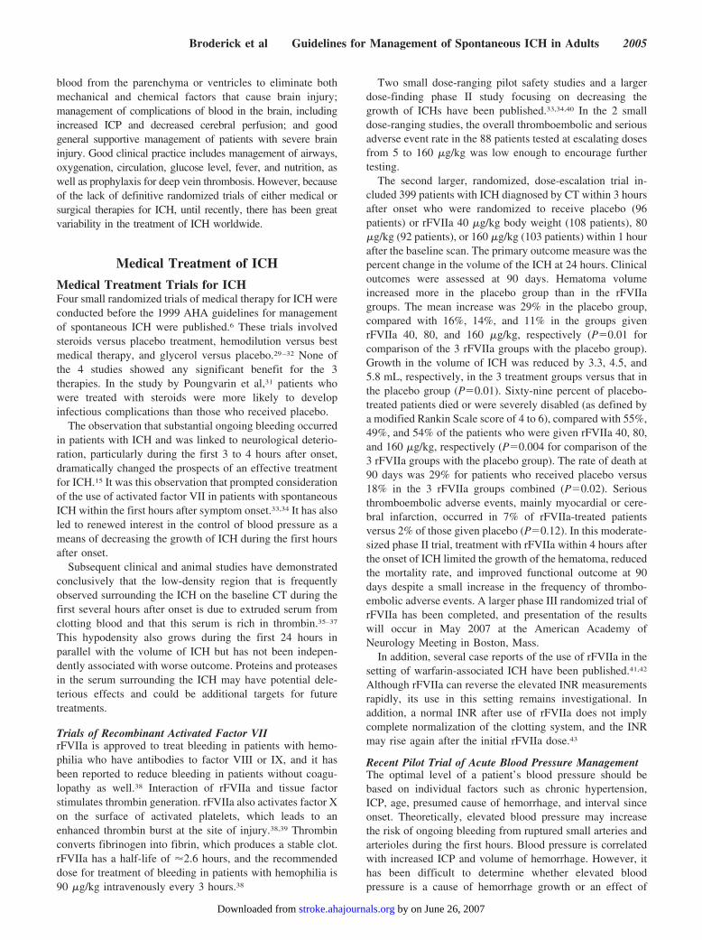
Trials of Recombinant Activated Factor VIIrFVIIa is approved to treat bleeding in patients with hemo-philia who have antibodies to factor VIII or IX, and it hasbeen reported to reduce bleeding in patients without coagu-lopathy as well.38 Interaction of rFVIIa and tissue factorstimulates thrombin generation. rFVIIa also activates factor Xon the surface of activated platelets, which leads to anenhanced thrombin burst at the site of injury.38,39 Thrombinconverts fibrinogen into fibrin, which produces a stable clot.rFVIIa has a half-life of �2.6 hours, and the recommendeddose for treatment of bleeding in patients with hemophilia is90 �g/kg intravenously every 3 hours.38
Two small dose-ranging pilot safety studies and a largerdose-finding phase II study focusing on decreasing thegrowth of ICHs have been published.33,34,40 In the 2 smalldose-ranging studies, the overall thromboembolic and seriousadverse event rate in the 88 patients tested at escalating dosesfrom 5 to 160 �g/kg was low enough to encourage furthertesting.
The second larger, randomized, dose-escalation trial in-cluded 399 patients with ICH diagnosed by CT within 3 hoursafter onset who were randomized to receive placebo (96patients) or rFVIIa 40 �g/kg body weight (108 patients), 80�g/kg (92 patients), or 160 �g/kg (103 patients) within 1 hourafter the baseline scan. The primary outcome measure was thepercent change in the volume of the ICH at 24 hours. Clinicaloutcomes were assessed at 90 days. Hematoma volumeincreased more in the placebo group than in the rFVIIagroups. The mean increase was 29% in the placebo group,compared with 16%, 14%, and 11% in the groups givenrFVIIa 40, 80, and 160 �g/kg, respectively (P�0.01 forcomparison of the 3 rFVIIa groups with the placebo group).Growth in the volume of ICH was reduced by 3.3, 4.5, and5.8 mL, respectively, in the 3 treatment groups versus that inthe placebo group (P�0.01). Sixty-nine percent of placebo-treated patients died or were severely disabled (as defined bya modified Rankin Scale score of 4 to 6), compared with 55%,49%, and 54% of the patients who were given rFVIIa 40, 80,and 160 �g/kg, respectively (P�0.004 for comparison of the3 rFVIIa groups with the placebo group). The rate of death at90 days was 29% for patients who received placebo versus18% in the 3 rFVIIa groups combined (P�0.02). Seriousthromboembolic adverse events, mainly myocardial or cere-bral infarction, occurred in 7% of rFVIIa-treated patientsversus 2% of those given placebo (P�0.12). In this moderate-sized phase II trial, treatment with rFVIIa within 4 hours afterthe onset of ICH limited the growth of the hematoma, reducedthe mortality rate, and improved functional outcome at 90days despite a small increase in the frequency of thrombo-embolic adverse events. A larger phase III randomized trial ofrFVIIa has been completed, and presentation of the resultswill occur in May 2007 at the American Academy ofNeurology Meeting in Boston, Mass.
In addition, several case reports of the use of rFVIIa in thesetting of warfarin-associated ICH have been published.41,42
Although rFVIIa can reverse the elevated INR measurementsrapidly, its use in this setting remains investigational. Inaddition, a normal INR after use of rFVIIa does not implycomplete normalization of the clotting system, and the INRmay rise again after the initial rFVIIa dose.43
Recent Pilot Trial of Acute Blood Pressure ManagementThe optimal level of a patient’s blood pressure should bebased on individual factors such as chronic hypertension,ICP, age, presumed cause of hemorrhage, and interval sinceonset. Theoretically, elevated blood pressure may increasethe risk of ongoing bleeding from ruptured small arteries andarterioles during the first hours. Blood pressure is correlatedwith increased ICP and volume of hemorrhage. However, ithas been difficult to determine whether elevated bloodpressure is a cause of hemorrhage growth or an effect of
Broderick et al Guidelines for Management of Spontaneous ICH in Adults 2005
by on June 26, 2007 stroke.ahajournals.orgDownloaded from

increasing volumes of ICH and increased ICP. A prospectiveobservational study of growth in the volume of ICH did notdemonstrate a relationship between baseline blood pressureand subsequent growth of ICH, but frequent early use ofhypertensive agents in that study may have obscured anyrelationship.15 Conversely, overaggressive treatment of bloodpressure may decrease cerebral perfusion pressure (CPP) andtheoretically worsen brain injury, particularly in the setting ofincreased ICP.
Powers and colleagues44 studied 14 patients with acutesupratentorial ICH 1 to 45 mL in size at 6 to 22 hours afteronset. Cerebral blood flow (CBF) was measured withpositron emission tomography and [15O]water. After comple-tion of the first CBF measurement, patients were randomizedto receive either nicardipine or labetalol to reduce meanarterial pressure by 15%, and the CBF study was repeated.Mean arterial pressure was lowered by �16.7�5.4% from143�10 to 119�11 mm Hg. No significant change wasobserved in either global CBF or periclot CBF. The authorsconcluded that in patients with small- to medium-sized acuteICHs, autoregulation of CBF was preserved with arterialblood pressure reductions in the range studied.
Recent Trial of Hyperosmolar Therapy for Treatment ofIncreased ICP in ICHResults of a trial of mannitol for spontaneous ICH werepublished in 2005.45 One hundred twenty-eight patients withprimary supratentorial ICH within 6 days of onset wererandomized to low-dose mannitol or sham therapy. The studygroup received mannitol 20%, 100 mL every 4 hours for 5days, tapered in the next 2 days. The control group receivedsham infusion. At 1 month, 16 patients (25% in each group)died in each group. The primary (P�0.80) and secondary endpoints were not significantly different between the 2 groups.At 3 months, the primary outcome was not significantlydifferent (P�0.25) between the groups. In the study group, 23patients had poor, 18 had partial, and 8 had completerecovery, and in the control group, 18 had poor, 20 hadpartial, and 9 had complete recovery.
Specific Medical Therapies
Blood Pressure ManagementThe previous AHA recommendation for the management ofblood pressure after ICH outlined the important concept ofselecting a target blood pressure on the basis of individualpatient factors,6 such as baseline blood pressure, presumedcause of hemorrhage, age, and elevated ICP. The primaryrationale for lowering the blood pressure is to avoid hemor-rhagic expansion from potential sites of bleeding. This isespecially true for hemorrhage resulting from a rupturedaneurysm or arteriovenous malformation, in which the risk ofcontinued bleeding or rebleeding is presumed to be highest.However, in primary ICH, in which a specific large-vesselvasculopathy is not apparent, the risk of hemorrhagic expan-sion with mild blood pressure elevation may be lower andmust be balanced with the theoretical risks of inducingcerebral ischemia in the edematous region that surrounds thehemorrhage. This theoretical risk has been somewhat
muted by prospective observational studies in both animalsand human beings44,46 that have dispelled the concept ofmajor ischemia in the edematous tissue surrounding thehemorrhage. Nevertheless, some controversy persists onthe basis of human MRI–apparent diffusion coefficientstudies of the perihemorrhagic region,47 which indicate arim of tissue at risk for secondary ischemia in largehematomas with elevated ICP.
Nonetheless, for primary ICH, little prospective evidenceexists to support a specific blood pressure threshold. Theprevious recommendation was to maintain a systolic bloodpressure �180 mm Hg and/or mean arterial pressure�130 mm Hg. The evidence to support any specific recom-mendation can be briefly summarized as follows: (1) Isolatedsystolic blood pressure �210 mm Hg is not clearly related tohemorrhagic expansion or to neurological worsening.48 (2)Reduction in mean arterial pressure by 15% (mean 142�10to 119�11 mm Hg) does not result in CBF reduction inhumans as measured by positron emission tomography.44 (3)In one prospective observational study,49 reduction of systolicblood pressure to a target �160/90 mm Hg was associatedwith neurological deterioration in 7% of patients and withhemorrhagic expansion in 9% but was associated with a trendtoward improved outcome in those patients in whom systolicblood pressure was lowered within 6 hours of hemorrhageonset. (4) Baseline blood pressure was not associated withgrowth of ICH in the largest prospective study of ICH growthand in the Recombinant Activated Factor VII IntracerebralHemorrhage Trial.15,16,50 (5) Hemorrhagic enlargement oc-curs more frequently in patients with elevated systolic bloodpressure, but it is not known whether this is an effect ofincreased growth of ICH with associated increases in ICP ora contributing cause to the growth of ICH.51 (6) A rapiddecline in blood pressure during the acute hospitalization wasassociated with increased death rate in one retrospectivestudy.52 (7) Experience in traumatic brain hemorrhage, aswell as spontaneous ICH, supports preservation of the CPP�60 mm Hg.53–56
Thus, whether more aggressive control of blood pressureduring the first hours after onset of ICH can decrease bleedingwithout compromising the perfusion of brain surrounding theICH remains unknown. The Antihypertensive Treatment inAcute Cerebral Hemorrhage (ATACH) Pilot Study, begun in2005, has been funded by the National Institute of Neurolog-ical Disorders and Stroke to investigate the control of bloodpressure in patients with ICH. This study will involve a3-dose–tiered trial of reducing systolic blood pressure to 3predetermined levels: 170 to 200, 140 to 170, and 110 to140 mm Hg. In addition, the phase III randomized interna-tional INTERACT study (Intensive Blood Pressure Reduc-tion in Acute Cerebral Hemorrhage) is planned to begin in2006. The goal of this study is to determine whether loweringhigh blood pressure levels after the start of ICH will reducethe chances of a person dying or surviving with a long-termdisability. The study includes patients with acute stroke dueto spontaneous ICH with at least 2 systolic blood pressuremeasurements of �150 mm Hg and �220 mm Hg recorded 2or more minutes apart and who are able to commence a
2006 Stroke June 2007
by on June 26, 2007 stroke.ahajournals.orgDownloaded from

randomly assigned blood pressure–lowering regimen within 6hours of ICH onset.
Brain Supportive Therapy in ICH: ICP andCPP/Glucose Control/Prevention and Treatment ofSeizures/Body Temperature
Monitoring of Neurological andCardiopulmonary FunctionThe sudden eruption of an intracranial hemorrhage destroysand displaces brain tissue and can induce an increase in ICP.The dynamics of ICH after appearance of the primary lesioninclude hematoma growth, perihematomal edema and/orischemia, hydrocephalus, or secondary IVH. All of thesecomplications also can potentially increase ICP and masseffect, resulting in neurological deterioration.56
The patient’s neurological status should be assessed fre-quently with the use of standard stroke scales such as theNational Institutes of Health Stroke Scale (NIHSS)57 andcoma scales such as the GCS.58 Blood pressure can bemonitored adequately with an automated cuff, whereas con-tinuous monitoring of systemic arterial pressure should beconsidered in patients who require continuous intravenousadministration of antihypertensive medications and in pa-tients whose neurological status is deteriorating. Airway andoxygenation can be assessed per respiratory status and pulseoximetry. Cardiopulmonary instability in association withincreased ICP is to be avoided to minimize deleterious effectsin patients with limited autoregulatory capacity. The vastmajority of ICH patients are admitted to intensive care unitsbecause of their impaired consciousness, elevated bloodpressure, and frequent need for intubation. It has beenreported that admission of ICH patients to a neuroscienceintensive care unit may result in a reduced mortality rate.59
Although these findings are preliminary and require furtherstudy, they have important implications for decisions aboutorganization of critical care services provided by hospitals.
Multimodal monitoring to assess metabolic and hemody-namic variables can provide crucial information at the cellu-lar level. Continuous or frequent assessment of variables interms of CBF, brain tissue oxygenation, and intracerebralmicrodialysis provide critical basic physiological informationabout brain function in patients with acute brain injury, butthe efficacy of these measures in patients with ICH has notbeen tested in randomized clinical trials.
The information provided by standard CT or MRI is static,and it is not practical to perform frequent imaging studies inthese patients. Fiberoptic ICP monitors within the brainparenchyma and ventricular catheters can detect dynamicchanges but are typically preferentially used in patients inwhom there is a high suspicion of ICP or who are clinicallydeteriorating.60
Transcranial Doppler sonography has the potential toassess mass effect and track ICP changes.61 Increased ICPand decreased CPP give rise to typical changes in the Dopplerwaveform obtained by transcranial insonation (ie, a decreasein diastolic velocity and an increase in the pulsatility index).Information on the relation of radiological data to 1 or morespecific transcranial Doppler variables and on the clinicalutility of radiological data is still sparse. Whether an increase
in pulsatility index reflects intracranial hypertension or masseffect in patients with ICH requires confirmation by othermeans.
Treatment of ICPTreatment of intracranial hypertension has evolved aroundpatients with head injuries and may not apply to thespecifics of patients with ICH. The “Lund protocol”assumes a disruption of the blood– brain barrier andrecommends manipulations to decrease the hydrostatic andincrease the osmotic forces that favor maintenance of fluidwithin the vascular compartment.62 The other primaryapproach, CPP-guided therapy, focuses on maintaining aCPP of �70 mm Hg to minimize reflex vasodilation orischemia and has become a popular treatment for intracra-nial hypertension.55,56,63,64 However, cerebral ischemia andhypoxia may still occur with CPP-guided therapy, andconcern remains that blood pressure elevation to maintainCPP may advance intracranial hypertension. A recentstudy on this matter concluded that the majority of patientsdid have increases in ICP when their mean arterial pressurewas elevated therapeutically.65
Despite long-standing debates, no controlled clinical trialhas demonstrated the superiority of either approach. Intoday’s neurological critical care environment, various potenttreatments to combat intracranial hypertension are available,but these are far from perfect and are associated with seriousadverse events. Nonselective hyperventilation may enhancesecondary brain injury; mannitol can cause intravascularvolume depletion, renal failure, and rebound intracranialhypertension; barbiturates are associated with cardiovascularand respiratory depression and prolonged coma; and cerebro-spinal fluid (CSF) drainage via intraventricular catheterinsertion may result in intracranial bleeding and infection andtissue shifts. Systemic cooling to 34°C can be effective inlowering refractory intracranial hypertension but is associatedwith a relatively high rate of complications, including pul-monary, infectious, coagulation, and electrolyte problems.66
A significant rebound in ICP also appears to occur wheninduced hypothermia is reversed.67
The exact frequency of increased ICP in patients with ICHis not known. Many patients with smaller ICHs will likely nothave increased ICP and require no measures to decrease ICP,as is the case for many patients with ischemic stroke.However, for those patients with clinical evidence of in-creased ICP, a balanced approach to ICP makes use of any ofthe approaches detailed below, with appropriate monitoringsafeguards in a critical care unit. A balanced approach beginswith simple and less aggressive measures, such as headpositioning, analgesia, and sedation, and then progresses tomore aggressive measures as clinically indicated. In general,the more aggressive the measures, the more critical is theneed to monitor ICP and CPP. No randomized clinical trialhas demonstrated the efficacy of monitoring ICP and CPP inthe setting of ICH.
Head-of-Bed ElevationElevation of the head of the bed to 30° improves jugularvenous outflow and lowers ICP. The head should be midline,and head turning to either side should be avoided. In patients
Broderick et al Guidelines for Management of Spontaneous ICH in Adults 2007
by on June 26, 2007 stroke.ahajournals.orgDownloaded from

who are hypovolemic, elevation of the head of the bed may beassociated with a fall in blood pressure and an overall fall inCPP; therefore, care must be taken initially to excludehypovolemia. The position of the arterial pressure transducerwill also need to be adjusted to ensure reliable measurementsof CPP.
CSF DrainageThe role of ventriculostomy has never been studied prospec-tively, and its use has been associated with very highmortality68 and morbidity69 rates. When an intraventricularcatheter is used to monitor ICP, CSF drainage is an effectivemethod for lowering ICP, particularly in the setting ofhydrocephalus. When an intraventricular catheter is used tomonitor ICP, CSF drainage is an effective method forlowering ICP. This can be accomplished by intermittentdrainage for short periods in response to elevations in ICP.The principal risks associated with ventriculostomy are in-fection and hemorrhage. Most studies report rates of bacterialcolonization rather than symptomatic infection that rangefrom 0% to 19%.60,70 The incidence of ventriculostomy-associated bacterial meningitis varies between 6% and22%.70,71
Analgesia and SedationIntravenous sedation is needed in unstable patients who areintubated for maintenance of ventilation and control ofairways, as well as for other procedures. Sedation should betitrated to minimize pain and increases in ICP, yet shouldenable evaluation of the patient’s clinical status. This isusually accomplished with intravenous propofol, etomidate,or midazolam for sedation and morphine or alfentanil foranalgesia and antitussive effect.
Neuromuscular BlockadeMuscle activity may further raise ICP by increasing intratho-racic pressure and obstructing cerebral venous outflow. If thepatient is not responsive to analgesia and sedation alone,neuromuscular blockade is considered. However, the prophy-lactic use of neuromuscular blockade in patients withoutproven intracranial hypertension has not been shown toimprove outcome. It is associated with an increased risk ofcomplications such as pneumonia and sepsis and can obscureseizure activity.
Osmotic TherapyThe most commonly used agent is mannitol, an intravascularosmotic agent that can draw fluid from both edematous andnonedematous brain tissue. In addition, it increases cardiacpreload and CPP, thus decreasing ICP through cerebralautoregulation. Mannitol decreases blood viscosity, whichresults in reflex vasoconstriction and decreased cerebrovas-cular volume. The major problems associated with mannitoladministration are hypovolemia and the induction of a hyper-osmotic state. Target serum osmolality has often been rec-ommended as 300 to 320 mOsm/kg, but definitive data on theeffectiveness of specific thresholds are lacking.
The use of hypertonic saline solutions has been shown toreduce ICP in a variety of conditions, even in cases refractoryto treatment with hyperventilation and mannitol. In terms ofhypertonic saline, many issues remain to be clarified, includ-
ing its exact mechanism of action, the best mode of admin-istration, and the concentration to be given.72,73
HyperventilationHyperventilation is one of the most effective methods avail-able for the rapid reduction of ICP. The CO2 reactivity ofintracerebral vessels is one of the normal mechanisms in-volved in the regulation of CBF. Experimental studies usinga pial window technique have clearly demonstrated that theaction of CO2 on cerebral vessels is exerted via changes inextracellular fluid pH.74 Molecular CO2 and bicarbonate ionsdo not have independent vasoactivity on these vessels. As aresult, hyperventilation consistently lowers ICP. Despite theeffectiveness of hyperventilation in lowering ICP, broad andaggressive use of this treatment modality to substantiallylower PCO2 levels has fallen out of favor, primarily because ofthe simultaneous effect on lowering CBF. Another character-istic of hyperventilation that limits its usefulness as a treat-ment modality for intracranial hypertension is the transientnature of its effect. Because the extracellular space of the brainrapidly accommodates to the pH change induced by hyperven-tilation, the effects on CBF and on ICP are short-lived. In fact,after a patient has been hyperventilated for �6 hours, rapidnormalization of arterial PCO2 can cause a significant reboundincrease in ICP. The target levels of CO2 for hyperventilation are30 to 35 mm Hg. Lower levels of CO2 are not recommended.75
Barbiturate ComaBarbiturates in high doses are effective in lowering refractoryintracranial hypertension but ineffective or potentially harm-ful as a first-line or prophylactic treatment in patients withbrain injuries. High-dose barbiturate treatment acts by de-pressing cerebral metabolic activity. This results in a reduc-tion in CBF, which is coupled to metabolism, and a fall inICP. The use of barbiturates in the treatment of refractoryintracranial hypertension requires intensive monitoring and isassociated with a significant risk of complications,76 the mostcommon being hypotension. Cerebral electrical activityshould ideally be monitored during high-dose barbituratetreatment, preferably on a continuous basis, with burst sup-pression activity providing a physiological end point for dosetitration.
Management of GlucoseHigh blood glucose on admission predicts an increased28-day case-fatality rate in both nondiabetic and diabeticpatients with ICH.24 In some studies, hyperglycemia in acutestroke has been considered a manifestation of premorbiddiabetic glucose metabolism77,78 or a stress reaction or hasbeen associated with other mechanisms.79 The amount ofevidence to support the stress hypothesis of poststroke hy-perglycemia is increasing.
The AHA guidelines for the early management of ischemicstroke published in 2003 indicated that there is generalagreement to recommend control of hypoglycemia or hyper-glycemia after stroke.80 Until further data from ongoing trialsbecame available, a judicious approach to management ofhyperglycemia was recommended. The only target providedin these guidelines was that markedly elevated glucose levelsbe lowered to �300 mg/dL (�16.63 mmol/L). More aggres-
2008 Stroke June 2007
by on June 26, 2007 stroke.ahajournals.orgDownloaded from

sive lowering of hyperglycemia in acute stroke is currentlybeing tested in randomized trials.
Antiepileptic DrugsSeizures occur commonly after ICH and may be nonconvul-sive. The frequency of observed seizures after ICH dependson the extent of monitoring. In a recently published largeclinical series of 761 subsequent patients, early seizuresoccurred in 4.2% of patients, and 8.1% had seizures within 30days after onset. Lobar location was significantly associatedwith the occurrence of early seizures.81 In a cohort of ICHpatients undergoing continuous electrophysiological monitor-ing in a neurocritical care unit, electrographic seizures oc-curred in 18 (28%) of 63 patients with ICH during the initial72 hours after admission. Seizures were independently asso-ciated with increased midline shift after intraparenchymalhemorrhage.82 ICH-related seizures are often nonconvulsiveand are associated with higher NIHSS scores, a midline shift,and a trend toward poor outcome.82
Treatment of clinical seizures in ICH patients during thehospitalization should include intravenous medications tocontrol seizures quickly, as for any hospitalized patient.Initial choice of medications includes benzodiazepines suchas lorazepam or diazepam, followed directly by intravenousfos-phenytoin or phenytoin. The European Federation ofNeurological Societies guidelines provide a detailed stepapproach to address more refractory cases of statusepilepticus.83
A brief period of antiepileptic therapy soon after ICH onsetmay reduce the risk of early seizures, particularly in patientswith lobar hemorrhage.81 Choice of medication for prophy-laxis should include one that can be administered intrave-nously as needed during the hospitalization and orally afterdischarge.
Temperature ManagementCerebral temperature has been recognized as a strong factorin ischemic brain damage. Laboratory investigations haveshown that hypothermia ameliorates brain damage.84 Theclassic mechanism proposed for this protection is redistribu-tion of oxygen and lowering of glucose consumption suffi-cient to permit tolerance to prolonged periods of oxygendeprivation. In terms of applying therapeutic cooling topatients with ICH, experimental research indicates thatthrombin-induced brain edema formation is significantlyreduced by induced hypothermia in the rat.85,86 Inhibition ofthrombin-induced blood–brain barrier breakdown and in-flammatory response by hypothermia appear to contribute tobrain protection in this model. Therapeutic cooling mayprovide an approach to potentially reduce edema after ICH. Inanother experimental study, delayed and prolonged coolingfailed to reduce residual blood volume and improve func-tional outcome in the rat. Although these results do notexclude possible beneficial effects of hypothermia, such asICP reduction, tissue that is quickly lost after ICH will notlikely be salvaged.87
By contrast, fever worsens outcome in several experimen-tal models of brain injury.88,89 The incidence of fever afterbasal ganglionic and lobar ICH is high, especially in patientswith ventricular hemorrhage. In patients surviving the first 72
hours after hospital admission, the duration of fever is relatedto outcome and appears to be an independent prognosticfactor in these patients.17 Rossi et al90 found that fever isassociated with increases in intracranial volume homeostasis,which causes intracranial hypertension. These data provide arationale for aggressive treatment of fever to normal levels inpatients with ICH.
Therapeutic cooling has been investigated in acute braininjuries in terms of controlling ICP and as a possibleneuroprotectant strategy.91 Cooling to 32°C to 34°C can beeffective in lowering refractory intracranial hypertension but,particularly with longer-term use (24 to 48 hours), is associ-ated with a relatively high rate of complications, includingpulmonary, infectious, coagulation, and electrolyte prob-lems.92 There also appears to be a risk for rebound intracra-nial hypertension when induced hypothermia is reversed tooquickly.67
Recommendations for Initial Medical Therapy
Class I
1. Monitoring and management of patients with an ICHshould take place in an intensive care unit settingbecause of the acuity of the condition, frequent eleva-tions in ICP and blood pressure, frequent need forintubation and assisted ventilation, and multiple com-plicating medical issues (Class I, Level of Evidence B).
2. Appropriate antiepileptic therapy should always beused for treatment of clinical seizures in patients withICH (Class I, Level of Evidence B).
3. It is generally agreed that sources of fever should betreated and antipyretic medications should be admin-istered to lower temperature in febrile patients withstroke (Class I, Level of Evidence C).
4. As for patients with ischemic stroke,93 early mobiliza-tion and rehabilitation are recommended in patientswith ICH who are clinically stable (Class I, Level ofEvidence C).
Class II
1. Treatment of elevated ICP should include a balancedand graded approach that begins with simple mea-sures, such as elevation of the head of the bed andanalgesia and sedation. More aggressive therapies todecrease elevated ICP, such as osmotic diuretics (man-nitol and hypertonic saline solution), drainage of CSFvia ventricular catheter, neuromuscular blockade, andhyperventilation, generally require concomitant monitor-ing of ICP and blood pressure with a goal to maintainCPP >70 mm Hg (Class IIa, Level of Evidence B).
2. Evidence indicates that persistent hyperglycemia(>140 mg/dL) during the first 24 hours after stroke isassociated with poor outcomes, and thus it is generallyagreed that hyperglycemia should be treated in pa-tients with acute stroke. Guidelines for ischemic strokesuggest that elevated glucose concentrations (>185mg/dL and possibly >140 mg/dL) probably shouldtrigger administration of insulin, similar to the proce-dure in other acute situations accompanied by hyper-glycemia. Use of these guidelines for ICH as well isreasonable. The results of ongoing research should
Broderick et al Guidelines for Management of Spontaneous ICH in Adults 2009
by on June 26, 2007 stroke.ahajournals.orgDownloaded from

clarify the management of hyperglycemia after stroke(Class IIa, Level of Evidence C).
3. Until ongoing clinical trials of blood pressure interven-tion for ICH are completed, physicians must manageblood pressure on the basis of the present incompleteevidence. Current suggested recommendations for tar-get blood pressures in various situations and potentialmedications are listed in Tables 2 and 3 and may beconsidered (Class IIb, Level of Evidence C).
4. Treatment with rFVIIa within the first 3 to 4 hoursafter onset to slow progression of bleeding has shownpromise in one moderate-sized phase II trial; however,the efficacy and safety of this treatment must beconfirmed in phase III trials before its use in patientswith ICH can be recommended outside of a clinicaltrial (Class IIb, Level of Evidence B).
5. A brief period of prophylactic antiepileptic therapysoon after ICH onset may reduce the risk of earlyseizures in patients with lobar hemorrhage (Class IIb,Level of Evidence C).
Prevention of Deep Vein Thrombosis andPulmonary EmbolismDeep vein thrombosis and pulmonary emboli are relativelycommon preventable causes of morbidity and mortality inpatients with acute ICH. In a prospective randomized rFVIIatrial, 2 (2.1%) of the 96 patients who received placebodeveloped a pulmonary embolism on days 7 to 11 (1 fatal).34
Four (1.3%) of the 303 patients treated with rFVIIa hadpulmonary embolism (1 fatal), and an additional patient hada deep vein thrombosis. In a retrospective study of 1926patients with ICH, 1.6% had a clinical diagnosis of venousthromboembolism as documented by the International Clas-sification of Diseases—9th Revision clinical modification(ICD-9-CM) codes.94 Studies using fibrinogen scanning orMRI to detect occult venous thrombosis report high frequen-cies (10% to 50%) of deep vein thrombosis in acute strokepatients with hemiplegia.95
The question is how to prevent and treat these venousthromboembolic complications without increasing the risk ofintracranial rebleeding. Anticoagulants, platelet antiag-gregants, unfractionated heparin and low-molecular-weightheparins/heparinoids, and use of mechanical methods such as
intermittent pneumatic compression and graduated compres-sion stockings are options with varying strengths of evidencefor preventing venous thromboembolism in patients withischemic stroke. However, almost all of the evidence forthese various options comes from studies of patients withischemic stroke or other causes of immobility. One recenttrial randomized patients with ICH to compression stockingsversus compression stockings plus intermittent pneumaticcompression. Asymptomatic deep vein thrombosis by ultra-sonography was detected at day 10 in 15.9% of patientswearing elastic stockings alone and in 4.7% in the combinedgroup (relative risk 0.29 [95% confidence interval {CI} 0.08to 1.00]).96
Boeer and colleagues97 reported a small randomized trialcomparing 68 patients with ICH who were randomized toreceive low-dose heparin (5000 U of heparin-sodium subcu-taneously 3 times per day) beginning on the tenth day oftreatment (group 1), the fourth day (group 2), or the secondday (group 3) after ICH onset. Group 1 served as the controlgroup because heparin treatment beginning on day 10 was thestandard treatment protocol in the intensive care unit. Allpatients received basic intensive care medication and regulardiagnostic evaluations. Group 3 patients showed a statisti-cally significant reduction in the number of pulmonaryemboli when compared with the other 2 groups. No overallincrease in the incidence of rebleeding was observed in any ofthe 3 groups. The incidence of deep vein thrombosis washigher in the first few days of treatment than at 10 days, butthis difference was not significant.97
A separate issue from primary prevention of deep veinthrombosis and embolism is what to do for patients with ICHwho develop deep vein thrombosis or pulmonary embolism.The rate of recurrent ICH during the initial 3 months after anacute ICH is �1%.98–100 A theoretical analysis estimated thatanticoagulation increases the risk of recurrent ICH 2-foldcompared with the recurrence risk of ICH overall.100 How-ever, the challenge of selecting therapy for an individualpatient is to balance the risk of subsequent life-threateningthromboembolism against the risk of recurrence of ICH, inwhich the mortality rate is �50%. The risk of ICH recurrencealso likely varies by location and age, but few prospectivedata are available (eg, higher risk in patients with lobar ICHdue to suspected amyloid angiopathy).101
TABLE 2. Suggested Recommended Guidelines for TreatingElevated Blood Pressure in Spontaneous ICH
1. If SBP is �200 mm Hg or MAP is �150 mm Hg, then consideraggressive reduction of blood pressure with continuous intravenousinfusion, with frequent blood pressure monitoring every 5 minutes.
2. If SBP is �180 mm Hg or MAP is �130 mm Hg and there isevidence of or suspicion of elevated ICP, then consider monitoringICP and reducing blood pressure using intermittent or continuousintravenous medications to keep cerebral perfusion pressure �60to 80 mm Hg.
3. If SBP is �180 mm Hg or MAP is �130 mm Hg and there is notevidence of or suspicion of elevated ICP, then consider a modestreduction of blood pressure (eg, MAP of 110 mm Hg or target bloodpressure of 160/90 mm Hg) using intermittent or continuousintravenous medications to control blood pressure, and clinicallyreexamine the patient every 15 minutes.
SBP indicates systolic blood pressure; MAP, mean arterial pressure.
TABLE 3. Intravenous Medications That May Be Consideredfor Control of Elevated Blood Pressure in Patients With ICH
Drug Intravenous Bolus Dose Continuous Infusion Rate
Labetalol 5 to 20 mg every 15 min 2 mg/min (maximum 300 mg/d)
Nicardipine NA 5 to 15 mg/h
Esmolol 250 �g/kg IVP loading dose 25 to 300 �g � kg�1 � min�1
Enalapril 1.25 to 5 mg IVP every 6 h* NA
Hydralazine 5 to 20 mg IVP every 30 min 1.5 to 5 �g � kg�1 � min�1
Nipride NA 0.1 to 10 �g � kg�1 � min�1
Nitroglycerin NA 20 to 400 �g/min
IVP indicates intravenous push; NA, not applicable.*Because of the risk of precipitous blood pressure lowering, the enalapril
first test dose should be 0.625 mg.
2010 Stroke June 2007
by on June 26, 2007 stroke.ahajournals.orgDownloaded from

Another option is the interruption of the inferior vena cavaby the placement of a filter.102 Vena cava filters may reducethe incidence of pulmonary embolism in patients with prox-imal deep vein thrombosis in the first several weeks but havea longer-term risk of increased venous thromboembo-lism.102,103 No randomized clinical trial has compared venacava filters with anticoagulation in patients with ICH orischemic stroke.
During anticoagulation, good control of blood pressuresubstantially reduces the risk of recurrent ICH. The random-ized PROGRESS trial (Perindopril pROtection aGainst RE-current Stroke Study)104,105 documented a 50% reduction ofthe risk of recurrence among ICH survivors by loweringsystolic blood pressure by 11 mm Hg.
Recommendations for Prevention of Deep VeinThrombosis and Pulmonary Embolism
Class I
1. Patients with acute primary ICH and hemiparesis/hemiplegia should have intermittent pneumatic com-pression for prevention of venous thromboembolism(Class I, Level of Evidence B).
2. Treatment of hypertension should always be part oflong-term therapy because such therapy decreases therisk of recurrent ICH (Class I, Level of Evidence B).
Class II
1. After documentation of cessation of bleeding, low-dosesubcutaneous low-molecular-weight heparin or unfrac-tionated heparin may be considered in patients withhemiplegia after 3 to 4 days from onset (Class IIb, Levelof Evidence B).
2. Patients with an ICH who develop an acute proximalvenous thrombosis, particularly those with clinical orsubclinical pulmonary emboli, should be consideredfor acute placement of a vena cava filter (Class IIb,Level of Evidence C).
3. The decision to add long-term antithrombotic therapyseveral weeks or more after placement of a vena cavafilter must take into consideration the likely cause ofthe hemorrhage (amyloid [higher risk of recurrentICH] versus hypertension), associated conditions withincreased arterial thrombotic risk (eg, atrial fibrilla-tion [AF]), and the overall health and mobility of thepatient (Class IIb, Level of Evidence B).
ICH Related to Coagulation and Fibrinolysis:Management of Acute ICH and RestartingAntithrombotic TherapyIn recent reports, ICH occurs with a frequency of approxi-mately 0.3% to 0.6% per year in patients undergoing chronicwarfarin anticoagulation.105 Furthermore, warfarin accountsfor a substantial proportion of the cases of ICH who presentto general hospitals: Among patients with supratentorial ICHadmitted to the Massachusetts General Hospital over a 7-yearperiod, 23.4% were taking warfarin106; figures between 6%and 16% were reported in earlier series.107–109
For warfarin-related ICH, the main risk factors are age,history of hypertension, intensity of anticoagulation,110,111
and associated conditions such as cerebral amyloid angiopa-
thy112 and leukoaraiosis.113 An elevation of the INR above thetherapeutic range of 2 to 3 has been associated with a steadyincrease in the frequency of ICH, especially above values of3.5 to 4.5111,114: The risk of ICH nearly doubles for eachincrease of 0.5 points in the INR above 4.5.114 The degree ofelevation of the INR also correlates with hematoma expan-sion and prognosis (death and functional outcome). Althoughthe increase of ICH risk with excessive prolongation of theINR is well documented, most warfarin-related ICHs occurwith INRs in the recommended therapeutic range.110 Cerebralamyloid angiopathy is probably a common underlying pathol-ogy in elderly patients with warfarin-related lobar ICH.112
Leukoaraiosis, despite its high frequency in patients withcerebrovascular disease and hypertension, is a likely riskfactor for warfarin-related ICH. It was present in 92% of aseries of patients with ICH taking warfarin (which had beenprescribed after an episode of ischemic stroke), comparedwith 48% of a control group of patients without ICH takingwarfarin after ischemic stroke.113 The management issues inwarfarin-related ICH are the need to rapidly reverse thecoagulation defect to minimize further hematoma growth andthe need for and feasibility of reinstituting oral anticoagula-tion. The measures available to counteract the warfarin effectinclude the use of vitamin K1, fresh frozen plasma (FFP),prothrombin complex concentrate, and rFVIIa. Vitamin K1 isgiven intravenously at a dose of 10 mg.115 The intravenousinjection entails a small risk of anaphylaxis, which is reducedby the slower-acting subcutaneous injection route.116 VitaminK1 should not be used alone because it takes hours (at least 6)for vitamin K1 to normalize the INR.117 FFP can be given toreplenish the vitamin K–dependent coagulation factors inhib-ited by warfarin. It is an effective way of correcting the INR,and it acts more quickly than vitamin K1; however, its use atthe recommended dose of 15 to 20 mL/kg involves theinfusion of potentially large volumes of plasma, which notonly may take several hours to be infused (with the potentialfor continuing hematoma enlargement) but can also lead tovolume overload and heart failure.118 In addition, the concen-tration of clotting factors in FFP varies substantially, and thusthe degree of effectiveness of different batches of FFP isunpredictable. Finally, circulating levels of factor IX mayremain low (and thus result in incomplete hemostasis) despitereplacement of all other clotting factors with FFP.119 Theselimitations, particularly the sometimes lengthy process ofnormalizing the INR in the emergency life-threatening situ-ation of warfarin-related ICH, make the FFP approachimpractical.
This has stimulated the search for better options. Prothrom-bin complex concentrate contains high levels of vitaminK–dependent factors (II, VII, and X), and factor IX complexconcentrate contains factors II, VII, IX, and X. These prep-arations have the advantage of requiring smaller volumes ofinfusion than FFP and correcting the coagulopathy fast-er.120,121 Their disadvantage is the risk of inducing thrombo-embolic complications, ranging from superficial thrombo-phlebitis, deep vein thrombosis and pulmonary embolism,and arterial thrombosis to disseminated intravascular coagu-lation. Concerns about viral transmission have been mini-mized by the current rigorous screening of blood products.
Broderick et al Guidelines for Management of Spontaneous ICH in Adults 2011
by on June 26, 2007 stroke.ahajournals.orgDownloaded from

The ability of rFVIIa to rapidly normalize the INR insubjects anticoagulated with warfarin43 and the recent reportof a beneficial effect in patients with spontaneous ICH34,41,42
suggest that this option should be tested in warfarin-relatedICH. A small study of 7 patients with warfarin-related ICHtreated with rFVIIa at a dose between 15 and 90 �g/kgshowed a rapid reduction of INR after single injections.41
Because of the short half-life of rFVIIa (2.6 hours),122 theinitial rapid response at times required repeated injections ofthe factor to maintain the INR in the normal range. Given thesignificant increase in thromboembolic complications (7%versus 2% in the control group) in patients with “spontane-ous” ICH treated with rFVIIa,34 concern is warranted about apotentially larger risk with the use of this procoagulant agentin subjects prone to embolism, such as those with prostheticheart valves or chronic AF. Randomized controlled trials ofrFVIIa, as well as the other various options for treatment ofwarfarin-related ICH, are needed.
In instances of ICH that result from the use of intravenousheparin, management involves rapid normalization of theactivated partial thromboplastin time by protamine sulfate.The recommended dose is 1 mg per 100 U heparin, and thedose needs to be adjusted according to the time elapsed sincethe last heparin dose. If heparin is stopped for 30 to 60minutes, the protamine sulfate dose is 0.5 to 0.75 mg per 100U heparin, down to 0.375 to 0.5 mg per 100 U heparin after60 to 120 minutes off heparin and 0.25 to 0.375 mg per 100U heparin if it was stopped �120 minutes from the time ofthe protamine sulfate injection. Protamine sulfate is given byslow intravenous injection, not to exceed 5 mg/min, with atotal dose not to exceed 50 mg. A faster rate of infusion canproduce severe systemic hypotension.
The issue of reinstitution of anticoagulation after warfarin-related ICH applies primarily to those who began takingwarfarin for the prevention of cardiogenic embolism associ-ated with either prosthetic heart valves or chronic AF. In viewof the documented rates of cerebral embolism of 5% per yearin patients who have nonvalvular AF without history ofstroke,123 12% per year in patients who have AF with priorischemic stroke events,124 and at least 4% per year in patientswith prosthetic mechanical heart valves,125 re-anticoagulationwith warfarin is often a consideration after warfarin-relatedICH. This difficult decision should balance the prevention ofischemic stroke and the risk of recurrent ICH.126 Unfortu-nately, no data are available on rates of ICH recurrence whilewarfarin treatment is being given. In an aggregate group of114 patients with ICH in 3 clinical series,127–129 reversal ofanticoagulation with FFP and discontinuation of warfarinafter ICH for a mean of 7 to 10 days was associated withembolism in 6 patients (5%). Rebleeding on reinstitution ofanticoagulation between 7 and 10 days from ICH onsetoccurred in 1 patient (0.8%). The use of prothrombin com-plex concentrate for reversal of anticoagulation in an aggre-gate group of 78 patients from 7 clinical series119,130–135
resulted in 4 thromboembolic events (5%), and continuedhematoma expansion occurred in 5 subjects (6%). Data on therate of thromboembolism with the use of rFVIIa in thissetting are not currently available. These limited data suggestthat reversal of anticoagulation with FFP or prothrombin
complex concentrate after ICH in patients with prostheticheart valves or chronic nonvalvular AF is associated with alow frequency of embolic events over periods of 7 to 10 days,after which reinstitution of warfarin anticoagulation appearsto be safe.136
One decision analysis, using quality-of-life years as theoutcome, compared the risk of restarting anticoagulation inpatients with chronic AF who had a lobar or deep hemor-rhage.100 In general, elderly patients with a lobar hemorrhagelikely due to amyloid angiopathy had a much higher projectedrisk of a poor outcome with continuation of warfarin. Inpatients with a small deep ICH, the risk was similar forrestarting and withholding warfarin.
The clinical dilemma of whether and when to restartanticoagulants in patients with ICH who have cardioembolicrisk will not be solved until prospectively generated data onrates of ICH recurrence after warfarin reinstitution becomeavailable. However, for patients with a lower risk of cerebralinfarction (eg, AF without prior ischemic stroke) and a higherrisk of amyloid angiopathy (eg, an elderly patient with lobarICH, particularly with evidence of small microbleeds onMRI), antiplatelet agents may be a better choice for preven-tion of ischemic stroke than warfarin.
ICH Related to FibrinolysisThrombolytic treatment for acute ischemic stroke was fol-lowed by symptomatic ICH in 3% to 9% of patients treatedintravenously with tissue-type plasminogen activator(tPA),77,137–139 6% of patients treated with a combination ofintravenous and intra-arterial tPA,140 and 10.9% of those whowere treated with intra-arterial prourokinase in a controlledclinical trial.141 In addition, ICH occurred in 0.5% to 0.6% ofpatients treated with thrombolytic agents for other acutearterial and venous occlusions, with higher rates among theelderly.142,143
The onset of ICH after fibrinolysis carries a poor prognosisbecause the hemorrhages tend to be massive, can be multi-focal, and are associated with a 30-day death rate of 60% ormore.138,144 No reliable data are available to guide theclinician in the choice of effective measures to control ICH inthis setting.145 Current recommended therapy includes theinfusion of platelets (6 to 8 U) and cryoprecipitate thatcontains factor VIII to rapidly correct the systemic fibrino-lytic state created by tPA.80,146 The guidelines for the surgicaltreatment of ICH after fibrinolysis for acute ischemic strokeare the same as those followed for ICH in general but shouldbe initiated only after a sufficient infusion of platelets andcryoprecipitate has stabilized intracranial bleeding.
Recommendations for the Management of ICHRelated to Coagulation and Fibrinolysis
Class I
1. Protamine sulfate should be used to reverse heparin-associated ICH, with the dose depending on the timefrom cessation of heparin (Class I, Level of Evidence B).
2. Patients with warfarin-associated ICH should betreated with intravenous vitamin K to reverse the
2012 Stroke June 2007
by on June 26, 2007 stroke.ahajournals.orgDownloaded from

effects of warfarin and with treatment to replaceclotting factors (Class I, Level of Evidence B).
Class II
1. Prothrombin complex concentrate, factor IX complexconcentrate, and rFVIIa normalize the laboratory ele-vation of the INR very rapidly and with lower volumesof fluid than FFP but with greater potential of throm-boembolism. FFP is another potential choice but isassociated with greater volumes and much longerinfusion times (Class IIb, Level of Evidence B).
2. The decision to restart antithrombotic therapy afterICH related to antithrombotic therapy depends on therisk of subsequent arterial or venous thromboembo-lism, the risk of recurrent ICH, and the overall state ofthe patient. For patients with a comparatively lowerrisk of cerebral infarction (eg, AF without prior ische-mic stroke) and a higher risk of amyloid angiopathy(eg, elderly patients with lobar ICH) or with very pooroverall neurological function, an antiplatelet agentmay be an overall better choice for prevention ofischemic stroke than warfarin. In patients with a veryhigh risk of thromboembolism in whom restartingwarfarin is considered, warfarin therapy may be re-started at 7 to 10 days after onset of the original ICH(Class IIb, Level of Evidence B).
3. Treatment of patients with ICH related to thrombolyt-ic therapy includes urgent empirical therapies to re-place clotting factors and platelets (Class IIb, Level ofEvidence B).
Surgical Treatment of ICH/IVHCraniotomyOf all the surgical therapies described for treating ICH,craniotomy has been the most extensively studied, with 7 ofthe 9 randomized controlled surgical trials reporting resultswith craniotomy either primarily or exclusively. Two of thesestudies were conducted with limited access to contemporarymedical and surgical technologies, which limits their rele-vance. Of the remaining 5, all but one are small, single-centerstudies that randomized fewer than 125 patients in total.Although none of these small studies found convincingevidence of surgical benefit, one concluded that for patientspresenting with mild to moderate alterations in consciousness(GCS score 7 to 10), surgery might reduce the risk of deathwithout improving functional outcome,147 and another sug-gested that ultra-early evacuation might improve the 3-monthNIHSS score.148
The insights of these smaller trials are important in light ofthe single large multicenter trial, the International SurgicalTrial in Intracerebral Haemorrhage (STICH), which random-ized 1033 patients from 107 centers over an 8-year period,beginning in 1995.149 Patients were eligible if randomizedwithin 72 hours and operated on within 96 hours of ictus fora clot �2 cm in diameter. Patients in very poor condition(GCS score �5) were excluded. Patients were randomized ifthe neurosurgeon was uncertain of the benefit of surgery, with50% randomly assigned to a policy of either early surgery orinitial medical management. Primary outcomes were theincidence of death and disability as measured by the extended
Glasgow Outcome Scale (GOS) at 6 months, and secondaryoutcomes were death, the Barthel Index (BI), and the modi-fied Rankin scale (mRS) at 6 months. To increase studypower, patients with an expected poor prognosis on the basisof age, admission GCS score, and hemorrhage volume wereanalyzed with different extended GOS, mRS, and BI cutoffs.In addition, several prespecified subgroup analyses wereincluded.
Five hundred six patients were randomized to surgery and530 to medical therapy, with groups being well matched forall known variables. Twenty-six percent of the medical armultimately crossed over to surgery. This crossover was due torebleeding or deterioration in 85% of crossover subjects, andcraniotomy was used in 85% of those subjects who crossedover to surgery. By contrast, only 75% of patients in theprimary surgical arm underwent craniotomy, with the othersbeing treated with less invasive surgical techniques. Ninety-three percent of patients were available for analysis at 6months.
In an intention-to-treat analysis, surgery within 96 hours ofictus was associated with a statistically insignificant absolutebenefit of 2.3% (95% CI �3.2% to 7.7%) in 6-monthprognosis-dichotomized extended GOS. Death (absolute ben-efit 1.2% [�4.9% to 7.2%]), mRS (absolute benefit 4.7%[�1.2% to 10.5%]), and BI (absolute benefit 4.1% [�1.4% to9.5%]) showed similar statistically insignificant trends infavor of surgery. Subgroup analysis identified those subjectswith GCS score of 9 to 12, those with lobar clots, and thosewith clots �1 cm from the surface that may have been helpedby early surgery, but this did not reach statistical significance.In contrast, those presenting in deep coma (GCS score 5 to 8)tended to do better with medical management. Together, thedata from both STICH and the other smaller trials suggestthat surgery does not appear to be helpful in treating mostsupratentorial ICH and is probably harmful in those patientspresenting in coma. Having said this, surgery, particularlycraniotomy, may be helpful in treating those lobar clotswithin 1 cm of the surface that present in patients with milderdeficits (GCS score �9), because both craniotomy andsurface location were associated with a 29% relative benefitin functional outcome when compared with medical manage-ment. Confirmation of these conclusions will require furthertrials.
These randomized trials of surgery did not include patientswith cerebellar hemorrhage. As discussed in the 1999 AHAguidelines for management of spontaneous ICH,6 nonran-domized treatment series of patients with cerebellar hemor-rhage report good outcomes for surgically treated patientswho have large (�3 cm) cerebellar hemorrhages or cerebellarhemorrhages with brain stem compression or hydrocepha-lus.150–156 In these patients, medical management alone oftenresults in bad outcomes. Smaller cerebellar hemorrhageswithout brain stem compression that are managed medicallydo reasonably well in the case series. For these reasons,neurosurgeons and neurologists have advocated that largecerebellar hemorrhages with compression of the brain stem orobstruction of the fourth ventricle should be removed surgi-cally as soon as possible.
Broderick et al Guidelines for Management of Spontaneous ICH in Adults 2013
by on June 26, 2007 stroke.ahajournals.orgDownloaded from
Minimally Invasive SurgeryThe purported advantages of minimally invasive clot evacu-ation over conventional craniotomy include (1) reducedoperative time, (2) the possibility of performance under localanesthesia, and (3) reduced tissue trauma, especially for deeplesions. Together, these advantages may also facilitate earlierevacuation of ICH than is possible or practical with conven-tional craniotomy. On the other hand, the reduced surgicalexposure, the inability to treat structural lesions (arterio-venous malformation or aneurysm), the potential for rebleed-ing related to the use of fibrinolytics, and the possibility of anincreased risk of infection related to prolonged indwellingcatheters are limitations of this approach. Despite the fact thatfewer data exist for these techniques than for craniotomy, wepresent an overview of the various minimally invasivetechniques.
The STICH trial suggests that subjects treated with anynoncraniotomy approach in the trial had a worse outcomethan those treated with conservative management (OR 1.3),but the confidence interval included 1 (95% CI 0.78 to 2.35).It is unclear whether the pathology chosen for these ap-proaches was less ideal for intervention, because patients withdeep hemorrhages and those in poor neurological condition(both of which fared worse in the trial) were likely those mostcommonly chosen for minimally invasive techniques.
Endoscopic AspirationEndoscopic aspiration of supratentorial hemorrhage has beenstudied in a small, single-center randomized trial.157 Onehundred patients between 30 and 80 years of age, withhemorrhages at least 10 mL in volume, received treatmentwithin 48 hours of onset via burr hole and continuousneuroendoscopic lavage of the hematoma cavity with artifi-cial CSF at a pressure of 10 to 15 mm Hg. The mixture ofblood clots and blood-stained CSF was removed by suction atregular intervals. Oozing vessels in the wall of the hematomawere coagulated with a laser built into the system, and theentire procedure was under direct visual control. More than90% of the clot was evacuated in 15% of patients andbetween 70% and 90% in 30% of patients, with all patientshaving at least a 50% reduction in size. At 6 months, themortality rate of the surgical group (42%) was significantlylower than that of the medical group (70%, P�0.01). A goodoutcome with minimal or no deficit was also seen morefrequently in the surgically treated group. In patients withlarge hematomas (50 mL), quality of life was not affected bysurgery, but the mortality rate was significantly lower. Bycontrast, endoscopic evacuation of smaller hematomas led toa significantly better quality of life versus those treatedmedically, but survival was similar for the 2 groups. More-over, the benefit was mainly limited to patients with lobarhematomas and patients �60 years of age.
Thrombolytic Therapy and Aspiration of ClotsZuccarello et al148 reported a single-center randomized pilottrial of conservative therapy (11 subjects) or very earlysurgical removal of the ICH (9 subjects) within 24 hours ofonset and 3 hours of randomization. Subjects with deephemorrhages who underwent surgery were treated with instil-lation of urokinase into the bed of the clot (n�4).148 Patients
who underwent stereotactic removal of the ICH did well(3-month BI scores of 100, 100, 90, and 85), which suggeststhat this approach was, as the animal studies had shown,158
safe and feasible. The effect of an infused thrombolytic onoutcome has been demonstrated by Niizuma et al,159 whoreported 81% of 175 patients with putaminal hemorrhageresumed useful lives. A number of nonrandomized studiesusing this approach reported aspiration rates ranging from30% to 90% of the initial hematoma157,160–162 and rebleedingrates comparable to those seen with conventional cranioto-my163 (range 0% to 10%; mean 4% of 392 cases).157,159–162,164
Finally, a multicenter randomized controlled trial (n�71patients)165 examined the utility of stereotactic urokinaseinfusion when administered within 72 hours for those patientspresenting with GCS score �5 and clots of �10 mL. Treatedpatients received 5000 IU of urokinase every 6 hours for amaximum of 48 hours. Primary end points were death anddegree of functional handicap (measured with the mRS) at 6months. The median reduction in volume of ICH frombaseline was 40% in the surgical group and 18% in themedical group. The rebleeding rate was 35% in the urokinasegroup and 17% in the conservatively managed group. Asignificant reduction in death (40%) was found in the treatedgroup, but no statistically significant difference in functionaloutcome scores was detected at different intervals oftreatment.
In 1999, urokinase became unavailable in the United Statesbecause of reports of the potential for viral contaminationduring pharmacological preparation, which prompted inves-tigators to examine the use of intraventricular instillation oftPA for clot evacuation in patients with severe IVH. Rohde etal166 reported that IVH disappeared earlier (1 to 3 days) withtPA than with urokinase (5 to 8 days). Compared withventriculostomy alone, IVH treated with the addition of tPAdecreased the mortality rate from a range of 60% to 90% toonly 5%.166 Other similar studies have indicated that the useof intraventricular tPA might improve the prognosis inpatients with large IVHs.167,168 The review of the currentliterature suggests that the use of intraventricular fibrinolyticscarries a low incidence of complications, which usuallyconsist of infections and hemorrhage.167–170
More recently, tPA has been used in the treatment of ICH.In a pig model of ICH, Wagner et al171 reported thathematoma aspiration after fibrinolysis with tPA resulted in�70% removal of the clot and marked reduction of perihe-matoma edema. In pilot human trials, Lippitz et al,172 Schalleret al,173 and Vespa et al174 reported that daily administrationof tPA into the hematoma cavity beginning 12 to 24 hoursafter stereotactic placement of a catheter resulted in anaverage 85% reduction in the hematoma volume by 2 to 4days after onset. Lippitz et al172 used a standard dose of 3 mgof tPA dissolved in 3 mL of 0.9% saline solution; dependingon the volume of residual hematoma after the first injection,tPA was repeated every 24 hours for 1 to 3 days. Schaller etal173 calculated the dose of tPA relative to the maximumdiameter of the hematoma; 1 cm of hematoma diameterequaled 1 mg of tPA. Repeated doses of tPA were adminis-tered every 24 hours for an additional 48 hours if hematomaremained. Vespa et al174 described the improvement in
2014 Stroke June 2007
by on June 26, 2007 stroke.ahajournals.orgDownloaded from

midline shift and NIHSS score with tPA treatment of ICH, aswell as a lower mortality rate compared with a matchedhistorical control cohort. No systemic side effects, includingintracranial bleeding or rebleeding, were reported.
Given these results, a multicenter, randomized, controlledand stratified study comparing administration of tPA into theclot cavity versus conventional medical treatment has re-cently been funded by the National Institutes of Health. Thisstudy (Minimally Invasive Stereotactic Surgery � rt-PA forICH Evacuation [MISTIE]) will test the following hypothe-ses: (1) Early use of minimally invasive surgery plus tPA for3 days is safe for treatment of ICH and (2) early use ofminimally invasive surgery plus tPA for 3 days produces clotsize reduction compared with medically treated patients.
Other Mechanical Devices and Radiographic GuidanceIn 1978, Backlund and von Holst175 described a new instru-ment for stereotactic evacuation of hematomas, which con-sisted of a 4-mm cannula in which there was an Archimedesscrew. Suction was applied to aspirate the clot into thecannula, while the rotating screw would morcellate thehematoma. Using this technique, others were able to almostcompletely evacuate ICHs in 13 of 16 patients in theirseries176; however, 81% of these patients died. After thesepioneering reports, several modifications to the device weredescribed.177–179 Other innovative devices included a spe-cially designed ultrasonic aspirator,180 a modified nucle-otome,181,182 and a double-track aspiration system.183,184 Al-though some of these approaches showed interesting results,especially when combined with real-time radiographic con-firmation of clot removal with CT or ultrasound, they havenot become popular.161 This is somewhat surprising, becauseKanaya and Kuroda161 reported that rebleeding after surgerywas seen in 10% of patients who underwent craniotomy butin only 5% to 6% of patients who underwent CT- orultrasound-guided aspiration. Moreover, on average, CT-guided aspiration removed 71% of the original hematoma,and ultrasound-guided aspiration removed 81%. The percent-age of hemorrhage removed did not significantly vary withthe timing of the operation.161
Early Clot EvacuationThe decision about when to operate remains controversial.One key issue has been the lack of consensus on the timeframe of what constitutes “early surgery.” Clinical studieshave reported a wide variability in the timing of surgery,ranging from within 7 hours up to 72 hours from the onset ofsymptoms to time of operation.147–149,185,186 Such time vari-ance among the studies has made direct comparison andanalysis of the impact of surgical timing difficult. Althoughmost randomized and nonrandomized studies did not demon-strate statistical significance in the mortality and functionaloutcome between the surgical and conservative treatmentgroups, some clinical evidence indicates that performance ofsurgery in the “ultra-early” stage (�7 hours) may, in fact, bebeneficial.
“Ultra-Early” StageIn a retrospective review with historical controls, Kaneko andcolleagues186 reported the surgical removal of 100 putaminal
hemorrhages within 7 hours of symptom onset. Sixty of thehemorrhages were treated within 3 hours of onset. Thesepatients had a baseline GCS score of 6 to 13, and mostpatients had a hematoma volume of �20 to 30 mL. Patientswith mild symptoms or GCS scores of �5 were treatedconservatively. At 6 months, 7 (7%) had died, whereas 15(15%) had fully recovered, and 35 (35%) were living inde-pendently at home. On the basis of these encouragingfindings, a small, single-center randomized trial of cranioto-my within 4 hours was performed.187 Despite a mean time tothe operating room of 191 minutes (range 95 to 240 minutes),the 6-month death rate was higher for patients who underwentsurgery (36% versus 29%, P�NS); functional outcomes insurvivors were no different (P�0.88). Interestingly, 4 of 11who underwent ultra-early operation suffered acute rebleed-ing, of whom 75% died, which led the authors to suggest thatthe early rebleeding seen in the absence of operation mightactually be facilitated by craniotomy. They suggest thatsurgery with concomitant use of rFVIIa might solve thisproblem.188
Within 12 HoursAs part of the same ultra-early surgical trial above, a thirdarm of patients underwent craniotomy within 12 hours ofictus (n�17).187 Interestingly, the mortality rate was only18%, compared with the 29% seen in the medically managedgroup (P�NS), and early postoperative clot recurrence wasmarkedly reduced. Nevertheless, outcome was not noticeablyimproved.
In the small, randomized study by Zuccarello et al148 of 20subjects, those randomized to operation had the surgery at amean of 8 hours and 25 minutes from symptom onset. Thelikelihood of a good outcome (primary outcome measure:GOS score �3) for the surgical treatment group (56%) didnot differ significantly from the medical treatment group(36%), although it trended in favor of surgery. No significantdifference in mortality rate was seen at 3 months. Analysis ofthe secondary 3-month outcome measures showed a nonsig-nificant trend toward a better outcome in the surgical treat-ment group for the median GOS, BI, and mRS scores and asignificant difference in the NIHSS score (4 versus 14;P�0.04).
Within 24 HoursTan et al189 conducted a prospective trial within 24 hours,matching 34 patients with hypertensive basal ganglia hemor-rhage and comparable hematoma volume and GCS scores toeither surgery or conservative treatment. Seventeen patientswere treated surgically. Eight patients died (47%), and nostatistical difference was seen in patient survival. At 3, 6, and12 months, there was no statistical difference in functionaloutcomes (using the BI score) between the 2 groups.
Within 48 HoursJuvela et al147 reported 52 patients treated for supratentorialICH in a prospective randomized trial. Twenty-six patientswere assigned to the surgical group and underwent cranioto-my, with the median time from bleeding to operation being14.5 hours (range 6 to 48 hours). At 6 months, 12 patients(46%) had died, and only 1 patient (4%) was living indepen-
Broderick et al Guidelines for Management of Spontaneous ICH in Adults 2015
by on June 26, 2007 stroke.ahajournals.orgDownloaded from

dently at home. No statistically significant difference inmortality and morbidity rates was found between the treat-ment groups, and it was concluded that spontaneous supra-tentorial ICH should be treated conservatively.
Within 96 HoursIn the STICH trial, Mendelow and colleagues149 compared“early” surgery with initial conservative treatment for 1033patients with ICH. As noted above, the average time from theonset of symptoms to surgery was 30 hours (range 16 to 49hours), and the average time for only 16% (74 of 465patients) was fewer than 12 hours. At 6 months, goodfunctional outcome was 26% (n�122) for the surgical group,which was not significant when compared with the medicalgroup (24%; OR 0.89, 95% CI 0.66 to 1.19). The absolute2.3% (�3.2% to 7.7%) and relative 10% (�13% to 7.7%)benefits for early surgery over initial conservative medicaltreatment were also found to be nonsignificant.
Decompressive CraniotomyThis technique has been reported to be beneficial in a numberof conditions, including hemispheric stroke and ICH associ-ated with aneurysmal subarachnoid hemorrhage. Such sur-gery can effectively lower ICP but may reduce the risk ofmortality at the expense of unacceptably high levels ofmorbidity. To date, no prospective randomized controlledtrials show a convincing beneficial effect on outcome for anyof the possible indications, much less for spontaneous ICH,for which the literature review found only a single caseseries.190 In that series, 12 consecutive patients (mean age 50years [range 19 to 76 years]) with hypertensive ICH weretreated with decompressive hemicraniectomy. Eleven patients(92%) survived to discharge, and 6 of them (54.5%) had agood functional outcome, defined as an mRS of 0 to 3 (meanfollow-up 17.13 months, range 2 to 39 months). Of note, 3 ofthe 7 patients with pupillary abnormalities made a goodrecovery, as did 4 of the 8 with clots �60 mL. Although thesedata are uncontrolled and preliminary, they call for furtherrigorous controlled trials, which may identify a subgroup ofpatients in whom this technique might prove to beworthwhile.
Recommendations for Surgical Approaches
Class I
1. Patients with cerebellar hemorrhage >3 cm who aredeteriorating neurologically or who have brain stemcompression and/or hydrocephalus from ventricularobstruction should have surgical removal of the hemor-rhage as soon as possible (Class I, Level of Evidence B).
Class II
1. Although stereotactic infusion of urokinase into theclot cavity within 72 hours of ictus apparently reducesthe clot burden and risk of death, rebleeding is morecommon, and functional outcome is not improved;therefore, its usefulness is unknown (Class IIb, Level ofEvidence B).
2. Although theoretically attractive, the usefulness ofminimally invasive clot evacuation utilizing a variety of
mechanical devices and/or endoscopy awaits furthertesting in clinical trials; therefore, its current useful-ness is unknown (Class IIb, Level of Evidence B).
3. For patients presenting with lobar clots within 1 cm ofthe surface, evacuation of supratentorial ICH by stan-dard craniotomy might be considered (Class IIb, Levelof Evidence B).
Class III
1. The routine evacuation of supratentorial ICH by stan-dard craniotomy within 96 hours of ictus is not recom-mended (Class III, Level of Evidence A). (See possibleClass II exception above for patients presenting withlobar clots within 1 cm of the surface.)
Recommendations for Timing of Surgery
Class II
1. No clear evidence at present indicates that ultra-earlycraniotomy improves functional outcome or mortalityrate. Operative removal within 12 hours, particularlywhen performed by less-invasive methods, has the mostsupportive evidence, but the number of subjects treatedwithin this window is very small (Class IIb, Level ofEvidence B). Very early craniotomy may be associatedwith an increased risk of recurrent bleeding (Class IIb,Level of Evidence B).
Class III
1. Delayed evacuation by craniotomy appears to offerlittle if any benefit with a fairly high degree of cer-tainty. In those patients presenting in coma with deephemorrhages, removal of ICH by craniotomy may actu-ally worsen outcome and is not recommended (Class III,Level of Evidence A).
Recommendation for Decompressive Craniectomy
Class II
1. Too few data currently exist to comment on thepotential of decompressive craniectomy to improveoutcome in ICH (Class IIb, Level of Evidence C).
Withdrawal of Technological SupportSeveral published articles on prognosis of spontaneous su-pratentorial ICH have been published. They all share acommon limitation: They do not consider the use of earlydo-not-resuscitate (DNR), do-not-attempt-to-resuscitate, ornot-for-resuscitation orders. Although the exact terminologyvaries among different countries, the primary issue aboutwhen and how to withdraw technological support is the same.Because withdrawing life-sustaining support is the mostcommon immediate cause of death in ICH,191 the use of earlyDNR orders biases predictive models to make ICH outcomelook worse than it would if full care was provided.192
Although physicians may concur on the appropriate circum-stances for DNR use in ICH,193 these decisions are based onthese prediction papers that do not account for early DNRuse. Recently, several studies have documented how the useof early (within the first 24 to 48 hours) DNR orders biases
2016 Stroke June 2007
by on June 26, 2007 stroke.ahajournals.orgDownloaded from

predictive models of ICH outcome and may falsely lead to thebelief that outcome from ICH is universally bleak.192 DNRorders are extremely common in ICH.194
Although a DNR order by definition means no aggressivetreatment should a cardiopulmonary arrest occur, in practicaluse, it is associated with an overall lack of aggressiveness ofcare.195 Hemphill and colleagues196 found that a given hos-pital’s rate of early DNR usage was very predictive of patientdeath independent of the individual patient’s clinical charac-teristics or personal DNR status. This again implies that theoverall aggressiveness of ICH care at a hospital is criticallyimportant in determining patients’ outcome.
Recommendation for Withdrawal ofTechnological Support
Class II
1. We recommend careful consideration of aggressive fullcare during the first 24 hours after ICH onset andpostponement of new DNR orders during that time(Class IIb, Level of Evidence B). Patients with previousDNR orders are not included in this recommendation.In all cases, physicians and nurses caring for ICHpatients who are given DNR status should be remindedthat the designation relates only to the circumstance ofcardiopulmonary arrest and that patients should re-ceive all other appropriate medical and surgicalinterventions.
Prevention of Recurrent ICHThe high morbidity and mortality rates of ICH mandate arigorous identification of risk factors to decrease its recur-rence. Although age cannot be treated, age �65 years hasbeen found to have an OR of 2.8 for recurrence of ICH, whichprovides useful prognostic information for patients and per-haps points their physicians to the need for aggressive riskfactor reduction.197
Hypertension remains the most important target for ICHprevention. The OR for ICH in one study was 3.5 withuntreated hypertension but only 1.4 for treated hypertension,
which suggests that treatment of hypertension can preventICH.198 More direct evidence comes from a study of 4736patients �60 years of age with isolated systolic hypertension,wherein treatment resulted in an adjusted relative risk of 0.46for ICH, and the benefit was observed within 1 year.199 In thePROGRESS cohort of 6105 patients with prior cerebrovascularevents, treatment with a perindopril-based regimen reduced theICH risk from 2% to 1% over 3.9 years of follow-up.104
Data on how and when to switch from intravenous medi-cations used to control blood pressure in ICH patients duringthe hospitalization to oral long-term medications are notavailable. This change in antihypertensive regimen oftenbegins after the patient is clinically stable, able to swallowmedication or to take oral medications through a gastrointes-tinal tube, and near discharge from the acute care hospital.
Smoking (particularly in the young), heavy alcohol use,and cocaine use have also been associated with an increasedrisk of ICH and should be strongly discouraged afterICH.200–204
Recommendations for Prevention ofRecurrent ICHClass I
1. Treating hypertension in the nonacute setting is themost important step to reduce the risk of ICH andprobably recurrent ICH as well (Class I, Level ofEvidence A).
2. Smoking, heavy alcohol use, and cocaine use are riskfactors for ICH, and discontinuation should be recom-mended for prevention of ICH recurrence (Class I,Level of Evidence B).
Future ConsiderationsThe first scientifically proven treatments for acute ICH arelikely to become a reality during the next 5 years, andpossibly sooner for some. New trials of antihypertensivetherapy, surgical removal of ICH, and other adjunctivetherapies are ongoing, but sustained efforts are needed todecrease the high morbidity and mortality rates associatedwith this deadly type of stroke.
Broderick et al Guidelines for Management of Spontaneous ICH in Adults 2017
by on June 26, 2007 stroke.ahajournals.orgDownloaded from

Disclosures
Writing Group Disclosures
Writing GroupMember Employment
ResearchGrant
OtherResearchSupport
Speakers’Bureau/Honoraria
OwnershipInterest
Consultant/AdvisoryBoard Other
JosephBroderick
University ofCincinnati
None None Boehringer Ingelheim None Genentech; Novo Nordisk(Clinical Trial Steering
Committee)
None
SanderConnolly
ColumbiaUniversity
None None None None APT Therapeutics; BostonScientific; Johnson & Johnson;
Maxigen, Inc; Merck, Inc;Paragenics, Inc; Shinogi, Inc;
Thrombogenics, Inc
None
EdwardFeldmann
BrownUniversity
None None Bristol-MyersSquibb/Sanofi;
Boehringer Ingelheim
None Medical-legal consulting, forboth plaintiffs and defendants,
with regard to the causes,evaluation, and treatment of ICH
Medical-legal consulting forboth plaintiffs and defendants
regarding to the causes,evaluation, and treatment of
stroke
Daniel Hanley Johns HopkinsUniversity
Abbott; Genentech None Beecham; Bristol-MyersSquibb/Sanofi;
Boehringer Ingelheim;GlaxoSmithKline
None Bayer; Boehringer Ingelheim;Boston Scientific; Centocor-Lilly;
Genentech; Hoechst-MarionMerrill Dow; Janssen KnollPharmaceutical; Medivance;
NMT Medical; NeuronTherapeutics; Novartis;
Pharmacia; Pharmos; PharmaSolvay
None
Carlos Kase BostonUniversity
Medical Center
None None Boehringer Ingelheim None Aventis/Sanofi; Novo Nordisk;Provensis
None
Derk Krieger ClevelandClinic
Foundation
None None Bristol-Myers Squibb None Medcool, Inc; Boston Scientific,Inc; McNeil Pharmaceuticals
None
Marc Mayberg SeattleNeuroscience
Institute
None None None Pulsar Vascular Northstar Medical None
LewisMorgenstern
University ofMichigan
None None AstraZeneca; NovoNordisk
None Merck None
Christopher S.Ogilvy
MassachusettsGeneralHospital
Alberta Heritage Foundation;American Association of
NeurologicalSurgeons/Congress of
Neurological Surgeons; Centerfor Integration of Medicineand Innovative Technology
None None None None None
Paul Vespa University ofCalifornia, Los
Angeles
None None PDL None The Medicines Company None
MarioZuccarello
Mayfield Clinic None None None None None None
This table represents the relationships of writing group members that may be perceived as actual or reasonably perceived conflicts of interest as reported on the DisclosureQuestionnaire, which all members of the writing group are required to complete and submit.
Reviewer Disclosures
Reviewer EmploymentResearch
Grant
OtherResearchSupport
Speakers’Bureau/
HonorariaExpert
WitnessOwnership
Interest
Consultant/AdvisoryBoard Other
Michael Diringer Washington University School of Medicine NINDS None None None None Novo Nordisk None
Donald D. Heistad University of Iowa College of Medicine NIH None None None None None None
Edward C. Jauch University of Cincinnati College of Medicine Biosite None BoehringerIngelheim
None None AstraZeneca;Biosite; Genentech;Johnson & Johnson;
Novo Nordisk
None
Stephan Mayer Columbia University Novo Nordisk None Novo Nordisk;PDL BioPharma
None None Novo Nordisk;PDL BioPharma
None
This table represents the relationships of reviewers that may be perceived as actual or reasonably perceived conflicts of interest as reported on the DisclosureQuestionnaire, which all reviewers are required to complete and submit.
2018 Stroke June 2007
by on June 26, 2007 stroke.ahajournals.orgDownloaded from

References1. Broderick JP, Brott T, Tomsick T, Miller R, Huster G. Intracerebral
hemorrhage more than twice as common as subarachnoid hemorrhage.J Neurosurg. 1993;78:188–191.
2. Anderson CS, Chakera TM, Stewart-Wynne EG, Jamrozik KD.Spectrum of primary intracerebral haemorrhage in Perth, Western Aus-tralia, 1989–90: incidence and outcome. J Neurol Neurosurg Psy-chiatry. 1994;57:936–940.
3. Counsell C, Boonyakarnkul S, Dennis M, Sandercock P, Bamford J,Burn J, Warlow C. Primary intracerebral haemorrhage in theOxfordshire Community Stroke Project, 2: prognosis. Cerebrovasc Dis.1995;5:26–34.
4. Flaherty ML, Woo D, Haverbusch M, Sekar P, Khoury J, Sauerbeck L,Moomaw CJ, Schneider A, Kissela B, Kleindorfer D, Broderick JP.Racial variations in location and risk of intracerebral hemorrhage.Stroke. 2005;36:934–937.
5. Flaherty ML, Haverbusch M, Sekar P, Kissela B, Kleindorfer D,Moomaw CJ, Sauerbeck L, Schneider A, Broderick JP, Woo D.Long-term mortality after intracerebral hemorrhage. Neurology. 2006;66:1182–1186.
6. Broderick JP, Adams HP Jr, Barsan W, Feinberg W, Feldmann E, GrottaJ, Kase C, Krieger D, Mayberg M, Tilley B, Zabramski JM, ZuccarelloM. Guidelines for the management of spontaneous intracerebral hem-orrhage: a statement for healthcare professionals from a special writinggroup of the Stroke Council, American Heart Association. Stroke. 1999;30:905–915.
7. Major ongoing stroke trials. Stroke. 2004;35:e163–e173.8. Goldberg MP, ed. Stroke Trials Directory. Available at: http://
www.strokecenter.org/trials. Accessed 2005.9. Goldstein LB, Simel DL. Is this patient having a stroke? JAMA. 2005;
293:2391–2402.10. Broderick JP, Brott TG, Duldner JE, Tomsick T, Huster G. Volume of
intracerebral hemorrhage: a powerful and easy-to-use predictor of30-day mortality. Stroke. 1993;24:987–993.
11. Diringer MN, Edwards DF, Zazulia AR. Hydrocephalus: a previouslyunrecognized predictor of poor outcome from supratentorial intracere-bral hemorrhage. Stroke. 1998;29:1352–1357.
12. Castellanos M, Leira R, Tejada J, Gil-Peralta A, Davalos A, Castillo J;Stroke Project, Cerebrovascular Diseases Group of the Spanish Neuro-logical Society. Predictors of good outcome in medium to large spon-taneous supratentorial intracerebral haemorrhages. J Neurol NeurosurgPsychiatry. 2005;76:691–695.
13. Kazui S, Naritomi H, Yamamoto H, Sawada T, Yamaguchi T.Enlargement of spontaneous intracerebral hemorrhage: incidence andtime course. Stroke. 1996;27:1783–1787.
14. Fujii Y, Takeuchi S, Sasaki O, Minakawa T, Tanaka R. Multivariateanalysis of predictors of hematoma enlargement in spontaneous intra-cerebral hemorrhage. Stroke. 1998;29:1160–1166.
15. Brott T, Broderick J, Kothari R, Barsan W, Tomsick T, Sauerbeck L,Spilker J, Duldner J, Khoury J. Early hemorrhage growth in patientswith intracerebral hemorrhage. Stroke. 1997;28:1–5.
16. Broderick JP, Diringer MN, Hill MD, Brun NC, Mayer SA, Steiner T,Skolnick BE, Davis SM; Recombinant Activated Factor VII Intracere-bral Hemorrhage Trial Investigators. Determinants of intracerebral hem-orrhage growth: an exploratory analysis. Stroke. 2007;38:1072–1075.
17. Schwarz S, Hafner K, Aschoff A, Schwab S. Incidence and prognosticsignificance of fever following intracerebral hemorrhage. Neurology.2000;54:354–361.
18. Silva Y, Leira R, Tejada J, Lainez JM, Castillo J, Davalos A; StrokeProject, Cerebrovascular Diseases Group of the Spanish NeurologicalSociety. Molecular signatures of vascular injury are associated withearly growth of intracerebral hemorrhage. Stroke. 2005;36:86–91.
19. Hill MD, Silver FL, Austin PC, Tu JV. Rate of stroke recurrence inpatients with primary intracerebral hemorrhage. Stroke. 2000;31:123–127.
20. Kidwell CS, Chalela JA, Saver JL, Starkman S, Hill MD, Demchuk AM,Butman JA, Patronas N, Alger JR, Latour LL, Luby ML, Baird AE,Leary MC, Tremwel M, Ovbiagele B, Fredieu A, Suzuki S, VillablancaJP, Davis S, Dunn B, Todd JW, Ezzeddine MA, Haymore J, Lynch JK,Davis L, Warach S. Comparison of MRI and CT for detection of acuteintracerebral hemorrhage. JAMA. 2004;292:1823–1830.
21. Fiebach JB, Schellinger PD, Gass A, Kucinski T, Siebler M, VillringerA, Olkers P, Hirsch JG, Heiland S, Wilde P, Jansen O, Rother J, HackeW, Sartor K. Stroke magnetic resonance imaging is accurate in
hyperacute intracerebral hemorrhage: a multicenter study on the validityof stroke imaging. Stroke. 2004;35:502–506.
22. Singer OC, Sitzer M, du Mesnil de Rochemont R, Neumann-Haefelin T.Practical limitations of acute stroke MRI due to patient-relatedproblems. Neurology. 2004;62:1848–1849.
23. Zhu XL, Chan MS, Poon WS. Spontaneous intracranial hemorrhage:which patients need diagnostic cerebral angiography? A prospectivestudy of 206 cases and review of the literature. Stroke. 1997;28:1406–1409.
24. Fogelholm R, Murros K, Rissanen A, Avikainen S. Admission bloodglucose and short term survival in primary intracerebral haemorrhage: apopulation based study. J Neurol Neurosurg Psychiatry. 2005;76:349–353.
25. Flibotte JJ, Hagan N, O’Donnell J, Greenberg SM, Rosand J. Warfarin,hematoma expansion, and outcome of intracerebral hemorrhage. Neu-rology. 2004;63:1059–1064.
26. Leira R, Davalos A, Silva Y, Gil-Peralta A, Tejada J, Garcia M, CastilloJ; Stroke Project, Cerebrovascular Diseases Group of the Spanish Neu-rological Society. Early neurologic deterioration in intracerebral hem-orrhage: predictors and associated factors. Neurology. 2004;63:461–467.
27. Alvarez-Sabin J, Delgado P, Abilleira S, Molina CA, Arenillas J, RiboM, Santamarina E, Quintana M, Monasterio J, Montaner J. Temporalprofile of matrix metalloproteinases and their inhibitors after spon-taneous intracerebral hemorrhage: relationship to clinical and radio-logical outcome. Stroke. 2004;35:1316–1322.
28. Castillo J, Davalos A, Alvarez-Sabin J, Pumar JM, Leira R, Silva Y,Montaner J, Kase CS. Molecular signatures of brain injury after intra-cerebral hemorrhage. Neurology. 2002;58:624–629.
29. Italian Acute Stroke Study Group. Haemodilution in acute stroke: resultsof the Italian haemodilution trial. Lancet. 1988;1:318–321.
30. Yu YL, Kumana CR, Lauder IJ, Cheung YK, Chan FL, Kou M, ChangCM, Cheung RT, Fong KY. Treatment of acute cerebral hemorrhagewith intravenous glycerol: a double-blind, placebo-controlled, ran-domized trial. Stroke. 1992;23:967–971.
31. Poungvarin N, Bhoopat W, Viriyavejakul A, Rodprasert P, Buranasiri P,Sukondhabhant S, Hensley MJ, Strom BL. Effects of dexamethasone inprimary supratentorial intracerebral hemorrhage. N Engl J Med. 1987;316:1229–1233.
32. Tellez H, Bauer RB. Dexamethasone as treatment in cerebrovasculardisease, 1: a controlled study in intracerebral hemorrhage. Stroke. 1973;4:541–546.
33. Mayer SA, Brun NC, Broderick J, Davis S, Diringer MN, Skolnick BE,Steiner T; Europe/AustralAsia NovoSeven ICH Trial Investigators.Safety and feasibility of recombinant factor VIIa for acute intracerebralhemorrhage. Stroke. 2005;36:74–79.
34. Mayer SA, Brun NC, Begtrup K, Broderick J, Davis S, Diringer MN,Skolnick BE, Steiner T; Recombinant Activated Factor VII IntracerebralHemorrhage Trial Investigators. Recombinant activated factor VII foracute intracerebral hemorrhage. N Engl J Med. 2005;352:777–785.
35. Gebel JM, Brott TG, Sila CA, Tomsick TA, Jauch E, Salisbury S,Khoury J, Miller R, Pancioli A, Duldner JE, Topol EJ, Broderick JP.Decreased perihematomal edema in thrombolysis-related intracerebralhemorrhage compared with spontaneous intracerebral hemorrhage.Stroke. 2000;31:596–600.
36. Wagner KR, Xi G, Hua Y, Kleinholz M, de Courten-Myers GM, MyersRE, Broderick JP, Brott TG. Lobar intracerebral hemorrhage model inpigs: rapid edema development in perihematomal white matter. Stroke.1996;27:490–497.
37. Xi G, Wagner KR, Keep RF, Hua Y, de Courten-Myers GM, BroderickJP, Brott TG, Hoff JT, Muizelaar JP. Role of blood clot formation onearly edema development after experimental intracerebral hemorrhage.Stroke. 1998;29:2580–2586.
38. Roberts HR, Monroe DM, White GC. The use of recombinant factorVIIa in the treatment of bleeding disorders [published correction appearsin Blood. 2005;105:2257]. Blood. 2004;104:3858–3864.
39. Hedner U. Mechanism of action of factor VIIa in the treatment ofcoagulopathies. Semin Thromb Hemost. 2006;32(suppl 1):77–85.
40. Mayer SA, Brun NC, Broderick J, Davis SM, Diringer MN, SkolnickBE, Steiner T; United States NovoSeven ICH Trial Investigators.Recombinant activated factor VII for acute intracerebral hemorrhage:US phase IIA trial. Neurocrit Care. 2006;4:206–214.
41. Freeman WD, Brott TG, Barrett KM, Castillo PR, Deen HG Jr,Czervionke LF, Meschia JF. Recombinant factor VIIa for rapid reversal
Broderick et al Guidelines for Management of Spontaneous ICH in Adults 2019
by on June 26, 2007 stroke.ahajournals.orgDownloaded from

of warfarin anticoagulation in acute intracranial hemorrhage. Mayo ClinProc. 2004;79:1495–1500.
42. Brody DL, Aiyagari V, Shackleford AM, Diringer MN. Use of recom-binant factor VIIa in patients with warfarin-associated intracranial hem-orrhage. Neurocrit Care. 2005;2:263–267.
43. Deveras RA, Kessler CM. Reversal of warfarin-induced excessive anti-coagulation with recombinant human factor VIIa concentrate. AnnIntern Med. 2002;137:884–888.
44. Powers WJ, Zazulia AR, Videen TO, Adams RE, Yundt KD, AiyagariV, Grubb RL Jr, Diringer MN. Autoregulation of cerebral blood flowsurrounding acute (6 to 22 hours) intracerebral hemorrhage. Neurology.2001;57:18–24.
45. Misra UK, Kalita J, Ranjan P, Mandal SK. Mannitol in intracerebralhemorrhage: a randomized controlled study. J Neurol Sci. 2005;234:41–45.
46. Qureshi AI, Wilson DA, Hanley DF, Traystman RJ. Pharmacologicreduction of mean arterial pressure does not adversely affect regionalcerebral blood flow and intracranial pressure in experimental intracere-bral hemorrhage. Crit Care Med. 1999;27:965–971.
47. Kidwell CS, Saver JL, Mattiello J, Warach S, Liebeskind DS, StarkmanS, Vespa PM, Villablanca JP, Martin NA, Frazee J, Alger JR. Diffusion-perfusion MR evaluation of perihematomal injury in hyperacute intra-cerebral hemorrhage. Neurology. 2001;57:1611–1617.
48. Kazui S, Minematsu K, Yamamoto H, Sawada T, Yamaguchi T. Pre-disposing factors to enlargement of spontaneous intracerebralhematoma. Stroke. 1997;28:2370–2375.
49. Qureshi AI, Mohammad YM, Yahia AM, Suarez JI, Siddiqui AM,Kirmani JF, Suri MF, Kolb J, Zaidat OO. A prospective multicenterstudy to evaluate the feasibility and safety of aggressive antihyper-tensive treatment in patients with acute intracerebral hemorrhage.J Intensive Care Med. 2005;20:34–42.
50. Jauch EC, Lindsell CJ, Adeoye O, Khoury J, Barsan W, Broderick J,Pancioli A, Brott T. Lack of evidence for an association betweenhemodynamic variables and hematoma growth in spontaneous intrace-rebral hemorrhage. Stroke. 2006;37:2061–2065.
51. Ohwaki K, Yano E, Nagashima H, Hirata M, Nakagomi T, Tamura A.Blood pressure management in acute intracerebral hemorrhage: rela-tionship between elevated blood pressure and hematoma enlargement.Stroke. 2004;35:1364–1367.
52. Qureshi AI, Bliwise DL, Bliwise NG, Akbar MS, Uzen G, Frankel MR.Rate of 24-hour blood pressure decline and mortality after spontaneousintracerebral hemorrhage: a retrospective analysis with a random effectsregression model. Crit Care Med. 1999;27:480–485.
53. Vespa P. What is the optimal threshold for cerebral perfusion pressurefollowing traumatic brain injury? Neurosurg Focus. 2003;15:E4.Review.
54. Robertson CS, Valadka AB, Hannay HJ, Contant CF, Gopinath SP,Cormio M, Uzura M, Grossman RG. Prevention of secondary ischemicinsults after severe head injury. Crit Care Med. 1999;27:2086–2095.
55. Chambers IR, Banister K, Mendelow AD. Intracranial pressure within adeveloping intracerebral haemorrhage. Br J Neurosurg. 2001;15:140–141.
56. Fernandes HM, Siddique S, Banister K, Chambers I, Wooldridge T,Gregson B, Mendelow AD. Continuous monitoring of ICP and CPPfollowing ICH and its relationship to clinical, radiological and surgicalparameters. Acta Neurochir Suppl. 2000;76:463–466.
57. Brott T, Adams HP Jr, Olinger CP, Marler JR, Barsan WG, Biller J,Spilker J, Holleran R, Eberle R, Hertzberg V. Measurements of acutecerebral infarction: a clinical examination scale. Stroke. 1989;20:864–870.
58. Teasdale G, Jennett B. Assessment of coma and impaired consciousness:a practical scale. Lancet. 1974;2:81–84.
59. Diringer MN, Edwards DF. Admission to a neurologic/neurosurgicalintensive care unit is associated with reduced mortality rate after intra-cerebral hemorrhage. Crit Care Med. 2001;29:635–640.
60. Dunn LT. Raised intracranial pressure. J Neurol Neurosurg Psychiatry.2002;73(suppl 1):i23–i27.
61. Marti-Fabregas J, Belvis R, Guardia E, Cocho D, Munoz J, MarruecosL, Marti-Vilalta JL. Prognostic value of Pulsatility Index in acute intra-cerebral hemorrhage. Neurology. 2003;61:1051–1056.
62. Lundberg N. Continuous recording and control of ventricular fluidpressure in neurosurgical practice. Acta Psychiatr Scand Suppl. 1960:36:1–193.
63. Rosner MJ. Introduction to cerebral perfusion pressure management.Neurosurg Clin N Am. 1995;6:761–773.
64. Rosner MJ, Daughton S. Cerebral perfusion pressure management inhead injury. J Trauma. 1990;30:933–940.
65. Oertel M, Kelly DF, Lee JH, Glenn TC, Vespa PM, Martin NA. Is CPPtherapy beneficial for all patients with high ICP? Acta Neurochir Suppl.2002;81:67–68.
66. Nirula R, Gentilello LM. Detrimental effects of hypothermia. In:Tisherman SA, Sterz F, eds. Therapeutic Hypothermia. New York, NY:Springer; 2005:235–251.
67. Steiner T, Friede T, Aschoff A, Schellinger PD, Schwab S, Hacke W.Effect and feasibility of controlled rewarming after moderate hypo-thermia in stroke patients with malignant infarction of the middlecerebral artery. Stroke. 2001;32:2833–2835.
68. Adams RE, Diringer MN. Response to external ventricular drainage inspontaneous intracerebral hemorrhage with hydrocephalus. Neurology.1998;50:519–523.
69. Schade RP, Schinkel J, Visser LG, Van Dijk JM, Voormolen JH,Kuijper EJ. Bacterial meningitis caused by the use of ventricular orlumbar cerebrospinal fluid catheters. J Neurosurg. 2005;102:229–234.
70. Lozier AP, Sciacca RR, Romagnoli MF, Connolly ES Jr.Ventriculostomy-related infections: a critical review of the literature.Neurosurgery. 2002;51:170–181.
71. Halloway KL, Barnes T, Choi S, Bullock R, Marshall LF, EisenbergHM, Jane JA, Ward JD, Young HF, Marmarou A. Ventricolostomyinfections: the effect of monitoring duration and catheter exchange in584 patients. J Neurosurg. 1996;85:419–424.
72. Diringer MN, Zazulia AR. Osmotic therapy: fact and fiction. NeurocritCare. 2004;1:219–233.
73. Qureshi AI, Suarez JI. Use of hypertonic saline solutions in treatment ofcerebral edema and intracranial hypertension. Crit Care Med. 2000;28:3301–3313.
74. Kontos HA, Raper AJ, Patterson JL. Analysis of vasoactivity of localpH, PCO2 and bicarbonate on pial vessels. Stroke. 1977;8:358–360.
75. Stocchetti N, Maas AI, Chieregato A, van der Plas AA. Hyperventilationin head injury: a review. Chest. 2005;127:1812–1827.
76. Schwab S, Spranger M, Schwarz S, Hacke W. Barbiturate coma insevere hemispheric stroke: useful or obsolete? Neurology. 1997;48:1608–1613.
77. Demchuk AM, Morgenstern LB, Krieger DW, Linda Chi T, Hu W,Wein TH, Hardy RJ, Grotta JC, Buchan AM. Serum glucose level anddiabetes predict tissue plasminogen activator-related intracerebral hem-orrhage in acute ischemic stroke. Stroke. 1999;30:34–39.
78. van Kooten F, Hoogerbrugge N, Naarding P, Koudstaal PJ. Hyper-glycemia in the acute phase of stroke is not caused by stress. Stroke.1993;24:1129–1132.
79. Weir CJ, Murray GD, Dyker AG, Lees KR. Is hyperglycaemia anindependent predictor of poor outcome after acute stroke? Results of along-term follow up study. BMJ. 1997;314:1303–1306.
80. Adams HP Jr, Adams RJ, Brott T, del Zoppo GJ, Furlan A, GoldsteinLB, Grubb RL, Higashida R, Kidwell C, Kwiatkowski TG, Marler JR,Hademenos GJ; Stroke Council of the American Stroke Association.Guidelines for the early management of patients with ischemic stroke: ascientific statement from the Stroke Council of the American StrokeAssociation. Stroke. 2003;34:1056–1083.
81. Passero S, Rocchi R, Rossi S, Ulivelli M, Vatti G. Seizures afterspontaneous supratentorial intracerebral hemorrhage. Epilepsia. 2002;43:1175–1180.
82. Vespa PM, O’Phelan K, Shah M, Mirabelli J, Starkman S, Kidwell C,Saver J, Nuwer MR, Frazee JG, McArthur DA, Martin NA. Acuteseizures after intracerebral hemorrhage: a factor in progressive midlineshift and outcome. Neurology. 2003;60:1441–1446.
83. Meierkord H, Boon P, Engelsen B, Gocke K, Shorvon S, Tinuper P,Holtkamp M. EFNS guideline on the management of status epilepticus.Eur J Neurol. 2006;13:445–450.
84. Krieger DW, Yenari MA. Therapeutic hypothermia for acute ischemicstroke: what do laboratory studies teach us? Stroke. 2004;35:1482–1489.
85. Kawai N, Nakamura T, Okauchi M, Nagao S. Effects of hypothermia onintracranial hemodynamics and ischemic brain damage: studies in the ratacute subdural hematoma model. Acta Neurochir Suppl. 2000;76:529–533.
86. Kawai N, Nakamura T, Okauchi M, Nagao S. Effects of hypothermia onintracranial pressure and brain edema formation: studies in a rat acutesubdural hematoma model. J Neurotrauma. 2000;17:193–202.
87. MacLellan C, Shuaib A, Colbourne F. Failure of delayed and prolongedhypothermia to favorably affect hemorrhagic stroke in rats. Brain Res.2002;958:192–200.
2020 Stroke June 2007
by on June 26, 2007 stroke.ahajournals.orgDownloaded from

88. Michenfelder JD, Milde JH. The relationship among canine brain tem-perature, metabolism, and function during hypothermia. Anesthesiology.1991;75:130–136.
89. Takagi K. Body temperature in acute stroke. Stroke. 2002;33:2154–2155; author reply 2154–2155.
90. Rossi S, Zanier ER, Mauri I, Columbo A, Stocchetti N. Brain tem-perature, body core temperature, and intracranial pressure in acutecerebral damage. J Neurol Neurosurg Psychiatry. 2001;71:448–454.
91. Gupta R, Jovin TG, Krieger DW. Therapeutic hypothermia for stroke:do new outfits change an old friend? Expert Rev Neurother. 2005;5:235–246.
92. Schwab S, Georgiadis D, Berrouschot J, Schellinger PD, Graffagnino C,Mayer SA. Feasibility and safety of moderate hypothermia after massivehemispheric infarction. Stroke. 2001;32:2033–2035.
93. Adams HP Jr, del Zoppo G, Alberts MJ, Bhatt DL, Brass L, Furlan A,Grubb RL, Higashida RT, Jauch EC, Kidwell C, Lyden PD, Mor-genstern LB, Qureshi AI, Rosenwasser RH, Scott PA, Wijdicks EFM.Guidelines for the early management of adults with ischemic stroke: aguideline from the American Heart Association/American Stroke Asso-ciation Stroke Council, Clinical Cardiology Council, CardiovascularRadiology and Intervention Council, and the Atherosclerotic PeripheralVascular Disease and Quality of Care Outcomes in Research Interdis-ciplinary Working Groups. Stroke. 2007;38:1655–1711.
94. Gregory PC, Kuhlemeier KV. Prevalence of venous thromboembolismin acute hemorrhagic and thromboembolic stroke. Am J Phys MedRehabil. 2003;82:364–369.
95. Fraser DG, Moody AR, Morgan PS, Martel AL, Davidson I. Diagnosisof lower-limb deep vein thrombosis: a prospective blinded study ofmagnetic resonance direct thrombus imaging. Ann Intern Med. 2002;136:89–98.
96. Lacut K, Bressollette L, Le Gal G, Etienne E, De Tinteniac A, RenaultA, Rouhart F, Besson G, Garcia JF, Mottier D, Oger E; VICTORIAh(Venous Intermittent Compression and Thrombosis Occurrence Relatedto Intra-cerebral Acute hemorrhage) Investigators. Prevention of venousthrombosis in patients with acute intracerebral hemorrhage. Neurology.2005;65:865–869.
97. Boeer A, Voth E, Henze T, Prange HW. Early heparin therapy inpatients with spontaneous intracerebral haemorrhage. J Neurol Neu-rosurg Psychiatry. 1991;54:466–467.
98. Bailey RD, Hart RG, Benavente O, Pearce LA. Recurrent brain hemor-rhage is more frequent than ischemic stroke after intracranial hemor-rhage. Neurology. 2001;56:773–777.
99. Passero S, Burgalassi L, D’Andrea P, Battistini N. Recurrence of bleedingin patients with primary intracerebral hemorrhage. Stroke. 1995;26:1189–1192.
100. Eckman MH, Rosand J, Knudsen KA, Singer DE, Greenberg SM. Canpatients be anticoagulated after intracerebral hemorrhage? A decisionanalysis. Stroke. 2003;34:1710–1716.
101. Greenberg SM, Eng JA, Ning M, Smith EE, Rosand J. Hemorrhageburden predicts recurrent intracerebral hemorrhage after lobar hemor-rhage. Stroke. 2004;35:1415–1420.
102. Decousus H, Leizorovicz A, Parent F, Page Y, Tardy B, Girard P,Laporte S, Faivre R, Charbonnier B, Barral FG, Huet Y, Simonneau G;Prevention du Risque d’Embolie Pulmonaire par Interruption CaveStudy Group. A clinical trial of vena caval filters in the prevention ofpulmonary embolism in patients with proximal deep-vein thrombosis.N Engl J Med. 1998;338:409–415.
103. Kelly J, Hunt BJ, Lewis RR, Rudd A. Anticoagulation or inferior venacava filter placement for patients with primary intracerebral hemorrhagedeveloping venous thromboembolism? Stroke. 2003;34:2999–3005.
104. Chapman N, Huxley R, Anderson C, Bousser MG, Chalmers J, ColmanS, Davis S, Donnan G, MacMahon S, Neal B, Warlow C, Woodward M.Effects of a perindopril-based blood pressure-lowering regimen on therisk of recurrent stroke according to stroke subtype and medical history:the PROGRESS Trial. Stroke. 2004;35:116–121.
105. Hart RG, Tonarelli SB, Pearce LA. Avoiding central nervous systembleeding during antithrombotic therapy: recent data and ideas. Stroke.2005;36:1588–1593.
106. Rosand J, Eckman MH, Knudsen KA, Singer DE, Greenberg SM. Theeffect of warfarin and intensity of anticoagulation on outcome of intra-cerebral hemorrhage. Arch Intern Med. 2004;164:880–884.
107. Punthakee X, Doobay J, Anand SS. Oral-anticoagulant-related intrace-rebral hemorrhage. Thromb Res. 2002;108:31–36.
108. Woo D, Sauerbeck LR, Kissela BM, Khoury JC, Szaflarski JP, Gebel J,Shukla R, Pancioli AM, Jauch EC, Menon AG, Deka R, Carrozzella JA,
Moomaw CJ, Fontaine RN, Broderick JP. Genetic and environmentalrisk factors for intracerebral hemorrhage: preliminary results of apopulation-based study. Stroke. 2002;33:1190–1195.
109. Nilsson OG, Lindgren A, Stahl N, Brandt L, Saveland H. Incidence ofintracerebral and subarachnoid haemorrhage in southern Sweden.J Neurol Neurosurg Psychiatry. 2000;69:601–607.
110. Hart RG, Boop BS, Anderson DC. Oral anticoagulants and intracranialhemorrhage: facts and hypotheses. Stroke. 1995;26:1471–1477.
111. Fang MC, Chang Y, Hylek EM, Rosand J, Greenberg SM, Go AS,Singer DE. Advanced age, anticoagulation intensity, and risk for intra-cranial hemorrhage among patients taking warfarin for atrial fibrillation.Ann Intern Med. 2004;141:745–752.
112. Rosand J, Hylek EM, O’Donnell HC, Greenberg SM. Warfarin-associated hemorrhage and cerebral amyloid angiopathy: a genetic andpathologic study. Neurology. 2000;55:947–951.
113. Smith EE, Rosand J, Knudsen KA, Hylek EM, Greenberg SM. Leuko-araiosis is associated with warfarin-related hemorrhage following ische-mic stroke. Neurology. 2002;59:193–197.
114. The Stroke Prevention in Reversible Ischemia Trial (SPIRIT) StudyGroup. A randomized trial of anticoagulants versus aspirin after cerebralischemia of presumed arterial origin. Ann Neurol. 1997;42:857–865.
115. Steiner T, Rosand J, Diringer M. Intracerebral hemorrhage associatedwith oral anticoagulant therapy: current practices and unresolvedquestions. Stroke. 2006;37:256–262.
116. Raj G, Kumar R, McKinney WP. Time course of reversal of antico-agulant effect of warfarin by intravenous and subcutaneous phyton-adione. Arch Intern Med. 1999;159:2721–2724.
117. Guidelines on oral anticoagulation: third edition. Br J Haematol. 1998;101:374–387.
118. Schulman S. Clinical practice: care of patients receiving long-termanticoagulant therapy. N Engl J Med. 2003;349:675–683.
119. Makris M, Greaves M, Phillips WS, Kitchen S, Rosendaal FR, PrestonEF. Emergency oral anticoagulant reversal: the relative efficacy ofinfusions of fresh frozen plasma and clotting factor concentrate oncorrection of the coagulopathy. Thromb Haemost. 1997;77:477–480.
120. Lankiewicz MW, Hays J, Friedman KD, Tinkoff G, Blatt PM. Urgentreversal of warfarin with prothrombin complex concentrate. J ThrombHaemost. 2006;4:967–970.
121. Baker RI, Coughlin PB, Gallus AS, Harper PL, Salem HH, Wood EM.Warfarin reversal: consensus guidelines, on behalf of the AustralasianSociety of Thrombosis and Haemostasis [published correction appearsin Med J Aust. 2005;182:48]. Med J Aust. 2004;181:492–497.
122. Lindley CM, Sawyer WT, Macik BG, Lusher J, Harrison JF, Baird-CoxK, Birch K, Glazer S, Roberts HR. Pharmacokinetics and pharmacody-namics of recombinant factor VIIa. Clin Pharmacol Ther. 1994;55:638–648.
123. Risk factors for stroke and efficacy of antithrombotic therapy in atrialfibrillation: analysis of pooled data from five randomized controlledtrials. Arch Intern Med. 1994;154:1449–1457.
124. EAFT (European Atrial Fibrillation Trial) Study Group. Secondaryprevention in non-rheumatic atrial fibrillation after transient ischaemicattack or minor stroke. Lancet. 1993;342:1255–1262.
125. Cannegieter SC, Rosendaal FR, Briet E. Thromboembolic and bleedingcomplications in patients with mechanical heart valve prostheses. Cir-culation. 1994;89:635–641.
126. Hacke W. The dilemma of reinstituting anticoagulation for patients withcardioembolic sources and intracranial hemorrhage: how wide is thestrait between Skylla and Karybdis? Arch Neurol. 2000;57:1682–1684.
127. Butler AC, Tait RC. Restarting anticoagulation in prosthetic heart valvepatients after intracranial haemorrhage: a 2-year follow-up. Br JHaematol. 1998;103:1064–1066.
128. Wijdicks EF, Schievink WI, Brown RD, Mullany CJ. The dilemma ofdiscontinuation of anticoagulation therapy for patients with intracranialhemorrhage and mechanical heart valves. Neurosurgery. 1998;42:769–773.
129. Phan TG, Koh M, Wijdicks EF. Safety of discontinuation of anticoag-ulation in patients with intracranial hemorrhage at high thromboembolicrisk. Arch Neurol. 2000;57:1710–1713.
130. Fredriksson K, Norrving B, Stromblad LG. Emergency reversal ofanticoagulation after intracerebral hemorrhage. Stroke. 1992;23:972–977.
131. Boulis NM, Bobek MP, Schmaier A, Hoff JT. Use of factor IX complexin warfarin-related intracranial hemorrhage. Neurosurgery. 1999;45:1113–1118.
Broderick et al Guidelines for Management of Spontaneous ICH in Adults 2021
by on June 26, 2007 stroke.ahajournals.orgDownloaded from

132. Cartmill M, Dolan G, Byrne JL, Byrne PO. Prothrombin complexconcentrate for oral anticoagulant reversal in neurosurgical emergencies.Br J Neurosurg. 2000;14:458–461.
133. Bertram M, Bonsanto M, Hacke W, Schwab S. Managing the thera-peutic dilemma: patients with spontaneous intracerebral hemorrhage andurgent need for anticoagulation. J Neurol. 2000;247:209–214.
134. Sjoblom L, Hardemark HG, Lindgren A, Norrving B, Fahlen M, Sam-uelsson M, Stigendal L, Stockelberg D, Taghavi A, Wallrup L, WallvikJ. Management and prognostic features of intracerebral hemorrhageduring anticoagulant therapy: a Swedish multicenter study. Stroke. 2001;32:2567–2574.
135. Yasaka M, Sakata T, Minematsu K, Naritomi H. Correction of INR byprothrombin complex concentrate and vitamin K in patients withwarfarin related hemorrhagic complication. Thromb Res. 2002;108:25–30.
136. Estol CJ, Kase CS. Need for continued use of anticoagulants afterintracerebral hemorrhage. Curr Treat Options Cardiovasc Med. 2003;5:201–209.
137. Albers GW, Bates VE, Clark WM, Bell R, Verro P, Hamilton SA.Intravenous tissue-type plasminogen activator for treatment of acutestroke: the Standard Treatment with Alteplase to Reverse Stroke(STARS) study. JAMA. 2000;283:1145–1150.
138. The NINDS t-PA Stroke Study Group. Intracerebral hemorrhage afterintravenous t-PA therapy for ischemic stroke. Stroke. 1997;28:2109–2118.
139. Grotta JC, Burgin WS, El-Mitwalli A, Long M, Campbell M, Mor-genstern LB, Malkoff M, Alexandrov AV. Intravenous tissue-type plas-minogen activator therapy for ischemic stroke: Houston experience 1996to 2000. Arch Neurol. 2001;58:2009–2013.
140. Combined intravenous and intra-arterial recanalization for acute ische-mic stroke: the Interventional Management of Stroke Study. Stroke.2004;35:904–911.
141. Furlan A, Higashida R, Wechsler L, Gent M, Rowley H, Kase C, PessinM, Ahuja A, Callahan F, Clark WM, Silver F, Rivera F. Intra-arterialprourokinase for acute ischemic stroke: the PROACT II study: a ran-domized controlled trial: Prolyse in Acute Cerebral Thromboembolism.JAMA. 1999;282:2003–2011.
142. Gore JM, Sloan M, Price TR, Randall AM, Bovill E, Collen D, FormanS, Knatterud GL, Sopko G, Terrin ML. Intracerebral hemorrhage,cerebral infarction, and subdural hematoma after acute myocardialinfarction and thrombolytic therapy in the Thrombolysis in MyocardialInfarction Study: Thrombolysis in Myocardial Infarction, phase II, pilotand clinical trial. Circulation. 1991;83:448–459.
143. Sloan MA, Price TR, Petito CK, Randall AM, Solomon RE, Terrin ML,Gore J, Collen D, Kleiman N, Feit F, Babb J, Herman M, Roberts WC,Sopko G, Bovill E, Forman S, Knatterud GL, for the TIMI Investigators.Clinical features and pathogenesis of intracerebral hemorrhage afterrt-PA and heparin therapy for acute myocardial infarction: the Throm-bolysis in Myocardial Infarction (TIMI) II Pilot and RandomizedClinical Trial combined experience. Neurology. 1995;45:649–658.
144. Kase CS, Furlan AJ, Wechsler LR, Higashida RT, Rowley HA, HartRG, Molinari GF, Frederick LS, Roberts HC, Gebel JM, Sila CA, SchulzGA, Roberts RS, Gent M. Cerebral hemorrhage after intra-arterialthrombolysis for ischemic stroke: the PROACT II trial. Neurology.2001;57:1603–1610.
145. Practice advisory: thrombolytic therapy for acute ischemic stroke:summary statement: report of the Quality Standards Subcommittee ofthe American Academy of Neurology. Neurology. 1996;47:835–839.
146. Adams H, Adams R, Del Zoppo G, Goldstein LB. Guidelines for theearly management of patients with ischemic stroke: 2005 guidelinesupdate a scientific statement from the Stroke Council of the AmericanHeart Association/American Stroke Association. Stroke. 2005;36:916–923.
147. Juvela S, Heiskanen O, Poranen A, Valtonen S, Kuurne T, Kaste M,Troupp H. The treatment of spontaneous intracerebral hemorrhage: aprospective randomized trial of surgical and conservative treatment.J Neurosurg. 1989;70:755–758.
148. Zuccarello M, Brott T, Derex L, Kothari R, Sauerbeck L, Tew J, VanLoveren H, Yeh HS, Tomsick T, Pancioli A, Khoury J, Broderick J.Early surgical treatment for supratentorial intracerebral hemorrhage: arandomized feasibility study. Stroke. 1999;30:1833–1839.
149. Mendelow AD, Gregson BA, Fernandes HM, Murray GD, TeasdaleGM, Hope DT, Karimi A, Shaw MD, Barer DH. Early surgery versusinitial conservative treatment in patients with spontaneous supratentorialintracerebral haematomas in the International Surgical Trial in Intrace-
rebral Haemorrhage (STICH): a randomised trial. Lancet. 2005;365:387–397.
150. van Loon J, Van Calenbergh F, Goffin J, Plets C. Controversies in themanagement of spontaneous cerebellar haemorrhage: a consecutiveseries of 49 cases and review of the literature. Acta Neurochir (Wien).1993;122:187–193.
151. Firsching R, Huber M, Frowein RA. Cerebellar haemorrhage: man-agement and prognosis. Neurosurg Rev. 1991;14:191–194.
152. Da Pian R, Bazzan A, Pasqualin A. Surgical versus medical treatment ofspontaneous posterior fossa haematomas: a cooperative study on 205cases. Neurol Res. 1984;6:145–151.
153. Kase C. Cerebellar hemorrhage. In: Kase CS, Caplan LR, eds. Intrace-rebral Hemorrhage. Boston, Mass: Butterworth-Heinemann; 1994:425–443.
154. Sypert G, Arpin-Sypert E. Spontaneous posterior fossa hematomas. In:Kaufman H, ed. Intracerebral Hematomas. New York, NY: RavenPress; 1992:187–196.
155. Morioka J, Fujii M, Kato S, Fujisawa H, Akimura T, Suzuki M,Kobayashi S. Surgery for spontaneous intracerebral hemorrhage hasgreater remedial value than conservative therapy. Surg Neurol. 2006;65:67–72.
156. Kirollos RW, Tyagi AK, Ross SA, van Hille PT, Marks PV. Man-agement of spontaneous cerebellar hematomas: a prospective treatmentprotocol. Neurosurgery. 2001;49:1378–1386.
157. Auer LM, Deinsberger W, Niederkorn K, Gell G, Kleinert R, SchneiderG, Holzer P, Bone G, Mokry M, Korner E, Kleinert G, Hanusch S.Endoscopic surgery versus medical treatment for spontaneous intrace-rebral hematoma: a randomized study. J Neurosurg. 1989;70:530–535.
158. Mohadjer M, Braus DF, Myers A, Scheremet R, Krauss JK. CT-stereo-tactic fibrinolysis of spontaneous intracerebral hematomas. NeurosurgRev. 1992;15:105–110.
159. Niizuma H, Shimizu Y, Yonemitsu T, Nakasato N, Suzuki J. Results ofstereotactic aspiration in 175 cases of putaminal hemorrhage. Neuro-surgery. 1989;24:814–819.
160. Broderick JP, Brott T, Zuccarello M. Management of intracerebralhemorrhage. In: Batjer HH, ed. Cerebrovascular Disease. Philadelphia,Pa: Lippincott-Raven; 1997:611–627.
161. Kanaya H, Kuroda K. Development in neurosurgical approaches tohypertensive intracerebral hemorrhage in Japan. In: Kaufman HH, ed.Intracerebral Hematomas. New York, NY: Raven Press; 1992:197–210.
162. Kaufman HH. Stereotactic aspiration with fibrinolytic and mechanicalassistance. In: Kaufman HH, ed. Intracerebral Hematoma. New York,NY: Raven Press; 1992:182–185.
163. Fujii Y, Tanaka R, Takeuchi S, Koike T, Minakawa T, Sasaki O.Hematoma enlargement in spontaneous intracerebral hemorrhage.J Neurosurg. 1994;80:51–57.
164. Niizuma H, Yonemitsu T, Jokura H, Nakasato N, Suzuki J, YoshimotoT. Stereotactic aspiration of thalamic hematoma: overall results of 75aspirated and 70 nonaspirated cases. Stereotact Funct Neurosurg. 1990;54–55:438–444.
165. Teernstra OP, Evers SM, Lodder J, Leffers P, Franke CL, Blaauw G.Stereotactic treatment of intracerebral hematoma by means of a plas-minogen activator: a multicenter randomized controlled trial (SICHPA).Stroke. 2003;34:968–974.
166. Rohde V, Schaller C, Hassler WE. Intraventricular recombinant tissueplasminogen activator for lysis of intraventricular haemorrhage.J Neurol Neurosurg Psychiatry. 1995;58:447–451.
167. Findlay JM, Grace MG, Weir BK. Treatment of intraventricular hem-orrhage with tissue plasminogen activator. Neurosurgery. 1993;32:941–947.
168. Mayfrank L, Lippitz B, Groth M, Bertalanffy H, Gilsbach JM. Effect ofrecombinant tissue plasminogen activator on clot lysis and ventriculardilatation in the treatment of severe intraventricular haemorrhage. ActaNeurochir (Wien). 1993;122:32–38.
169. Schwarz S, Schwab S, Steiner HH, Hacke W. Secondary hemorrhageafter intraventricular fibrinolysis: a cautionary note: a report of twocases. Neurosurgery. 1998;42:659–662.
170. Naff NJ, Hanley DF, Keyl PM, Tuhrim S, Kraut M, Bederson J, BullockR, Mayer SA, Schmutzhard E. Intraventricular thrombolysis speedsblood clot resolution: results of a pilot, prospective, randomized, double-blind, controlled trial. Neurosurgery. 2004;54:577–583.
171. Wagner KR, Xi G, Hua Y, Zuccarello M, de Courten-Myers GM,Broderick JP, Brott TG. Ultra-early clot aspiration after lysis with tissueplasminogen activator in a porcine model of intracerebral hemorrhage:
2022 Stroke June 2007
by on June 26, 2007 stroke.ahajournals.orgDownloaded from

edema reduction and blood-brain barrier protection. J Neurosurg. 1999;90:491–498.
172. Lippitz BE, Mayfrank L, Spetzger U, Warnke JP, Bertalanffy H,Gilsbach JM. Lysis of basal ganglia haematoma with recombinant tissueplasminogen activator (rtPA) after stereotactic aspiration: initial results.Acta Neurochir (Wien). 1994;127:157–160.
173. Schaller C, Rohde V, Meyer B, Hassler W. Stereotactic puncture andlysis of spontaneous intracerebral hemorrhage using recombinant tissue-plasminogen activator. Neurosurgery. 1995;36:328–333.
174. Vespa P, McArthur D, Miller C, O’Phelan K, Frazee J, Kidwell C, SaverJ, Starkman S, Martin N. Frameless stereotactic aspiration andthrombolysis of deep intracerebral hemorrhage is associated withreduction of hemorrhage volume and neurological improvement. Neu-rocrit Care. 2005;2:274–281.
175. Backlund EO, von Holst H. Controlled subtotal evacuation of intrace-rebral haematomas by stereotactic technique. Surg Neurol. 1978;9:99–101.
176. Broseta J, Gonzalez-Darder J, Barcia-Salorio JL. Stereotactic evacuationof intracerebral hematomas. Appl Neurophysiol. 1982;45:443–448.
177. Kandel EI, Peresedov VV. Stereotaxic evacuation of spontaneous intra-cerebral hematomas. J Neurosurg. 1985;62:206–213.
178. Higgins AC, Nashold BS Jr. Stereotactic evacuation of large intracere-bral hematoma. Appl Neurophysiol. 1980;43:96–103.
179. Pan DH-C, Lee L-S, Chen M-S, Manns AG. Modified screw-and-suction technique for stereotactic evacuation of deep intracerebralhematomas. Surg Neurol. 1986;25:540–544.
180. Donauer E, Faubert C. Management of spontaneous intracerebral andcerebellar hemorrhage. In: Kaufman HH, ed. Intracerebral Hematomas.New York, NY: Raven Press; 1992:211–227.
181. Kaufman HH. Treatment of deep spontaneous intracerebral hematomas:a review. Stroke. 1993;24(suppl):I-101–I-106.
182. Nguyen JP, Decq P, Brugieres P, Yepes C, Melon E, Gaston A, KeravelY. A technique for stereotactic aspiration of deep intracerebralhematomas under computed tomographic control using a new device.Neurosurgery. 1992;31:330–334.
183. Tanikawa T, Amano K, Kawamura H, Kawabatake H, Notani M, IsekiH, Shiwaku T, Nagao T, Iwata Y, Taira T, Umezawa Y, Shimizu T,Kitamura K. CT-guided stereotactic surgery for evacuation of hyper-tensive intracerebral hematoma. Appl Neurophysiol. 1985;48:431–439.
184. Niizuma H, Suzuki J. Stereotactic aspiration of putaminal hemorrhageusing a double track aspiration technique. Neurosurgery. 1988;22:432–436.
185. Fernandes HM, Gregson B, Siddique S, Mendelow AD. Surgery inintracerebral hemorrhage: the uncertainty continues. Stroke. 2000;31:2511–2516.
186. Kaneko M, Tanaka K, Shimada T, Sato K, Uemura K. Long-termevaluation of ultra-early operation for hypertensive intracerebral hem-orrhage in 100 cases. J Neurosurg. 1983;58:838–842.
187. Morgenstern LB, Demchuk AM, Kim DH, Frankowski RF, Grotta JC.Rebleeding leads to poor outcome in ultra-early craniotomy for intrace-rebral hemorrhage. Neurology. 2001;56:1294–1299.
188. Brown DL, Morgenstern LB. Stopping the bleeding in intracerebralhemorrhage. N Engl J Med. 2005;352:828–830.
189. Tan SH, Ng PY, Yeo TT, Wong SH, Ong PL, Venketasubramanian N.Hypertensive basal ganglia hemorrhage: a prospective study comparingsurgical and nonsurgical management. Surg Neurol. 2001;56:287–292.
190. Murthy JM, Chowdary GV, Murthy TV, Bhasha PS, Naryanan TJ.Decompressive craniectomy with clot evacuation in large hemispherichypertensive intracerebral hemorrhage. Neurocrit Care. 2005;2:258–262.
191. Zurasky JA, Aiyagari V, Zazulia AR, Shackelford A, Diringer MN.Early mortality following spontaneous intracerebral hemorrhage. Neu-rology. 2005;64:725–727.
192. Becker KJ, Baxter AB, Cohen WA, Bybee HM, Tirschwell DL, NewellDW, Winn HR, Longstreth WT Jr. Withdrawal of support in intracere-bral hemorrhage may lead to self-fulfilling prophecies. Neurology. 2001;56:766–772.
193. Alexandrov AV, Pullicino PM, Meslin EM, Norris JW; Members of theCanadian and Western New York Stroke Consortiums. Agreement ondisease-specific criteria for do-not-resuscitate orders in acute stroke.Stroke. 1996;27:232–237.
194. Alexandrov AV, Bladin CF, Meslin EM, Norris JW. Do-not-resuscitateorders in acute stroke. Neurology. 1995;45:634–640.
195. Beach MC, Morrison RS. The effect of do-not-resuscitate orders onphysician decision-making. J Am Geriatr Soc. 2002;50:2057–2061.
196. Hemphill JC III, Newman J, Zhao S, Johnston SC. Hospital usage ofearly do-not-resuscitate orders and outcome after intracerebral hemor-rhage. Stroke. 2004;35:1130–1134.
197. Vermeer SE, Algra A, Franke CL, Koudstaal PJ, Rinkel GJ. Long-termprognosis after recovery from primary intracerebral hemorrhage. Neu-rology. 2002;59:205–209.
198. Woo D, Haverbusch M, Sekar P, Kissela B, Khoury J, Schneider A,Kleindorfer D, Szaflarski J, Pancioli A, Jauch E, Moomaw C, SauerbeckL, Gebel J, Broderick J. Effect of untreated hypertension on hemorrhagicstroke. Stroke. 2004;35:1703–1708.
199. Perry HM Jr, Davis BR, Price TR, Applegate WB, Fields WS, GuralnikJM, Kuller L, Pressel S, Stamler J, Probstfield JL. Effect of treatingisolated systolic hypertension on the risk of developing various typesand subtypes of stroke: the Systolic Hypertension in the Elderly Program(SHEP). JAMA. 2000;284:465–471.
200. Kurth T, Kase CS, Berger K, Schaeffner ES, Buring JE, Gaziano JM.Smoking and the risk of hemorrhagic stroke in men. Stroke. 2003;34:1151–1155.
201. Kurth T, Kase CS, Berger K, Gaziano JM, Cook NR, Buring JE.Smoking and risk of hemorrhagic stroke in women. Stroke. 2003;34:2792–2795.
202. Iso H, Baba S, Mannami T, Sasaki S, Okada K, Konishi M, Tsugane S.Alcohol consumption and risk of stroke among middle-aged men: theJPHC Study Cohort I. Stroke. 2004;35:1124–1129.
203. Thrift AG, Donnan GA, McNeil JJ. Heavy drinking, but not moderate orintermediate drinking, increases the risk of intracerebral hemorrhage.Epidemiology. 1999;10:307–312.
204. Feldmann E, Broderick JP, Kernan WN, Viscoli CM, Brass LM, BrottT, Morgenstern LB, Wilterdink JL, Horwitz RI. Major risk factors forintracerebral hemorrhage in the young are modifiable. Stroke. 2005;36:1881–1885.
Broderick et al Guidelines for Management of Spontaneous ICH in Adults 2023
by on June 26, 2007 stroke.ahajournals.orgDownloaded from