Agnese Cremaschi intervista il Dr. Andrea Marchesi - Tatto Ink-Related Pseudolymphoma
7
Dott. Andrea Marchesi Medico Chirurgo Specialista in Chirurgia Plastica, Ricostruttiva ed Estetica Medico di I livello – U.O. di Chirurgia Plastica I.R.C.C.S. Policlinico San Donato e-mail: [email protected] Studio: I.R.C.C.S. Policlinico San Donato: Piazza E. Malan, 20097, San Donato Milanese (MI) tel: 02 52774406 – 02 52774504. Centro Medico Mandelli: Piazza VII Martiri, 23, Terno D’isola (BG) tel: 035 904788 Il Dott. Andrea Marchesi, Medico chirurgo, laureato presso l’Università degli Studi di Milano-Bicocca (110 e lode) e Specializzato in Chirurgia Plastica e Ricostruttiva (50 e lode), attualmente lavora come Medico di I livello presso l’I.R.C.C.S. Policlinico San Donato a San Donato Milanese (MI) ed esercita la libera professione in provincia di Milano e Bergamo. E’ iscritto all’Ordine dei Medici di Bergamo (n. 06769). Dal 2009 collabora alle attività clinico-assistenziali, chirurgiche e scientifiche dell’U.O. di Chirurgia Plastica Ricostruttiva dell’I.R.C.C.S. Policlinico San Donato, eseguendo più di 1500 interventi chirurgici in regime ambulatoriale e partecipando a più di 700 interventi di chirurgia maggiore, di cui 150 da primo operatore. Avendo fatto fellowship professionalizzanti e lavorato presso diverse Strutture ad elevata specializzazione in Italia e all’estero (Belgio), ha esteso le proprie competenze ed esperienze nei diversi ambiti della Chirurgia Plastica, Ricostruttiva ed Estetica. Inoltre ha una buona esperienza in campo dermatologico (diagnosi e asportazione di tumori cutanei, trattamento chirurgico dell’idrosadenite suppurativa), in campo vulnologico (guarigione delle ferite difficili) e nei rapporti tra Chirurgia Plastica e Medicina Legale e delle Assicurazioni (visite e pareri specialistici a scopo medico-legale). Attraverso l’esperienza maturata in ambito chirurgico-traumatologico, si occupa sia del trattamento acuto dei traumi sia TATTOO INK-RELATED CUTANEOUS PSEUDOLYMPHOMA: A RARE BUT SIGNIFICANT COMPLICATION. CASE REPORT AND REVIEW OF THE LITERATURE LA DOTT.SSA AGNESE CREMASCHI INTERVISTA IL DOTT. ANDREA MARCHESI, MEDICO CHIRURGO SPECIALISTA IN CHIRURGIA PLASTICA, RICOSTRUTTIVA ED ESTETICA É un altro articolo pubblicato l’anno scorso sulla rivista americana Aesthetic Plastic Surgery. Riguarda le complicanze dei tatuaggi: come saprà meglio di me, ormai negli USA circa il 25% degli adulti ha almeno un tatuaggio. La gente conosce abbastanza bene i rischi delle malattie infettive che si possono trasmettere con un tatuaggio, ma ben poco sa circa le possibili complicanze di altra natura. Questo articolo parla nello specifico di una di esse: i cosiddetti pseudo linfomi, patologie rare che simulano i linfomi maligni.
-
Upload
agnese-cremaschi -
Category
Health & Medicine
-
view
81 -
download
0
Transcript of Agnese Cremaschi intervista il Dr. Andrea Marchesi - Tatto Ink-Related Pseudolymphoma

Dott. Andrea Marchesi Medico Chirurgo Specialista in Chirurgia Plastica, Ricostruttiva ed Estetica Medico di I livello – U.O. di Chirurgia Plastica I.R.C.C.S. Policlinico San Donato e-mail: [email protected]
Studio: I.R.C.C.S. Policlinico San Donato: Piazza E. Malan, 20097, San Donato Milanese (MI) tel: 02 52774406 – 02 52774504.
Centro Medico Mandelli: Piazza VII Martiri, 23, Terno D’isola (BG) tel: 035 904788
Il Dott. Andrea Marchesi, Medico chirurgo, laureato presso l’Università degli Studi di Milano-Bicocca (110 e lode) e Specializzato in Chirurgia Plastica e Ricostruttiva (50 e lode), attualmente lavora come Medico di I livello presso l’I.R.C.C.S. Policlinico San Donato a San Donato Milanese (MI) ed esercita la libera professione in provincia di Milano e Bergamo.
E’ iscritto all’Ordine dei Medici di Bergamo (n. 06769). Dal 2009 collabora alle attività clinico-assistenziali, chirurgiche e scientifiche dell’U.O. di Chirurgia Plastica Ricostruttiva dell’I.R.C.C.S. Policlinico San Donato, eseguendo più di 1500 interventi chirurgici in regime ambulatoriale e partecipando a più di 700 interventi di chirurgia maggiore, di cui 150 da primo operatore.
Avendo fatto fellowship professionalizzanti e lavorato presso diverse Strutture ad elevata specializzazione in Italia e all’estero (Belgio), ha esteso le proprie competenze ed esperienze nei diversi ambiti della Chirurgia Plastica, Ricostruttiva ed Estetica. Inoltre ha una buona esperienza in campo dermatologico (diagnosi e asportazione di tumori cutanei, trattamento chirurgico dell’idrosadenite suppurativa), in campo vulnologico (guarigione delle ferite difficili) e nei rapporti tra Chirurgia Plastica e Medicina Legale e delle Assicurazioni (visite e pareri specialistici a scopo medico-legale). Attraverso l’esperienza maturata in ambito chirurgico-traumatologico, si occupa sia del trattamento acuto dei traumi sia
TATTOO INK-RELATED CUTANEOUS PSEUDOLYMPHOMA: A RARE BUT SIGNIFICANT COMPLICATION. CASE REPORT AND REVIEW OF
THE LITERATURE
la Dott.ssa agnese CremasChi intervista il Dott. anDrea marChesi, meDiCo Chirurgo
speCialista in Chirurgia plastiCa, riCostruttiva eD estetiCa
É un altro articolo pubblicato l’anno scorso sulla rivista americana Aesthetic Plastic Surgery.
Riguarda le complicanze dei tatuaggi: come saprà meglio di me, ormai negli USA circa il 25% degli adulti ha almeno un tatuaggio. La gente conosce abbastanza bene i rischi delle malattie infettive che si possono trasmettere con un tatuaggio, ma ben poco sa circa le possibili complicanze di altra natura. Questo articolo parla nello specifico di una di esse: i cosiddetti pseudo linfomi, patologie rare che simulano i linfomi maligni.

Tattoo Ink-Related Cutaneous Pseudolymphoma: A Rare but Significant Complication. Case Report and Review of the Literature
Andrea Marchesi • Pier Camillo Parodi • Marco Brioschi • Matteo Marchesi • Barbara Bruni • Maria Giulia Cangi • Luca Vaienti
Received: 5 February 2013 / Accepted: 31 January 2014
Springer Science+Business Media New York and International Society of Aesthetic Plastic Surgery 2014
AbstractBackground The demand for decorative tattoos is steadily growing worldwide, and in the US it is estimated that up to 24 % of adults has one or more tattoos. Subsequently, the number of tattoo-related complications is increasing.Among these, lymphoproliferative disorders play a minor but important role. The aim of this article is to arouse the awareness of plastic surgeons and dermatologists about this rare but serious complication and to stimulate stricter clinical control of their tattooed patients.
Methods We report a new case of tattoo-related cutaneous pseudolymphoma (CPL) and perform a review of the last 30 years of literature on the topic in PubMed. Results Apart from this new case, only 18 cases of CPL have been reported in PubMed so far. In contrast to the classic knowledge, the T cell was the predominant phenotype in 68 % of cases. Red is confirmed to be the most involved ink. Topical and intralesional steroids, laser therapy, and surgery were used for treatment of CPL.Conclusions Even if CPL is a very rare and benign complication, we should not forget that in rare cases pseudolymphoma may evolve into a true lymphoma.Diagnosis is still difficult and is based on anamnestic, clinical, and histopathological data. From the review of the literature, the T cell predominance suggests a reclassification of tattoo-induced CPL and there is not a gold standard treatment yet. Finally, once a pseudolymphoma is diagnosed, there must be a long follow-up because of the possibility to transform into a malignancy.Level of Evidence V This journal requires that authors assign a level of evidence to each article. For a full description of these Evidence-Based Medicine ratings, please refer to the Table of Contents or the online Instructions to Authors www.springer.com/00266.
delle sequele post-traumatiche (cicatrici, deformità residue), attraverso tecniche di Chirurgia Ricostruttiva (innesti, lembi) o di Chirurgia Rigenerativa (lipofilling, sostituti dermici avanzati). Nel campo ricostruttivo ha maturato esperienza in campo di Chirurgia della Mano, sia per la gestione di traumi (lesioni cutanee, nervose, tendinee ed ossee), sia per le patologie infiammatorie-degenerative (sindrome del dito a scatto, sindrome del tunnel carpale, malattia di Dupuytren e di De Quervain). Nel campo della Chirurgia estetica, si occupa di Chirurgia estetica del seno, di Chirurgia estetica del volto e di Body-contouring, con particolare riguardo alle più aggiornate tecniche di Laser-liposuzione, Nell’ambito della Medicina estetica è esperto di filler (sia con acido ialuronico sia con grasso autologo), di biorivitalizzanti e di tossina botulinica (per il trattamento delle rughe del volto o dell’iperidrosi ascellare).
Ha partecipato come relatore/autore/moderatore a oltre 30 Congressi e Corsi nazionali e internazionali e pubblicato 77 lavori scientifici tra articoli su riviste nazionali e internazionali e capitoli di libro. Ha tenuto numerose lezioni presso l’Università degli Studi di Milano sia per il Corso di Laurea in Medicina e Chirurgia sia per le Scuole di Specializzazione in Chirurgia Generale e in Ortopedia. Per quanto concerne gli ambiti di ricerca in ambito Ricostruttivo, s’interessa dello studio dei sostituti dermici, della gestione dei grandi traumi degli arti e dello studio e della prevenzione del rischio professionale nella Chirurgia Plastica; in ambito di ricerca sul settore estetico, s’interessa dello studio delle più recenti tecniche di Laser-liposuzione.
E’ Socio della Società Italiana di Chirurgia Plastica, Ricostruttiva ed Estetica (SICPRE) e dell’Associazione Lombarda di Dermatologia e Venereologia (ALDeV)
Milano, Luglio 2015

IntroductionThe art of body-painting has become increasingly popular in recent years, and in the Western countries, the estimated percentage of adolescents and adults with one or more tattoos now varies from 4 to 24 % [1–3]. Concurrently, plastic surgeons and dermatologists are frequently called upon to treat tattoo-related complications or even remove the entire tattoo. In such a setting, it is fundamental to keep up to date with this growing field.With respect to tattoo-related complications, infectious diseases have been studied extensively and more sophisticated tattooing techniques have lowered but not abolished the risk of transmission of syphilis, tuberculosis, and hepatitis. Even allergic and granulomatous complications in connection with the process of tattooing have been described. Other diseases such as lupus erythematosus discoid, sarcoidosis, psoriasis, lichen planus, and skin tumors such as basal cell carcinoma, squamous cell carcinoma,and melanoma may be localized to only the tattooed area [4, 5].Despite the wide popularity of tattoos, the relationship between tattooed pictures and lymphoproliferative disorders has not been studied in depth and only a few cases of tattoo ink-related cutaneous pseudolymphoma (CPL) have been reported in the last 30 years; no data are available about the overall incidence and prevalence of tattoorelated CPL. CPL represents a benign T- or B-cell lymphoproliferativedisorder. Historically, the B-cell pattern has been more correlated to tattoo ink than the T-cellpattern [6]. The most involved pigment is red and the mechanism of development of CPL is still unknown. It is generally wise to be guarded in the diagnosis of CPL because in a number of cases clear progression fromapparent CPL to true cutaneous lymphoma have been recorded and one case was
Table 1 The reported cases of tattoo-related cutaneous pseudolymphoma (CPL) in the last 30 years of literature on PubMed
Case Study Sex,
age,
ethnicity
Time of
onset
Tattoo
ink
Clinical
presentation
Allergic test Histology Treatment Outcome
1 Gutermuth
et al. [27]
M, 57,
White
6 months Red Erythematous
plaques,
asymptomatic
Patch test?: nickel
Intradermal test?:
Premium 2000 red
Lymphoid infiltrate consisted
mainly of CD3?T
lymphocytes
Wait and see Complete remission
after 3 years
2 Chiang and
Romero
[20]
M, 67,
White
42 years Blue,
green
Pruritic
erythematous
papules
developed
– Lymphoid infiltrate consisted
mainly of CD3?T
lymphocytes
Q-switched Nd:YAG laser
and concomitant
intralesional triamcinolone
Improvement months
later
3 Munoz
et al. [28]
F, 36,
White
– Red ‘‘Embossing’’ of
the areas of the
tattoo, pruritic
Patch test?: metallic
mercury 0.5 % in
petrolatum
Pseudolymphoma with diffuse
lymphocytic dermal infiltrate
Surgical excision Complete remission
after 3 years.
4 Cruz et al.
[26]
M, 30,
White
1 year Red Hyperkeratotic
nodulations,
asymptomatic
– Dense lymphocytic infiltrate
around the pigment,
compatible with
pseudolymphoma
Topical occlusive clobetasol Response was poor
5 Campolmi
et al. [19]
M, 65,
White
10 years Black Swelling and
micronodular
lesions,
asymptomatic
Patch testing- Lymphoid infiltrate consisted
mainly of CD3?T
lymphocytes
Systemic methylprednisolone
acetate (40 mg) and
Q-switched Nd:YAG laser
Recurrence after
steroids; complete
resolution with
laser
6 Shin et al.
[25]
F, 46,
White
1 year Red Red-colored and
indurated
swelling,
asymptomatic
– Pseudolymphoma with T cell
predominant features
595-nm pulsed dye laser and
intralesional triamcinolone
injections
Partial recurrence
after 1 year
7 Kahofer
et al.
F, 34,
White
6 years Red Red nodular
infiltrate,
asymptomatic
– Lymphoid infiltrate consisted
mainly of CD3?T
lymphocytes
Topical steroids and surgical
excision
Complete remission
8 Blumental
et al.
(Case 1)
[10]
F, 49,
white
27 years Blue,
green
Several pruritic
nodules
– Intermingled small lymphoid
cells and histiocytes
Intralesional triamcinolone
acetonide
Complete resolution
after 2 years
9 Blumental
et al.
(Case 2)
[10]
F, 40,
white
20 years Red Red nodule,
asymptomatic
– Spiegler-Fendt
pseudolymphoma
– Complete resolution
after 2 years
10 Blumental
et al.
(Case 3)
[10]
M, 32,
White
17 years Red Several nodules,
asymptomatic
– Small lymphoid cells and
histiocyte
– Complete resolution
after 2 years
11 Zinberg
et al. [32]
M, 28,
White
– Red Nodular masses,
asymptomatic
– Lymphohistiocytic infiltrate Intralesional triamcinolone Several recurrences
Aesth
Plast
Surg
123
Table 1 continued
Case Study Sex,
age,
ethnicity
Time of
onset
Tattoo
ink
Clinical
presentation
Allergic test Histology Treatment Outcome
12 Chave et al.
[31]
M, 28,
White
28 years Red
and
blue
Asymptomatic
nodules,
indurated plaque
Patch testing- Lymphoid infiltrate consisted
mainly of CD3?T
lymphocytes
Topical clobetasol propionate
0.05 % cream
Flattening of the
indurated areas
13 Patrizi
et al. [21]
F, 35,
White
3 years Green Multiple dome-
shaped papules,
asymptomatic
– Lymphoid infiltrate consisted
mainly of CD3?T
lymphocytes
Hydroxychloroquine sulfate Complete resolution
after 1 year
14 Cristaudo
et al.
(Case 1)
F, 32,
White
1 year Red Papulonodular
lesions
Patch testing- Lymphoid infiltrate consisted
mainly of CD3?T
lymphocytes
Topical clobetasol propionate
0.05 % cream
No response
15 Cristaudo
et al.
(Case 2)
F, 36,
White
15 months Red Linear nodular
lesions
Patch testing- Lymphoid infiltrate consisted
mainly of CD3?T
lymphocytes
Topical clobetasol propionate
0.05 % cream
No response
16 Cristaudo
et al.
(Case 3)
M, 49,
White
2 years Red Nodular lesions Patch testing- Lymphoid infiltrate consisted
mainly of CD3?T
lymphocytes
Topical clobetasol propionate
0.05 % cream
No response
17 Camilot
et al.
(Case 1)
F, 39,
White
5 months Red Nodular lesions Lymphoid infiltrate consisted
mainly of CD3?T
lymphocytes
Surgical excision No follow-up
18 Camilot
et al.
(case 2)
M, 46,
White
4 months Red Nodular lesions Lymphoid infiltrate consisted
mainly of CD3?T
lymphocytes
Surgical excision No follow-up
19 Present
case
M, 35,
White
6 months Red Red pruritic
plaques
Patch testing- Lymphoid infiltrate consisted
mainly of CD3?T
lymphocytes
Surgical excision Complete resolution
at 3 month
Aesth
Plast
Surg
123
related to tattoo ink. Wedescribe here a new case of tattoo-related CPL and review all reported cases in the literature. The aim of this article was to increase the awareness of plastic surgeons about this possible diagnosis, which can easily be confused with more banal conditions such as hypertrophic or keloid scarring. Moreover, once CPL is diagnosed, it requires a long and strict follow-up.
Materials and MethodsTattoo-induced CPL is really a rare complication. We report one case and found 18 cases of tattoo-induced CPL in the last 30 years in the English literature in Pub Med(Table 1). Patients’ general characteristics, the anatomical site of the tattoo, delay of onset of symptoms from tattooing, clinical appearance of the lesion, the tattoo ink responsible for reaction, the allergic tests performed, the predominant lymphocyte population on histology, the type of management used, and the effectiveness of treatment were reviewed.
Case Report
A 35-year-old white male presented with a skin lesion localized to a tattoo gotten 1 year before on his right
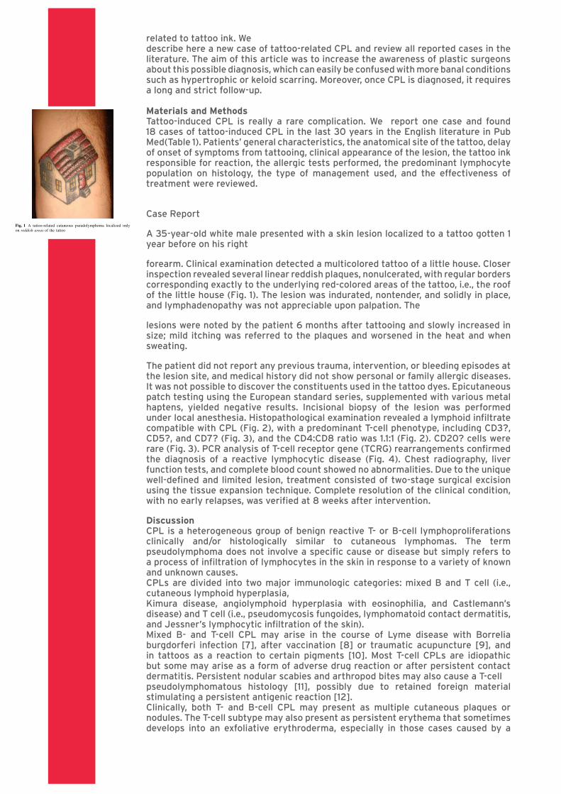
forearm. Clinical examination detected a multicolored tattoo of a little house. Closer inspection revealed several linear reddish plaques, nonulcerated, with regular borders corresponding exactly to the underlying red-colored areas of the tattoo, i.e., the roof of the little house (Fig. 1). The lesion was indurated, nontender, and solidly in place, and lymphadenopathy was not appreciable upon palpation. The
lesions were noted by the patient 6 months after tattooing and slowly increased in size; mild itching was referred to the plaques and worsened in the heat and when sweating.
The patient did not report any previous trauma, intervention, or bleeding episodes at the lesion site, and medical history did not show personal or family allergic diseases. It was not possible to discover the constituents used in the tattoo dyes. Epicutaneous patch testing using the European standard series, supplemented with various metal haptens, yielded negative results. Incisional biopsy of the lesion was performed under local anesthesia. Histopathological examination revealed a lymphoid infiltrate compatible with CPL (Fig. 2), with a predominant T-cell phenotype, including CD3?, CD5?, and CD7? (Fig. 3), and the CD4:CD8 ratio was 1.1:1 (Fig. 2). CD20? cells were rare (Fig. 3). PCR analysis of T-cell receptor gene (TCRG) rearrangements confirmed the diagnosis of a reactive lymphocytic disease (Fig. 4). Chest radiography, liver function tests, and complete blood count showed no abnormalities. Due to the unique well-defined and limited lesion, treatment consisted of two-stage surgical excision using the tissue expansion technique. Complete resolution of the clinical condition, with no early relapses, was verified at 8 weeks after intervention.
DiscussionCPL is a heterogeneous group of benign reactive T- or B-cell lymphoproliferations clinically and/or histologically similar to cutaneous lymphomas. The term pseudolymphoma does not involve a specific cause or disease but simply refers to a process of infiltration of lymphocytes in the skin in response to a variety of known and unknown causes. CPLs are divided into two major immunologic categories: mixed B and T cell (i.e., cutaneous lymphoid hyperplasia,Kimura disease, angiolymphoid hyperplasia with eosinophilia, and Castlemann’s disease) and T cell (i.e., pseudomycosis fungoides, lymphomatoid contact dermatitis,and Jessner’s lymphocytic infiltration of the skin).Mixed B- and T-cell CPL may arise in the course of Lyme disease with Borrelia burgdorferi infection [7], after vaccination [8] or traumatic acupuncture [9], and in tattoos as a reaction to certain pigments [10]. Most T-cell CPLs are idiopathic but some may arise as a form of adverse drug reaction or after persistent contact dermatitis. Persistent nodular scabies and arthropod bites may also cause a T-cellpseudolymphomatous histology [11], possibly due to retained foreign material stimulating a persistent antigenic reaction [12].Clinically, both T- and B-cell CPL may present as multiple cutaneous plaques or nodules. The T-cell subtype may also present as persistent erythema that sometimesdevelops into an exfoliative erythroderma, especially in those cases caused by a
estimated percentage of adolescents and adults with one
or more tattoos now varies from 4 to 24 % [1–3]. Con-
currently, plastic surgeons and dermatologists are fre-
quently called upon to treat tattoo-related complications or
even remove the entire tattoo. In such a setting, it is
fundamental to keep up to date with this growing field.
With respect to tattoo-related complications, infectious
diseases have been studied extensively and more sophis-
ticated tattooing techniques have lowered but not abol-
ished the risk of transmission of syphilis, tuberculosis, and
hepatitis. Even allergic and granulomatous complications
in connection with the process of tattooing have been
described. Other diseases such as lupus erythematosus
discoid, sarcoidosis, psoriasis, lichen planus, and skin
tumors such as basal cell carcinoma, squamous cell car-
cinoma, and melanoma may be localized to only the tat-
tooed area [4, 5].
Despite the wide popularity of tattoos, the relationship
between tattooed pictures and lymphoproliferative disor-
ders has not been studied in depth and only a few cases of
tattoo ink-related cutaneous pseudolymphoma (CPL) have
been reported in the last 30 years; no data are available
about the overall incidence and prevalence of tattoo-
related CPL. CPL represents a benign T- or B-cell lym-
phoproliferative disorder. Historically, the B-cell pattern
has been more correlated to tattoo ink than the T-cell
pattern [6]. The most involved pigment is red and the
mechanism of development of CPL is still unknown. It is
generally wise to be guarded in the diagnosis of CPL
because in a number of cases clear progression from
apparent CPL to true cutaneous lymphoma have been
recorded and one case was related to tattoo ink. We
describe here a new case of tattoo-related CPL and review
all reported cases in the literature. The aim of this article
was to increase the awareness of plastic surgeons about
this possible diagnosis, which can easily be confused with
more banal conditions such as hypertrophic or keloid
scarring. Moreover, once CPL is diagnosed, it requires a
long and strict follow-up.
Materials and Methods
Tattoo-induced CPL is really a rare complication. We
report one case and found 18 cases of tattoo-induced CPL
in the last 30 years in the English literature in PubMed
(Table 1). Patients’ general characteristics, the anatomical
site of the tattoo, delay of onset of symptoms from tat-
tooing, clinical appearance of the lesion, the tattoo ink
responsible for reaction, the allergic tests performed, the
predominant lymphocyte population on histology, the type
of management used, and the effectiveness of treatment
were reviewed.
Case Report
A 35-year-old white male presented with a skin lesion
localized to a tattoo gotten 1 year before on his right
forearm. Clinical examination detected a multicolored
tattoo of a little house. Closer inspection revealed several
linear reddish plaques, nonulcerated, with regular borders
corresponding exactly to the underlying red-colored areas
of the tattoo, i.e., the roof of the little house (Fig. 1). The
lesion was indurated, nontender, and solidly in place, and
lymphadenopathy was not appreciable upon palpation. The
lesions were noted by the patient 6 months after tattooing
and slowly increased in size; mild itching was referred to
the plaques and worsened in the heat and when sweating.
The patient did not report any previous trauma, interven-
tion, or bleeding episodes at the lesion site, and medical
history did not show personal or family allergic diseases. It
was not possible to discover the constituents used in the
tattoo dyes. Epicutaneous patch testing using the European
standard series, supplemented with various metal haptens,
yielded negative results. Incisional biopsy of the lesion was
performed under local anesthesia. Histopathological
examination revealed a lymphoid infiltrate compatible with
CPL (Fig. 2), with a predominant T-cell phenotype,
including CD3?, CD5?, and CD7? (Fig. 3), and the
CD4:CD8 ratio was 1.1:1 (Fig. 2). CD20? cells were rare
(Fig. 3). PCR analysis of T-cell receptor gene (TCRG)
rearrangements confirmed the diagnosis of a reactive
lymphocytic disease (Fig. 4). Chest radiography, liver
function tests, and complete blood count showed no
Fig. 1 A tattoo-related cutaneous pseudolymphoma localized only
on reddish areas of the tattoo
Aesth Plast Surg
123
reaction to a drug or contact dermatitis.The mixed B- and T-cell subtype may also be associated with palpable lymphadenopathy, adding to diagnostic confusion.It is fundamental to give the pathologist a good clinical history to help distinguish between true lymphoma and pseudolymphoma because the pathological, phenotypic,and molecular differentiations are not absolute. The inflammatory infiltrate may be bandlike, nodular, or diffuse and composed predominantly of lymphocytes and eventually with other inflammatory cells (i.e., plasma cells, eosinophils, mast cells, neutrophils, and histiocytic giant cells). Benign or reactive processes are usually considered polyclonal, whereas the presence of monoclonality generally implies a malignant process. Thus, PCR analysis of TCRG is usually recommended to distinguish between reactive and neoplastic cutaneous lymphocytic diseases.This distinction, however, is not always true. It is possible to find clonal T- or B-cell populations in CPL and maybe a subset more likely to develop into lymphoma [13–15].Indeed, it is estimated that lymphomas develop in 10–20 % of patients affected by CPL, typically many years after the original diagnosis [16]. This may explain the one published case of tattoo-induced CPL malignant transformation [17].Even if unlikely, physicians should be alerted to this occurrence and routinely follow patients for extracutaneous manifestations or local relapses. Unfortunately, malignant evolution cannot be predicted by clinical or histologic features, TCRG rearrangement, or DNA flow cytometry [18]. Nevertheless, in our case we found an abnormal CD4:CD8 ratio of 1.1:1, whereas normally it is approximately 2–3:1. This suggests that this patient should be followed to exclude the possibility of an evolving tattoorelated T-cell lymphoproliferative disorder. From the review of the literature (our case is included), 10 men and 9 women, all white and with a median age of 41 years, were evaluated. The mean time of onset of CPL
after tattooing was 9 years (range = 4 months to 42 years). It is confirmed that red ink was the most common causative agent (15 cases, 79 %), but black [19] and blue and green [20, 21] dyes also were able to induce CPL.Classical tattoo dyes contain metal compounds—reddish areas are mainly cinnabar, blue mainly cobalt salts, and green mainly chrome salts—but organic substances,including synthetic azo-dyes, are used in modern tattoo dyes.Even if the pathogenesis is not completely clear, it has been suggested that these components are the leading elicitors of delayed hypersensitivity; indeed, allergic reactions to red mercury-based dyes have been reported [22].A photosensitive reaction to cadmium yellow occurs occasionally [23], and when photosensitive reactions to red tattoos are investigated, it is found that the cause issometimes a cadmium salt [24]. In only one case were the constituents of tattoo ink provided by the manufacturer [25]: metal iron, copper fumes, metal manganese, andmetal cobalt.
abnormalities. Due to the unique well-defined and limited
lesion, treatment consisted of two-stage surgical excision
using the tissue expansion technique. Complete resolution
of the clinical condition, with no early relapses, was veri-
fied at 8 weeks after intervention.
Discussion
CPL is a heterogeneous group of benign reactive T- or
B-cell lymphoproliferations clinically and/or histologically
similar to cutaneous lymphomas. The term pseudolym-
phoma does not involve a specific cause or disease but
simply refers to a process of infiltration of lymphocytes in
the skin in response to a variety of known and unknown
causes.
CPLs are divided into two major immunologic catego-
ries: mixed B and T cell (i.e., cutaneous lymphoid hyper-
plasia, Kimura disease, angiolymphoid hyperplasia with
eosinophilia, and Castlemann’s disease) and T cell (i.e.,
pseudomycosis fungoides, lymphomatoid contact dermati-
tis, and Jessner’s lymphocytic infiltration of the skin).
Mixed B- and T-cell CPL may arise in the course of Lyme
disease with Borrelia burgdorferi infection [7], after vac-
cination [8] or traumatic acupuncture [9], and in tattoos as
a reaction to certain pigments [10]. Most T-cell CPLs are
idiopathic but some may arise as a form of adverse drug
reaction or after persistent contact dermatitis. Persistent
nodular scabies and arthropod bites may also cause a T-cell
pseudolymphomatous histology [11], possibly due to
retained foreign material stimulating a persistent antigenic
reaction [12].
Clinically, both T- and B-cell CPL may present as
multiple cutaneous plaques or nodules. The T-cell subtype
may also present as persistent erythema that sometimes
develops into an exfoliative erythroderma, especially in
those cases caused by a reaction to a drug or contact der-
matitis. The mixed B- and T-cell subtype may also be
Fig. 2 a Tattoo pigment in the
superficial reticular dermis and
diffuse lymphoid infiltrate
(hematoxylin and eosin; original
magnification, 910). Lymphoid
cells are almost equally positive
for CD4 (b) and CD8
(c) (original magnification,
910), with an abnormal
CD4:CD8 ratio of 1.1:1
Aesth Plast Surg
123
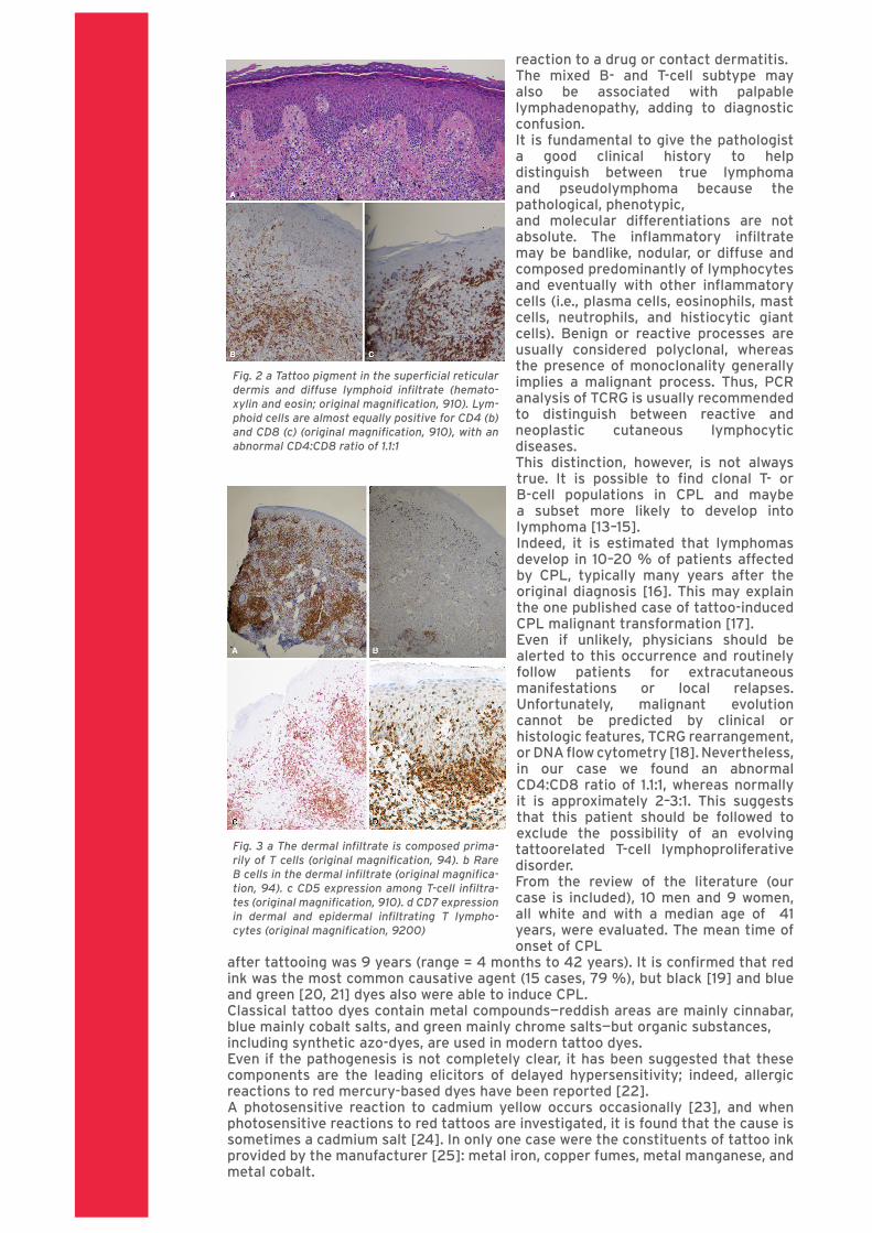
Fig. 2 a Tattoo pigment in the superficial reticular dermis and diffuse lymphoid infiltrate (hemato-xylin and eosin; original magnification, 910). Lym-phoid cells are almost equally positive for CD4 (b) and CD8 (c) (original magnification, 910), with an abnormal CD4:CD8 ratio of 1.1:1
associated with palpable lymphadenopathy, adding to
diagnostic confusion.
It is fundamental to give the pathologist a good clinical
history to help distinguish between true lymphoma and
pseudolymphoma because the pathological, phenotypic,
and molecular differentiations are not absolute. The
inflammatory infiltrate may be bandlike, nodular, or diffuse
and composed predominantly of lymphocytes and eventu-
ally with other inflammatory cells (i.e., plasma cells,
eosinophils, mast cells, neutrophils, and histiocytic giant
cells). Benign or reactive processes are usually considered
polyclonal, whereas the presence of monoclonality gener-
ally implies a malignant process. Thus, PCR analysis of
TCRG is usually recommended to distinguish between
reactive and neoplastic cutaneous lymphocytic diseases.
This distinction, however, is not always true. It is possible
to find clonal T- or B-cell populations in CPL and maybe a
subset more likely to develop into lymphoma [13–15].
Indeed, it is estimated that lymphomas develop in 10–20 %
of patients affected by CPL, typically many years after the
original diagnosis [16]. This may explain the one published
case of tattoo-induced CPL malignant transformation [17].
Even if unlikely, physicians should be alerted to this
occurrence and routinely follow patients for extracutaneous
manifestations or local relapses. Unfortunately, malignant
evolution cannot be predicted by clinical or histologic
features, TCRG rearrangement, or DNA flow cytometry
[18]. Nevertheless, in our case we found an abnormal
CD4:CD8 ratio of 1.1:1, whereas normally it is approxi-
mately 2–3:1. This suggests that this patient should be
followed to exclude the possibility of an evolving tattoo-
related T-cell lymphoproliferative disorder.
From the review of the literature (our case is included),
10 men and 9 women, all white and with a median age of
Fig. 3 a The dermal infiltrate is
composed primarily of T cells
(original magnification, 94).
b Rare B cells in the dermal
infiltrate (original
magnification, 94). c CD5
expression among T-cell
infiltrates (original
magnification, 910). d CD7
expression in dermal and
epidermal infiltrating T
lymphocytes (original
magnification, 9200)
Aesth Plast Surg
123
Fig. 3 a The dermal infiltrate is composed prima-rily of T cells (original magnification, 94). b Rare B cells in the dermal infiltrate (original magnifica-tion, 94). c CD5 expression among T-cell infiltra-tes (original magnification, 910). d CD7 expression in dermal and epidermal infiltrating T lympho-cytes (original magnification, 9200)

In most cases the infiltrate is localized to the tattoo area where pigment is deposited. It appears like erythematous nodules or plaques, sometimes causing itching, or a photosensitive reaction [26]. From the precise location on only specific-colored areas and its uniform appearance, CPL can be clinically distinguished from pathologic scarring or granulomatous reactions.However, diagnosis of CPL is based on histologic features, architecture of infiltration, and the results of immunophenotyping and genotyping of the lymphocytes.Thirteen of 19 cases (68 %) showed T-cell preponderance on histology, and the other six were described without using immunochemistry. In contrast to the literature, B-cell predominance was not demonstrated in the collected cases.This suggests that adjustment or reclassification of tattooinduced CPL may be necessary. To evaluate the evidence for a delayed hypersensitivity reaction to the pigment, patch testing was performed in eight cases. During the tattooing process, the dyes are pushed directly into the dermis: because the epidermal
Langerhans cells are avoided in this way, some of the allergic tests given in such cases may be negative. An intradermal reaction test was also performed in one case [27].There is no standard treatment for tattoo-induced pseudolymphoma since none of those proposed has yet been successful. As cautioned by Mun˜oz et al. [28], even ifmalignant transformation is rare, surgical excision of the tattoo should be performed whenever possible [27, 28, 31], including only excision of smaller tattoos or reconstruction with tissue expansion for larger tattoos. For those patients in whom a surgical option is not feasible (e.g., lip-liner tattoo), a wait-and-see approach can be proposed [25].However, regular follow-up is mandatory as progression of CPL to lymphoma has been reported [17]. Laser treatment, especially Nd:YAG, has been used with success [19, 20, 29] but the risk of spreading dyes in the dermis, worsening the hypersensitivity response, must be considered. Moreover, treatment with a laser may not remove the pigment completely and is therefore not recommended [30].Topicals such as clobetasol propionate 0.05 % [31] or intralesional injection of corticosteroids like triamcinolone [32] have been used but the results are variable. The use of different strengths and doses of steroids does not allow a standardized comparison and recurrences are common [26]. However, if nonsurgical treatment modalities are chosen, regular follow-up visits are advised [29].
ConclusionsWith the increasing popularity of tattoos, reactions to them are more likely to occur. Even if it is less common compared to lichenoid and granulomatous reactions in the tattoo area, CPL is a rare skin lesion to consider, since it can be easily confused with a pathologic scar or a granulomatous reaction and can, in rare cases, evolve into a malignancy. Different from previous data reported in dermatology textbooks, in our case and in most of the cases in PubMed, the T-cell pattern seems to be predominant and so further studies are needed. Finally, considering the long time between tattooing and the onset of lymphoproliferative disorders, we should expect to see a higher rate of tattoo-induced pseudolymphomas in the future.Conflict of interest The authors have no conflicts of interest to disclose.
41 years, were evaluated. The mean time of onset of CPL
after tattooing was 9 years (range = 4 months to
42 years). It is confirmed that red ink was the most com-
mon causative agent (15 cases, 79 %), but black [19] and
blue and green [20, 21] dyes also were able to induce CPL.
Classical tattoo dyes contain metal compounds—reddish
areas are mainly cinnabar, blue mainly cobalt salts, and
green mainly chrome salts—but organic substances,
including synthetic azo-dyes, are used in modern tattoo
dyes.
Even if the pathogenesis is not completely clear, it has
been suggested that these components are the leading
elicitors of delayed hypersensitivity; indeed, allergic reac-
tions to red mercury-based dyes have been reported [22].
A photosensitive reaction to cadmium yellow occurs
occasionally [23], and when photosensitive reactions to red
tattoos are investigated, it is found that the cause is
sometimes a cadmium salt [24]. In only one case were the
constituents of tattoo ink provided by the manufacturer
[25]: metal iron, copper fumes, metal manganese, and
metal cobalt.
In most cases the infiltrate is localized to the tattoo area
where pigment is deposited. It appears like erythematous
nodules or plaques, sometimes causing itching, or a pho-
tosensitive reaction [26]. From the precise location on only
specific-colored areas and its uniform appearance, CPL can
be clinically distinguished from pathologic scarring or
granulomatous reactions.
However, diagnosis of CPL is based on histologic fea-
tures, architecture of infiltration, and the results of immu-
nophenotyping and genotyping of the lymphocytes.
Thirteen of 19 cases (68 %) showed T-cell preponderance
on histology, and the other six were described without
using immunochemistry. In contrast to the literature, B-cell
predominance was not demonstrated in the collected cases.
This suggests that adjustment or reclassification of tattoo-
induced CPL may be necessary.
To evaluate the evidence for a delayed hypersensitivity
reaction to the pigment, patch testing was performed in
eight cases. During the tattooing process, the dyes are
pushed directly into the dermis: because the epidermal
Langerhans cells are avoided in this way, some of the
allergic tests given in such cases may be negative. An
intradermal reaction test was also performed in one case
[27].
There is no standard treatment for tattoo-induced pseu-
dolymphoma since none of those proposed has yet been
successful. As cautioned by Munoz et al. [28], even if
malignant transformation is rare, surgical excision of the
tattoo should be performed whenever possible [27, 28, 31],
including only excision of smaller tattoos or reconstruction
with tissue expansion for larger tattoos. For those patients
in whom a surgical option is not feasible (e.g., lip-liner
tattoo), a wait-and-see approach can be proposed [25].
However, regular follow-up is mandatory as progression of
CPL to lymphoma has been reported [17].
Laser treatment, especially Nd:YAG, has been used with
success [19, 20, 29] but the risk of spreading dyes in the
dermis, worsening the hypersensitivity response, must be
considered. Moreover, treatment with a laser may not
remove the pigment completely and is therefore not rec-
ommended [30].
Topicals such as clobetasol propionate 0.05 % [31] or
intralesional injection of corticosteroids like triamcinolone
[32] have been used but the results are variable. The use of
different strengths and doses of steroids does not allow a
standardized comparison and recurrences are common
[26]. However, if nonsurgical treatment modalities are
chosen, regular follow-up visits are advised [29].
Conclusions
With the increasing popularity of tattoos, reactions to them
are more likely to occur. Even if it is less common com-
pared to lichenoid and granulomatous reactions in the
Fig. 4 Profile of gene-scanning analysis of T-cell receptor gene
(TCRG) rearrangements (x axis, length of PCR product; y axis,
fluorescence intensity). a Polyclonal pattern of TCRG rearrangements
of the case, showing typical polyclonal Gaussian curve. b Positive
control of monoclonal TCRG rearrangements. PCR analysis was
performed as described in van Dongen et al. [33]
Aesth Plast Surg
123
Fig. 4 Profile of gene-scanning analysis of T-cell receptor gene (TCRG) rearrangements (x axis, length of PCR product; y axis, fluorescence in-tensity). a Polyclonal pattern of TCRG rearran-gements of the case, showing typical polyclonal Gaussian curve. b Positive control of monoclonal TCRG rearrangements. PCR analysis was perfor-med as described in van Dongen et al. [33]
References1. Bosello R, Favaro A, Zanetti T, Soave M, Vidotto G, Huon G,Santonastaso P (2010) Tattoos and piercings in adolescents: family conflicts and temperament. Riv Psichiatr 45(2):102–1062. Mayers LB, Judelson DA, Moriarty BW, Rundell KW (2002)Prevalence of body art (body piercing and tattooing) in university undergraduates and incidence of medical complications. Mayo Clin Proc 77(1):29–34
3. Laumann AE, Derick AJ (2006) Tattoos and body piercings in the United States: a national data set. J Am Acad Dermatol 55(3):413–4214. Kluger N, Koljonen V (2012) Tattoos, inks, and cancer. Lancet Oncol 13(4):e161–e16185. Kazandjieva J, Tsankov N (2007) Tattoos: dermatological complications. Clin Dermatol 25:375–3826. Van Vloten WA, Willemze R (2003) The many faces of lymphocytoma cutis. J Eur Acad Dermatol Venereol

Agnese Cremaschi
17(1):3–67. Garbe C, Stein H, Dienemann D, Orfanos C (1991) Borrelia burdorferi-associated cutaneous B-cell lymphoma. J Am Acad Dermatol 24:584–590 8. Lanzafame S, Micali G (1993) Cutaneous lymphoid hyperplasia (pseudolymphoma) secondary to vaccination. Pathologica 85:555–5569. Kim K, Lee M, Choi J et al (2002) CD30-positive T-cell rich pseudolyphoma induced by gold acupuncture. Br J Dermatol 146:882–88410. Blumental G, Okun MR, Ponitch A (1982) Pseudolymphomatous reactions to tattoos. J Am Acad Dermatol 6:485–48811. Walton S, Bottomley WW, Wyatt EH, Bury HP (1991) Pseudo T-cell lymphoma due to scabies in a patient with Hodgkin’s disease. Br J Dermatol 124:277–27812. Burg G, Dummer R, Kadin M (2001) From inflammation to neoplasia. Arch Dermatol 137:949–95213. Ploysangam T, Breneman DL, Mutasim DF (1998) Cutaneous pseudolymphomas. J Am Acad Dermatol 38:877–90514. Halevy S, Sandbank M (1987) Transformation of lymphocytoma cutis into malignant lymphoma. Acta Derm Venereol 67:172–17515. Nakayama H, Mihara M, Shimao S (1987) Malignant transformation of lymphadenosis benigna cutis: a possibly transformed case and B-cell lymphoma. J Dermatol 4:266–26916. Chott A, Vonderheid EC, Olbricht S et al (1996) The same dominant T-cell clone is present in multiple regressing skin lesions and associated T-cell lymphomas of patients with lymphomatoid papulosis. J Invest Dermatol 106:696–70017. Sangueza OP, Yadav S, White CR et al (1992) Evolution of B-cell lymphoma from pseudolymphoma: a multidisciplinary approach using histology, immunochemistry and Southern blot analysis. Am J Dermatopathol 14:408–41318. el-Azhary RA, Gibson LE, Kurtin PJ et al (1994) Lymphomatoid papulosis: a clinical and histologic review of 53 cases with leukocyte immunophenotyping, DNA flow cytometry, and T-cell receptor gene rearrangement studies. J Am Acad Dermatol 30:210–21819. Campolmi P, Bassi A, Bonan P et al (2011) Cutaneous pseudolymphoma localized to black tattoo. J Am Acad Dermatol 65(5):e155–E15720. Chiang C, Romero L (2009) Cutaneous lymphoid hyperplasia (pseudolymphoma) in a tattoo after far infrared light. Dermatol Surg 35:1434–1438
21. Patrizi A, Raone B, Savoia F, Bacci F, Pileri A, Gurioli C, Neri I (2009) Tattoo-associated pseudolymphomatous reaction and its successful treatment with hydroxychloroquine. Acta Derm Venereol 89(3):327–32822. Sowden JM, Byrne JP, Smith AG, Hiley C, Suarez V, Wagner B, Slater DN (1991) Red tattoo reactions: X-ray microanalysis and patch-test studies. Br J Dermatol 124(6):576–58023. Bjo¨rnberg A (1963) Reactions to light in yellow tattoos from cadmium sulphide. Arch Dermatol 88:267–27124. Yazdian-Tehrani H, Shibu MM, Carver NC (2001) Reaction in a red tattoo in the absence of mercury. Br J Plast Surg 54:555–55625. Shin JB, Seo SH, Kim BK, Kim IH, Son SW (2009) Cutaneous T cell pseudolymphoma at the site of a semipermanent lip-liner tattoo. Dermatology 218:75–7826. Cruz FA, Lage D, Frige´rio RM, Zaniboni MC, Arruda LH (2010) Reactions to the different pigments in tattoos: a report of two cases. An Bras Dermatol 85(5):708–71127. Gutermuth J, Hein R, Fend F, Ring J, Jakob T (2007) Cutaneous pseudolymphoma arising after tattoo placement. Eur Acad Dermatol Venereol 21:536–57828. Mun˜oz C, Guilabert A, Mascaro´ JM Jr, Lopez-Lerma I, Vilaplana J (2005) An embossed tattoo. Clin Exp Dermatol 31:309–31029. Antony FC, Harland CC (2003) Red ink tattoo reactions: successful treatment with the Q-switched 532 nm Nd:YAG laser. Br J Dermatol 149:94–9830. Zelickson BD, Mehregan DA, Zarrin AA, Coles C, Hartwig P, Olson S, Leaf-Davis J (1994) Clinical, histologic, and ultrastructural evaluation of tattoos treated with three laser systems. Laser Surg Med 15:364–37231. Chave A, Mortimer NJ, Johnston GA (2004) Simultaneous pseudolymphomatous and lichenoid tattoo reactions triggered by re-tattooing. Clin Exp Dermatol 29:196–20532. Zinberg M, Heilman E, Glickman F (1982) Cutaneous pseudolymphoma resulting from a tattoo. J Dermatol Surg Oncol 8(11):955–95833. van Dongen JJ, Langerak AW, Bru¨ggemann M et al (2003) Design and standardization of PCR primers and protocols for detection of clonal immunoglobulin and T-cell receptor gene recombinations in suspect lymphoproliferations: report of the BIOMED-2 concerted action BMH4-CT98-3936. Leukemia 17:2257–2317 Aesth Plast Surg 123