Aging of the Skeleton: Osteoporosis An Evolutionary and Biocultural Perspective.
47
Aging of the Skeleton: Aging of the Skeleton: Osteoporosis Osteoporosis An Evolutionary and Biocultural Perspective
-
date post
20-Dec-2015 -
Category
Documents
-
view
215 -
download
1
Transcript of Aging of the Skeleton: Osteoporosis An Evolutionary and Biocultural Perspective.

Aging of the Skeleton: Aging of the Skeleton: OsteoporosisOsteoporosis
An Evolutionary and Biocultural Perspective

Aging and Skeleton
• Aging and Senescence: signs and the process of becoming old, less capable of homeostasis function (body cannot heal and is susceptible)
• Decline in many body functions: cardiac output, pulmonary and renal function, immune system
• Increase in chronic and degenerative diseases: osteoarthritis
• Bone loss: osteoporosis
OsteoporosisOsteoporosis• A reduction in bone mass and deterioration bone
microstructure
• Clinically recognized as bone fragility with an increased susceptibility to fracture
• Osteopenia vs. osteoporosis
• Serious health concern in western aging populations: 1/2 women and 1/4 men > age of 50 will have an osteoporosis-related fracture. Women 4x times more

OsteoporosisOsteoporosis• Bone mineral density (BMD)
WHO definition: BDM 2.5 SD’s below young adult mean
• Measured with densitometry
• Problems: population specific, overlap in fracture and non-fracture BMD, only looks at one measure of bone (quantity)

OsteoporosisOsteoporosis
• Factures hip, distal forearm, vertebral and hip fractures
• bone resorption > bone formation
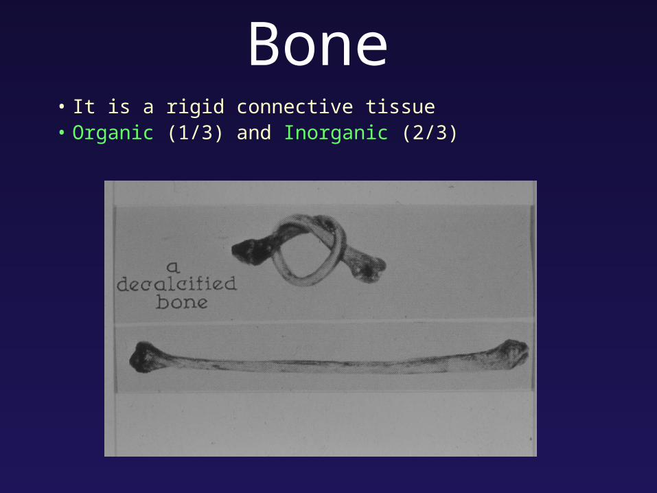
Bone• It is a rigid connective tissue• Organic (1/3) and Inorganic (2/3)
Bone has • structural function: - framework for soft tissue, muscles,
protects vital organs• physiological function: - reservoir marrow and hemapoetic
tissue (rbc, white bc, platelets) - storage for adipose - stores Ca and Ph, linked to blood
system - 90% of Ca and Ph is in the bone
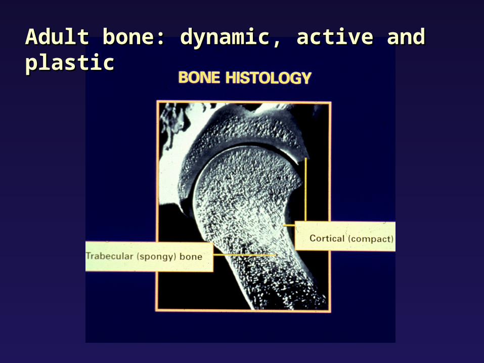
Adult bone: dynamic, active and plasticAdult bone: dynamic, active and plastic
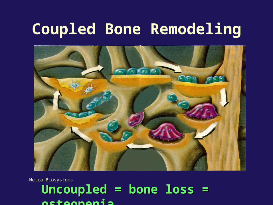
Metra Biosystems
Coupled Bone Remodeling
Uncoupled = bone loss = osteopeniaUncoupled = bone loss = osteopenia
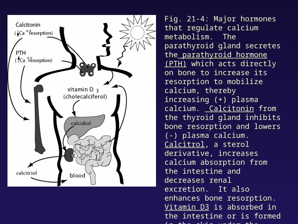
Fig. 21-4: Major hormones that regulate calcium metabolism. The parathyroid gland secretes the parathyroid hormone (PTH) which acts directly on bone to increase its resorption to mobilize calcium, thereby increasing (+) plasma calcium. Calcitonin from the thyroid gland inhibits bone resorption and lowers (-) plasma calcium. Calcitrol, a sterol derivative, increases calcium absorption from the intestine and decreases renal excretion. It also enhances bone resorption. Vitamin D3 is absorbed in the intestine or is formed in the skin under the influence of ultraviolet radiation. In the liver, vitamin D3 is converted to cacidiol, which in turn, is activated in the kidney to calcitirol, the most active form in calcium regulation.
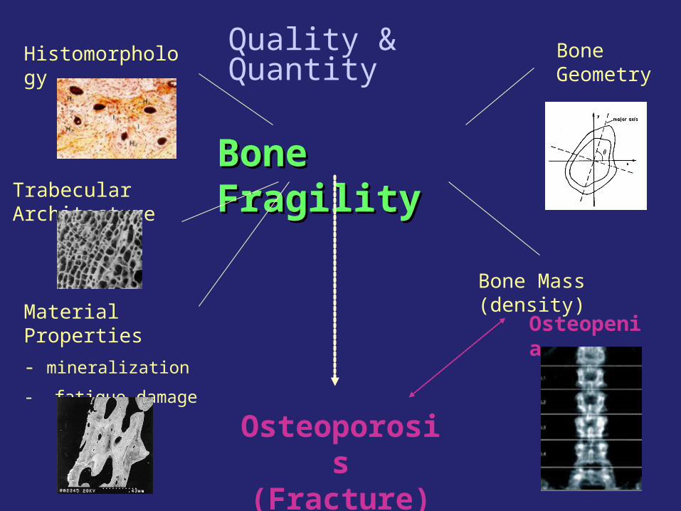
Bone FragilityBone Fragility
Histomorphology
Trabecular Architecture
Material Properties
- mineralization
- fatigue damage
Bone Geometry
Bone Mass (density)
Osteoporosis (Fracture)
Osteopenia
Quality & Quantity

What causes What causes bone loss and bone loss and fragility?fragility?
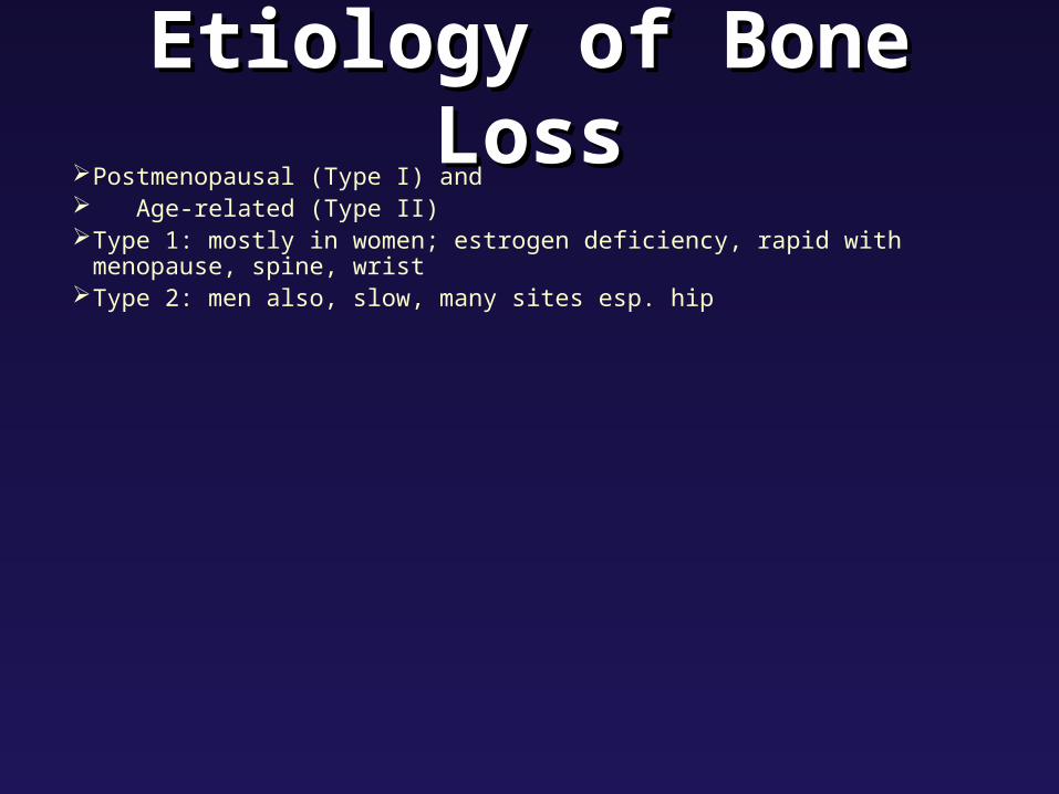
Etiology of Bone LossEtiology of Bone LossPostmenopausal (Type I) and Age-related (Type II)Type 1: mostly in women; estrogen deficiency, rapid with menopause, spine, wrist Type 2: men also, slow, many sites esp. hip
Age-related Bone LossAge-related Bone Loss• Senescence of osteoblasts (impaired function)• Also increase in PTH levels (due to decrease renal and Ca re-
absorption and intestinal Ca absorption that occurs in elderly - as less Vit D exposure, less synthesis of calcitrol, or vitD resisance?)

Fig. 21-3: Calcium metabolism. Note the different daily requirements of calcium with age; they almost double in women after 50, as compared to young adults.

Postmenopausal and Age-Related Bone Loss Integrated• cannot look at bone loss in females
without thinking of post-menopausal loss and life history

Multifactorial EtiologyMultifactorial EtiologyNumber of factors that play a role in
peak bone mass and loss: Genetics and ethnicity Lifestyle: alcohol, smoking, drugs diet/nutrition mechanical usage (physical activity) reproductive factors (pregnancy, parity and
lactation)
• Since multifactorial interest to look at its evolution and variation in different populations

Paleopathology and Paleopathology and OsteoporosisOsteoporosis
• The study of health and disease in the past = paleopathology
• Secondary Sources: documentary and iconographic data
• Primary Sources: skeletal remains
- usually only direct evidence left in the archaeological record to examine health and disease in past populations

Biocultural ApproachBiocultural Approach
biocultural approach = emphasizes that environmental, cultural, and lifestyle factors have impact on the biology of disease.

OP in Past PopulationsOP in Past Populations
• Historical populations unique model to study bone loss
• Many studies of bone loss in various archaeological populations:
- pattern of bone loss and fragility in the past is different than in clinical osteoporosis today
- mostly bone mass
- few studies of bone quality
trabecular architecture

Young normal
Elderly osteoporotic
Trabecular ArchitectureTrabecular Architecture

MaterialsMaterials• trabecular architecture
• several medieval British archaeological populations
• 4th lumbar vertebrae
• 3 adult age groups: 17-25, 26-45, 45+
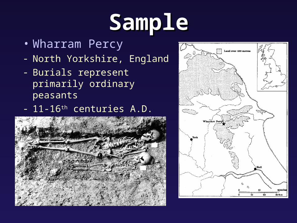
SampleSample• Wharram Percy- North Yorkshire, England
- Burials represent primarily ordinary peasants
- 11-16th centuries A.D.

Image AnalysisImage Analysis• trabecular architecture • x-rays 5mm thick lumbar sections• image processing and analysis, obtained
binary and skeletonized images

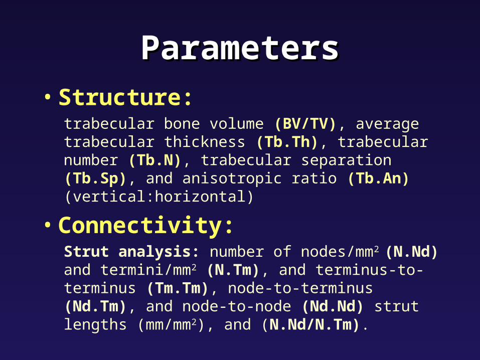
ParametersParameters• Structure:
trabecular bone volume (BV/TV), average trabecular thickness (Tb.Th), trabecular number (Tb.N), trabecular separation (Tb.Sp), and anisotropic ratio (Tb.An) (vertical:horizontal)
• Connectivity:Strut analysis: number of nodes/mm2 (N.Nd) and termini/mm2 (N.Tm), and terminus-to-terminus (Tm.Tm), node-to-terminus (Nd.Tm), and node-to-node (Nd.Nd) strut lengths (mm/mm2), and (N.Nd/N.Tm).

ParametersParameters• Structure:
trabecular bone volume (BV/TV), average trabecular thickness (Tb.Th), trabecular number (Tb.N), trabecular separation (Tb.Sp)
Connectivity:Connectivity:2-D Star Volume 2-D Star Volume (2D Star)(2D Star) strut analysis: number of strut analysis: number of nodes/mmnodes/mm2 2 (N.Nd) (N.Nd) and termini/mmand termini/mm22 (N.Tm)(N.Tm), and , and terminus-to-terminus terminus-to-terminus (Tm.Tm)(Tm.Tm), node-to-terminus , node-to-terminus (Nd.Tm)(Nd.Tm), and node-to-node , and node-to-node (Nd.Nd)(Nd.Nd) strut lengths strut lengths (mm/mm(mm/mm22), and (), and (N.Nd/N.Tm)N.Nd/N.Tm)..
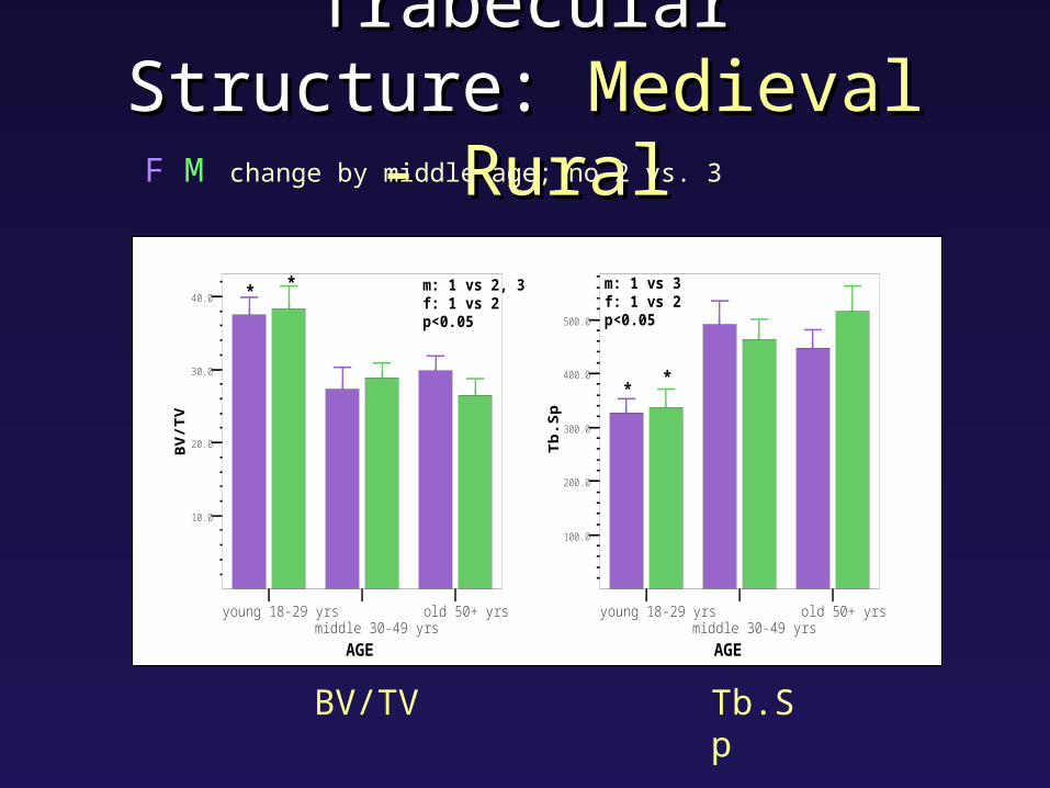
Trabecular Structure: Trabecular Structure: Medieval - RuralMedieval - Rural
F M change by middle age; no 2 vs. 3
* * m: 1 vs 2, 3f: 1 vs 2p<0.05
young 18-29 yrsmiddle 30-49 yrs
old 50+ yrs
AGE
10.0
20.0
30.0
40.0
**
m: 1 vs 3f: 1 vs 2p<0.05
young 18-29 yrsmiddle 30-49 yrs
old 50+ yrs
AGE
100.0
200.0
300.0
400.0
500.0
BV/TV Tb.Sp
ParametersParameters• Structure:
trabecular bone volume (BV/TV), average trabecular thickness (Tb.Th), trabecular number (Tb.N), trabecular separation (Tb.Sp), and anisotropic ratio (Tb.An) (vertical:horizontal)
• Connectivity:Strut analysis: number of nodes/mm2 (N.Nd) and termini/mm2 (N.Tm), and terminus-to-terminus (Tm.Tm), node-to-terminus (Nd.Tm), and node-to-node (Nd.Nd) strut lengths (mm/mm2), and (N.Nd/N.Tm).

ParametersParameters• Structure:Structure:
trabecular bone volumetrabecular bone volume (BV/TV) (BV/TV), average trabecular , average trabecular thicknessthickness (Tb.Th) (Tb.Th), trabecular number, trabecular number (Tb.N) (Tb.N),, trabecular separationtrabecular separation (Tb.Sp) (Tb.Sp), and anisotropic ratio, and anisotropic ratio (Tb.An) (Tb.An) (vertical:horizontal)(vertical:horizontal)
• Connectivity:Strut analysis: number of nodes/mm2 (N.Nd) and termini/mm2 (N.Tm), and terminus-to-terminus (Tm.Tm), node-to-terminus (Nd.Tm), and node-to-node (Nd.Nd) strut lengths (mm/mm2), and (N.Nd/N.Tm).

Strut AnalysisStrut Analysis
terminus
terminus
terminus
terminus
node-terminus strut
nodenode
node-node strut
terminus-terminus strut

Connectivity: Connectivity: RuralRuralM decrease by middle age, no 2 vs. 3
F no difference between age groups
*m: 1 vs 3
young 18-29 yrsmiddle 30-49 yrs
old 50+ yrs
age
0.0
0.5
1.0
1.5
2.0
* m: 1 vs 2, 3p<0.05
young 18-29 yrsmiddle 30-49 yrs
old 50+ yrs
age
0.0
0.3
0.5
0.8
1.0
N.Nd Nd.Nd
19-39 40-59 60-80
(%)
of
TS
L
0
5
10
15
20
25
30
35
40
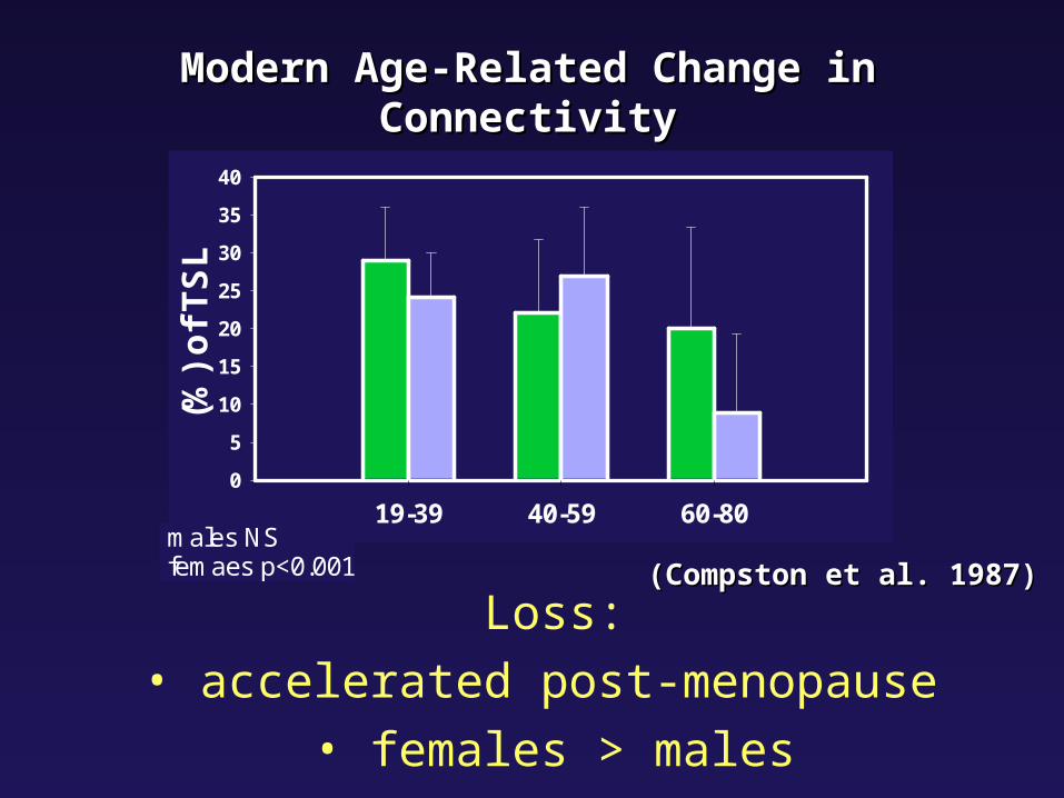
males NS femaes p<0.001 (Compston et al. 1987)(Compston et al. 1987)
Modern Age-Related Change in ConnectivityModern Age-Related Change in Connectivity
Loss:
• accelerated post-menopause
• females > males
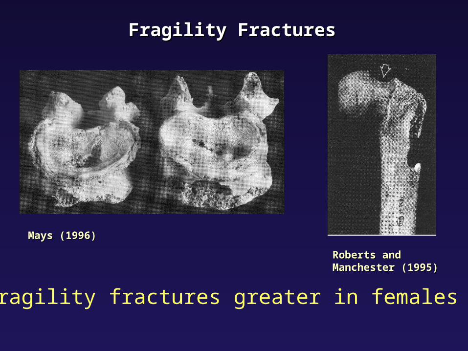
Fragility FracturesFragility Fractures
Mays (1996)Mays (1996)
Roberts and Roberts and Manchester (1995)Manchester (1995)
• fragility fractures greater in females

Medieval ResultsMedieval Results
• Both sexes: significant change in trabecular architecture by middle age
• No sex difference
• Low prevalence of fracture
• Different patterns than seen in modern populations
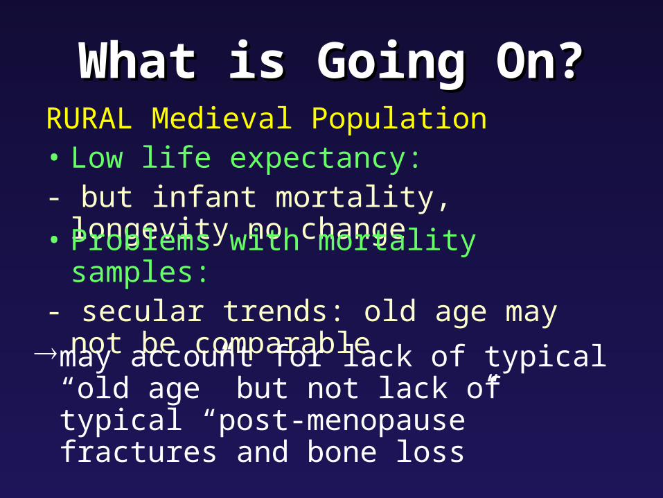
What is Going On?What is Going On?RURAL Medieval Population• Low life expectancy: - but infant mortality, longevity no change
• Problems with mortality samples:- secular trends: old age may not be
comparablemay account for lack of typical “old age”
but not lack of typical “post-menopause” fractures and bone loss
• Nutrition/diet:- maybe sub-optimal conditions- no evidence for calcium or vitamin D deficiency - Vitamin D is likely important: Vitamin D insufficiency- Is Calcium really the big story in bone loss???
What is Going On?What is Going On?
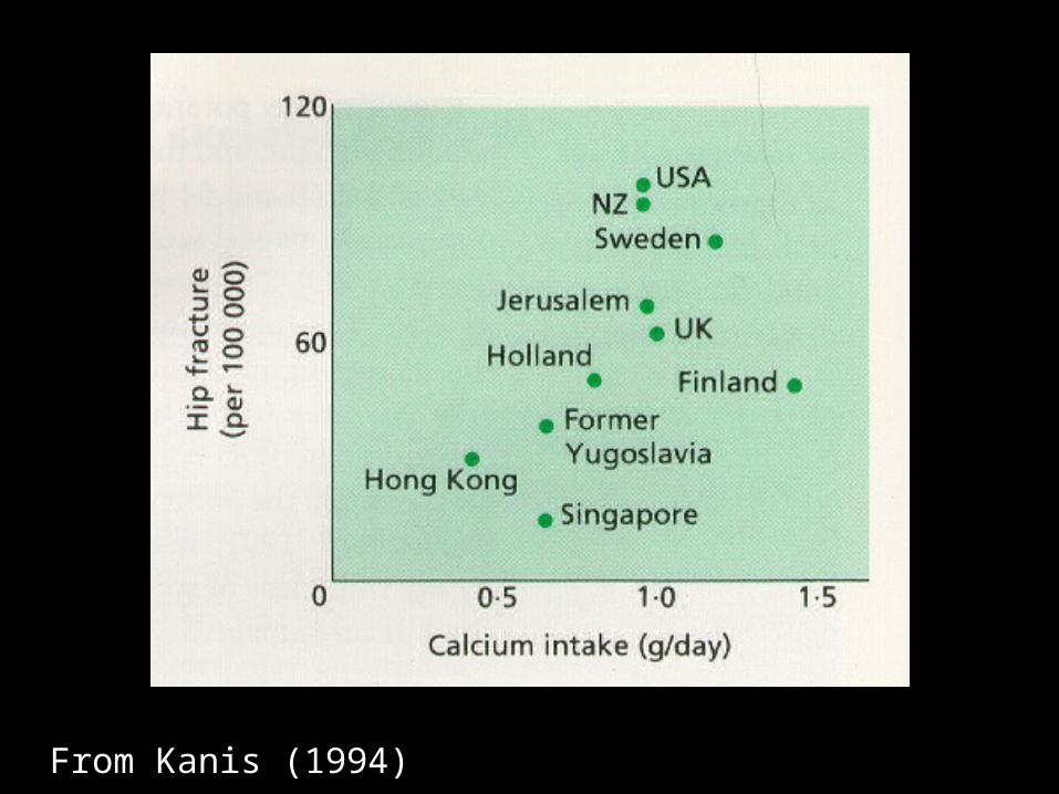
From Kanis (1994)
What is Going On?What is Going On?
• Physical activity:- rural farming population: both sexes
involved in similar arduous activity- Could have protected both sexes from
bone loss in old age

What is Going On?What is Going On?Both diet and activity do not fully explain atypical patterns seen in females
• Reproductive Factors: pregnancy and lactation
Pregnancy and LactationPregnancy and Lactation
• metabolically active states in bone
• bone loss during pregnancy
• long-term bone fragility? unlikely
• With lactation recovery of bone loss
• Evolutionary perspective: reproduction should not be bad for skeleton

Nonhuman PrimatesNonhuman Primates• plasticity in primate reproductive patterns
• our genetically closest primate relatives gorillas and chimps:
first birth soon after menarche lactation 3-4 yrs with frequent nursing ~5 offspring
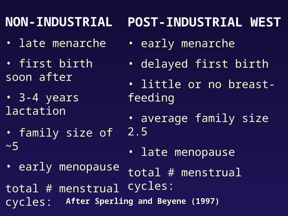
NON-INDUSTRIAL
• late menarche
• first birth soon after
• 3-4 years lactation
• family size of ~5
• early menopause
total # menstrual cycles:
4 years (48 cycles)
POST-INDUSTRIAL WEST
• early menarche
• delayed first birth
• little or no breast-feeding
• average family size 2.5
• late menopause
total # menstrual cycles:
35 years (420 cycles)After Sperling and Beyene (1997)After Sperling and Beyene (1997)
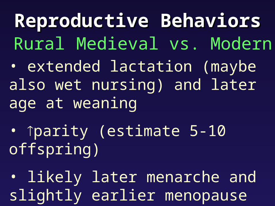
Reproductive BehaviorsReproductive Behaviors Rural Medieval vs. Modern
• extended lactation (maybe also wet nursing) and later age at weaning
• parity (estimate 5-10 offspring)
• likely later menarche and slightly earlier menopause
=different hormonal environment
DiscussionDiscussion
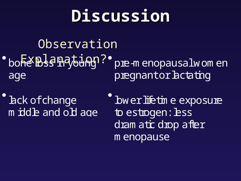
bone loss in youngage
lack of changemiddle and old age
pre-menopausal womenpregnant or lactating
lower lifetime exposureto estrogen: lessdramatic drop aftermenopause
Observation Explanation?

ConclusionsConclusions• Skeletal remains offer unique evidence to
study health and disease in past populations with a biocultural approach
• Patterns of bone loss and fragility are different in the past than those seen in modern Western populations
• Maybe due to lifestyle and layers of life history: reproductive factors
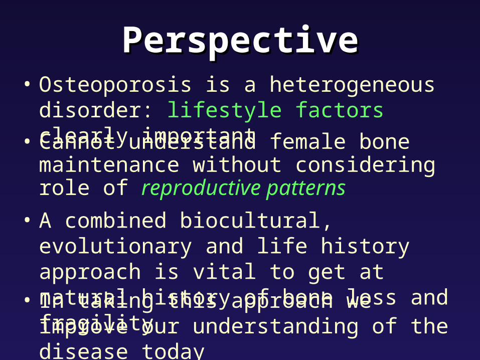
PerspectivePerspective
• Cannot understand female bone maintenance without considering role of reproductive patterns
• In taking this approach we improve our understanding of the disease today
• Osteoporosis is a heterogeneous disorder: lifestyle factors clearly important
• A combined biocultural, evolutionary and life history approach is vital to get at natural history of bone loss and fragility