Agilent Oligonucleotide Array-Based CGH for Genomic DNA ... · Agilent Technologies Agilent...
98
Agilent Technologies Agilent Oligonucleotide Array-Based CGH for Genomic DNA Analysis Enzymatic Labeling for Blood, Cells or Tissues (with a High Throughput option) Protocol Version 6.2, October 2009 Research Use Only. Not for Diagnostic Procedures.
Transcript of Agilent Oligonucleotide Array-Based CGH for Genomic DNA ... · Agilent Technologies Agilent...
Agilent Oligonucleotide Array-Based CGH for Genomic DNA Analysis
Enzymatic Labeling for Blood, Cells or Tissues (with a High Throughput option)
ProtocolVersion 6.2, October 2009
Research Use Only. Not for Diagnostic Procedures.
Agilent Technologies
Array-Based CGH for Genomic DNA Analysis - Enzymatic Labeling
Notices© Agilent Technologies, Inc. 2009
No part of this manual may be reproduced in any form or by any means (including elec-tronic storage and retrieval or translation into a foreign language) without prior agree-ment and written consent from Agilent Technologies, Inc. as governed by United States and international copyright laws.
Manual Part NumberG4410-90010
EditionVersion 6.2, October 2009
Printed in USA
Agilent Technologies, Inc.5301 Stevens Creek Blvd. Santa Clara, CA 95051 USA
WarrantyThe material contained in this docu-ment is provided “as is,” and is sub-ject to being changed, without notice, in future editions. Further, to the max-imum extent permitted by applicable law, Agilent disclaims all warranties, either express or implied, with regard to this manual and any information contained herein, including but not limited to the implied warranties of merchantability and fitness for a par-ticular purpose. Agilent shall not be liable for errors or for incidental or consequential damages in connec-tion with the furnishing, use, or per-formance of this document or of any information contained herein. Should Agilent and the user have a separate written agreement with warranty terms covering the material in this document that conflict with these terms, the warranty terms in the sep-arate agreement shall control.
Technology Licenses The hardware and/or software described in this document are furnished under a license and may be used or copied only in accor-dance with the terms of such license.
Restricted Rights LegendU.S. Government Restricted Rights. Soft-ware and technical data rights granted to the federal government include only those rights customarily provided to end user cus-tomers. Agilent provides this customary commercial license in Software and techni-cal data pursuant to FAR 12.211 (Technical Data) and 12.212 (Computer Software) and, for the Department of Defense, DFARS 252.227-7015 (Technical Data - Commercial Items) and DFARS 227.7202-3 (Rights in Commercial Computer Software or Com-puter Software Documentation).
Safety Notices
CAUTION
A CAUTION notice denotes a haz-ard. It calls attention to an operat-ing procedure, practice, or the like that, if not correctly performed or adhered to, could result in damage to the product or loss of important data. Do not proceed beyond a CAUTION notice until the indicated conditions are fully understood and met.
WARNING
A WARNING notice denotes a hazard. It calls attention to an operating procedure, practice, or the like that, if not correctly per-formed or adhered to, could result in personal injury or death. Do not proceed beyond a WARNING notice until the indicated condi-tions are fully understood and met.
Technical SupportTechnical product support may be obtained by contacting your local Agilent Support Services representative. Agilent’s world-wide sales and support center telephone numbers can be obtained at the following Web site: www.agilent.com/chem/contactus
or send an e-mail to:[email protected]
Notice to PurchaserThese products are intended for research use only. Agilent products may not be resold, modified for resale, or used to manu-facture commercial products without writ-ten approval of Agilent Technologies, Inc.
In This Guide…This guide describes Agilent's recommended operational procedures to analyze DNA copy number variations using Agilent 60- mer oligonucleotide microarrays for array- based comparative genomic hybridization (aCGH) analysis. This protocol is specifically developed and optimized to enzymatically label DNA from blood, cells or frozen tissues. For processing FFPE samples, follow the Agilent Oligonucleotide Array- Based CGH for Genomic DNA Analysis (ULS Labeling for Blood, Cells, Tissues or FFPE) Protocol v3.1 (p/n G4410- 90020).
1 Before You Begin
This chapter contains information (such as procedural notes, safety information, required reagents and equipment) that you should read and understand before you start an experiment.
2 DNA Isolation
This chapter describes the method to isolate genomic DNA (gDNA) from blood, cells, or frozen tissues.
3 Sample Preparation
This chapter describes the two methods to process genomic DNA (gDNA) prior to labeling.
4 Sample Labeling
This chapter describes the steps to differentially label the gDNA samples with fluorescent- labeled nucleotides.
5 Microarray Processing and Feature Extraction
This chapter describes the steps to hybridize, wash and scan Agilent CGH microarrays and to extract data using the Agilent Feature Extraction Software.
Array-Based CGH for Genomic DNA Analysis - Enzymatic Labeling 3
6 Troubleshooting
This chapter contains the causes for above- threshold DLRSD (Derivative Log Ratio Standard Deviation). A poor DLRSD score reflects high probe- to- probe log ratio noise.
7 Reference
This chapter contains reference information related to the protocol.
What’s New in Version 6.2The Microcon YM- 30 filter units are replaced with the Amicon Ultra- 0.5, Ultracel- 30 Membrane, 30 kDa centrifugal filters in the clean- up of Labeled Genomic DNA step.
What’s New in Version 6.1• The use of the Agilent Ozone- Barrier Slide Cover is
described.
• Additional part numbers are added.
• Additional guidelines on yield and specific activity after labeling.
• Guidelines are expanded to enable skipping of the restriction digestion step.
4 Array-Based CGH for Genomic DNA Analysis - Enzymatic Labeling
Content
1 Before You Begin 7
Procedural Notes 8Safety Notes 9Agilent Oligo CGH Microarray Kit Contents 10Required Equipment 15Required Reagents 18Required Hardware and Software 19
2 DNA Isolation 21
Step 1. gDNA Extraction 23Step 2. gDNA Quantitation and Quality Analysis 26
3 Sample Preparation 29
Direct Method 30
Restriction Digestion 31
Amplification Method 34
Step 1. Fragmentation 36Step 2. Library Preparation 37Step 3. Amplification 38Step 4. Purification of PCR products 39Step 5. Quantitation of Amplified-Purified gDNA 40Step 6. Preparation of Amplified-Purified gDNA before Labeling 40
4 Sample Labeling 41
Step 1. Fluorescent Labeling of Genomic DNA 42Step 2. Clean-up of Labeled Genomic DNA 46To determine yield , degree of labeling or specific activity 51Step 3. Preparation of Labeled Genomic DNA for Hybridization 53
Array-Based CGH for Genomic DNA Analysis - Enzymatic Labeling 5
Contents
5 Microarray Processing and Feature Extraction 57
Step 1. Microarray Hybridization 58Step 2. Wash Preparation 60Step 3. Microarray Washing 64Step 4. Microarray Scanning using Agilent C, B or A Scanner or GenePix
Scanner 70Step 5. Data Extraction using Feature Extraction Software 72
6 Troubleshooting 81
If you have an OD260/230 or OD260/280 value below 1.8 82If you have poor sample quality due to residual RNA 82If you get poor sample quality due to degradation 83If the estimated concentration is too high or low 83If you have low specific activity or degree of labeling not due to poor sample
quality 84If you have low yield not due to poor sample quality 84If you have post-labeling signal loss 85If you have high BGNoise values 86If you have poor reproducibility 86
7 Reference 87
Supporting User Guides 88Agilent Microarray Layout and Orientation 89Array/Sample tracking on a 8x array slide 92Agilent Information Assets Access Agreement 93
6 Array-Based CGH for Genomic DNA Analysis - Enzymatic Labeling
Agilent Oligonucleotide Array-Based CGH for Genomic DNA Analysis Protocol
1Before You Begin
Procedural Notes 8
Safety Notes 9
Agilent Oligo CGH Microarray Kit Contents 10
Required Equipment 15
Required Reagents 18
Required Hardware and Software 19
Make sure that you read and understand the information in this chapter and have the necessary equipment and reagents listed before you start an experiment.
7Agilent Technologies

1 Before You BeginProcedural Notes
Procedural Notes
• Follow the procedure described in this document to isolate gDNA from blood, cells, or frozen tissues, to increase the likelihood of a successful experiment. For processing FFPE samples, refer to the Agilent Oligonucleotide Array- Based CGH for Genomic DNA Analysis (ULS Labeling for Blood, Cells, Tissues or FFPE) Protocol v3.1 (p/n G4410- 90020).
• If the DNA isolation procedure described in this document cannot be followed, make sure that the DNA is free of RNA and protein contamination.
• To prevent contamination of reagents by nucleases, always wear powder- free laboratory gloves, and use dedicated solutions and pipettors with nuclease- free aerosol- resistant tips.
• Maintain a clean work area.
• Do not mix stock solutions and reactions containing gDNA or enzymes on a vortex mixer. Instead, mix the solutions and reactions by gently tapping the tube with your finger.
• Avoid repeated freeze- thaw cycles of solutions containing gDNA or enzymes.
• When preparing frozen reagent stock solutions for use:
1 Thaw the aliquot as quickly as possible without heating above room temperature.
2 Mix briefly on a vortex mixer, then spin in a microcentrifuge for 5 to 10 seconds to drive the contents off the walls and lid.
3 Store on ice or in a cold block until use.
• In general, follow Biosafety Level 1 (BL1) safety rules.
8 Array-Based CGH for Genomic DNA Analysis - Enzymatic Labeling
Before You Begin 1Safety Notes
Safety Notes
CAUTION Wear appropriate personal protective equipment (PPE) when working in the laboratory.
WARNING • Cyanine 3-dUTP and Cyanine 5-dUTP are considered hazardous by the OSHA Hazard Communication Standard (29 CFR 1910.1200). Contains material that causes damage to the following organs: kidneys, liver, cardiovascular system, respiratory tract, skin, eye lens or cornea, stomach. May be harmful if swallowed. Avoid contact with eyes, skin and clothing.
• 2X Hi-RPM Hybridization Buffer is considered hazardous by the OSHA Hazard Communication Standard (29 CFR 1910.1200). Contains material that causes damage to the following organs: skin, central nervous system. May be harmful if swallowed. Avoid contact with eyes, skin and clothing.
• Triton is harmful if swallowed. Risk of serious damage to eyes. Wear suitable PPE. Triton is a component of Agilent’s 2X Hi-RPM Hybridization Buffer.
• Agilent Stabilization and Drying Solution is considered hazardous by the OSHA Hazard Communication Standard (29 CFR 1910.1200). Flammable liquid and vapor. Keep away from heat, sparks and flame. Keep container closed. Use only with adequate ventilation. This solution contains material which causes damage to the following organs: kidneys, liver, cardiovascular system, upper respiratory tract, skin, central nervous system (CNS), eye, lens or cornea.
Array-Based CGH for Genomic DNA Analysis - Enzymatic Labeling 9
1 Before You BeginAgilent Oligo CGH Microarray Kit Contents
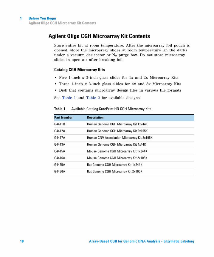
Agilent Oligo CGH Microarray Kit Contents
Store entire kit at room temperature. After the microarray foil pouch is opened, store the microarray slides at room temperature (in the dark) under a vacuum desiccator or N2 purge box. Do not store microarray slides in open air after breaking foil.
Catalog CGH Microarray Kits
• Five 1- inch x 3- inch glass slides for 1x and 2x Microarray Kits
• Three 1- inch x 3- inch glass slides for 4x and 8x Microarray Kits
• Disk that contains microarray design files in various file formats
See Table 1 and Table 2 for available designs.
Table 1 Available Catalog SurePrint HD CGH Microarray Kits
Part Number Description
G4411B Human Genome CGH Microarray Kit 1x244K
G4412A Human Genome CGH Microarray Kit 2x10⁵K
G4417A Human CNV Association Microarray Kit 2x10⁵K
G4413A Human Genome CGH Microarray Kit 4x44K
G4415A Mouse Genome CGH Microarray Kit 1x244K
G4416A Mouse Genome CGH Microarray Kit 2x10⁵K
G4435A Rat Genome CGH Microarray Kit 1x244K
G4436A Rat Genome CGH Microarray Kit 2x10⁵K
10 Array-Based CGH for Genomic DNA Analysis - Enzymatic Labeling
Before You Begin 1Agilent Oligo CGH Microarray Kit Contents
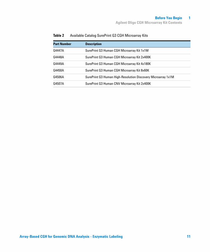
Table 2 Available Catalog SurePrint G3 CGH Microarray Kits
Part Number Description
G4447A SurePrint G3 Human CGH Microarray Kit 1x1M
G4448A SurePrint G3 Human CGH Microarray Kit 2x400K
G4449A SurePrint G3 Human CGH Microarray Kit 4x180K
G4450A SurePrint G3 Human CGH Microarray Kit 8x60K
G4506A SurePrint G3 Human High-Resolution Discovery Microarray 1x1M
G4507A SurePrint G3 Human CNV Microarray Kit 2x400K
Array-Based CGH for Genomic DNA Analysis - Enzymatic Labeling 11
1 Before You BeginAgilent Oligo CGH Microarray Kit Contents
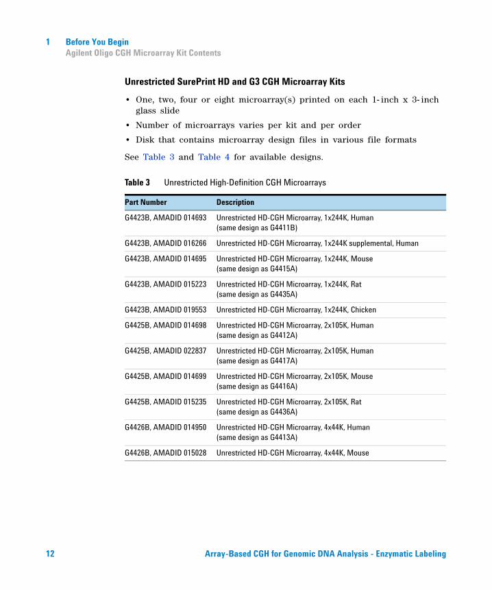
Unrestricted SurePrint HD and G3 CGH Microarray Kits
• One, two, four or eight microarray(s) printed on each 1- inch x 3- inch glass slide
• Number of microarrays varies per kit and per order
• Disk that contains microarray design files in various file formats
See Table 3 and Table 4 for available designs.
Table 3 Unrestricted High-Definition CGH Microarrays
Part Number Description
G4423B, AMADID 014693 Unrestricted HD-CGH Microarray, 1x244K, Human (same design as G4411B)
G4423B, AMADID 016266 Unrestricted HD-CGH Microarray, 1x244K supplemental, Human
G4423B, AMADID 014695 Unrestricted HD-CGH Microarray, 1x244K, Mouse (same design as G4415A)
G4423B, AMADID 015223 Unrestricted HD-CGH Microarray, 1x244K, Rat (same design as G4435A)
G4423B, AMADID 019553 Unrestricted HD-CGH Microarray, 1x244K, Chicken
G4425B, AMADID 014698 Unrestricted HD-CGH Microarray, 2x10⁵K, Human (same design as G4412A)
G4425B, AMADID 022837 Unrestricted HD-CGH Microarray, 2x10⁵K, Human (same design as G4417A)
G4425B, AMADID 014699 Unrestricted HD-CGH Microarray, 2x10⁵K, Mouse(same design as G4416A)
G4425B, AMADID 015235 Unrestricted HD-CGH Microarray, 2x10⁵K, Rat(same design as G4436A)
G4426B, AMADID 014950 Unrestricted HD-CGH Microarray, 4x44K, Human(same design as G4413A)
G4426B, AMADID 015028 Unrestricted HD-CGH Microarray, 4x44K, Mouse
12 Array-Based CGH for Genomic DNA Analysis - Enzymatic Labeling
Before You Begin 1Agilent Oligo CGH Microarray Kit Contents
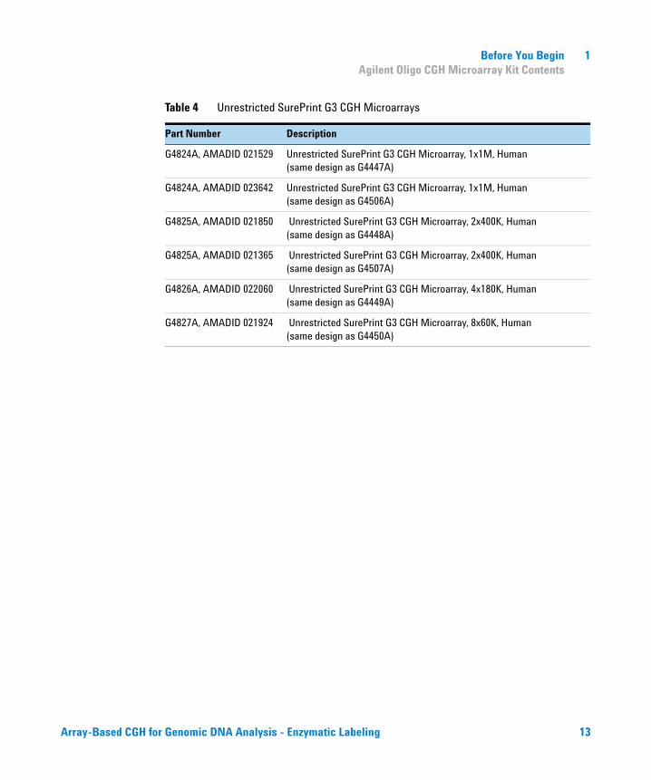
Table 4 Unrestricted SurePrint G3 CGH Microarrays
Part Number Description
G4824A, AMADID 021529 Unrestricted SurePrint G3 CGH Microarray, 1x1M, Human(same design as G4447A)
G4824A, AMADID 023642 Unrestricted SurePrint G3 CGH Microarray, 1x1M, Human(same design as G4506A)
G4825A, AMADID 021850 Unrestricted SurePrint G3 CGH Microarray, 2x400K, Human (same design as G4448A)
G4825A, AMADID 021365 Unrestricted SurePrint G3 CGH Microarray, 2x400K, Human (same design as G4507A)
G4826A, AMADID 022060 Unrestricted SurePrint G3 CGH Microarray, 4x180K, Human (same design as G4449A)
G4827A, AMADID 021924 Unrestricted SurePrint G3 CGH Microarray, 8x60K, Human (same design as G4450A)
Array-Based CGH for Genomic DNA Analysis - Enzymatic Labeling 13
1 Before You BeginAgilent Oligo CGH Microarray Kit Contents
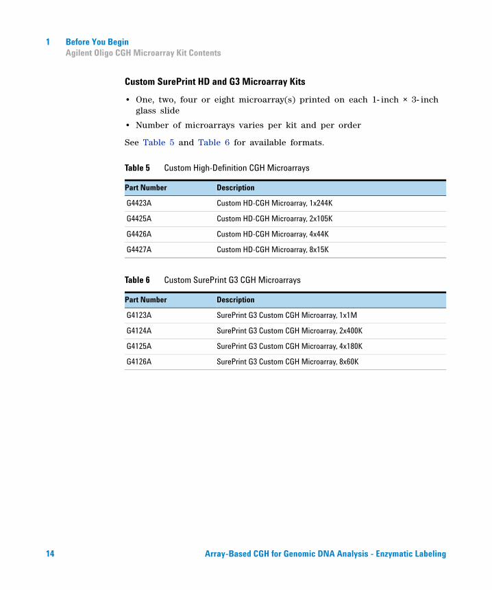
Custom SurePrint HD and G3 Microarray Kits
• One, two, four or eight microarray(s) printed on each 1- inch × 3- inch glass slide
• Number of microarrays varies per kit and per order
See Table 5 and Table 6 for available formats.
Table 5 Custom High-Definition CGH Microarrays
Part Number Description
G4423A Custom HD-CGH Microarray, 1x244K
G4425A Custom HD-CGH Microarray, 2x10⁵K
G4426A Custom HD-CGH Microarray, 4x44K
G4427A Custom HD-CGH Microarray, 8x15K
Table 6 Custom SurePrint G3 CGH Microarrays
Part Number Description
G4123A SurePrint G3 Custom CGH Microarray, 1x1M
G4124A SurePrint G3 Custom CGH Microarray, 2x400K
G4125A SurePrint G3 Custom CGH Microarray, 4x180K
G4126A SurePrint G3 Custom CGH Microarray, 8x60K
14 Array-Based CGH for Genomic DNA Analysis - Enzymatic Labeling
Before You Begin 1Required Equipment
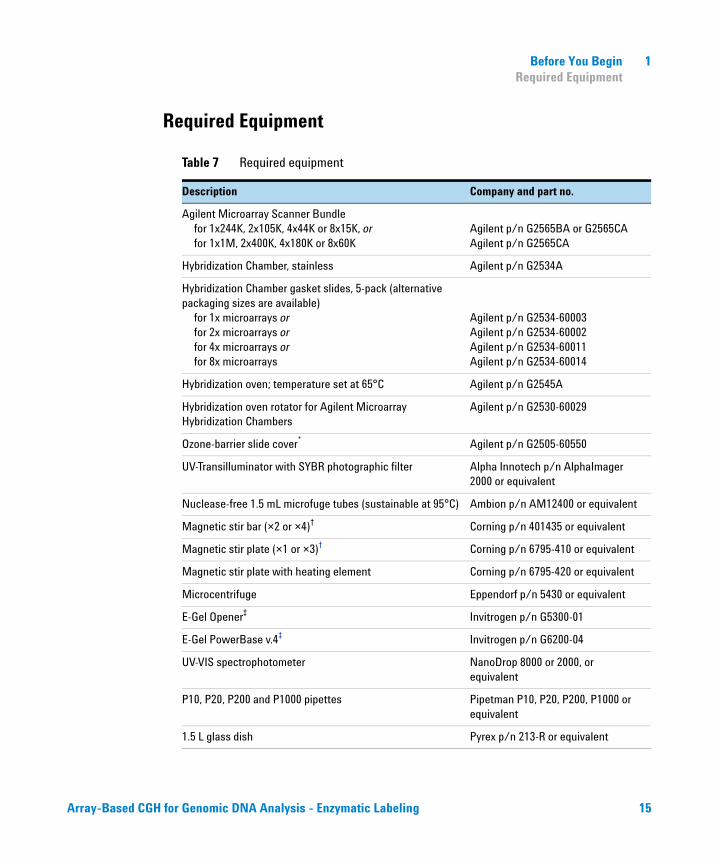
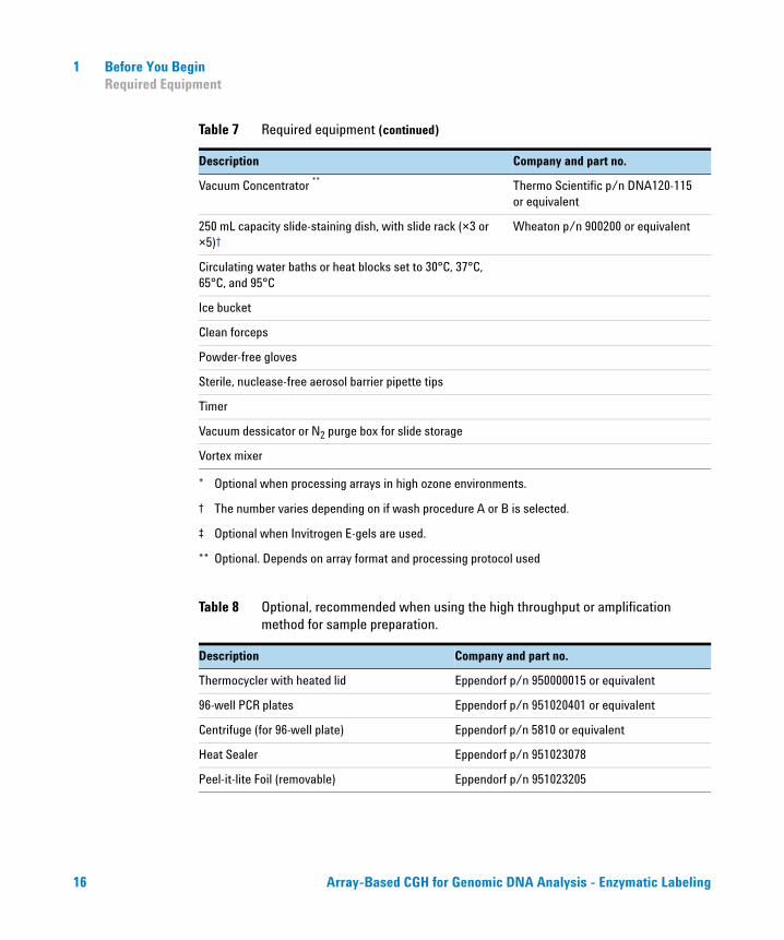
Required Equipment
Table 7 Required equipment
Description Company and part no.
Agilent Microarray Scanner Bundlefor 1x244K, 2x10⁵K, 4x44K or 8x15K, orfor 1x1M, 2x400K, 4x180K or 8x60K
Agilent p/n G2565BA or G2565CAAgilent p/n G2565CA
Hybridization Chamber, stainless Agilent p/n G2534A
Hybridization Chamber gasket slides, 5-pack (alternative packaging sizes are available)
for 1x microarrays or for 2x microarrays orfor 4x microarrays orfor 8x microarrays
Agilent p/n G2534-60003Agilent p/n G2534-60002Agilent p/n G2534-60011Agilent p/n G2534-60014
Hybridization oven; temperature set at 65°C Agilent p/n G2545A
Hybridization oven rotator for Agilent Microarray Hybridization Chambers
Agilent p/n G2530-60029
Ozone-barrier slide cover* Agilent p/n G2505-60550
UV-Transilluminator with SYBR photographic filter Alpha Innotech p/n AlphaImager 2000 or equivalent
Nuclease-free 1.5 mL microfuge tubes (sustainable at 95°C) Ambion p/n AM12400 or equivalent
Magnetic stir bar (×2 or ×4)† Corning p/n 401435 or equivalent
Magnetic stir plate (×1 or ×3)† Corning p/n 6795-410 or equivalent
Magnetic stir plate with heating element Corning p/n 6795-420 or equivalent
Microcentrifuge Eppendorf p/n 5430 or equivalent
E-Gel Opener‡ Invitrogen p/n G5300-01
E-Gel PowerBase v.4‡ Invitrogen p/n G6200-04
UV-VIS spectrophotometer NanoDrop 8000 or 2000, or equivalent
P10, P20, P200 and P1000 pipettes Pipetman P10, P20, P200, P1000 or equivalent
1.5 L glass dish Pyrex p/n 213-R or equivalent
Array-Based CGH for Genomic DNA Analysis - Enzymatic Labeling 15
1 Before You BeginRequired Equipment
Vacuum Concentrator ** Thermo Scientific p/n DNA120-115 or equivalent
250 mL capacity slide-staining dish, with slide rack (×3 or ×5)†
Wheaton p/n 900200 or equivalent
Circulating water baths or heat blocks set to 30°C, 37°C, 65°C, and 95°C
Ice bucket
Clean forceps
Powder-free gloves
Sterile, nuclease-free aerosol barrier pipette tips
Timer
Vacuum dessicator or N2 purge box for slide storage
Vortex mixer
* Optional when processing arrays in high ozone environments.
† The number varies depending on if wash procedure A or B is selected.
‡ Optional when Invitrogen E-gels are used.
** Optional. Depends on array format and processing protocol used
Table 8 Optional, recommended when using the high throughput or amplification method for sample preparation.
Description Company and part no.
Thermocycler with heated lid Eppendorf p/n 950000015 or equivalent
96-well PCR plates Eppendorf p/n 951020401 or equivalent
Centrifuge (for 96-well plate) Eppendorf p/n 5810 or equivalent
Heat Sealer Eppendorf p/n 951023078
Peel-it-lite Foil (removable) Eppendorf p/n 951023205
Table 7 Required equipment (continued)
Description Company and part no.
16 Array-Based CGH for Genomic DNA Analysis - Enzymatic Labeling
Before You Begin 1Required Equipment
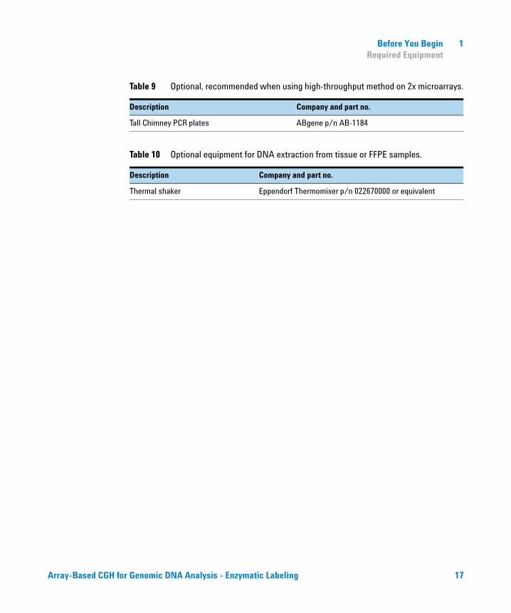
Table 9 Optional, recommended when using high-throughput method on 2x microarrays.
Description Company and part no.
Tall Chimney PCR plates ABgene p/n AB-1184
Table 10 Optional equipment for DNA extraction from tissue or FFPE samples.
Description Company and part no.
Thermal shaker Eppendorf Thermomixer p/n 022670000 or equivalent
Array-Based CGH for Genomic DNA Analysis - Enzymatic Labeling 17
1 Before You BeginRequired Reagents
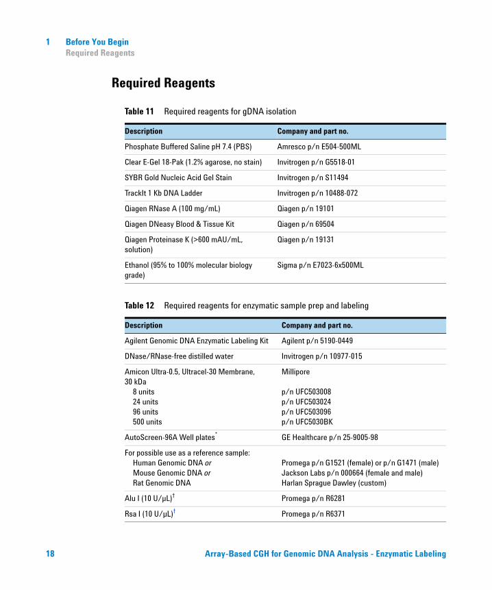
Required Reagents
Table 11 Required reagents for gDNA isolation
Description Company and part no.
Phosphate Buffered Saline pH 7.4 (PBS) Amresco p/n E504-500ML
Clear E-Gel 18-Pak (1.2% agarose, no stain) Invitrogen p/n G5518-01
SYBR Gold Nucleic Acid Gel Stain Invitrogen p/n S11494
TrackIt 1 Kb DNA Ladder Invitrogen p/n 10488-072
Qiagen RNase A (100 mg/mL) Qiagen p/n 19101
Qiagen DNeasy Blood & Tissue Kit Qiagen p/n 69504
Qiagen Proteinase K (>600 mAU/mL, solution)
Qiagen p/n 19131
Ethanol (95% to 100% molecular biology grade)
Sigma p/n E7023-6x500ML
Table 12 Required reagents for enzymatic sample prep and labeling
Description Company and part no.
Agilent Genomic DNA Enzymatic Labeling Kit Agilent p/n 5190-0449
DNase/RNase-free distilled water Invitrogen p/n 10977-015
Amicon Ultra-0.5, Ultracel-30 Membrane, 30 kDa
8 units24 units96 units500 units
Millipore
p/n UFC503008p/n UFC503024p/n UFC503096p/n UFC5030BK
AutoScreen-96A Well plates* GE Healthcare p/n 25-9005-98
For possible use as a reference sample:Human Genomic DNA orMouse Genomic DNA orRat Genomic DNA
Promega p/n G1521 (female) or p/n G1471 (male)Jackson Labs p/n 000664 (female and male)Harlan Sprague Dawley (custom)
Alu I (10 U/µL)† Promega p/n R6281
Rsa I (10 U/µL)† Promega p/n R6371
18 Array-Based CGH for Genomic DNA Analysis - Enzymatic Labeling
Before You Begin 1Required Hardware and Software
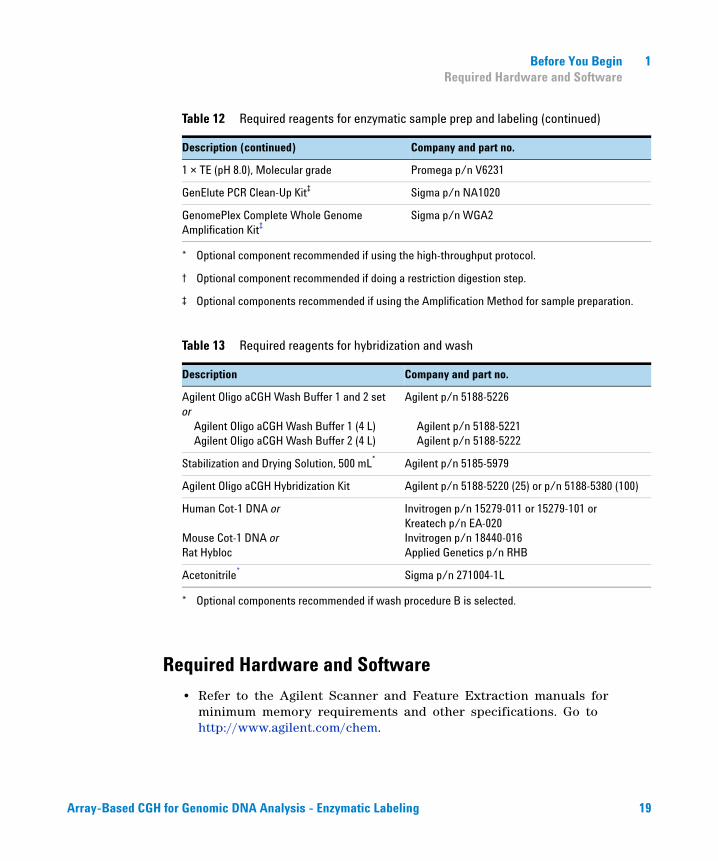
Required Hardware and Software
• Refer to the Agilent Scanner and Feature Extraction manuals for minimum memory requirements and other specifications. Go to http://www.agilent.com/chem.
1 × TE (pH 8.0), Molecular grade Promega p/n V6231
GenElute PCR Clean-Up Kit‡ Sigma p/n NA1020
GenomePlex Complete Whole Genome Amplification Kit‡
Sigma p/n WGA2
* Optional component recommended if using the high-throughput protocol.
† Optional component recommended if doing a restriction digestion step.
‡ Optional components recommended if using the Amplification Method for sample preparation.
Table 13 Required reagents for hybridization and wash
Description Company and part no.
Agilent Oligo aCGH Wash Buffer 1 and 2 set or
Agilent Oligo aCGH Wash Buffer 1 (4 L)Agilent Oligo aCGH Wash Buffer 2 (4 L)
Agilent p/n 5188-5226
Agilent p/n 5188-5221Agilent p/n 5188-5222
Stabilization and Drying Solution, 500 mL*
* Optional components recommended if wash procedure B is selected.
Agilent p/n 5185-5979
Agilent Oligo aCGH Hybridization Kit Agilent p/n 5188-5220 (25) or p/n 5188-5380 (100)
Human Cot-1 DNA or
Mouse Cot-1 DNA orRat Hybloc
Invitrogen p/n 15279-011 or 15279-101 orKreatech p/n EA-020Invitrogen p/n 18440-016Applied Genetics p/n RHB
Acetonitrile* Sigma p/n 271004-1L
Table 12 Required reagents for enzymatic sample prep and labeling (continued)
Description (continued) Company and part no.
Array-Based CGH for Genomic DNA Analysis - Enzymatic Labeling 19
1 Before You BeginRequired Hardware and Software
20 Array-Based CGH for Genomic DNA Analysis - Enzymatic Labeling
Agilent Oligonucleotide Array-Based CGH for Genomic DNA Analysis Protocol
2DNA Isolation
Step 1. gDNA Extraction 23
Step 2. gDNA Quantitation and Quality Analysis 26
Agilent’s array- based Comparative Genomic Hybridization (aCGH) application uses a “two- color” process to measure DNA copy number changes in an experimental sample relative to a reference sample. The type of sample used as a reference is a matter of experimental choice; however, many experimenters use normal commercial gDNA as a reference sample.
This chapter describes Agilent’s recommended procedure to isolate genomic DNA (gDNA) from blood, cells, or frozen tissues using the Qiagen DNeasy Blood & Tissue Kit (p/n 69504).
For processing FFPE samples, follow the Agilent Oligonucleotide Array- Based CGH for Genomic DNA Analysis (ULS Labeling for Blood, Cells, Tissues or FFPE) Protocol v3.1 (p/n G4410- 90020).
NOTE Agilent cannot guarantee microarray performance and does not provide technical support to those who use non-Agilent protocols in processing Agilent microarrays.
21Agilent Technologies
2 DNA Isolation
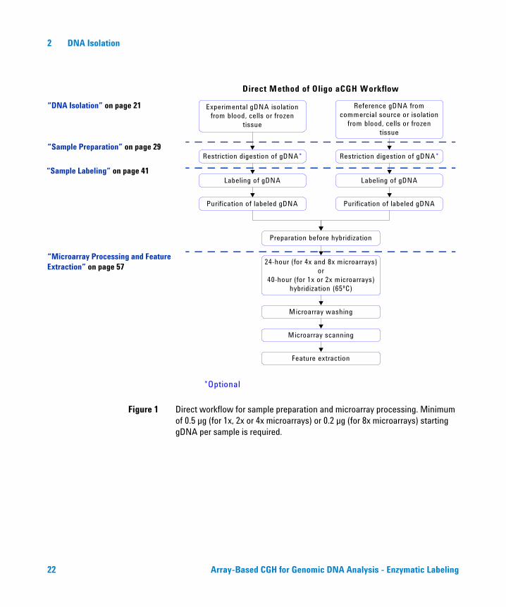
Figure 1 Direct workflow for sample preparation and microarray processing. Minimum of 0.5 µg (for 1x, 2x or 4x microarrays) or 0.2 µg (for 8x microarrays) starting gDNA per sample is required.
Experimental gDNA isolation from blood, cells or frozen
tissue
Restriction digestion of gDNA*
Labeling of gDNA
Reference gDNA from commercial source or isolation
from blood, cells or frozen tissue
Restriction digestion of gDNA*
Labeling of gDNA
Purification of labeled gDNA Purification of labeled gDNA
Preparation before hybridization
24-hour (for 4x and 8x microarrays) or
40-hour (for 1x or 2x microarrays) hybridization (65ºC)
Microarray washing
Microarray scanning
Feature extraction
Direct Method of Oligo aCGH Workflow
*Optional
“DNA Isolation” on page 21
“Sample Preparation” on page 29
“Sample Labeling” on page 41
“Microarray Processing and Feature Extraction” on page 57
22 Array-Based CGH for Genomic DNA Analysis - Enzymatic Labeling
DNA Isolation 2Step 1. gDNA Extraction
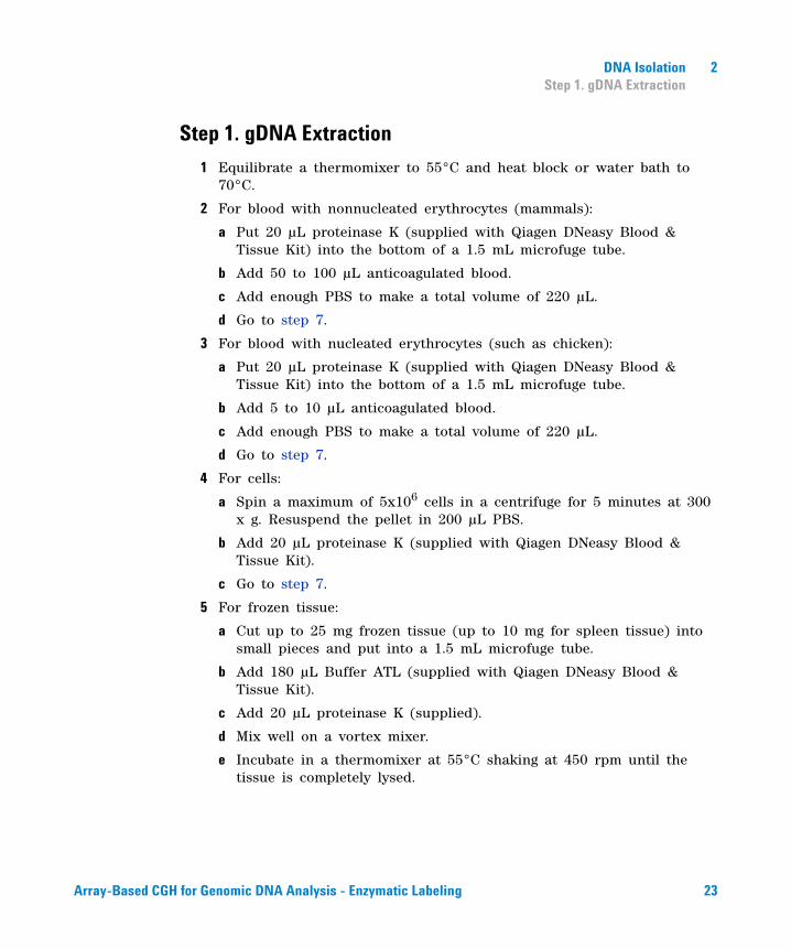
Step 1. gDNA Extraction
1 Equilibrate a thermomixer to 55°C and heat block or water bath to 70°C.
2 For blood with nonnucleated erythrocytes (mammals):
a Put 20 µL proteinase K (supplied with Qiagen DNeasy Blood & Tissue Kit) into the bottom of a 1.5 mL microfuge tube.
b Add 50 to 100 µL anticoagulated blood.
c Add enough PBS to make a total volume of 220 µL.
d Go to step 7.
3 For blood with nucleated erythrocytes (such as chicken):
a Put 20 µL proteinase K (supplied with Qiagen DNeasy Blood & Tissue Kit) into the bottom of a 1.5 mL microfuge tube.
b Add 5 to 10 µL anticoagulated blood.
c Add enough PBS to make a total volume of 220 µL.
d Go to step 7.
4 For cells:
a Spin a maximum of 5x10⁶ cells in a centrifuge for 5 minutes at 300 x g. Resuspend the pellet in 200 µL PBS.
b Add 20 µL proteinase K (supplied with Qiagen DNeasy Blood & Tissue Kit).
c Go to step 7.
5 For frozen tissue:
a Cut up to 25 mg frozen tissue (up to 10 mg for spleen tissue) into small pieces and put into a 1.5 mL microfuge tube.
b Add 180 µL Buffer ATL (supplied with Qiagen DNeasy Blood & Tissue Kit).
c Add 20 µL proteinase K (supplied).
d Mix well on a vortex mixer.
e Incubate in a thermomixer at 55°C shaking at 450 rpm until the tissue is completely lysed.
Array-Based CGH for Genomic DNA Analysis - Enzymatic Labeling 23

2 DNA IsolationStep 1. gDNA Extraction
Lysis time varies depending on the type of tissue processed. Usually lysis is complete in 1 to 3 hours. If it is more convenient, samples can be lysed overnight.
f Let the sample cool to room temperature and spin in a microcentrifuge for 30 seconds at 6,000 x g to drive the contents off the walls and lid.
g Go to step 7.
6 For further purification of extracted DNA:
a Take a maximum 25 µg of DNA.
b Add enough PBS to make a total volume of 220 µL.
c Add 20 µL proteinase K (supplied with Qiagen DNeasy Blood & Tissue Kit).
7 Add 4 µL RNase A (100 mg/mL), mix on a vortex mixer, and incubate for 2 minutes at room temperature. Spin in a microcentrifuge for 30 seconds at 6,000 x g to drive the contents off the walls and lid.
8 Add 200 µL Buffer AL (supplied) to each sample, mix thoroughly on a vortex mixer, and incubate at 70°C for 10 minutes in a heat block or water bath. Spin in a microcentrifuge for 30 seconds at 6,000 x g to drive the contents off the walls and lid.
9 Add 200 µL 100% ethanol to each sample, and mix thoroughly on a vortex mixer. Spin in a microcentrifuge for 30 seconds at 6,000 x g to drive the contents off the walls and lid.
10 Transfer the sample mixture onto a DNeasy Mini spin column in a 2 mL collection tube (supplied). Spin in a centrifuge at 6,000 x g for 1 minute. Discard the flow- through and collection tube. Put the DNeasy Mini spin column in a new 2 mL collection tube (supplied).
11 Before using for the first time, prepare Buffer AW1 by adding 100% ethanol to the Buffer AW1 bottle (supplied; see bottle label for volume). Mark the appropriate check box to indicate that ethanol was added to the bottle.
12 Add 500 µL Buffer AW1 onto the column, and spin in a microcentrifuge for 1 minute at 6,000 x g. Discard the flow- through and collection tube. Put the DNeasy Mini spin column in a new 2 mL collection tube (supplied).
13 Before using for the first time, prepare Buffer AW2 by adding 100% ethanol to the Buffer AW2 bottle (supplied; see bottle label for volume).
24 Array-Based CGH for Genomic DNA Analysis - Enzymatic Labeling
DNA Isolation 2Step 1. gDNA Extraction
Mark the appropriate check box to indicate that ethanol was added to the bottle.
14 Add 500 µL Buffer AW2 onto the column, and spin in a centrifuge for 3 minutes at 20,000 x g to dry the DNeasy membrane. Discard the flow- through and collection tube.
15 Put the DNeasy Mini spin column in a clean 1.5 mL microcentrifuge tube, and pipette 200 µL of Buffer AE (supplied) directly onto the center of the DNeasy column membrane.
16 Incubate at room temperature for 1 minute, and then spin in a microcentrifuge for 1 minute at 6,000 x g to elute the DNA.
17 Repeat elution with Buffer AE once as described in step 15 and step 16. Combine the duplicate samples in one microcentrifuge tube for a final volume of 400 µL.
Array-Based CGH for Genomic DNA Analysis - Enzymatic Labeling 25
2 DNA IsolationStep 2. gDNA Quantitation and Quality Analysis
Step 2. gDNA Quantitation and Quality Analysis
Accurate assessment of gDNA quantity and quality are crucial to the success of an Agilent Oligo aCGH experiment. High quality gDNA should be free of contaminants such as carbohydrates, proteins, and traces of organic solvents, and should also be intact with minimal degradation.
See “FFPE Tissues” in the Agilent Oligonucleotide Array- Based CGH for Genomic DNA Analysis (ULS Labeling for Blood, Cells, Tissues or FFPE) Protocol v3.1 (p/n G4410- 90020) for details on how to isolate gDNA from FFPE tissues.
Use the NanoDrop ND- 1000 UV- VIS Spectrophotometer (or equivalent) to assess gDNA concentration and purity. Use agarose gel electrophoresis to assess gDNA intactness and the average molecular weight for each sample.
UV-VIS Spectrophotometry
1 In the Nanodrop program menu, select Nucleic Acid Measurement, then select Sample Type to be DNA- 50.
2 Use 1.5 µL of elution buffer to blank the instrument.
3 Use 1.5 µL of each gDNA sample to measure DNA concentration. Record the gDNA concentration (ng/µL) for each sample. Calculate the yield as
4 Record the A260/A280 and A260/A230 ratios. High- quality gDNA samples should have an A260/A280 ratio of 1.8 to 2.0, indicating the absence of contaminating proteins, and an A260/A230 ratio of >2.0, indicating the absence of other organic compounds such as guanidinium isothiocyanate, alcohol and phenol as well as cellular contaminants such as carbohydrates.
Yield (µg) DNAconcentration (ng/µL) Sample Volume (µL)1000 ng/µg
--------------------------------------------------------------------------------------------------------------------------=
26 Array-Based CGH for Genomic DNA Analysis - Enzymatic Labeling
DNA Isolation 2Step 2. gDNA Quantitation and Quality Analysis
Agarose Gel Electrophoresis
1 Load 20 ng gDNA for each sample in a volume of 10 µL nuclease- free water in the well of a single- comb 1.2% Clear E- Gel. (You do not need to add loading buffer in this system).
2 As a control, load 20 ng of commercial Human Genomic DNA in a volume of 10 µL nuclease free water in one of the wells of the E- Gel.
3 Mix 5 µL TrackIt 1 Kb DNA Ladder with 95 µL deionized water and load 10 µL of the diluted ladder in one of the wells of the E- Gel.
4 Run the gel for 30 minutes as described in Invitrogen's instructions.
5 Open the gel cassette with E- Gel Opener as described in Invitrogen’s instructions.
6 Stain the gel with SYBR Gold Nucleic Acid Gel Stain (diluted 1:10,000 by adding 10 µL of SYBR Gold Nucleic Acid Gel Stain to 100 mL of nuclease- free water) in a plastic tray for 15 minutes.
7 Visualize the gel on the UV- transilluminator using a SYBR Gold photographic filter.
Array-Based CGH for Genomic DNA Analysis - Enzymatic Labeling 27
2 DNA IsolationStep 2. gDNA Quantitation and Quality Analysis
28 Array-Based CGH for Genomic DNA Analysis - Enzymatic Labeling
Agilent Oligonucleotide Array-Based CGH for Genomic DNA Analysis Protocol
3Sample Preparation
Direct Method 30
Restriction Digestion 31
Amplification Method 34
Step 1. Fragmentation 36
Step 2. Library Preparation 37
Step 3. Amplification 38
Step 4. Purification of PCR products 39
Step 5. Quantitation of Amplified-Purified gDNA 40
Step 6. Preparation of Amplified-Purified gDNA before Labeling 40
This chapter describes Agilent’s two recommended options to process genomic DNA (gDNA) prior to labeling.
You can choose between two methods for sample preparation prior to labeling: “Direct Method” on page 30 and “Amplification Method” on page 34. Figure 1 on page 22 and Figure 2 on page 35 show the respective workflows.
29Agilent Technologies
3 Sample Preparation
Direct Method
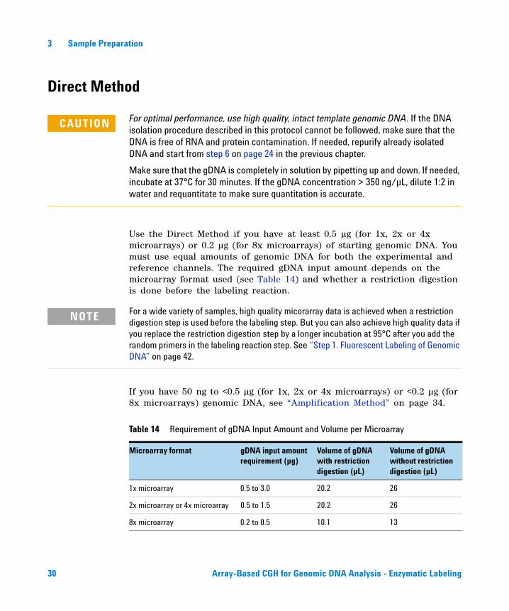
Use the Direct Method if you have at least 0.5 µg (for 1x, 2x or 4x microarrays) or 0.2 µg (for 8x microarrays) of starting genomic DNA. You must use equal amounts of genomic DNA for both the experimental and reference channels. The required gDNA input amount depends on the microarray format used (see Table 14) and whether a restriction digestion is done before the labeling reaction.
If you have 50 ng to <0.5 µg (for 1x, 2x or 4x microarrays) or <0.2 µg (for 8x microarrays) genomic DNA, see “Amplification Method” on page 34.
CAUTION For optimal performance, use high quality, intact template genomic DNA. If the DNA isolation procedure described in this protocol cannot be followed, make sure that the DNA is free of RNA and protein contamination. If needed, repurify already isolated DNA and start from step 6 on page 24 in the previous chapter.
Make sure that the gDNA is completely in solution by pipetting up and down. If needed, incubate at 37°C for 30 minutes. If the gDNA concentration > 350 ng/µL, dilute 1:2 in water and requantitate to make sure quantitation is accurate.
NOTE For a wide variety of samples, high quality micorarray data is achieved when a restriction digestion step is used before the labeling step. But you can also achieve high quality data if you replace the restriction digestion step by a longer incubation at 95°C after you add the random primers in the labeling reaction step. See “Step 1. Fluorescent Labeling of Genomic DNA” on page 42.
Table 14 Requirement of gDNA Input Amount and Volume per Microarray
Microarray format gDNA input amount requirement (µg)
Volume of gDNA with restriction digestion (µL)
Volume of gDNA without restriction digestion (µL)
1x microarray 0.5 to 3.0 20.2 26
2x microarray or 4x microarray 0.5 to 1.5 20.2 26
8x microarray 0.2 to 0.5 10.1 13
30 Array-Based CGH for Genomic DNA Analysis - Enzymatic Labeling
Sample Preparation 3Restriction Digestion
Restriction Digestion
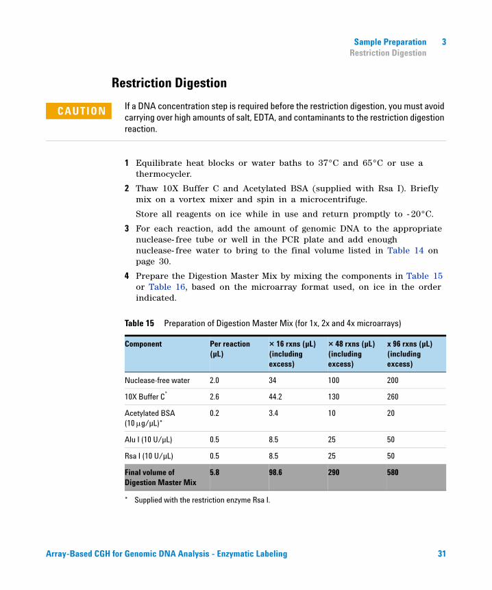
1 Equilibrate heat blocks or water baths to 37°C and 65°C or use a thermocycler.
2 Thaw 10X Buffer C and Acetylated BSA (supplied with Rsa I). Briefly mix on a vortex mixer and spin in a microcentrifuge.
Store all reagents on ice while in use and return promptly to - 20°C.
3 For each reaction, add the amount of genomic DNA to the appropriate nuclease- free tube or well in the PCR plate and add enough nuclease- free water to bring to the final volume listed in Table 14 on page 30.
4 Prepare the Digestion Master Mix by mixing the components in Table 15 or Table 16, based on the microarray format used, on ice in the order indicated.
CAUTION If a DNA concentration step is required before the restriction digestion, you must avoid carrying over high amounts of salt, EDTA, and contaminants to the restriction digestion reaction.
Table 15 Preparation of Digestion Master Mix (for 1x, 2x and 4x microarrays)
Component Per reaction (µL)
× 16 rxns (µL)(including excess)
× 48 rxns (µL)(including excess)
x 96 rxns (µL)(including excess)
Nuclease-free water 2.0 34 100 200
10X Buffer C*
* Supplied with the restriction enzyme Rsa I.
2.6 44.2 130 260
Acetylated BSA (10 g/µL)*
0.2 3.4 10 20
Alu I (10 U/µL) 0.5 8.5 25 50
Rsa I (10 U/µL) 0.5 8.5 25 50
Final volume of Digestion Master Mix
5.8 98.6 290 580
Array-Based CGH for Genomic DNA Analysis - Enzymatic Labeling 31
3 Sample PreparationRestriction Digestion
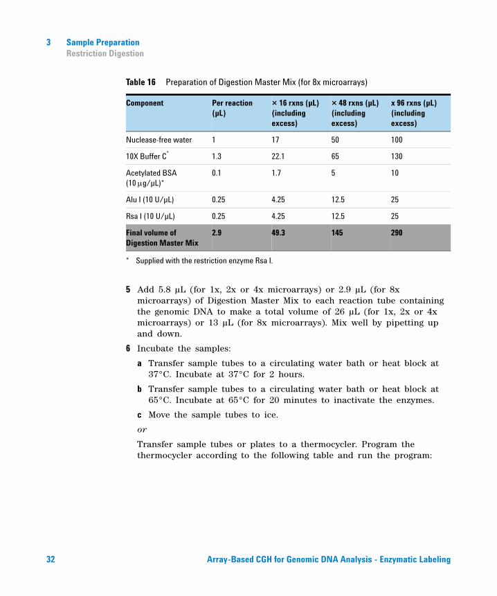
5 Add 5.8 µL (for 1x, 2x or 4x microarrays) or 2.9 µL (for 8x microarrays) of Digestion Master Mix to each reaction tube containing the genomic DNA to make a total volume of 26 µL (for 1x, 2x or 4x microarrays) or 13 µL (for 8x microarrays). Mix well by pipetting up and down.
6 Incubate the samples:
a Transfer sample tubes to a circulating water bath or heat block at 37°C. Incubate at 37°C for 2 hours.
b Transfer sample tubes to a circulating water bath or heat block at 65°C. Incubate at 65°C for 20 minutes to inactivate the enzymes.
c Move the sample tubes to ice.
or
Transfer sample tubes or plates to a thermocycler. Program the thermocycler according to the following table and run the program:
Table 16 Preparation of Digestion Master Mix (for 8x microarrays)
Component Per reaction (µL)
× 16 rxns (µL)(including excess)
× 48 rxns (µL)(including excess)
x 96 rxns (µL)(including excess)
Nuclease-free water 1 17 50 100
10X Buffer C* 1.3 22.1 65 130
Acetylated BSA (10 g/µL)*
0.1 1.7 5 10
Alu I (10 U/µL) 0.25 4.25 12.5 25
Rsa I (10 U/µL) 0.25 4.25 12.5 25
Final volume of Digestion Master Mix
2.9 49.3 145 290
* Supplied with the restriction enzyme Rsa I.
32 Array-Based CGH for Genomic DNA Analysis - Enzymatic Labeling
Sample Preparation 3Restriction Digestion
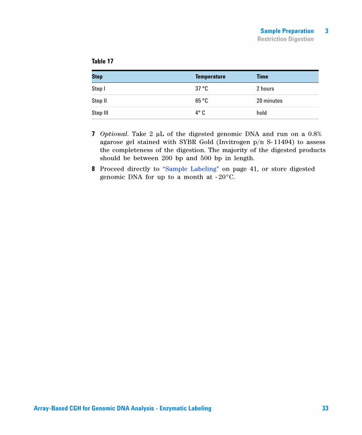
7 Optional. Take 2 µL of the digested genomic DNA and run on a 0.8% agarose gel stained with SYBR Gold (Invitrogen p/n S- 11494) to assess the completeness of the digestion. The majority of the digested products should be between 200 bp and 500 bp in length.
8 Proceed directly to “Sample Labeling” on page 41, or store digested genomic DNA for up to a month at - 20°C.
Table 17
Step Temperature Time
Step I 37 °C 2 hours
Step II 65 °C 20 minutes
Step III 4° C hold
Array-Based CGH for Genomic DNA Analysis - Enzymatic Labeling 33

3 Sample PreparationRestriction Digestion
Amplification Method
Use the Amplification Method if you have limited amounts of genomic DNA. If you have 0.5 µg (for 1x, 2x or 4x microarrays) or 0.2 µg (for 8x microarrays) or more genomic DNA, see “Direct Method” on page 30.
Reference
GenomePlex Whole Genome Amplification (WGA) Kit. Product Code WGA2. Technical Bulletin. Sigma- Aldrich. 2006. TR/PHC 06/05- 1
Genomic Amplification
The Sigma GenomePlex Whole Genome Amplification (WGA) kit allows you to generate a representative amplification of genomic DNA. The kit uses a linker mediated primer PCR amplification technology based upon random fragmentation of genomic DNA and conversion of the resulting small fragments to PCR- amplifiable OmniPlex Library molecules flanked by universal priming sites. The OmniPlex library is then PCR amplified using universal oligonucleotide primers and a limited number of cycles. It is suitable to use with purified genomic DNA from a variety of sources including fresh frozen tissues and cultured cell lines.
CAUTION For optimal performance, use high quality, intact template genomic DNA. If the DNA isolation procedure described in this protocol cannot be followed, make sure that the DNA is free of RNA and protein contamination. If needed, repurify already isolated DNA and start from step 6 on page 24 in the previous chapter.
Make sure that the gDNA is completely in solution by pipetting up and down. If needed, incubate at 37°C for 30 minutes.
GenomePlex can be used on degraded samples if the extracted DNA is 500 bp or greater in size. However, greater quantities (up to 100 ng) of damaged DNA are required to get acceptable yield of final product. DNA isolated from FFPE samples is often severely degraded and damaged and is not always suitable for GenomePlex amplification.
34 Array-Based CGH for Genomic DNA Analysis - Enzymatic Labeling
Sample Preparation 3Restriction Digestion
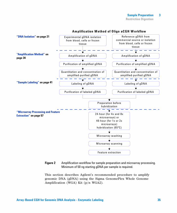
Figure 2 Amplification workflow for sample preparation and microarray processing. Minimum of 50 ng starting gDNA per sample is required.
This section describes Agilent’s recommended procedure to amplify genomic DNA (gDNA) using the Sigma GenomePlex Whole Genome Amplification (WGA) Kit (p/n WGA2).
Experimental gDNA isolationfrom blood, cells or frozen
tissue
Am plification of gDNA
Labeling of gDNA
Reference gDNA fromcom mercial source or isolation
from blood, cells or frozentissue
Amplification of gDNA
Labeling of gDNA
Purification of labeled gDNA Purification of labeled gDNA
Preparation before hybridization
24-hour (for 4x and 8x m icroarrays) or
40-hour (for 1x or 2x m icroarrays)
hybridization (65ºC)
M icroarray washing
M icroarray scanning
Feature extraction
Am plification M ethod of Oligo aCGH W orkflow
Purification of am plified gDNA Purification of amplified gDNA
Quantitation and concentration of am plified-purified gDNA
Quantitation and concentration of amplified-purified gDNA
“DNA Isolation” on page 21
“Amplification Method” on page 34
“Microarray Processing and Feature Extraction” on page 57
“Sample Labeling” on page 41
Array-Based CGH for Genomic DNA Analysis - Enzymatic Labeling 35
3 Sample PreparationStep 1. Fragmentation
Step 1. Fragmentation
1 Add 50 ng genomic DNA to a 0.2 mL nuclease- free PCR tube or plate. Add nuclease- free water to bring to a final volume of 10 µL.
2 Add 1 µL of 10X Fragmentation Buffer to each reaction tube containing the genomic DNA to make a total volume of 11 µL and mix well by pipetting up and down.
3 Place the tube or plate in a thermocycler with heated lid at 95°C for exactly 4 minutes.
4 Immediately cool the sample on ice, then spin briefly in a centrifuge to drive the contents off the walls and lid.
CAUTION The incubation is very time sensitive. Any deviation may alter results.
CAUTION You must continue to “Step 2. Library Preparation” without interruption. The ends of the library DNA can degrade.
36 Array-Based CGH for Genomic DNA Analysis - Enzymatic Labeling
Sample Preparation 3Step 2. Library Preparation
Step 2. Library Preparation
1 Add 2 µL of 1X Library Preparation Buffer to each reaction tube.
2 Add 1 µL of Library Stabilization Solution to each reaction tube.
3 Mix thoroughly, spin briefly in a centrifuge to drive the contents of the walls and lid and place in a thermocycler with heated lid at 95°C for 2 minutes.
4 Cool the sample on ice, spin briefly in a centrifuge to drive the contents off the walls and lid, and return to ice.
5 Add 1 µL Library Preparation Enzyme to make a total volume of 15 µL. Mix thoroughly, and spin briefly in a centrifuge to drive the contents of the walls and lid.
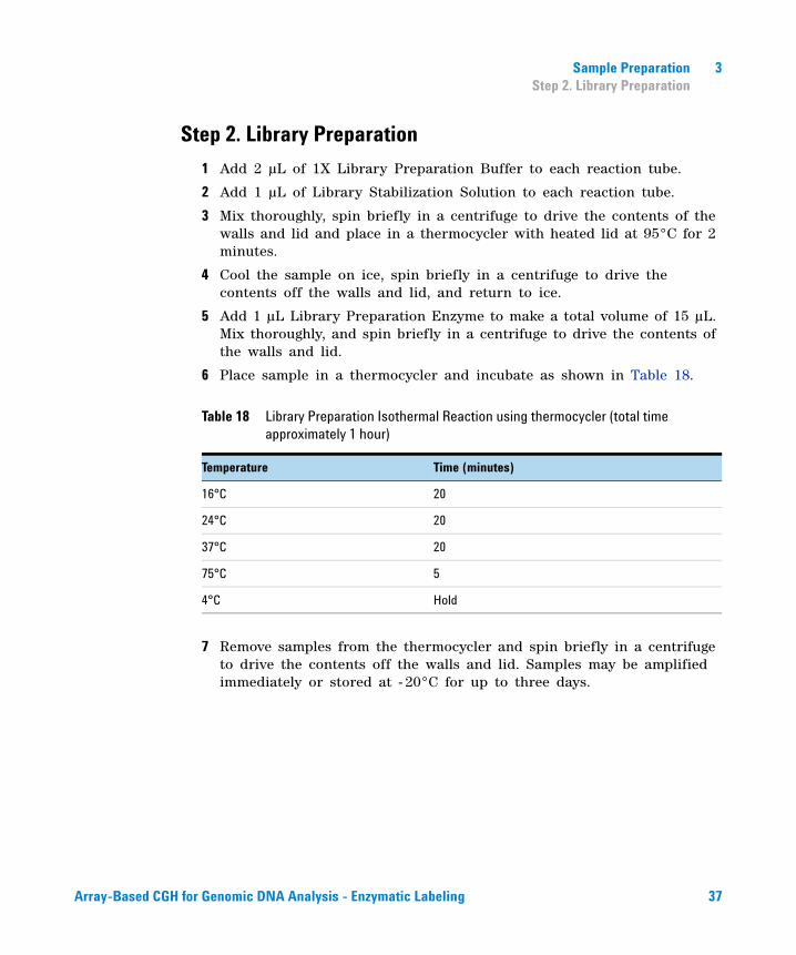
6 Place sample in a thermocycler and incubate as shown in Table 18.
7 Remove samples from the thermocycler and spin briefly in a centrifuge to drive the contents off the walls and lid. Samples may be amplified immediately or stored at - 20°C for up to three days.
Table 18 Library Preparation Isothermal Reaction using thermocycler (total time approximately 1 hour)
Temperature Time (minutes)
16°C 20
24°C 20
37°C 20
75°C 5
4°C Hold
Array-Based CGH for Genomic DNA Analysis - Enzymatic Labeling 37
3 Sample PreparationStep 3. Amplification
Step 3. Amplification
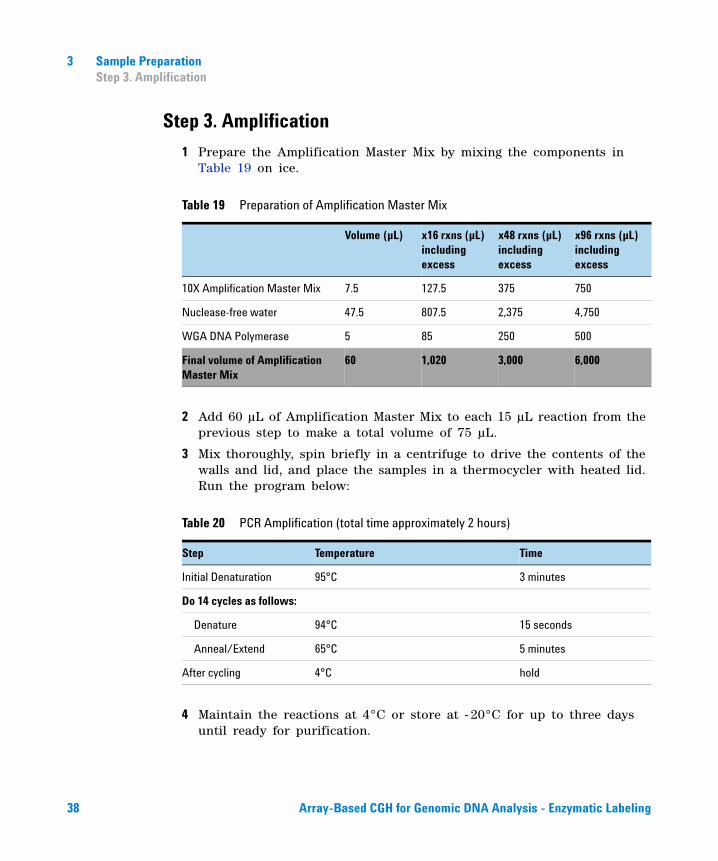
1 Prepare the Amplification Master Mix by mixing the components in Table 19 on ice.
2 Add 60 µL of Amplification Master Mix to each 15 µL reaction from the previous step to make a total volume of 75 µL.
3 Mix thoroughly, spin briefly in a centrifuge to drive the contents of the walls and lid, and place the samples in a thermocycler with heated lid. Run the program below:
4 Maintain the reactions at 4°C or store at - 20°C for up to three days until ready for purification.
Table 19 Preparation of Amplification Master Mix
Volume (µL) x16 rxns (µL) including excess
x48 rxns (µL)including excess
x96 rxns (µL)including excess
10X Amplification Master Mix 7.5 127.5 375 750
Nuclease-free water 47.5 807.5 2,375 4,750
WGA DNA Polymerase 5 85 250 500
Final volume of Amplification Master Mix
60 1,020 3,000 6,000
Table 20 PCR Amplification (total time approximately 2 hours)
Step Temperature Time
Initial Denaturation 95°C 3 minutes
Do 14 cycles as follows:
Denature 94°C 15 seconds
Anneal/Extend 65°C 5 minutes
After cycling 4°C hold
38 Array-Based CGH for Genomic DNA Analysis - Enzymatic Labeling
Sample Preparation 3Step 4. Purification of PCR products
Step 4. Purification of PCR products
Use Sigma’s GenElute PCR Clean- Up Kit for the purification of amplified gDNA.
1 Before using for the first time, dilute the Wash Solution Concentrate with 48 ml of 100% ethanol.
2 Insert a GenElute Miniprep Binding Column (with a blue O- ring) into a provided collection tube, if not already assembled. Add 0.5 mL of the Column Preparation Solution to each miniprep column and spin in a centrifuge at 12,000 g for 30 seconds to 1 minute. Discard the eluate, but keep the collection tube.
The Column Preparation Solution maximizes binding of the DNA to the membrane resulting in more consistent yields.
3 Add 375 µL of Binding Solution to each 75 µL sample. Transfer the solution into the binding column. Spin the column in a centrifuge at maximum speed (12,000 to 16,000 x g) for 1 minute. Discard the eluate, but keep the collection tube.
4 Place the binding column into the same collection tube. Apply 0.5 mL of diluted Wash Solution to the column and spin in a centrifuge at maximum speed for 1 minute. Discard the eluate, but retain the collection tube.
5 Place the column into the same collection tube. Spin the column in a centrifuge at maximum speed for 2 minutes, without any additional wash solution, to remove excess ethanol. Discard any residual eluate as well as the collection tube.
6 Transfer the column to a fresh 2 mL collection tube. Apply 50 µL of Elution Solution (10 mM Tris, pH 8.0) to the center of each column. Incubate at room temperature for 1 minute.
7 To elute the DNA, spin the column in a centrifuge at maximum speed for 1 minute.
The PCR amplification product is now present in the eluate and is ready for quantitation and labeling without restriction enzyme digestion. The final amplified DNA can be stored at - 20°C.
Array-Based CGH for Genomic DNA Analysis - Enzymatic Labeling 39
3 Sample PreparationStep 5. Quantitation of Amplified-Purified gDNA
Step 5. Quantitation of Amplified-Purified gDNA
Quantitate amplified- purified gDNA using the NanoDrop ND- 1000 UV-VIS Spectrophotometer or equivalent.
1 Select Nucleic Acid Measurement, then select Sample Type to be DNA- 50.
2 Use 1.5 µL of Elution Solution to blank the instrument.
3 Use 1.5 µL of each purified gDNA to measure DNA concentration. Record the DNA concentration (ng/µL) for each sample.
4 Calculate the amplification yield (µg) as
Step 6. Preparation of Amplified-Purified gDNA before Labeling
1 Add 2 µg of amplified- purified gDNA to a 1.5 mL nuclease- free tube or well in the PCR plate and bring to a final volume of 26 µL (1x, 2x, 4x microarrays) or 13 µL (8x microarrays) with nuclease- free water.
Both the experimental and reference channels require equal amounts of amplified- purified gDNA for the subsequent labeling reaction.
2 If the gDNA sample volume exceeds 26 µL (for 1x, 2x and 4x microarrays) or 13 µL (for 8x microarrays), concentrate the amplified- purified gDNA using a vacuum concentrator (such as a Speed Vac).
You can concentrate the gDNA to dryness and resuspend in water. Do not excessively dry the gDNA because the pellets will become difficult to resuspend.
Proceed directly to “Sample Labeling” on page 41 or store amplified- purified DNA at - 20°C.
Yield (µg) DNAconcentration (ng/µL) Sample Volume (µL)1000 ng/µg
--------------------------------------------------------------------------------------------------------------------------=
40 Array-Based CGH for Genomic DNA Analysis - Enzymatic Labeling
Agilent Oligonucleotide Array-Based CGH for Genomic DNA Analysis Protocol
4Sample Labeling
Step 1. Fluorescent Labeling of Genomic DNA 42
Step 2. Clean-up of Labeled Genomic DNA 46
To determine yield , degree of labeling or specific activity 51
Step 3. Preparation of Labeled Genomic DNA for Hybridization 53
The Agilent Genomic DNA Enzymatic Labeling Kit (Agilent p/n 5190- 0449) uses random primers and the exo- Klenow fragment to differentially label genomic DNA samples with fluorescent- labeled nucleotides. For Agilent's Oligo aCGH application, the experimental sample is labeled with one dye while the reference sample is labeled with the other dye. The “polarity” of the sample labeling is a matter of experimental choice.
41Agilent Technologies
4 Sample LabelingStep 1. Fluorescent Labeling of Genomic DNA
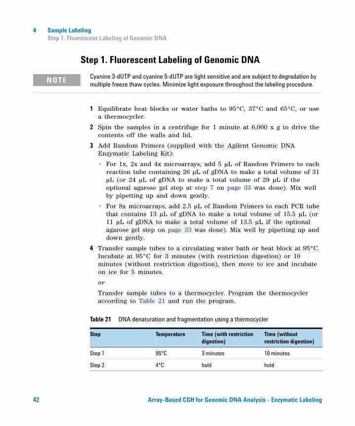
Step 1. Fluorescent Labeling of Genomic DNA
1 Equilibrate heat blocks or water baths to 95°C, 37°C and 65°C, or use a thermocycler.
2 Spin the samples in a centrifuge for 1 minute at 6,000 x g to drive the contents off the walls and lid.
3 Add Random Primers (supplied with the Agilent Genomic DNA Enzymatic Labeling Kit):
• For 1x, 2x and 4x microarrays, add 5 µL of Random Primers to each reaction tube containing 26 µL of gDNA to make a total volume of 31 µL (or 24 µL of gDNA to make a total volume of 29 µL if the optional agarose gel step at step 7 on page 33 was done). Mix well by pipetting up and down gently.
• For 8x microarrays, add 2.5 µL of Random Primers to each PCR tube that contains 13 µL of gDNA to make a total volume of 15.5 µL (or 11 µL of gDNA to make a total volume of 13.5 µL if the optional agarose gel step on page 33 was done). Mix well by pipetting up and down gently.
4 Transfer sample tubes to a circulating water bath or heat block at 95°C. Incubate at 95°C for 3 minutes (with restriction digestion) or 10 minutes (without restriction digestion), then move to ice and incubate on ice for 5 minutes.
or
Transfer sample tubes to a thermocycler. Program the thermocycler according to Table 21 and run the program.
Cyanine 3-dUTP and cyanine 5-dUTP are light sensitive and are subject to degradation by multiple freeze thaw cycles. Minimize light exposure throughout the labeling procedure.
NOTE
Table 21 DNA denaturation and fragmentation using a thermocycler
Step Temperature Time (with restriction digestion)
Time (without restriction digestion)
Step 1 95°C 3 minutes 10 minutes
Step 2 4°C hold hold
42 Array-Based CGH for Genomic DNA Analysis - Enzymatic Labeling
Sample Labeling 4Step 1. Fluorescent Labeling of Genomic DNA
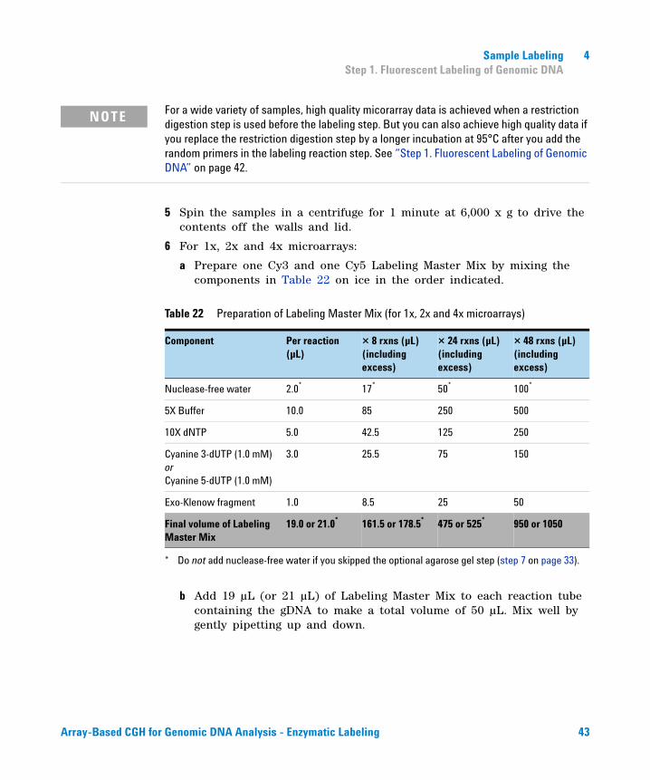
5 Spin the samples in a centrifuge for 1 minute at 6,000 x g to drive the contents off the walls and lid.
6 For 1x, 2x and 4x microarrays:
a Prepare one Cy3 and one Cy5 Labeling Master Mix by mixing the components in Table 22 on ice in the order indicated.
b Add 19 µL (or 21 µL) of Labeling Master Mix to each reaction tube containing the gDNA to make a total volume of 50 µL. Mix well by gently pipetting up and down.
NOTE For a wide variety of samples, high quality micorarray data is achieved when a restriction digestion step is used before the labeling step. But you can also achieve high quality data if you replace the restriction digestion step by a longer incubation at 95°C after you add the random primers in the labeling reaction step. See “Step 1. Fluorescent Labeling of Genomic DNA” on page 42.
Table 22 Preparation of Labeling Master Mix (for 1x, 2x and 4x microarrays)
Component Per reaction (µL)
× 8 rxns (µL)(including excess)
× 24 rxns (µL)(including excess)
× 48 rxns (µL)(including excess)
Nuclease-free water 2.0*
* Do not add nuclease-free water if you skipped the optional agarose gel step (step 7 on page 33).
17* 50* 100*
5X Buffer 10.0 85 250 500
10X dNTP 5.0 42.5 125 250
Cyanine 3-dUTP (1.0 mM) or Cyanine 5-dUTP (1.0 mM)
3.0 25.5 75 150
Exo-Klenow fragment 1.0 8.5 25 50
Final volume of Labeling Master Mix
19.0 or 21.0* 161.5 or 178.5* 475 or 525* 950 or 1050
Array-Based CGH for Genomic DNA Analysis - Enzymatic Labeling 43
4 Sample LabelingStep 1. Fluorescent Labeling of Genomic DNA
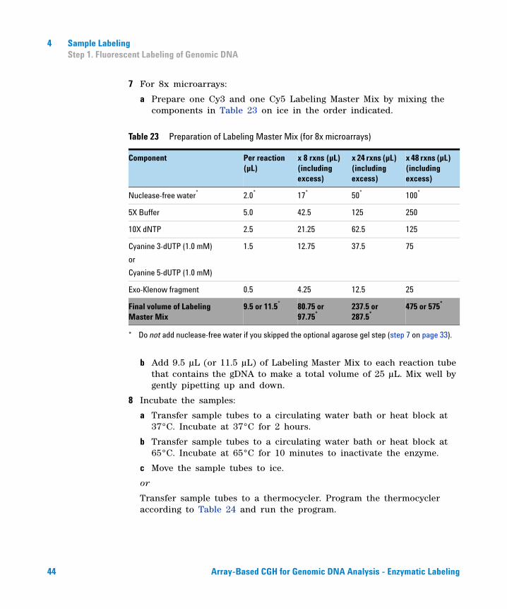
7 For 8x microarrays:
a Prepare one Cy3 and one Cy5 Labeling Master Mix by mixing the components in Table 23 on ice in the order indicated.
b Add 9.5 µL (or 11.5 µL) of Labeling Master Mix to each reaction tube that contains the gDNA to make a total volume of 25 µL. Mix well by gently pipetting up and down.
8 Incubate the samples:
a Transfer sample tubes to a circulating water bath or heat block at 37°C. Incubate at 37°C for 2 hours.
b Transfer sample tubes to a circulating water bath or heat block at 65°C. Incubate at 65°C for 10 minutes to inactivate the enzyme.
c Move the sample tubes to ice.
or
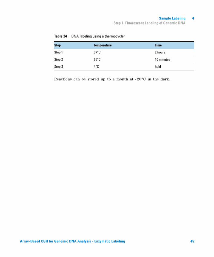
Transfer sample tubes to a thermocycler. Program the thermocycler according to Table 24 and run the program.
Table 23 Preparation of Labeling Master Mix (for 8x microarrays)
Component Per reaction (µL)
x 8 rxns (µL) (including excess)
x 24 rxns (µL) (including excess)
x 48 rxns (µL) (including excess)
Nuclease-free water*
* Do not add nuclease-free water if you skipped the optional agarose gel step (step 7 on page 33).
2.0* 17* 50* 100*
5X Buffer 5.0 42.5 125 250
10X dNTP 2.5 21.25 62.5 125
Cyanine 3-dUTP (1.0 mM)
or
Cyanine 5-dUTP (1.0 mM)
1.5 12.75 37.5 75
Exo-Klenow fragment 0.5 4.25 12.5 25
Final volume of Labeling Master Mix
9.5 or 11.5* 80.75 or 97.75*
237.5 or 287.5*
475 or 575*
44 Array-Based CGH for Genomic DNA Analysis - Enzymatic Labeling
Sample Labeling 4Step 1. Fluorescent Labeling of Genomic DNA
Reactions can be stored up to a month at - 20°C in the dark.
Table 24 DNA labeling using a thermocycler
Step Temperature Time
Step 1 37°C 2 hours
Step 2 65°C 10 minutes
Step 3 4°C hold
Array-Based CGH for Genomic DNA Analysis - Enzymatic Labeling 45
4 Sample LabelingStep 2. Clean-up of Labeled Genomic DNA
Step 2. Clean-up of Labeled Genomic DNA
Labeled genomic DNA can be purified using individual Amicon 30kDA filters or GE Healthcare 96- well plates.
Amicon 30kDa filters
1 Spin the labeled genomic DNA samples in a centrifuge for 1 minute at 6,000 x g to drive the contents off the walls and lid.
2 Add 430 µL of 1X TE (pH 8.0) to each reaction tube.
3 Place an Amicon 30kDa filter into a 1.5- mL microfuge tube (supplied) and load each labeled gDNA into the filter. Spin 10 minutes at 14,000 × g in a microcentrifuge at room temperature. Discard the flow- through.
4 Add 480 µL of 1X TE (pH 8.0) to each filter. Spin for 10 minutes at 14,000 × g in a microcentrifuge at room temperature. Discard the flow- through.
5 Invert the filter into a fresh 1.5- mL microfuge tube (supplied). Spin for 1 minute at 1,000 × g in a microcentrifuge at room temperature to collect purified sample.
The volume per sample will be approximately 21 µL.
6 Add 1X TE or use a concentrator to bring to the sample volume in Table 25.
If needed, you can concentrate the gDNA sample to dryness and resuspend in the volume listed in Table 25. Do not excessively dry the gDNA because the pellets will become difficult to resuspend.
7 Take 1.5 µL of each sample to determine yield and specific activity. See “To determine yield , degree of labeling or specific activity” on page 51. Refer to Table 27 on page 52 for expected yield of labeled genomic DNA and specific activity after labeling and clean- up, when starting with high quality genomic DNA.
NOTE Keep Cyanine-3 and Cyanine-5 labeled genomic DNA samples separated throughout this clean-up step.
46 Array-Based CGH for Genomic DNA Analysis - Enzymatic Labeling
Sample Labeling 4Step 2. Clean-up of Labeled Genomic DNA
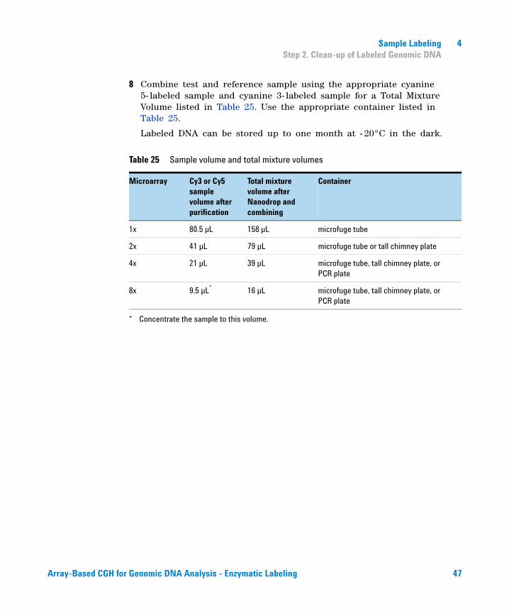
8 Combine test and reference sample using the appropriate cyanine 5- labeled sample and cyanine 3- labeled sample for a Total Mixture Volume listed in Table 25. Use the appropriate container listed in Table 25.
Labeled DNA can be stored up to one month at - 20°C in the dark.
Table 25 Sample volume and total mixture volumes
Microarray Cy3 or Cy5 sample volume after purification
Total mixture volume after Nanodrop and combining
Container
1x 80.5 µL 158 µL microfuge tube
2x 41 µL 79 µL microfuge tube or tall chimney plate
4x 21 µL 39 µL microfuge tube, tall chimney plate, or PCR plate
8x 9.5 µL*
* Concentrate the sample to this volume.
16 µL microfuge tube, tall chimney plate, or PCR plate
Array-Based CGH for Genomic DNA Analysis - Enzymatic Labeling 47
4 Sample LabelingStep 2. Clean-up of Labeled Genomic DNA
GE Healthcare 96-well plates
For 1x, 2x, 4x you can use two wells of the AutoScreen- 96A Well plates (GE Healthcare p/n 25- 9005- 98) per sample or concentrate the samples down to 25 µL with a vacuum concentrator (such as a Speed Vac). You need to supply two 96- well PCR plates (Eppendorf p/n 951020401 or equivalent). Label one plate "wash plate" and the other plate "collection plate". The wash plate can be re- used in next experiments.
1 Remove the purification plates from the foil storage pouch.
If the purification plates were stored at 4°C, allow them to equilibrate to ambient temperature before use (approximately 2 hours).
2 Carefully remove the top and bottom seal of the purification plates.
Once the bottom seal is removed, keep the purification plates on top of a wash plate. Do not allow the bottom surface to come in contact with laboratory benchtop liners, wipes, or other materials.
3 Place the purification plates in re- usable wash plates.
4 Pre- spin the purification plates in a centrifuge for 5 minutes at 910 x g.
5 Discard the flow- through from the wash plates, and place the purification plates back to the same wash plates.
6 Add the 150 µL nuclease free water to the purification plates.
7 Spin again in a centrifuge for 5 minutes at 910 x g.
8 Discard the flow- through.
9 Transfer the purification plates to the sample collection plate.
10 Add labeled genomic DNA to the purification plates:
• For 1x, 2x, 4x microarray samples that were not concentrated to 25 µL, add 2x25 µL labeled genomic DNA to two separate wells.
• For 1x, 2x, 4x microarray samples that were concentrated to 25 µL with concentrator and for 8x microarray samples, add 1x25 µL labeled genomic DNA to one well.
NOTE Use the same centrifuge speed and length for all three spinning steps (step 4, step 7 and step 11). If you spin only one plate, make sure you counterbalance.
48 Array-Based CGH for Genomic DNA Analysis - Enzymatic Labeling
Sample Labeling 4Step 2. Clean-up of Labeled Genomic DNA
11 Spin in a centrifuge for 5 minutes at 910 x g to collect the purified labeled gDNA in the sample collection plate. The volume per sample will be approximately 20 µL.
12 For 1x, 2x and 4x microarray samples that were not concentrated prior to purification, combine the duplicate samples for a total volume of approximately 40 µL.
13 Take 1.5 µL of each sample to determine the yield and specific activity. See “To determine yield , degree of labeling or specific activity” on page 51.
Refer to Table 27 on page 52 for expected yield of labeled genomic DNA and specific activity after labeling and clean- up, when starting with high quality genomic DNA.
14 Combine the test and reference sample using the appropriate cyanine 5- labeled sample and cyanine 3- labeled sample. Use the appropriate container listed in Table 26. Add 1X TE or use a concentrator to bring to the Total Mixture Volume in Table 26.
If needed, you can concentrate the combined Cy5- and Cy3- labeled gDNA mixture to dryness and resuspend in water to the final volume listed in Table 26. Do not excessively dry the samples because the pellets will become difficult to resuspend.
Array-Based CGH for Genomic DNA Analysis - Enzymatic Labeling 49
4 Sample LabelingStep 2. Clean-up of Labeled Genomic DNA
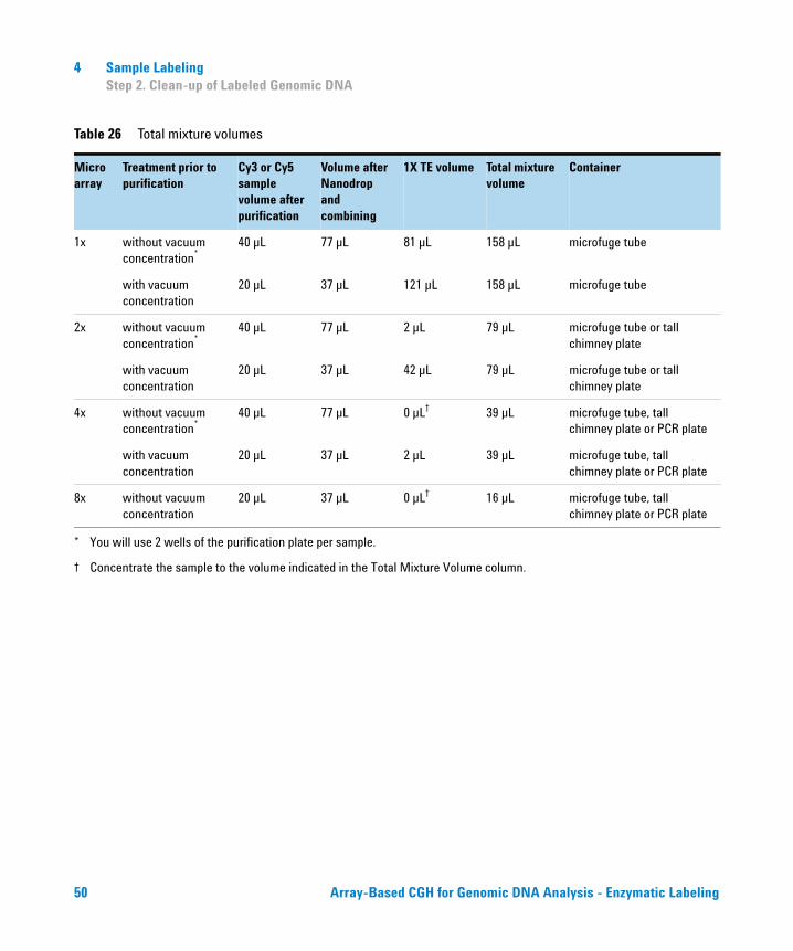
Table 26 Total mixture volumes
Microarray
Treatment prior to purification
Cy3 or Cy5 sample volume after purification
Volume after Nanodrop and combining
1X TE volume Total mixture volume
Container
1x without vacuum concentration*
* You will use 2 wells of the purification plate per sample.
40 µL 77 µL 81 µL 158 µL microfuge tube
with vacuum concentration
20 µL 37 µL 121 µL 158 µL microfuge tube
2x without vacuum concentration*
40 µL 77 µL 2 µL 79 µL microfuge tube or tall chimney plate
with vacuum concentration
20 µL 37 µL 42 µL 79 µL microfuge tube or tall chimney plate
4x without vacuum concentration*
40 µL 77 µL 0 µL†
† Concentrate the sample to the volume indicated in the Total Mixture Volume column.
39 µL microfuge tube, tall chimney plate or PCR plate
with vacuum concentration
20 µL 37 µL 2 µL 39 µL microfuge tube, tall chimney plate or PCR plate
8x without vacuum concentration
20 µL 37 µL 0 µL† 16 µL microfuge tube, tall chimney plate or PCR plate
50 Array-Based CGH for Genomic DNA Analysis - Enzymatic Labeling
Sample Labeling 4To determine yield , degree of labeling or specific activity
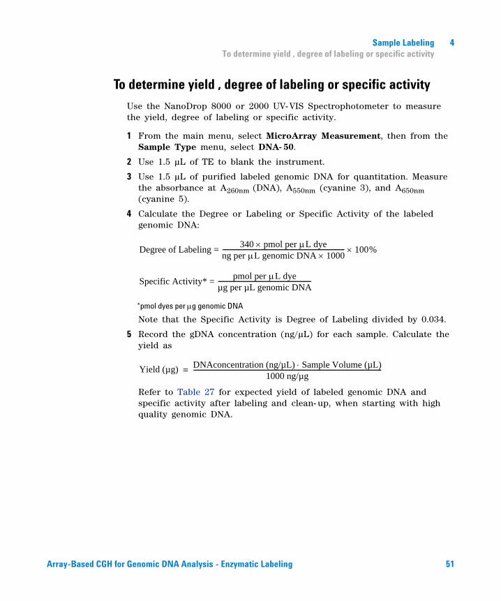
To determine yield , degree of labeling or specific activity
Use the NanoDrop 8000 or 2000 UV-VIS Spectrophotometer to measure the yield, degree of labeling or specific activity.
1 From the main menu, select MicroArray Measurement, then from the Sample Type menu, select DNA- 50.
2 Use 1.5 µL of TE to blank the instrument.
3 Use 1.5 µL of purified labeled genomic DNA for quantitation. Measure the absorbance at A260nm (DNA), A550nm (cyanine 3), and A650nm (cyanine 5).
4 Calculate the Degree or Labeling or Specific Activity of the labeled genomic DNA:
*pmol dyes per g genomic DNA
Note that the Specific Activity is Degree of Labeling divided by 0.034.
5 Record the gDNA concentration (ng/µL) for each sample. Calculate the yield as
Refer to Table 27 for expected yield of labeled genomic DNA and specific activity after labeling and clean- up, when starting with high quality genomic DNA.
Degree of Labeling = 340 pmol per L dyeng per L genomic DNA 1000------------------------------------------------------------------------------- 100%
Specific Activity* = pmol per L dyeµg per µL genomic DNA------------------------------------------------------------
Yield (µg) DNAconcentration (ng/µL) Sample Volume (µL)1000 ng/µg
--------------------------------------------------------------------------------------------------------------------------=
Array-Based CGH for Genomic DNA Analysis - Enzymatic Labeling 51
4 Sample LabelingTo determine yield , degree of labeling or specific activity
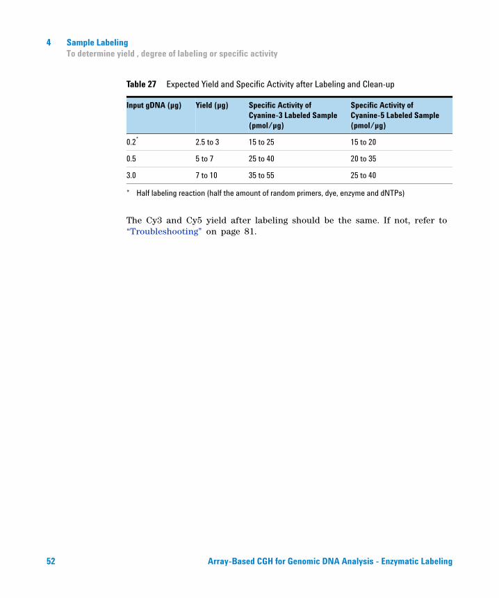
The Cy3 and Cy5 yield after labeling should be the same. If not, refer to “Troubleshooting” on page 81.
Table 27 Expected Yield and Specific Activity after Labeling and Clean-up
Input gDNA (µg) Yield (µg) Specific Activity of Cyanine-3 Labeled Sample (pmol/µg)
Specific Activity of Cyanine-5 Labeled Sample (pmol/µg)
0.2*
* Half labeling reaction (half the amount of random primers, dye, enzyme and dNTPs)
2.5 to 3 15 to 25 15 to 20
0.5 5 to 7 25 to 40 20 to 35
3.0 7 to 10 35 to 55 25 to 40
52 Array-Based CGH for Genomic DNA Analysis - Enzymatic Labeling
Sample Labeling 4Step 3. Preparation of Labeled Genomic DNA for Hybridization
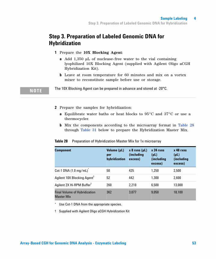
Step 3. Preparation of Labeled Genomic DNA for Hybridization
1 Prepare the 10X Blocking Agent:
a Add 1,350 µL of nuclease- free water to the vial containing lyophilized 10X Blocking Agent (supplied with Agilent Oligo aCGH Hybridization Kit).
b Leave at room temperature for 60 minutes and mix on a vortex mixer to reconstitute sample before use or storage.
2 Prepare the samples for hybridization:
a Equilibrate water baths or heat blocks to 95°C and 37°C or use a thermocycler.
b Mix the components according to the microarray format in Table 28 through Table 31 below to prepare the Hybridization Master Mix.
NOTE The 10X Blocking Agent can be prepared in advance and stored at -20°C.
Table 28 Preparation of Hybridization Master Mix for 1x microarray
Component Volume (µL) per hybridization
x 8 rxns (µL) (including excess)
x 24 rxns (µL) (including excess)
x 48 rxns (µL) (including excess)
Cot-1 DNA (1.0 mg/mL)*
* Use Cot-1 DNA from the appropriate species.
50 425 1,250 2,500
Agilent 10X Blocking Agent†
† Supplied with Agilent Oligo aCGH Hybridization Kit
52 442 1,300 2,600
Agilent 2X Hi-RPM Buffer† 260 2,210 6,500 13,000
Final Volume of Hybridization Master Mix
362 3,077 9,050 18,100
Array-Based CGH for Genomic DNA Analysis - Enzymatic Labeling 53
4 Sample LabelingStep 3. Preparation of Labeled Genomic DNA for Hybridization
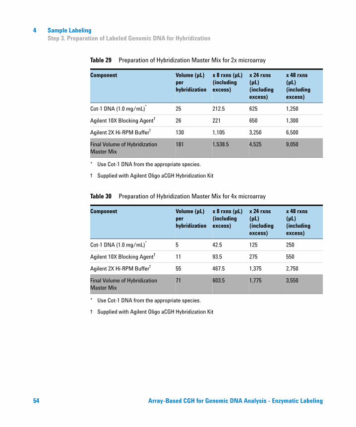
Table 29 Preparation of Hybridization Master Mix for 2x microarray
Component Volume (µL) per hybridization
x 8 rxns (µL) (including excess)
x 24 rxns (µL) (including excess)
x 48 rxns (µL) (including excess)
Cot-1 DNA (1.0 mg/mL)*
* Use Cot-1 DNA from the appropriate species.
25 212.5 625 1,250
Agilent 10X Blocking Agent†
† Supplied with Agilent Oligo aCGH Hybridization Kit
26 221 650 1,300
Agilent 2X Hi-RPM Buffer† 130 1,105 3,250 6,500
Final Volume of Hybridization Master Mix
181 1,538.5 4,525 9,050
Table 30 Preparation of Hybridization Master Mix for 4x microarray
Component Volume (µL) per hybridization
x 8 rxns (µL) (including excess)
x 24 rxns (µL) (including excess)
x 48 rxns (µL) (including excess)
Cot-1 DNA (1.0 mg/mL)*
* Use Cot-1 DNA from the appropriate species.
5 42.5 125 250
Agilent 10X Blocking Agent†
† Supplied with Agilent Oligo aCGH Hybridization Kit
11 93.5 275 550
Agilent 2X Hi-RPM Buffer† 55 467.5 1,375 2,750
Final Volume of Hybridization Master Mix
71 603.5 1,775 3,550
54 Array-Based CGH for Genomic DNA Analysis - Enzymatic Labeling
Sample Labeling 4Step 3. Preparation of Labeled Genomic DNA for Hybridization
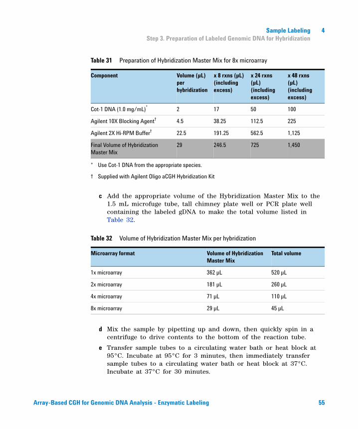
c Add the appropriate volume of the Hybridization Master Mix to the 1.5 mL microfuge tube, tall chimney plate well or PCR plate well containing the labeled gDNA to make the total volume listed in Table 32.
d Mix the sample by pipetting up and down, then quickly spin in a centrifuge to drive contents to the bottom of the reaction tube.
e Transfer sample tubes to a circulating water bath or heat block at 95°C. Incubate at 95°C for 3 minutes, then immediately transfer sample tubes to a circulating water bath or heat block at 37°C. Incubate at 37°C for 30 minutes.
Table 31 Preparation of Hybridization Master Mix for 8x microarray
Component Volume (µL) per hybridization
x 8 rxns (µL) (including excess)
x 24 rxns (µL) (including excess)
x 48 rxns (µL) (including excess)
Cot-1 DNA (1.0 mg/mL)*
* Use Cot-1 DNA from the appropriate species.
2 17 50 100
Agilent 10X Blocking Agent†
† Supplied with Agilent Oligo aCGH Hybridization Kit
4.5 38.25 112.5 225
Agilent 2X Hi-RPM Buffer† 22.5 191.25 562.5 1,125
Final Volume of Hybridization Master Mix
29 246.5 725 1,450
Table 32 Volume of Hybridization Master Mix per hybridization
Microarray format Volume of Hybridization Master Mix
Total volume
1x microarray 362 µL 520 µL
2x microarray 181 µL 260 µL
4x microarray 71 µL 110 µL
8x microarray 29 µL 45 µL
Array-Based CGH for Genomic DNA Analysis - Enzymatic Labeling 55
4 Sample LabelingStep 3. Preparation of Labeled Genomic DNA for Hybridization
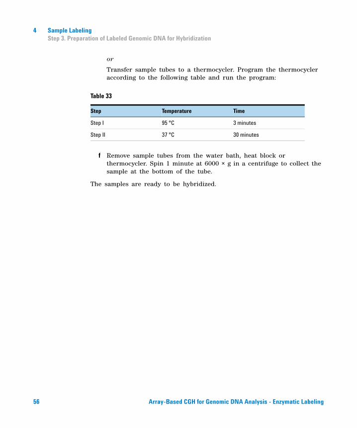
or
Transfer sample tubes to a thermocycler. Program the thermocycler according to the following table and run the program:
f Remove sample tubes from the water bath, heat block or thermocycler. Spin 1 minute at 6000 × g in a centrifuge to collect the sample at the bottom of the tube.
The samples are ready to be hybridized.
Table 33
Step Temperature Time
Step I 95 °C 3 minutes
Step II 37 °C 30 minutes
56 Array-Based CGH for Genomic DNA Analysis - Enzymatic Labeling
Agilent Oligonucleotide Array-Based CGH for Genomic DNA Analysis Protocol
5Microarray Processing and Feature Extraction
Step 1. Microarray Hybridization 58
Step 2. Wash Preparation 60
Step 3. Microarray Washing 64
Step 4. Microarray Scanning using Agilent C, B or A Scanner or GenePix Scanner 70
Step 5. Data Extraction using Feature Extraction Software 72
Microarray processing consists of hybridization, washing, and scanning.
Feature Extraction is the process by which data is extracted from the scanned microarray image (.tif) and translated into log ratios, allowing researchers to measure DNA copy number changes in their experiments in conjunction with Agilent Genomic Workbench Software.
57Agilent Technologies
5 Microarray Processing and Feature ExtractionStep 1. Microarray Hybridization
Step 1. Microarray Hybridization
Microarray Handling Tips
Each microarray is printed on the side of the glass slide containing the “Agilent”- labeled barcode. This side is called the “active side”. The numeric barcode is on the “inactive side” of the glass slide.
The hybridization sample mixture is applied directly to the gasket slide and not to the microarray slide. Then the active side of the microarray slide is put on top of the gasket slide to form a “sandwich slide pair”.
To avoid damaging the microarray, always handle glass slides carefully by their edges. Wear powder- free gloves. Never touch the surfaces of the slides. If you do, you may cause irreparable damage to the microarray.
Never allow the microarray surface to dry out during the hybridization process and washing steps.
Hybridization Assembly
1 Load a clean gasket slide into the Agilent SureHyb chamber base with the gasket label facing up and aligned with the rectangular section of the chamber base. Ensure that the gasket slide is flush with the chamber base and is not ajar.
2 Slowly dispense 490 µL (for 1x microarray), 245 µL (for 2x microarray), 100 µL (for 4x microarray) or 40 µL (for 8x microarray) of hybridization sample mixture onto the gasket well in a “drag and dispense” manner. For multi- pack microarray formats (i.e. 2x, 4x or 8x microarray), load all gasket wells before you load the microarray slide.
NOTE Familiarize yourself with the assembly and disassembly instructions for use with the Agilent microarray hybridization chamber and gasket slides. Please refer to the Agilent Microarray Hybridization Chamber User Guide (G2534-90001) for in-depth instructions on how to load samples, assemble and disassemble chambers, as well as other helpful tips. This user guide can be downloaded from the Agilent Web site at www.agilent.com/chem/dnamanuals-protocols.
CAUTION Keep the temperature of hybridization sample mixtures as close to 37°C as possible. To do this, process them in small batches and/or put them on a heat block, thermocycler or in an oven.
58 Array-Based CGH for Genomic DNA Analysis - Enzymatic Labeling
Microarray Processing and Feature Extraction 5Step 1. Microarray Hybridization
3 Put a microarray slide “active side” down onto the gasket slide, so the numeric barcode side is facing up and the “Agilent”- labeled barcode is facing down. Assess that the sandwich- pair is properly aligned.
4 Put the SureHyb chamber cover onto the sandwiched slides and slide the clamp assembly onto both pieces.
5 Hand- tighten the clamp onto the chamber.
6 Vertically rotate the assembled chamber to wet the slides and assess the mobility of the bubbles. Tap the assembly on a hard surface if necessary to move stationary bubbles.
7 Put assembled slide chamber in the rotator rack in a hybridization oven set to 65°C. Set your hybridization rotator to rotate at 20 rpm.
8 Hybridize at 65°C for 40 hours (for 1x or 2x microarrays) or for 24 hours (for 4x or 8x microarrays).
9 Hybridize at 65°C:
• 24 hours for blood, cell and tissue samples (4x and 8x microarrays)
• 40 hours for blood, cell and tissue samples (1x and 2x microarrays)
• 40 hours for FFPE samples (1x, 2x, 4x and 8x microarrays)
For more information on the effects of hybridization temperature and time, as well as the rotation speed on the final microarray results, please refer to the application note titled “60- mer Oligo- Based Comparative Genomic Hybridization” (publication 5989- 4848EN) from the Agilent Web site at www.agilent.com/chem/dnaapplications.
CAUTION If you are not loading all the available positions on the hybridization rotator rack, be sure to balance the loaded hybridization chambers on the rack similar to a centrifuge to prevent unnecessary strain on the oven motor.
Array-Based CGH for Genomic DNA Analysis - Enzymatic Labeling 59
5 Microarray Processing and Feature ExtractionStep 2. Wash Preparation
Step 2. Wash Preparation
Before you begin, determine which wash procedure to use:
Equipment Preparation
• Always use clean equipment when conducting the wash procedures.
NOTE Cyanine 5 has been shown to be sensitive to ozone degradation. Ozone levels as low as 5 ppb (approximately 10 µg/m3) can affect Cyanine 5 signal and compromise microarray results. The Agilent Stabilization and Drying Solution and the Ozone-Barrier Slide Cover are designed to protect against ozone-induced degradation of Cyanine dyes. Use these when working with Agilent oligo-based microarrays in high ozone environments. Note that the Ozone-Barrier Slide covers are compatible with the B and C scanner slide holders only.
Another option to address ozone-induced Cyanine-5 degradation is to use Carbon Loaded Nonwoven Filters to remove ozone from the air. These filters can be installed in either your HVAC system, or as part of small Ozone Controlled Enclosures. These free-standing enclosures can be installed either on a lab bench or as a walk-in room within your lab. These products are available through filter suppliers listed in Agilent Technical Note 5989-0875EN.
Table 34 Wash procedure to follow
Ozone level in your lab
Wash Procedure Ozone-Barrier Slide Cover
< 5 ppb “Wash Procedure A (without Stabilization and Drying Solution)” on page 64
No
> 5 ppb < 10 ppb “Wash Procedure A (without Stabilization and Drying Solution)” on page 64
Yes
> 10 ppb “Wash Procedure B (with Stabilization and Drying Solution)” on page 67
Yes
CAUTION Do not use detergent to wash the staining dishes as some detergents may leave fluorescent residue on the dishes. If you do, you must ensure that all traces are removed by thoroughly rinsing with Milli-Q water.
60 Array-Based CGH for Genomic DNA Analysis - Enzymatic Labeling
Microarray Processing and Feature Extraction 5Step 2. Wash Preparation
• Use only dishes that are designated and dedicated for use in Agilent oligo aCGH experiments.
Cleaning with Milli-Q Water Wash
Rinse slide- staining dishes, slide racks and stir bars thoroughly with high- quality Milli- Q water before use and in between washing groups.
a Run copious amounts of Milli- Q water through the slide- staining dishes, slide racks and stir bars.
b Empty out the water collected in the dishes at least five times.
c Repeat step a and step b until all traces of contaminating material are removed.
Cleaning with Acetonitrile Wash (Wash Procedure B Only)
Acetonitrile wash removes any remaining residue of Agilent Stabilization and Drying Solution from slide- staining dishes, slide racks and stir bars that were used in previous experiments with “Wash Procedure B (with Stabilization and Drying Solution)” on page 67.
a Add the slide rack and stir bar to the slide- staining dish, and transfer to a magnetic stir plate.
b Fill the slide- staining dish with 100% acetonitrile.
c Turn on the magnetic stir plate and adjust the speed to a setting of 4 (medium speed).
d Wash for 5 minutes at room temperature.
e Discard the acetonitrile as is appropriate for your site.
f Repeat step a through step e.
g Air dry everything in the vented fume hood.
h Continue with the Milli- Q water wash as previously instructed.
WARNING Do acetonitrile washes in a vented fume hood. Acetonitrile is highly flammable and toxic.
Array-Based CGH for Genomic DNA Analysis - Enzymatic Labeling 61
5 Microarray Processing and Feature ExtractionStep 2. Wash Preparation
Prewarming Oligo aCGH Wash Buffer 2 (Overnight)
The temperature of Oligo aCGH Wash Buffer 2 must be at 37°C for optimal performance.
1 Add the volume of buffer required to a disposable plastic bottle and warm overnight in an incubator or circulating water bath set to 37°C.
2 Put a slide- staining dish into a 1.5 L glass dish three- fourths filled with water and warm to 37°C by storing overnight in an incubator set to 37°C.
Prewarming Stabilization and Drying Solution (Wash Procedure B Only)
The Agilent Stabilization and Drying Solution contains an ozone scavenging compound dissolved in acetonitrile. The compound in solution is present in saturating amounts and may precipitate from the solution under normal storage conditions. If the solution shows visible precipitation, warming of the solution will be necessary to redissolve the compound. Washing slides using Stabilization and Drying Solution showing visible precipitation will have profound adverse affects on array performance.
1 Put a clean magnetic stir bar into the Stabilization and Drying Solution bottle and recap.
2 Partially fill a plastic bucket with hot water at approximately 40°C to 45°C (for example from a hot water tap).
WARNING The Agilent Stabilization and Drying Solution is a flammable liquid. Warming the solution will increase the generation of ignitable vapors. Use gloves and eye/face protection in every step of the warming procedures.
WARNING Do not use a hot plate, oven, an open flame or a microwave. Do not increase temperature rapidly. Warm and mix the material away from ignition sources.
WARNING Failure to follow the outlined process will increase the potential for fire, explosion, and possible personal injury.
62 Array-Based CGH for Genomic DNA Analysis - Enzymatic Labeling
Microarray Processing and Feature Extraction 5Step 2. Wash Preparation
3 Put the Stabilization and Drying Solution bottle into the hot water in the plastic bucket.
4 Put the plastic bucket on a magnetic stirrer (not a hot- plate) and stir.
5 The hot water cools to room temperature. If the precipitate has not all dissolved replenish the cold water with hot water.
6 Repeat step 5 until the solution is clear.
7 After the precipitate is completely dissolved, allow the solution to equilibrate to room temperature prior to use.
CAUTION Do not filter the Stabilization and Drying solution, or the concentration of the ozone scavenger may vary.
Array-Based CGH for Genomic DNA Analysis - Enzymatic Labeling 63
5 Microarray Processing and Feature ExtractionStep 3. Microarray Washing
Step 3. Microarray Washing
Before you begin, determine which wash procedure to use:
Wash Procedure A (without Stabilization and Drying Solution)
Always use fresh Oligo aCGH Wash Buffer 1 and Oligo aCGH Wash Buffer 2 for each wash group (up to five slides).
Table 36 lists the wash conditions for the Wash Procedure A without Stabilization and Drying Solution.
1 Completely fill slide- staining dish #1 with Oligo aCGH Wash Buffer 1 at room temperature.
2 Put a slide rack into slide- staining dish #2. Add a magnetic stir bar. Fill slide- staining dish #2 with enough Oligo aCGH Wash Buffer 1 at room temperature to cover the slide rack. Put this dish on a magnetic stir plate.
Table 35 Wash procedure to follow
Ozone level in your lab
Wash Procedure Ozone-Barrier Slide Cover
< 5 ppb “Wash Procedure A (without Stabilization and Drying Solution)” on page 64
No
> 5 ppb < 10 ppb “Wash Procedure A (without Stabilization and Drying Solution)” on page 64
Yes
> 10 ppb “Wash Procedure B (with Stabilization and Drying Solution)” on page 67
Yes
Table 36 Wash conditions
Dish Wash buffer Temperature Time
Disassembly #1 Oligo aCGH Wash Buffer 1 Room temperature
1st wash #2 Oligo aCGH Wash Buffer 1 Room temperature 5 minutes
2nd wash #3 Oligo aCGH Wash Buffer 2 37°C 1 minute
64 Array-Based CGH for Genomic DNA Analysis - Enzymatic Labeling
Microarray Processing and Feature Extraction 5Step 3. Microarray Washing
3 Put the prewarmed 1.5 L glass dish filled with water and containing slide- staining dish #3 on a magnetic stir plate with heating element. Fill the slide- staining dish #3 approximately three- fourths full with Oligo aCGH Wash Buffer 2 (warmed to 37°C). Add a magnetic stir bar. Turn on the heating element and maintain temperature of Oligo aCGH Wash Buffer 2 at 37°C; monitor using a thermometer.
4 Remove one hybridization chamber from incubator and record time. Record whether bubbles formed during hybridization and if all bubbles are rotating freely.
5 Prepare the hybridization chamber disassembly.
a Put the hybridization chamber assembly on a flat surface and loosen the thumbscrew, turning counter- clockwise.
b Slide off the clamp assembly and remove the chamber cover.
c With gloved fingers, remove the array- gasket sandwich from the chamber base by grabbing the slides from their ends. Keep the microarray slide numeric barcode facing up as you quickly transfer the sandwich to slide- staining dish #1.
d Without letting go of the slides, submerge the array- gasket sandwich into slide- staining dish #1 containing Oligo aCGH Wash Buffer 1.
6 With the sandwich completely submerged in Oligo aCGH Wash Buffer 1, pry the sandwich open from the barcode end only. Do this by slipping one of the blunt ends of the forceps between the slides and then gently turn the forceps upwards or downwards to separate the slides. Let the gasket slide drop to the bottom of the staining dish. Remove the microarray slide and put into slide rack in the slide- staining dish #2 containing Oligo aCGH Wash Buffer 1 at room temperature. Minimize exposure of the slide to air. Touch only the barcode portion of the microarray slide or its edges!
7 Repeat step 4 through step 6 for up to four additional slides in the group. A maximum of five disassembly procedures yielding five microarray slides is advised at one time in order to facilitate uniform washing.
8 When all slides in the group are put into the slide rack in slide- staining dish #2, stir using setting 4 for 5 minutes. Adjust the setting to get good but not vigorous mixing.
9 Transfer slide rack to slide- staining dish #3 containing Oligo aCGH Wash Buffer 2 at 37°C, and stir using setting 4 for 1 minute.
Array-Based CGH for Genomic DNA Analysis - Enzymatic Labeling 65
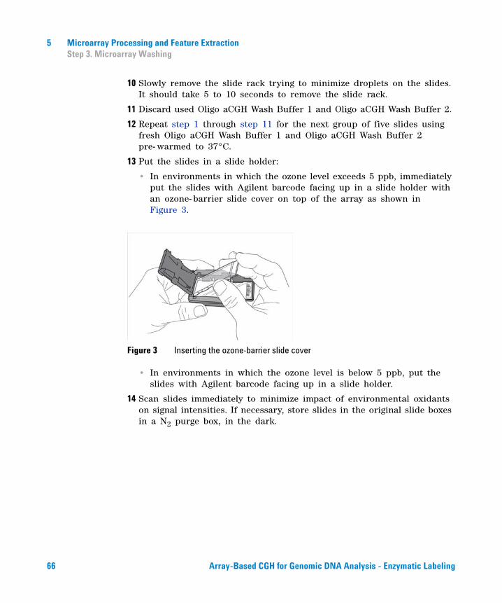
5 Microarray Processing and Feature ExtractionStep 3. Microarray Washing
10 Slowly remove the slide rack trying to minimize droplets on the slides. It should take 5 to 10 seconds to remove the slide rack.
11 Discard used Oligo aCGH Wash Buffer 1 and Oligo aCGH Wash Buffer 2.
12 Repeat step 1 through step 11 for the next group of five slides using fresh Oligo aCGH Wash Buffer 1 and Oligo aCGH Wash Buffer 2 pre- warmed to 37°C.
13 Put the slides in a slide holder:
• In environments in which the ozone level exceeds 5 ppb, immediately put the slides with Agilent barcode facing up in a slide holder with an ozone- barrier slide cover on top of the array as shown in Figure 3.
Figure 3 Inserting the ozone-barrier slide cover
• In environments in which the ozone level is below 5 ppb, put the slides with Agilent barcode facing up in a slide holder.
14 Scan slides immediately to minimize impact of environmental oxidants on signal intensities. If necessary, store slides in the original slide boxes in a N2 purge box, in the dark.
66 Array-Based CGH for Genomic DNA Analysis - Enzymatic Labeling
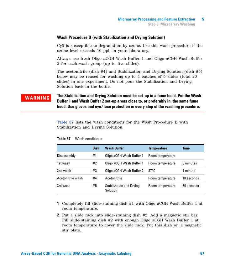
Microarray Processing and Feature Extraction 5Step 3. Microarray Washing
Wash Procedure B (with Stabilization and Drying Solution)
Cy5 is susceptible to degradation by ozone. Use this wash procedure if the ozone level exceeds 10 ppb in your laboratory.
Always use fresh Oligo aCGH Wash Buffer 1 and Oligo aCGH Wash Buffer 2 for each wash group (up to five slides).
The acetonitrile (dish #4) and Stabilization and Drying Solution (dish #5) below may be reused for washing up to 4 batches of 5 slides (total 20 slides) in one experiment. Do not pour the Stabilization and Drying Solution back in the bottle.
Table 37 lists the wash conditions for the Wash Procedure B with Stabilization and Drying Solution.
1 Completely fill slide- staining dish #1 with Oligo aCGH Wash Buffer 1 at room temperature.
2 Put a slide rack into slide- staining dish #2. Add a magnetic stir bar. Fill slide- staining dish #2 with enough Oligo aCGH Wash Buffer 1 at room temperature to cover the slide rack. Put this dish on a magnetic stir plate.
WARNING The Stabilization and Drying Solution must be set-up in a fume hood. Put the Wash Buffer 1 and Wash Buffer 2 set-up areas close to, or preferably in, the same fume hood. Use gloves and eye/face protection in every step of the washing procedure.
Table 37 Wash conditions
Dish Wash Buffer Temperature Time
Disassembly #1 Oligo aCGH Wash Buffer 1 Room temperature
1st wash #2 Oligo aCGH Wash Buffer 1 Room temperature 5 minutes
2nd wash #3 Oligo aCGH Wash Buffer 2 37°C 1 minute
Acetonitrile wash #4 Acetonitrile Room temperature 10 seconds
3rd wash #5 Stabilization and Drying Solution
Room temperature 30 seconds
Array-Based CGH for Genomic DNA Analysis - Enzymatic Labeling 67
5 Microarray Processing and Feature ExtractionStep 3. Microarray Washing
3 Put the prewarmed 1.5 L glass dish filled with water and containing slide- staining dish #3 on a magnetic stir plate with heating element. Fill the slide- staining dish #3 approximately three- fourths full with Oligo aCGH Wash Buffer 2 (warmed to 37°C). Add a magnetic stir bar. Turn on the heating element and maintain temperature of Oligo aCGH Wash Buffer 2 at 37°C; monitor using a thermometer.
4 In the fume hood, fill slide- staining dish #4 approximately three- fourths full with acetonitrile. Add a magnetic stir bar and put this dish on a magnetic stir plate.
5 In the fume hood, fill slide- staining dish #5 approximately three- fourths full with Stabilization and Drying Solution. Add a magnetic stir bar and put this dish on a magnetic stir plate.
6 Remove one hybridization chamber from incubator and record time. Record whether bubbles formed during hybridization, and if all bubbles are rotating freely.
7 Prepare the hybridization chamber disassembly.
a Put the hybridization chamber assembly on a flat surface and loosen the thumbscrew, turning counter- clockwise.
b Slide off the clamp assembly and remove the chamber cover.
c With gloved fingers, remove the array- gasket sandwich from the chamber base by grabbing the slides from their ends. Keep the microarray slide numeric barcode facing up as you quickly transfer the sandwich to slide- staining dish #1.
d Without letting go of the slides, submerge the array- gasket sandwich into slide- staining dish #1 containing Oligo aCGH Wash Buffer 1.
8 With the sandwich completely submerged in Oligo aCGH Wash Buffer 1, pry the sandwich open from the barcode end only. Do this by slipping one of the blunt ends of the forceps between the slides and then gently turn the forceps upwards or downwards to separate the slides. Let the gasket slide drop to the bottom of the staining dish. Remove the microarray slide and put into slide rack in the slide- staining dish #2 containing Oligo aCGH Wash Buffer 1 at room temperature. Minimize exposure of the slide to air. Touch only the barcode portion of the microarray slide or its edges!
9 Repeat step 6 through step 8 for up to four additional slides in the group. A maximum of five disassembly procedures yielding five microarray slides is advised at one time in order to facilitate uniform washing.
68 Array-Based CGH for Genomic DNA Analysis - Enzymatic Labeling
Microarray Processing and Feature Extraction 5Step 3. Microarray Washing
10 When all slides in the group are placed into the slide rack in slide- staining dish #2, stir using setting 4 for 5 minutes. Adjust the setting to get good but not vigorous mixing.
11 Transfer slide rack to slide- staining dish #3 containing Oligo aCGH Wash Buffer 2 at 37°C, and stir using setting 4 for 1 minute.
12 Remove the slide rack from Oligo aCGH Wash Buffer 2 and tilt the rack slightly to minimize wash buffer carry- over. Immediately transfer the slide rack to slide- staining dish #4 containing acetonitrile, and stir using setting 4 for 10 seconds.
13 Transfer slide rack to slide- staining dish #5 filled with Stabilization and Drying Solution, and stir using setting 4 for 30 seconds.
14 Slowly remove the slide rack trying to minimize droplets on the slides. It should take 5 to 10 seconds to remove the slide rack.
15 Discard used Oligo aCGH Wash Buffer 1 and Oligo aCGH Wash Buffer 2.
16 Repeat step 1 through step 15 for the next group of five slides using fresh Oligo aCGH Wash Buffer 1 and Oligo aCGH Wash Buffer 2 prewarmed to 37°C.
17 Immediately put the slides with Agilent barcode facing up in a slide holder with an ozone- barrier slide cover on top of the array as shown in Figure 3 on page 66.
18 Scan slides immediately to minimize impact of environmental oxidants on signal intensities. If necessary, store slides in original slide boxes in a N2 purge box, in the dark.
19 Dispose of acetonitrile and Stabilization and Drying Solution as flammable solvents.
NOTE The acetonitrile and the Stabilization and Drying Solution may be reused for washing of up to four batches of five slides (that is, total 20 microarray slides) in one experiment. Do not pour the Stabilization and Drying Solution back in the bottle. After each use, rinse the slide rack and the slide-staining dish that were in contact with the Stabilization and Drying Solution with acetonitrile followed by a rinse in Milli-Q water.
Array-Based CGH for Genomic DNA Analysis - Enzymatic Labeling 69

5 Microarray Processing and Feature ExtractionStep 4. Microarray Scanning using Agilent C, B or A Scanner or GenePix Scanner
Step 4. Microarray Scanning using Agilent C, B or A Scanner or GenePix Scanner
Agilent C Scanner Settings
An Agilent C- scanner and Agilent Scanner Control software v8.3 or higher is required for 1x1M, 2x400K, 4x180K and 8x60K density microarrays and is optional for 1x244K, 2x10⁵K, 4x44K and 8x15K density microarrays.
1 Put assembled slide holders with or without the ozone- barrier slide cover into scanner carousel.
2 Select Start Slot m End Slot n where the letter m represents the Start slot where the first slide is located and the letter n represents the End slot where the last slide is located.
3 Select Profile Agilent G3_CGH for 1x1M, 2x400K, 4x180K and 8x60K microarrays. Select Profile Agilent HD_CGH for 1x244K, 2x10⁵K, 4x44K and 8x15K microarrays.
4 Verify that
• Slide ID is set to <Auto Detect>.
• Channels is set to R+G
• Scan region is set to Agilent HD (61 × 21.6 mm).
• Resolution (µm) is set to 3 µm for 1x1M, 2x400K, 4x180K and 8x60K microarrays, and 5 µm for 1x244K, 2x10⁵K, 4x44K and 8x15K microarrays.
• Tiff is set to 16 bit
• R PMT is set to 100%.
• G PMT is set to 100%.
• XDR is set to <No XDR>.
• Output Path Browse is set for desired location.
5 Verify that the Scanner status in the main window says Scanner Ready.
6 Click Scan Slot m- n on the Scan Control main window where the letter m represents the Start slot where the first slide is located and the letter n represents the End slot where the last slide is located.
70 Array-Based CGH for Genomic DNA Analysis - Enzymatic Labeling
Microarray Processing and Feature Extraction 5Step 4. Microarray Scanning using Agilent C, B or A Scanner or GenePix Scanner
Agilent A and B Scanner Settings
Agilent Scanner Control software v7.0 is recommended for 5 µm scans of 1x, 2x, 4x and 8x density microarrays.
1 Assemble slides into appropriate slide holders:
• For version B and A slide holders, put slide into slide holder, with or without the ozone- barrier slide cover, with Agilent barcode facing up.
• For version A slide holders, check that slides are seated parallel to the bottom of the slide holder.
2 Put assembled slide holders into scanner carousel.
3 Verify Default Scan Settings (click Settings > Modify Default Settings).
• Scan region is set to Scan Area (61 × 21.6 mm).
• Scan resolution (µm) is set to 5 for 1x244K, 2x10⁵K, 4x44K and 8x15K microarrays.
• Dye channel is set to Red & Green.
• Green PMT is set to 100%.
• Red PMT is set to 100%.
4 Select settings for the automatic file naming.
• Prefix1 is set to Instrument Serial Number.
• Prefix2 is set to Array Barcode.
5 Clear the eXtended Dynamic Range check box.
6 Verify that the Scanner status in the main window says Scanner Ready.
7 Click Scan Slot m- n on the Scan Control main window where the letter m represents the Start slot where the first slide is located and the letter n represents the End slot where the last slide is located.
GenePix Scanner Settings
Agilent 1x244K, 2x10⁵K, 4x44K and 8x15K CGH microarrays require 5 µm scan resolution, which is only supported in the GenePix 4000B scanner.
Agilent 1x1M, 2x400K, 4x180K and 8x60K CGH microarrays require 3 µm scan resolution, which is not supported in the GenePix 4000B scanner.
• Refer to the manufacturer's user guide for appropriate scanner settings.
• Refer to “Agilent Microarray Layout and Orientation” on page 89 for appropriate slide layout and orientation in GenePix scanner.
Array-Based CGH for Genomic DNA Analysis - Enzymatic Labeling 71
5 Microarray Processing and Feature ExtractionStep 5. Data Extraction using Feature Extraction Software
Step 5. Data Extraction using Feature Extraction Software
The Feature Extraction (FE) software v10.5 or higher supports extraction of microarray TIFF images (.tif) of Agilent CGH microarrays scanned on the Agilent C Scanner but does not support extraction of Agilent CGH microarrays on the GenePix 4000B scanner.
The Feature Extraction (FE) software v9.5 supports extraction of microarray TIFF images (.tif) of Agilent CGH microarrays scanned on the Agilent Scanner B and GenePix 4000B Scanner.
Feature Extraction version 10.7 or later can automatically download Grid Templates, protocols and QC metrics (QCMT) from eArray if configured appropriately. See “Automatic Download from eArray” on page 76 for configuration.
Figure 4 shows an example of Agilent 1M CGH microarray image opened in Feature Extraction software v10.5 in both full and zoomed view.
72 Array-Based CGH for Genomic DNA Analysis - Enzymatic Labeling
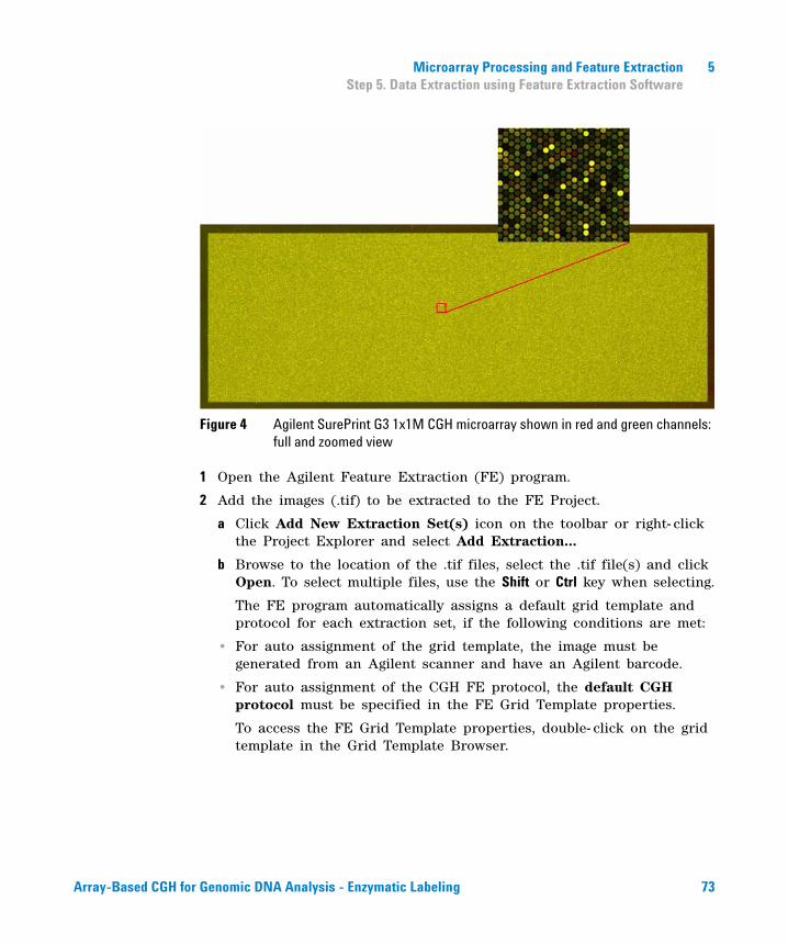
Microarray Processing and Feature Extraction 5Step 5. Data Extraction using Feature Extraction Software
Figure 4 Agilent SurePrint G3 1x1M CGH microarray shown in red and green channels: full and zoomed view
1 Open the Agilent Feature Extraction (FE) program.
2 Add the images (.tif) to be extracted to the FE Project.
a Click Add New Extraction Set(s) icon on the toolbar or right- click the Project Explorer and select Add Extraction...
b Browse to the location of the .tif files, select the .tif file(s) and click Open. To select multiple files, use the Shift or Ctrl key when selecting.
The FE program automatically assigns a default grid template and protocol for each extraction set, if the following conditions are met:
• For auto assignment of the grid template, the image must be generated from an Agilent scanner and have an Agilent barcode.
• For auto assignment of the CGH FE protocol, the default CGH protocol must be specified in the FE Grid Template properties.
To access the FE Grid Template properties, double- click on the grid template in the Grid Template Browser.
Array-Based CGH for Genomic DNA Analysis - Enzymatic Labeling 73
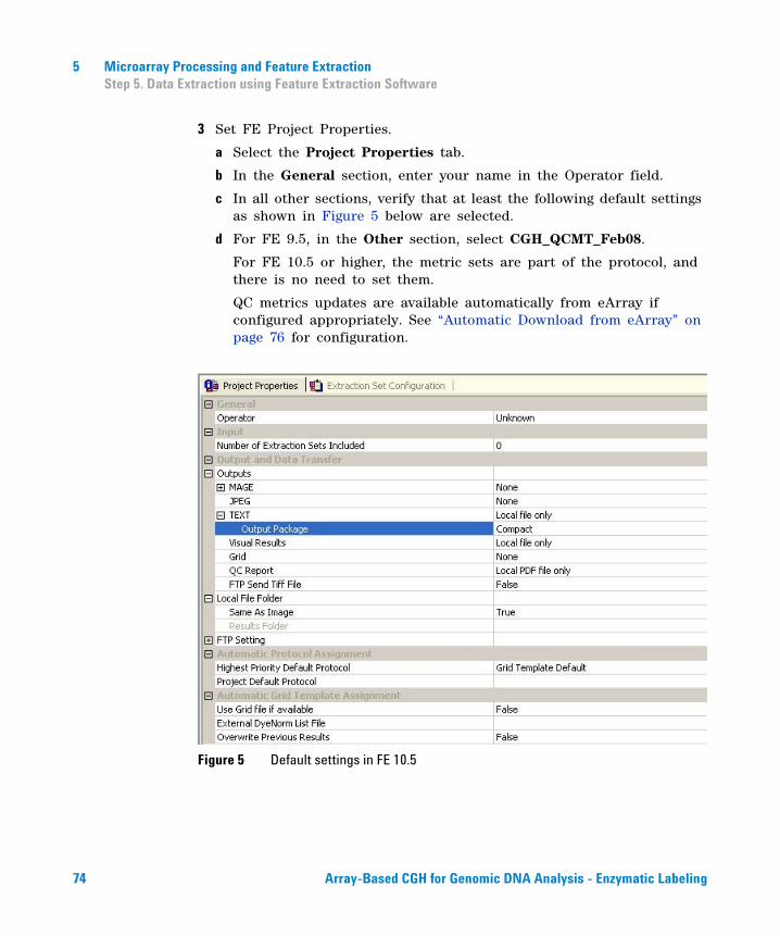
5 Microarray Processing and Feature ExtractionStep 5. Data Extraction using Feature Extraction Software
3 Set FE Project Properties.
a Select the Project Properties tab.
b In the General section, enter your name in the Operator field.
c In all other sections, verify that at least the following default settings as shown in Figure 5 below are selected.
d For FE 9.5, in the Other section, select CGH_QCMT_Feb08.
For FE 10.5 or higher, the metric sets are part of the protocol, and there is no need to set them.
QC metrics updates are available automatically from eArray if configured appropriately. See “Automatic Download from eArray” on page 76 for configuration.
Figure 5 Default settings in FE 10.5
74 Array-Based CGH for Genomic DNA Analysis - Enzymatic Labeling

Microarray Processing and Feature Extraction 5Step 5. Data Extraction using Feature Extraction Software
4 Check the Extraction Set Configuration.
a Select the Extraction Set Configuration tab.
b Verify that the correct grid template is assigned to each extraction set in the Grid Name column. To assign a different grid template to an extraction set, select one from the pull down menu.
If a grid template is not available to select from the pull down menu, you must add it to the Grid Template Browser. To add, right- click inside the Grid Template Browser, select Add. Look for the design file (.xml) and click Open to load grid template into the FE database.
To update to the latest grid templates via Online Update, right- click Grid Template Browser and select Online Update. You can also download the latest grid templates from Agilent Web site at www.agilent.com/chem/downloaddesignfiles. After downloading, you must add the grid templates to the Grid Template Browser.
After a new grid template is added to the Grid Template Browser, remember to specify the default protocol for the new grid template if you want the Feature Extraction program to automatically assign an FE protocol to an extraction set.
c Verify that the CGH- v4_95_Feb07 (in FE 9.5), CGH_105_Dec08 (in FE 10.5) or CGH_107_Sep09 (in FE 10.7) protocol is assigned to each extraction set in the Protocol Name column.
If a protocol is not available to select from the pull down menu, you must import it to the FE Protocol Browser. To import, right- click the FE Protocol Browser, select Import. Browse for the FE protocol (.xml) and click Open to load the protocol into the FE database. Visit Agilent Web site at www.agilent.com/chem/feprotocols to download the latest protocols.
Protocols are also available automatically from eArray if configured appropriately. See “Automatic Download from eArray” on page 76 for configuration.
5 Save the FE Project (.fep) by selecting File > Save As and browse for desired location.
6 Verify that the icons for the image files in the FE Project Window no longer have a red X through them. A red X through the icon indicates that an extraction protocol was not selected. If needed, reselect the extraction protocol for that image file.
7 Select Project > Start Extracting.
Array-Based CGH for Genomic DNA Analysis - Enzymatic Labeling 75
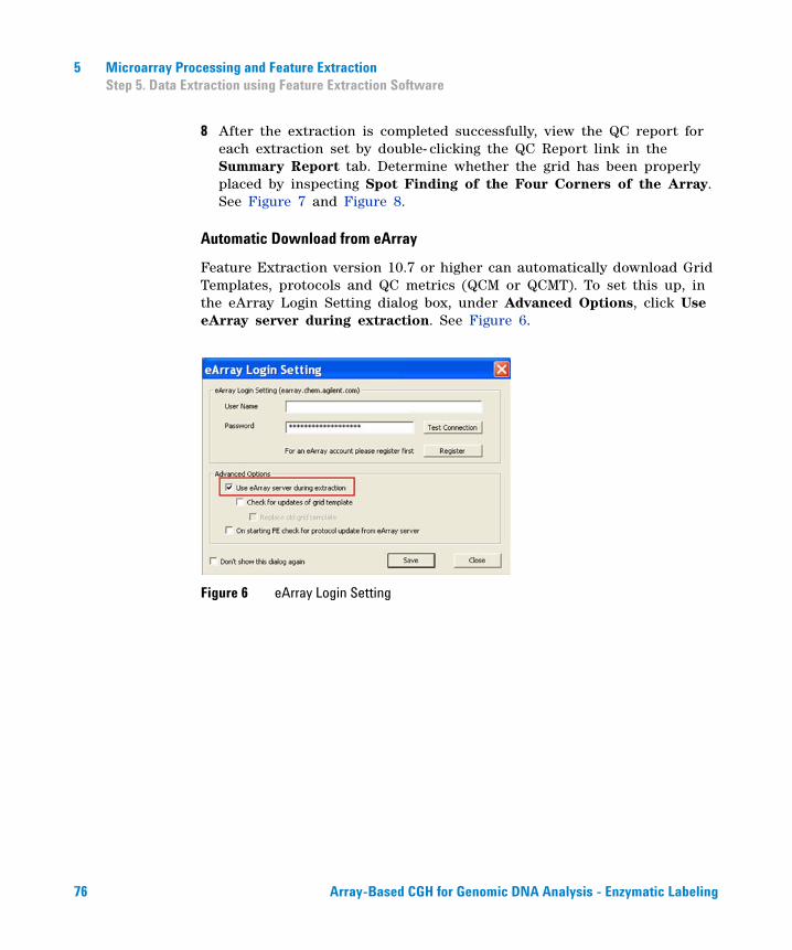
5 Microarray Processing and Feature ExtractionStep 5. Data Extraction using Feature Extraction Software
8 After the extraction is completed successfully, view the QC report for each extraction set by double- clicking the QC Report link in the Summary Report tab. Determine whether the grid has been properly placed by inspecting Spot Finding of the Four Corners of the Array. See Figure 7 and Figure 8.
Automatic Download from eArray
Feature Extraction version 10.7 or higher can automatically download Grid Templates, protocols and QC metrics (QCM or QCMT). To set this up, in the eArray Login Setting dialog box, under Advanced Options, click Use eArray server during extraction. See Figure 6.
Figure 6 eArray Login Setting
76 Array-Based CGH for Genomic DNA Analysis - Enzymatic Labeling
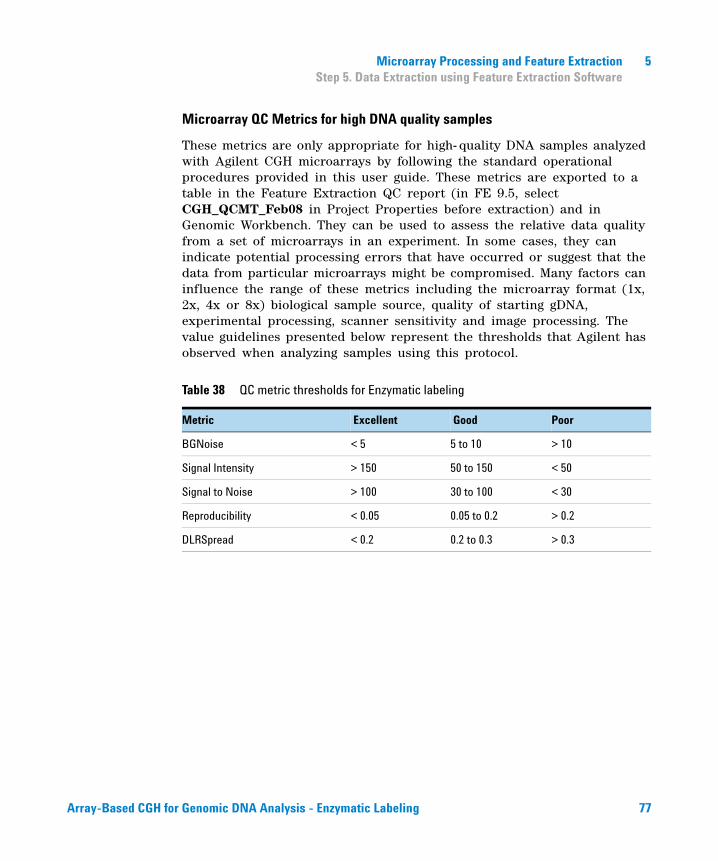
Microarray Processing and Feature Extraction 5Step 5. Data Extraction using Feature Extraction Software
Microarray QC Metrics for high DNA quality samples
These metrics are only appropriate for high- quality DNA samples analyzed with Agilent CGH microarrays by following the standard operational procedures provided in this user guide. These metrics are exported to a table in the Feature Extraction QC report (in FE 9.5, select CGH_QCMT_Feb08 in Project Properties before extraction) and in Genomic Workbench. They can be used to assess the relative data quality from a set of microarrays in an experiment. In some cases, they can indicate potential processing errors that have occurred or suggest that the data from particular microarrays might be compromised. Many factors can influence the range of these metrics including the microarray format (1x, 2x, 4x or 8x) biological sample source, quality of starting gDNA, experimental processing, scanner sensitivity and image processing. The value guidelines presented below represent the thresholds that Agilent has observed when analyzing samples using this protocol.
Table 38 QC metric thresholds for Enzymatic labeling
Metric Excellent Good Poor
BGNoise < 5 5 to 10 > 10
Signal Intensity > 150 50 to 150 < 50
Signal to Noise > 100 30 to 100 < 30
Reproducibility < 0.05 0.05 to 0.2 > 0.2
DLRSpread < 0.2 0.2 to 0.3 > 0.3
Array-Based CGH for Genomic DNA Analysis - Enzymatic Labeling 77
5 Microarray Processing and Feature ExtractionStep 5. Data Extraction using Feature Extraction Software
QC Chart Tool
At times the Feature Extraction program is used in a production environment, where the biological samples are similar, microarray processing protocols are identical and monitoring run- to- run consistency is an important goal. The Feature Extraction program can help monitor this consistency with the optional QC Chart Tool. The QC Chart Tool extracts summary statistics from a set of Feature Extraction output text files and can be used to generate metric sets that can be imported into the Feature Extraction program for analysis of each batch of microarrays processed. Only one metric set can be assigned to a Feature Extraction project. When that project is run, the Feature Extraction program summarizes the metric statistics on each microarray’s QC report and shows if the thresholds (if any) were exceeded. In addition, at the end of the project, a summary chart can be opened to display graphically what the results are for each metric for each microarray. The QC Chart Tool can be downloaded at www.agilent.com/chem/FEQCMetrics.
78 Array-Based CGH for Genomic DNA Analysis - Enzymatic Labeling
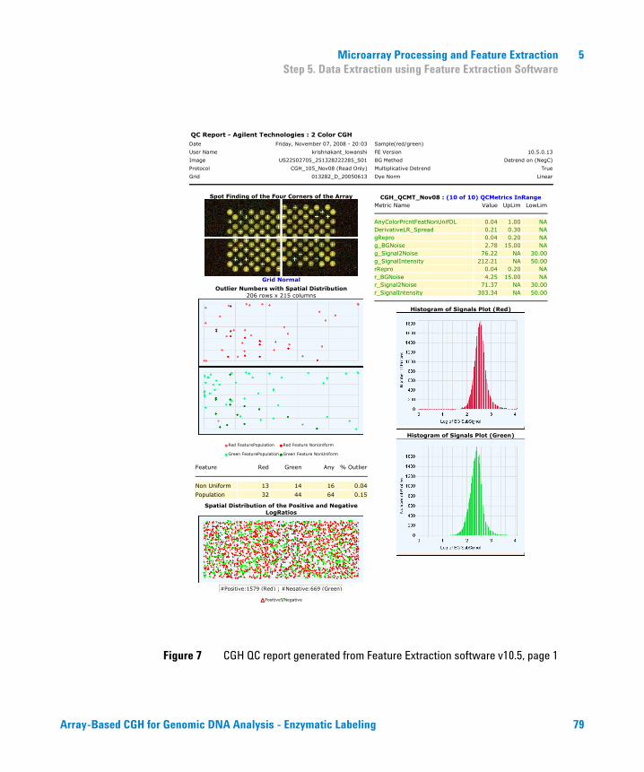
Microarray Processing and Feature Extraction 5Step 5. Data Extraction using Feature Extraction Software
Figure 7 CGH QC report generated from Feature Extraction software v10.5, page 1
QC Report - Agilent Technologies : 2 Color CGHDate Friday, November 07, 2008 - 20:03 Sample(red/green)
User Name krishnakant_lowanshi FE Version 10.5.0.13
Image US22502705_251328222285_S01 BG Method Detrend on (NegC)
Protocol CGH_105_Nov08 (Read Only) Multiplicative Detrend True
Grid 013282_D_20050613 Dye Norm Linear
Spot Finding of the Four Corners of the Array
Grid Normal
Outlier Numbers with Spatial Distribution206 rows x 215 columns
•Red FeaturePopulation •Red Feature NonUniform
•Green FeaturePopulation•Green Feature NonUniform
Feature Red Green Any % Outlier
Non Uniform 13 14 16 0.04
Population 32 44 64 0.15
Spatial Distribution of the Positive and Negative LogRatios
#Positive:1579 (Red) ; #Negative:669 (Green)
�Positive Negative
CGH_QCMT_Nov08 : (10 of 10) QCMetrics InRange Metric Name Value UpLim LowLim
AnyColorPrcntFeatNonUnifOL 0.04 1.00 NADerivativeLR_Spread 0.21 0.30 NAgRepro 0.04 0.20 NAg_BGNoise 2.78 15.00 NAg_Signal2Noise 76.22 NA 30.00g_SignalIntensity 212.21 NA 50.00rRepro 0.04 0.20 NAr_BGNoise 4.25 15.00 NAr_Signal2Noise 71.37 NA 30.00r_SignalIntensity 303.34 NA 50.00
Histogram of Signals Plot (Red)
Histogram of Signals Plot (Green)
Array-Based CGH for Genomic DNA Analysis - Enzymatic Labeling 79
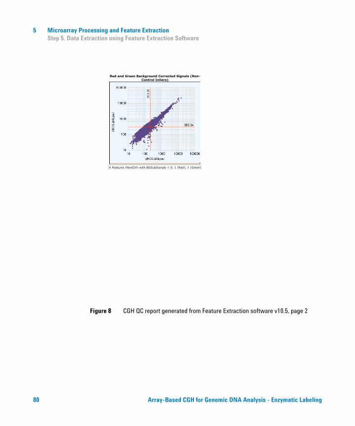
5 Microarray Processing and Feature ExtractionStep 5. Data Extraction using Feature Extraction Software
Figure 8 CGH QC report generated from Feature Extraction software v10.5, page 2
Red and Green Background Corrected Signals (Non-Control Inliers)
# Features (NonCtrl) with BGSubSignals < 0: 1 (Red); 1 (Green)
80 Array-Based CGH for Genomic DNA Analysis - Enzymatic Labeling
Agilent Oligonucleotide Array-Based CGH for Genomic DNA Analysis Protocol
6Troubleshooting
If you have an OD260/230 or OD260/280 value below 1.8 82
If you have poor sample quality due to residual RNA 82
If you get poor sample quality due to degradation 83
If the estimated concentration is too high or low 83
If you have low specific activity or degree of labeling not due to poor sample quality 84
If you have low yield not due to poor sample quality 84
If you have post-labeling signal loss 85
If you have high BGNoise values 86
If you have poor reproducibility 86
This chapter contains the causes for above- threshold DLRSD (Derivative Log Ratio Standard Deviation). A poor DLRSD score reflects high probe- to- probe log ratio noise.
81Agilent Technologies
6 TroubleshootingIf you have an OD260/230 or OD260/280 value below 1.8
If you have an OD260/230 or OD260/280 value below 1.8
A low OD260/230 value can indicate contaminants, such as residual phenol or salt. A low OD260/280 value indicates residual protein. Either condition can result in low specific activity (pmol dye/µg DNA) or Degree of Labeling. See “To determine yield , degree of labeling or specific activity” on page 51.
✔ Repurify the DNA using the Qiagen DNeasy protocol. See “DNA Isolation” on page 21. This procedure includes a proteinase K treatment.
✔ If you must do a phenol/chloroform DNA extraction, do not get too close to the interface.
If you have poor sample quality due to residual RNA
The input amount of DNA for the experimental labeling reaction must be the same as for the reference sample labeling reaction. RNA absorbs at the same wavelength as DNA, which makes an accurate measurement of the DNA concentration in an RNA- contaminated sample impossible.
✔ Repurify the DNA using the Qiagen DNeasy protocol. See “DNA Isolation” on page 21. This procedure includes a RNase A treatment.
82 Array-Based CGH for Genomic DNA Analysis - Enzymatic Labeling
Troubleshooting 6If you get poor sample quality due to degradation
If you get poor sample quality due to degradation
For non- FFPE samples: On a 1 to 1.5% agarose gel, intact genomic DNA should appear as a compact, high- molecular weight band with no lower molecular weight smears. Degraded DNA results in biased labeling.
✔ Check DNA on a 1 to 1.5% agarose gel, if DNA that was isolated from cells, blood or frozen tissue, is degraded then repurify the DNA using the Qiagen DNeasy protocol. See “DNA Isolation” on page 21.
✔ For processing FFPE samples, refer to the Agilent Oligonucleotide Array- Based CGH for Genomic DNA Analysis (ULS Labeling for Blood, Cells, Tissues or FFPE) Protocol v3.0 (p/n G4410- 90020).
If the estimated concentration is too high or low
The input amount of DNA for the experimental labeling reaction must be the same as for the reference sample labeling reactions. Precipitated DNA or DNA that is at a very high concentration cannot be quantitated accurately.
Contaminants such as organic solvents and RNA also absorb at 260 nm, which results in an inaccurate DNA quantitation.
✔ Make sure that the gDNA is completely in solution by pipetting up and down. If needed, incubate at 37°C for 30 minutes. If the gDNA concentration is > 350 ng/µL, dilute 1:2 in water and re- quantitate to make sure quantitation is accurate.
✔ If needed, repurify the DNA using the Qiagen DNeasy protocol. See “DNA Isolation” on page 21.
Array-Based CGH for Genomic DNA Analysis - Enzymatic Labeling 83
6 TroubleshootingIf you have low specific activity or degree of labeling not due to poor sample quality
If you have low specific activity or degree of labeling not due to poor sample quality
Low specific activity or degree of labeling can result from sub- optimal labeling conditions such as Cyanine dUTP with too many freeze thaws, enzyme degradation due to being left warm for too long, wrong temperatures or times, volume mistakes, or too much exposure to light or air.
✔ Store Cyanine dUTP at 4°C. Keep enzymes on ice and return to - 20°C as quickly as possible.
✔ Double check incubation times and temperatures (use a calibrated thermometer), and use a thermocycler with heated lid.
✔ Evaporation can be a problem when you process samples in 96- well plates. Use a plate heat sealer (Eppendorf p/n 951023078) to avoid evaporation.
✔ Make sure that the pipettors are not out of calibration.
✔ Make sure that the gDNA, reagents, and master mixes are well mixed. Tap the tube with your finger or use a pipette to move the entire volume up and down. Then spin in a microcentrifuge for 5 to 10 seconds to drive the contents off the walls and lid. Do not mix the stock solutions and reactions that contain gDNA or enzymes on a vortex mixer.
If you have low yield not due to poor sample quality
Possible sample loss during clean- up after labeling.
✔ See “Step 2. Clean- up of Labeled Genomic DNA” on page 46 to remove unreacted dye. Many other columns result in the loss of shorter fragments.
84 Array-Based CGH for Genomic DNA Analysis - Enzymatic Labeling
Troubleshooting 6If you have post-labeling signal loss
If you have post-labeling signal loss
Signal loss can be due to wash or hyb conditions that are too stringent, or degradation of the Cyanine 5 signal.
Cyanine 5 signal degradation can be caused by ozone or NOx compounds coming from pollution and/or compressors and centrifuges. Cyanine 5 signal degradation can result in less red signal around the edges of the features, a visible gradient of significant Cy5/Cy3 positive ratios and more significant Cy5/Cy3 negative ratios – especially on the left side of the slide and on slides scanned later in a batch.
✔ Check oven and Wash 2 temperature.
✔ Check that Wash 2 was not accidentally used instead of Wash 1.
✔ Wash and scan slides in an ozone controlled environment (<5 ppb), such as an ozone tent.
✔ Use small batches that can be washed and scanned in about 40 minutes to minimize exposure to air.
✔ Use the Agilent Ozone- Barrier Slide Cover (p/n G2505- 60550).
✔ Use the Stabilization and Drying Solution as described in “Wash Procedure B (with Stabilization and Drying Solution)” on page 67.
Array-Based CGH for Genomic DNA Analysis - Enzymatic Labeling 85
6 TroubleshootingIf you have high BGNoise values
If you have high BGNoise values
High BGNoise can cause lower signal- to- noise values (see Table 38 on page 77 for thresholds) and higher DLRSD values. BGNoise is defined as the standard deviation of the signals on the negative controls. If the BGNoise is high, examine the array image for visible non- uniformities. High BGNoise is often introduced during the washes.
✔ Make sure that wash dishes, racks and stir bars are clean. Do not use tap water or detergents to clean wash equipment. If needed, rinse wash equipment with acetonitrile followed by rinses with MilliQ water.
✔ If needed, wash the slides with acetonitrile:
1 In the fume hood, fill a slide- staining dish approximately three- fourths full with acetonitrile.
2 Add a magnetic stir bar and put this dish on a magnetic stir plate.
3 Put the slides in a slide rack and transfer the slide rack to the slide- staining dish containing acetonitrile, and stir using setting 4 for 1 minute.
4 Slowly remove the slide rack and scan the slides immediately.
If you have poor reproducibility
Poor reproducibility (see Table 38 on page 77 for thresholds), defined as high CVs of signals of replicated probes may indicate that the hybridization volume was too low or that the oven stopped rotating during the hybridization. Only very high scores on this metric will affect the DLRSD.
✔ When setting up the gasket- slide hybridization sandwich dispense the hybridization sample mixture slowly in a “drag and dispense” manner to prevent spills.
✔ Check that the oven is rotating.
86 Array-Based CGH for Genomic DNA Analysis - Enzymatic Labeling
Agilent Oligonucleotide Array-Based CGH for Genomic DNA Analysis Protocol
7Reference
Supporting User Guides 88
Agilent Microarray Layout and Orientation 89
Array/Sample tracking on a 8x array slide 92
Agilent Information Assets Access Agreement 93
This chapter contains reference information that pertains to this protocol.
87Agilent Technologies
7 ReferenceSupporting User Guides
Supporting User Guides
If you are a first- time user of Agilent’s oligo microarray system, please refer to the following user guides for detailed descriptions and operation recommendations for each of the hardware and software components used in the Oligo aCGH application workflow.
The user guides can be downloaded from the Agilent Web site at www.agilent.com/chem/dnamanuals- protocols.
• Agilent Microarray Hybridization Chamber User Guide (p/n G2534- 90001)
• Agilent Ozone- Barrier Slide Cover User Guide (p/n G2505- 90050)
• Agilent Technical Note “Improving microarray results by preventing ozone- mediated fluorescent signal degradation” (p/n 5989- 0875EN)
• Agilent G2545A Hybridization Oven User Manual (p/n G2545- 80001)
• Agilent G2565AA and G2565BA Microarray Scanner System User Manual
• Agilent G2565CA Microarray Scanner System User Manual
• Agilent Microarray Format Technical Drawings with Tolerances (p/n G4502- 90001)
• Agilent Feature Extraction Software Quick Start Guide
• Agilent Feature Extraction Software User Guide
• Agilent Feature Extraction Software Reference Guide
88 Array-Based CGH for Genomic DNA Analysis - Enzymatic Labeling
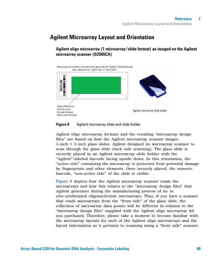
Reference 7Agilent Microarray Layout and Orientation
Agilent Microarray Layout and Orientation
Agilent oligo microarray (1 microarray/slide format) as imaged on the Agilent microarray scanner (G2565CA)
Figure 9 Agilent microarray slide and slide holder
Agilent oligo microarray formats and the resulting “microarray design files” are based on how the Agilent microarray scanner images 1- inch × 3- inch glass slides. Agilent designed its microarray scanner to scan through the glass slide (back side scanning). The glass slide is securely placed in an Agilent microarray slide holder with the “Agilent”- labeled barcode facing upside down. In this orientation, the “active side” containing the microarray is protected from potential damage by fingerprints and other elements. Once securely placed, the numeric barcode, “non- active side” of the slide is visible.
Figure 9 depicts how the Agilent microarray scanner reads the microarrays and how this relates to the “microarray design files” that Agilent generates during the manufacturing process of its in situ- synthesized oligonucleotide microarrays. Thus, if you have a scanner that reads microarrays from the “front side” of the glass slide, the collection of microarray data points will be different in relation to the “microarray design files” supplied with the Agilent oligo microarray kit you purchased. Therefore, please take a moment to become familiar with the microarray layouts for each of the Agilent oligo microarrays and the layout information as it pertains to scanning using a “front side” scanner.
Microarrays are printed on the side of the glass with the “Agilent”-labeled barcode (also referenced as "active side" or "front side").
Agilent MicroarrayScanner scansthrough the glass.(Back side scanning.)
00116789
Agilent microarray slide holder
Array-Based CGH for Genomic DNA Analysis - Enzymatic Labeling 89
7 ReferenceAgilent Microarray Layout and Orientation
Non-Agilent Front Side Microarray Scanners
When scanning Agilent oligo microarray slides, the user must determine:
• If the scanner images the microarrays by reading them on the “front side” of the glass slide (“Agilent”- labeled barcode side of the slide) and
• If the microarray image produced by the non- Agilent scanner is oriented in a “portrait” or “landscape” mode, and “Agilent”- labeled barcode is on the left- side, right- side, up or down, as viewed as an image in the imaging software (see Figure 10).
This changes the feature numbering and location as it relates to the “microarray design files” found on the disk in each Agilent oligo microarray kit.
90 Array-Based CGH for Genomic DNA Analysis - Enzymatic Labeling
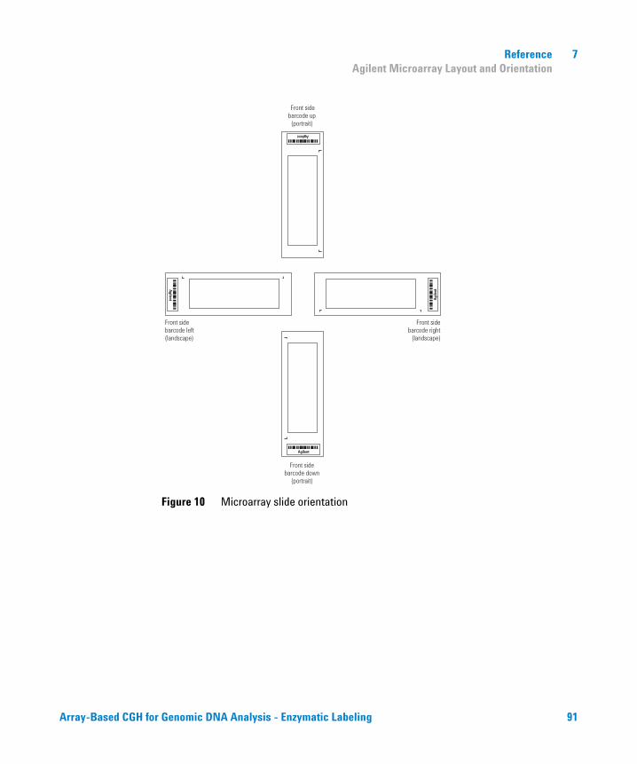
Reference 7Agilent Microarray Layout and Orientation
Figure 10 Microarray slide orientation
Front sidebarcode left(landscape)
Front sidebarcode right
(landscape)
Front sidebarcode up(portrait)
Front sidebarcode down
(portrait)
Agilent
Agi
lent
Agilent
Agilent
Array-Based CGH for Genomic DNA Analysis - Enzymatic Labeling 91
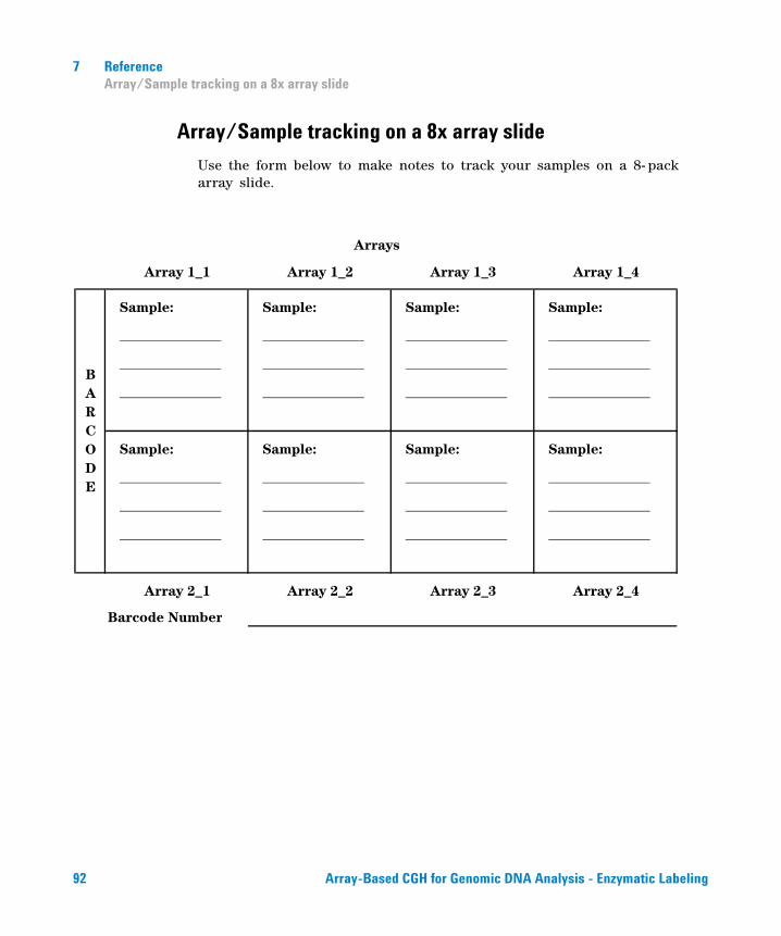
7 ReferenceArray/Sample tracking on a 8x array slide
Array/Sample tracking on a 8x array slide
Use the form below to make notes to track your samples on a 8- pack array slide.
Array 1_1 Array 1_2 Array 1_3 Array 1_4
Sample: Sample: Sample: Sample:
Sample: Sample: Sample: Sample:
Array 2_1 Array 2_2 Array 2_3 Array 2_4
Barcode Number
Arrays
BARCODE
92 Array-Based CGH for Genomic DNA Analysis - Enzymatic Labeling
Reference 7Agilent Information Assets Access Agreement
Agilent Information Assets Access Agreement
I. PURPOSE
1 Agilent Technologies (“Agilent”) and User have entered into a business relationship that would be fostered by granting User access to certain of Agilent's Information Assets (“Assets”). Accordingly, User and Agilent have entered into this Information Assets Access Agreement (IAAA) in order to protect the confidentiality, integrity, availability, and physical security of Agilent 's various information assets and to prevent unauthorized individuals from gaining access to Agilent's various networks, computers and information assets. Access is granted only for the purposes of fulfilling the specific business relationship between User and Agilent which gives rise to this agreement. The use of Agilent information for any other purpose is expressly prohibited.
2 User and Agilent acknowledge that they have entered into other agreements or contractual arrangements defining their business relationship. These agreements are independent of this IAAA and in the event of any conflict between them, the terms of this IAAA shall prevail only as applies to this IAAA.
II. PHYSICAL SECURITY OF AGILENT INFORMATION ASSETS
1 User agrees to exercise no less than a reasonable degree of care to ensure that access to Agilent's information assets is limited solely to its authorized personnel who have been granted access by Agilent to specific information assets. Under no circumstance may User allow an individual to access an Agilent information asset without a properly authorized grant of access from Agilent for that individual and for that information asset. Only those Users provided access to the information assets via an Agilent authorized authentication procedure may access the information assets.
USE OF AGILENT INFORMATION ASSETS FOR OTHER THAN INTERNAL USE AND/OR FOR COMMERCIAL PURPOSES WITHOUT AGILENT'S PRIOR WRITTEN CONSENT IS EXPRESSLY PROHIBITED.
Use of any other computer systems or Agilent information assets is expressly prohibited in the absence of written authorization from Agilent. This prohibition applies even when an information resource which User is authorized to access serves as a gateway to other information resources outside of User's scope of authorization. These prohibitions apply even if Agilent fails to implement physical or logical security measures which prevent the access.
Array-Based CGH for Genomic DNA Analysis - Enzymatic Labeling 93
7 ReferenceAgilent Information Assets Access Agreement
USE OF AGILENT INFORMATION ASSETS BY OTHER INDIVIDUALS NOT AUTHORIZED IN WRITING OR VIA ELECTRONIC MAIL BY AGILENT IS EXPRESSLY PROHIBITED.
2 Without limiting the foregoing, User specifically agrees to:
a Control physical access to passwords, tokens, log- on scripts and other such hardware/data as may be used to access Assets. No passwords, security tokens or other authentication information may be shared between individuals without written approval from Agilent. User agrees to access Agilent information resources only from the specific locations that have been approved for access by Agilent management. For access outside of Agilent premises, Agilent will specify in writing or via electronic mail the specific network connections that will be used to access Agilent information assets.
b Periodically review personnel granted access under this IAAA to insure that access remains necessary to the underlying business purpose.
c Coordinate within User's organization the proper implementation of this agreement and ensure that all the rules and obligations contained in this agreement are strictly followed and complied with at all times in User's organization.
3 In its sole discretion and without incurring liability, Agilent reserves the right to deny, limit, or terminate access of any person or firm or access to any system(s) without notice or cause.
III. PROTECTION OF ELECTRONIC CONFIDENTIAL INFORMATION
1 User and its employees or agents (collectively referred to as User, unless the context requires otherwise) shall not disclose any Electronic Confidential Information (ECI) received from Agilent to any other person without the express prior written consent of Agilent. User shall use such information only for purposes necessary for its relationship with Agilent or consistent with this Agreement, and shall use reasonable care to avoid unauthorized disclosure or use of such ECI.
2 Electronic Confidential Information obtained from Assets shall be defined as any information marked as confidential or proprietary, and any information, while not marked as confidential or proprietary, which is of such a nature that a reasonable person would believe it to be confidential or proprietary. Agilent shall take reasonable precautions to prevent the User from receiving unmarked confidential or proprietary information. However, User acknowledges that due to the nature of
94 Array-Based CGH for Genomic DNA Analysis - Enzymatic Labeling
Reference 7Agilent Information Assets Access Agreement
electronic systems, such information may be delivered to User inadvertently or with an inadvertent omission of the markings. If User receives information that a reasonable person would believe to be confidential or proprietary, User shall have an affirmative duty to inquire of Agilent as to the confidentiality of the information before treating it as non- confidential information.
3 This Agreement imposes no obligation upon User with respect to any confidential or proprietary information disclosed to User pursuant to the terms of a separately executed Confidential Disclosure Agreement or other contractual arrangement specifically addressing such information. The obligations of the parties with respect to such information shall be governed solely by the terms of such Confidential Disclosure Agreement or contractual arrangement.
IV. LIMITED RIGHTS TO PUBLISH IN PEER- REVIEWED JOURNALS ONLY
1 User may publish the following only in peer- reviewed scientific journals [“Publication.”]
2 User may publish only fifty (50) probe- sequences in any single Publication.
3 User or its permitted publisher, including without limitation the investigator, scientists, and/or researchers, hereby agrees to add a statement in a prominent location within such publication; provided User publishes the specific design file number that Agilent shall provide, stating the following: ANY PUBLICATION OR PUBLIC DISCLOSURE OF OLIGONUCLEOTIDE SEQUENCES FOR USE WITH MICROARRAYS MAY ONLY BE REPRODUCED OR MANUFACTURED FOR YOU BY AGILENT TECHNOLOGIES, INC. BY REFERENCING THE CATALOG MICROARRAY PRODUCT NUMBER OR THE CUSTOM MICROARRAY DESIGN IDENTIFICATION NUMBER SUPPLIED BY YOU BY AGILENT.
4 User hereby grants Agilent all rights to reproduce products based upon User's Confidential Information for Third Parties requesting such Products.
Array-Based CGH for Genomic DNA Analysis - Enzymatic Labeling 95
7 ReferenceAgilent Information Assets Access Agreement
V. ADMINISTRATION
1 Audit: Upon reasonable notice, User shall allow Agilent access to such information as may reasonably be necessary to ascertain User's compliance with the obligations contained herein.
2 Loss/Damage: User shall indemnify and hold Agilent harmless from all loss, damage, cost and expense arising from User's breach of this Agreement.
3 User warrants and represents that each employee or agent of User who is granted access under this agreement has been informed of the obligations contained herein and has agreed in writing to be bound by them. Without limiting the foregoing, each individual must be informed of the specific information assets he is authorized to access and the duty to refrain from accessing other information assets outside his scope of authorization even if logical or physical security measures are not in place to prevent the access.
VI. MISCELLANEOUS
1 This Agreement is made under and shall be construed according to the laws of the State or Country in which the signing Agilent entity is located.
2 This Agreement constitutes the entire and exclusive agreement of the parties regarding the subject matter of this Agreement. This Agreement may not be varied.
96 Array-Based CGH for Genomic DNA Analysis - Enzymatic Labeling

Agilent Technologies, Inc. 2009
Printed in USA Version 6.2, October 2009
*G4410-90010*G4410-90010
www.agilent.com
Agilent Technologies
In This Book
This guide contains information to run the Oligonucleotide Array- Based CGH for Genomic DNA Analysis (enzymatic labeling) protocol.