Age-related maculopathy and the impact of blue light hazard · from blue light (peak 430 nm) ......
12
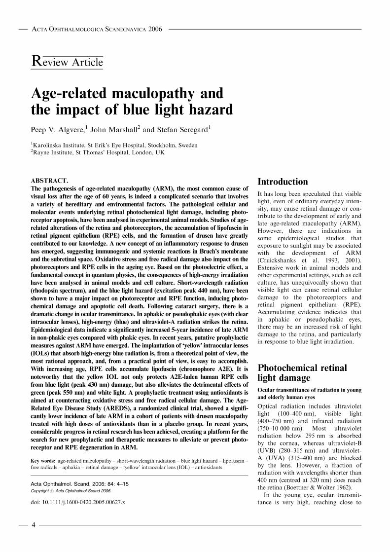
R eview Article Age-related maculopathy and the impact of blue light hazard Peep V. Algvere, 1 John Marshall 2 and Stefan Seregard 1 1 Karolinska Institute, St Erik’s Eye Hospital, Stockholm, Sweden 2 Rayne Institute, St Thomas’ Hospital, London, UK ABSTRACT. The pathogenesis of age-related maculopathy (ARM), the most common cause of visual loss after the age of 60 years, is indeed a complicated scenario that involves a variety of hereditary and environmental factors. The pathological cellular and molecular events underlying retinal photochemical light damage, including photo- receptor apoptosis, have been analysed in experimental animal models. Studies of age- related alterations of the retina and photoreceptors, the accumulation of lipofuscin in retinal pigment epithelium (RPE) cells, and the formation of drusen have greatly contributed to our knowledge. A new concept of an inflammatory response to drusen has emerged, suggesting immunogenic and systemic reactions in Bruch’s membrane and the subretinal space. Oxidative stress and free radical damage also impact on the photoreceptors and RPE cells in the ageing eye. Based on the photoelectric effect, a fundamental concept in quantum physics, the consequences of high-energy irradiation have been analysed in animal models and cell culture. Short-wavelength radiation (rhodopsin spectrum), and the blue light hazard (excitation peak 440 nm), have been shown to have a major impact on photoreceptor and RPE function, inducing photo- chemical damage and apoptotic cell death. Following cataract surgery, there is a dramatic change in ocular transmittance. In aphakic or pseudophakic eyes (with clear intraocular lenses), high-energy (blue) and ultraviolet-A radiation strikes the retina. Epidemiological data indicate a significantly increased 5-year incidence of late ARM in non-phakic eyes compared with phakic eyes. In recent years, putative prophylactic measures against ARM have emerged. The implantation of ‘yellow’ intraocular lenses (IOLs) that absorb high-energy blue radiation is, from a theoretical point of view, the most rational approach, and, from a practical point of view, is easy to accomplish. With increasing age, RPE cells accumulate lipofuscin (chromophore A2E). It is noteworthy that the yellow IOL not only protects A2E-laden human RPE cells from blue light (peak 430 nm) damage, but also alleviates the detrimental effects of green (peak 550 nm) and white light. A prophylactic treatment using antioxidants is aimed at counteracting oxidative stress and free radical cellular damage. The Age- Related Eye Disease Study (AREDS), a randomized clinical trial, showed a signifi- cantly lower incidence of late ARM in a cohort of patients with drusen maculopathy treated with high doses of antioxidants than in a placebo group. In recent years, considerable progress in retinal research has been achieved, creating a platform for the search for new prophylactic and therapeutic measures to alleviate or prevent photo- receptor and RPE degeneration in ARM. Key words: age-related maculopathy – short-wavelength radiation – blue light hazard – lipofuscin – free radicals – aphakia – retinal damage – ‘yellow’ intraocular lens (IOL) – antioxidants Acta Ophthalmol. Scand. 2006: 84: 4–15 Copyright # Acta Ophthalmol Scand 2006. doi: 10.1111/j.1600-0420.2005.00627.x Introduction It has long been speculated that visible light, even of ordinary everyday inten- sity, may cause retinal damage or con- tribute to the development of early and late age-related maculopathy (ARM). However, there are indications in some epidemiological studies that exposure to sunlight may be associated with the development of ARM (Cruickshanks et al. 1993, 2001). Extensive work in animal models and other experimental settings, such as cell culture, has unequivocally shown that visible light can cause retinal cellular damage to the photoreceptors and retinal pigment epithelium (RPE). Accumulating evidence indicates that in aphakic or pseudophakic eyes, there may be an increased risk of light damage to the retina, and particularly in response to blue light irradiation. Photochemical retinal light damage Ocular transmittance of radiation in young and elderly human eyes Optical radiation includes ultraviolet light (100–400 nm), visible light (400–750 nm) and infrared radiation (750–10 000 nm). Most ultraviolet radiation below 295 nm is absorbed by the cornea, whereas ultraviolet-B (UVB) (280–315 nm) and ultraviolet- A (UVA) (315–400 nm) are blocked by the lens. However, a fraction of radiation with wavelengths shorter than 400 nm (centred at 320 nm) does reach the retina (Boettner & Wolter 1962). In the young eye, ocular transmit- tance is very high, reaching close to ACTA OPHTHALMOLOGICA SCANDINAVICA 2006 4
-
Upload
hoangkhuong -
Category
Documents
-
view
214 -
download
0
Transcript of Age-related maculopathy and the impact of blue light hazard · from blue light (peak 430 nm) ......
Review Article
Age-related maculopathy andthe impact of blue light hazard
Peep V. Algvere,1 John Marshall2 and Stefan Seregard1
1Karolinska Institute, St Erik’s Eye Hospital, Stockholm, Sweden2Rayne Institute, St Thomas’ Hospital, London, UK
ABSTRACT.
The pathogenesis of age-related maculopathy (ARM), the most common cause of
visual loss after the age of 60 years, is indeed a complicated scenario that involves
a variety of hereditary and environmental factors. The pathological cellular and
molecular events underlying retinal photochemical light damage, including photo-
receptor apoptosis, have been analysed in experimental animal models. Studies of age-
related alterations of the retina and photoreceptors, the accumulation of lipofuscin in
retinal pigment epithelium (RPE) cells, and the formation of drusen have greatly
contributed to our knowledge. A new concept of an inflammatory response to drusen
has emerged, suggesting immunogenic and systemic reactions in Bruch’s membrane
and the subretinal space. Oxidative stress and free radical damage also impact on the
photoreceptors and RPE cells in the ageing eye. Based on the photoelectric effect, a
fundamental concept in quantum physics, the consequences of high-energy irradiation
have been analysed in animal models and cell culture. Short-wavelength radiation
(rhodopsin spectrum), and the blue light hazard (excitation peak 440 nm), have been
shown to have a major impact on photoreceptor and RPE function, inducing photo-
chemical damage and apoptotic cell death. Following cataract surgery, there is a
dramatic change in ocular transmittance. In aphakic or pseudophakic eyes (with clear
intraocular lenses), high-energy (blue) and ultraviolet-A radiation strikes the retina.
Epidemiological data indicate a significantly increased 5-year incidence of late ARM
in non-phakic eyes compared with phakic eyes. In recent years, putative prophylactic
measures against ARM have emerged. The implantation of ‘yellow’ intraocular lenses
(IOLs) that absorb high-energy blue radiation is, from a theoretical point of view, the
most rational approach, and, from a practical point of view, is easy to accomplish.
With increasing age, RPE cells accumulate lipofuscin (chromophore A2E). It is
noteworthy that the yellow IOL not only protects A2E-laden human RPE cells
from blue light (peak 430 nm) damage, but also alleviates the detrimental effects of
green (peak 550 nm) and white light. A prophylactic treatment using antioxidants is
aimed at counteracting oxidative stress and free radical cellular damage. The Age-
Related Eye Disease Study (AREDS), a randomized clinical trial, showed a signifi-
cantly lower incidence of late ARM in a cohort of patients with drusen maculopathy
treated with high doses of antioxidants than in a placebo group. In recent years,
considerable progress in retinal research has been achieved, creating a platform for the
search for new prophylactic and therapeutic measures to alleviate or prevent photo-
receptor and RPE degeneration in ARM.
Key words: age-related maculopathy – short-wavelength radiation – blue light hazard – lipofuscin –
free radicals – aphakia – retinal damage – ‘yellow’ intraocular lens (IOL) – antioxidants
Acta Ophthalmol. Scand. 2006: 84: 4–15Copyright # Acta Ophthalmol Scand 2006.
doi: 10.1111/j.1600-0420.2005.00627.x
Introduction
It has long been speculated that visiblelight, even of ordinary everyday inten-sity, may cause retinal damage or con-tribute to the development of early andlate age-related maculopathy (ARM).However, there are indications insome epidemiological studies thatexposure to sunlight may be associatedwith the development of ARM(Cruickshanks et al. 1993, 2001).Extensive work in animal models andother experimental settings, such as cellculture, has unequivocally shown thatvisible light can cause retinal cellulardamage to the photoreceptors andretinal pigment epithelium (RPE).Accumulating evidence indicates thatin aphakic or pseudophakic eyes,there may be an increased risk of lightdamage to the retina, and particularlyin response to blue light irradiation.
Photochemical retinallight damage
Ocular transmittance of radiation in young
and elderly human eyes
Optical radiation includes ultravioletlight (100–400 nm), visible light(400–750 nm) and infrared radiation(750–10 000 nm). Most ultravioletradiation below 295 nm is absorbedby the cornea, whereas ultraviolet-B(UVB) (280–315 nm) and ultraviolet-A (UVA) (315–400 nm) are blockedby the lens. However, a fraction ofradiation with wavelengths shorter than400 nm (centred at 320 nm) does reachthe retina (Boettner & Wolter 1962).
In the young eye, ocular transmit-tance is very high, reaching close to
ACTA OPHTHALMOLOGICA SCANDINAVICA 2006
4

90% at 450 nm. The transmittance ofelderly lenses is much lower and hasconsiderable interindividual variation,but generally does not reach 70–80%until wavelengths of 540 nm. This is incontrast with aphakic eyes, in whichsubstantial energy from radiation ofwavelengths longer than 310 nm, thusincluding UVA, will strike the retina.
The photoelectric effect
At the birth of quantum physics a cen-tury ago, it was suggested that theenergy of a photon is proportional toits radiation frequency: E ¼ nf (wheren ¼ Planck’s constant, f ¼ radiationfrequency) (Planck 1900; quoted inBenson 1991). The photoelectric effect(Einstein 1905; quoted in Benson 1991)demonstrates that the high energy ofshort-wavelength radiation, such asX-rays, UV and blue light, has thecapacity to induce an electric currentfrom certain metal bodies. In contrast,longer wavelengths with low energyquanta (e.g. > 600 nm) are insufficientto produce a photoelectric effect in theexperimental setting (Fig. 1). In recentyears, the biological effects of differentfrequencies of radiation to the eye havebeen analysed and defined in somedetail.
Photochemical class I damage
It was demonstrated long ago that rela-tively low light intensities coulddamage the retina in the experimentalanimal when long exposure duration
was used. In the rat, this phototoxicityhad the spectrum of rhodopsin andscotopic vision and displayed a degen-eration of the rod photoreceptors(Noell et al. 1966). The energy distribu-tion of sunlight reaching the earth ishigh in the rhodopsin spectrum(Fig. 2).
Photochemical class II damage
When light of relatively high intensityis used with short exposure time (min-utes, hours), retinal damage willincrease in response to shorter wave-lengths, that is the high energy portionof the visible spectrum (400–500 nm).This has been referred to as the ‘bluelight hazard’, and has an excitationpeak around 440 nm (Fig. 2). In pri-mates, the resulting retinal damagehas been described as located in theRPE layer (Ham et al. 1978). Thedegree of retinal injury is dependenton oxygen concentration and antioxi-dants seem to reduce the light damage(Organisciak & Winkler 1994).
Thus, both photoreceptors and RPEcells may be damaged by light, but thetype of injury is modified by severalfactors, such as intensity, duration,intermittence of exposure to light, andspectral distribution. Laube et al.(2004) reported that a small fractionof UV radiation (around 300 nm) istransmitted by the crystalline lens(Fig. 3). Generally, the first alterationsoccur in the photoreceptor outer seg-ments, followed by changes in the RPEcells (Kuwabara & Funahashi 1976).Whether rods or cones are affectedfirst seems to vary between different
species. In the rat, which has a predo-minantly rod-based retina, the conephotoreceptors survive despite a wide-spread degeneration of rods (Cicerone1976). However, in pigeons, whichhave a predominantly cone-basedretina, the cone photoreceptors degen-erate early, but this phenomenon mayreflect the slower renewal of cone outersegments versus that of rods (Marshall1985). Blue cones are particularly vul-nerable when exposed to blue light(Sperling & Johnson 1980).
Age-related alterations ofthe retina
Population-based studies on the preva-lence of ARM show a strong positivecorrelation with both age and smoking(Thornton et al. 2005). Retinal pigmentepithelium cells are post-mitotic cellsand should persist for the entire life ofthe individual. Under normal condi-tions, RPE cells do not divide, butthey are able to proliferate under cer-tain pathological conditions, such asretinal detachment. It has been calcu-lated that each RPE cell will phagocy-tose about 3 billion outer segment discsduring a 70-year lifetime (Marshall1987).
There is an age-related loss of RPEcells in the retina, and the number ofRPE cells is reduced, particularly in themacular centre. The phagocytoticworkload must then be carried out byneighbouring RPE cells in an attemptto compensate for functional loss. Inaddition, the density of melanosomesin RPE cells decreases with age.Melanin plays a role in scavenging
200
nm
300
400
500
600
700
10–18 J
00 2 4 6 8 10 12 14
0.2
0.4
0.6
1014 Hz
Visible light4.0
eV
3.0
2.0
1.0
Fig. 1. The photoelectric effect demonstrates
that, in a certain experimental setting, high-
energy quanta induce an electric current in
a metallic body, but low energy photons
(550–700 nm) are unable to induce a current.
Energy quanta in the blue light spectrum
(400–500 nm) have a much higher capacity to
remove electrons from molecules, creating
reactive oxygen species, than visible light in
the 500–600 nm range is able to. Radiation
frequency expressed in Hertz (Hz), energy in
joules (J) and electron volts (eV), wavelength
in nanometres (nm).
300 400 500 600 700 800 900 1000 1100
1.3
1.1
0.9
0.7
0.5
0.3
0.1Spe
ctra
l Ene
rgy
Dis
trib
utio
n
NoonNoon
Wavelength (nm)
UV VIS IR
Fig. 2. Spectrum of sunlight reaching the
earth. The visible light has a spectral range of
400–750 nm. The energy of the sunlight is high
in the rhodopsin spectrum. The high-energy
(short-wavelength) part of the spectrum (verti-
cal arrow) comprises blue and violet light and
is responsible for blue light hazard (excitation
peak around 440 nm).
100%UV-C UV-B UV-A
90%80%70%60%50%40%30%20%10%
0%200 nm 300 nm 400 nm 500 nm
lenscorneavitreousaqueous
Fig. 3. Transmittance of ultraviolet radiation
in the eye. The cornea, vitreous and aqueous
transmit blue light and UVA radiation to the
retina. The crystalline lens absorbs part of the
blue light and most of the UVA radiation but
transmits a small fraction of UVA and UVB at
around 300 nm. (Reprinted by permission of
Laube et al.; Ophthalmology 2004; 111 : 884.)
ACTA OPHTHALMOLOGICA SCANDINAVICA 2006
5
free radicals and is responsible forabsorption of light, thus minimizingstray light (Holz et al. 2004a). There isa continuous increase in intracellularlipofuscin in the RPE cells over time,which deteriorates cellular functionand makes the retina more sensitive toradiation damage (Schutt et al. 2002).Furthermore, the formation of drusenin Bruch’s membrane may compromiseRPE cell function by impeding fluidtransport (Starita et al. 1997) andinducing inflammatory responses(Hageman et al. 2001). In animalexperiments, an increased susceptibilityto light damage was reported in olderrats compared with young control rats(O’Steen & Anderson 1974), indicatingthat the defence mechanisms in theelderly retina are insufficient to preventcellular light damage. Accordingly, thisscenario makes the ageing humanretina, particularly the fovea, sensitiveto light damage, and all the more so asthe maximum irradiance falls on themacular area, as has been previouslydescribed (Algvere & Seregard 2002).
Rod photoreceptors seem to be morevulnerable and suffer more significantloss during ageing compared withcones (Gao & Hollyfield 1992). Inhuman eyes, the number of rods wasfound to reduce by approximately 30%with age, whereas the number of conesdid not change significantly (Curcioet al. 1993, 2000). Morphologicalstudies have indicated a greater lossof rods in the macula than in the peri-phery. This concept is supported byrecent investigations into the expres-sion of the rod photoreceptor proteinsrhodopsin and arrestin, which showeda significant decline in expression in themacula, but not in the periphery, withprogression of ARM (Ethen et al.2005). Whether this decline in rod den-sity is genetic or whether it is related togeneral senescence, radiation (light)damage or other factors remains to bedetermined.
Yellow macular pigments
The macula displays yellow pigmentsof the carotenoid family, such as luteinand zeaxanthine. These pigments arelocated mainly within the Henle fibrelayer and have an absorbance spectrumpeaking at 460 nm; thus they act asfilters for blue light and UV radiation(Krinsky et al. 2003). These carotenoids
are also excellent free radical scaven-gers. Approximately one half of thetotal carotenoids in the retina arefound in the macula. However, theconcentration of macular pigmentsdrops to half its value 1.5 degreesfrom the fixation centre, thus confer-ring substantial protection to the foveaonly (Kilbride et al. 1989).
It has been reported that the amountof carotenoids is reduced in elderly eyes(Gellermann et al. 2002). However, inrecent studies, no significant differencesin macular pigment density were foundwith age or between the various stages ofARM (Berendschot et al. 2002; Ciulla &Hammond 2004; Jahn et al. 2005). It wasconcluded that all findings to date areassociative only and there seems to beno direct evidence that high macular pig-ment levels confer a protective effect(Davies & Morland 2004).
Lipofuscin
Lipofuscin is a hallmark of ageing, aproduct accumulating in post-mitoticcells, such as neurons, cardiac musclecells and RPE cells (Terman et al.2004). Lipofuscin represents incomple-tely degraded membrane material andwaste products. Ageing of cells is asso-ciated with accumulation of lipofuscinand decreasing activity of enzyme sys-tems. Lipofuscin is thus considered abiomarker for cellular ageing (autop-hagy) and cumulative oxidativedamage (Schutt et al. 2002).
There is a continuous accumulationof lipofuscin in RPE cells with ageingand deteriorating cellular function. Bythe ninth decade of age, lipofuscingranules outnumber melanin granulesand a fusion of melanolipofuscin com-monly occurs, occupying a substantialpart of the cytoplasmic space (Feeney-Burns et al. 1980). The maximal distri-bution of lipofuscin coincides with themaximal distribution of rods (Marshall1987). Accordingly, the fovea centralishas a lower density of lipofuscin thanthe parafoveal region, which in turn,displays much more lipofuscin thanthe peripheral retina (Delori et al.2001). This fact may reflect the highercellular density coupled with the pro-nounced irradiance of the maculararea, as compared with the peripheralretina.
In areas with elevated concentrationof lipofuscin in the retina, RPE atrophymay develop in human ARM (Holz
et al. 2001; Bindewald et al. 2005). Aconcomitant reduced retinal sensitivityin areas with elevated auto fluorescencecan be demonstrated on microperimetry(Holz et al. 2004a), thus confirming thecellular damage in this area of the retina.By the use of autofluorescence in vivo,the presence of lipofuscin was found toprecede subsequent development of exu-dative ARM (Spaide 2003a; Schmitz-Valkenberg 2004).
Lipofuscin contains different fluoro-phores. A2E is a major retinoidfluorophore, derived from vitaminA-aldehyde and ethanolamine, and mostprobably originating from oxidativedamage to the photoreceptor outer seg-ments. Other components of lipofuscinseem to be derived from free radical-induced oxidation of polyunsaturatedlipids (Fig. 4).
Lipofuscin is associated with severaldeleterious effects on cellular function.It contains substances that inhibit lyso-somal degradation (Eldred 1998), andexpress photoreactivity (Wihlmarket al. 1997). Lipofuscin generates reac-tive oxygen species on photoexcitation(Boulton et al. 1993; Gaillard et al.1995) and is phototoxic to RPE cells inculture (Davies et al. 2001). In addition,it was recently reported that chloro-form- insoluble components of humanRPE lipofuscin significantly increasewith age, and that these componentsare likely to mediate the formation ofreactive oxygen species, particularly inresponse to blue light (Rozanowskaet al. 2004).
Drusen
There is a conspicuous accumulation ofdebris or waste products in the extra-cellular matrix between the RPE andthe choroid (Bruch’s membrane).Although they are mostly randomlydistributed in the macula, drusenare likely to form in the perifovealretina, where the concentration of rodphotoreceptors is comparatively high.Accordingly, at the edge of a geo-graphic atrophy the formation of dru-sen is often conspicuous, which isbelieved to reflect rod damage (Holzet al. 2004a).
Non-degraded material is extrudedfrom the RPE cells. Basal laminardeposit is located between the RPEplasma membrane and the basallamina. Basal linear deposit accumu-lates between the RPE basal lamina
ACTA OPHTHALMOLOGICA SCANDINAVICA 2006
6
and the inner collagenous layer ofBruch’s membrane (Fig. 4B). Basallinear deposit is the ultrastructuralcorrelate to soft drusen (Green &Enger 1993; Curcio & Millican 1999).The thickening of Bruch’s membranewith cross-linking between the collagenfibres and increasing lipid incorpora-tion render a decreased hydraulic con-ductivity and the membrane becomesmore hydrophobic, impeding fluidtransport (Starita et al. 1997; Guoet al. 1999). The main resistance devel-ops in the inner collagenous layer ofBruch’s membrane, most likely due toa progressive accumulation of lipiddeposits in this layer (Pauleikoff et al.1990; Starita et al. 1997; Holz et al.2004a). These lipids contain phospholi-pids and neutral fats and show a con-tinuous increase with age. Theperoxidized lipids seem to be derived,at least partly, from polyunsaturatedfatty acids in the photoreceptor outersegments (Spaide et al. 2003b).
Inflammatory response inARM
There is accumulating evidence thatlocal inflammation may exacerbate theeffects of the primary extracellulardeposits in Bruch’s membrane clini-cally recognized as drusen. It isknown from general pathology thatforeign material deposited in the extra-cellular space, sooner or later, is likelyto induce an inflammatory or immuno-genic response. In surgically excised
subretinal fibrovascular membranesfrom human eyes with late ARM,inflammatory cells, such as macro-phages and lymphocytes, are foundregularly (Seregard et al. 1994; Spraulet al. 1999; Czaky et al. 2004). An activa-tion of monocytes that express the angio-genic tumour necrosis factor (TNF-a)was recently demonstrated in patientswith ARM (Cousins et al. 2004).
Morphological investigations indi-cate that cellular remnants, melaningranules, lipofuscin and other debrisderived from RPE cells are extrudedto the inner collagenous layer ofBruch’s membrane (Johnson et al.2003). A new concept has emerged,suggesting that immunogenic cells,such as dendritic cells, participate inthe cellular response and that severalcomplement components are activated(Hageman et al. 2001).
In an animal model of ARM, com-plement activation and immune com-plex deposition (IgG) in the RPE andchoroid accompanied senescence inmice. These pathological events mayinduce an infiltration of macrophagesthat are supposed to aid in the clear-ance of drusen. However, it was sug-gested that impaired macrophagerecruitment may allow the accumula-tion of complement C5a and IgG,which induces vascular endothelialgrowth factor (VEGF) production bythe RPE and mediates the developmentof choroidal neovascularization (CNV)(Ambati et al. 2003). This experimentalmodel implicates that impaired macro-phage function in vivo allows the accre-tion of proteins associated with
complement activation and immunecomplexes in areas of basal lineardeposits and drusen. Impaired macro-phage mobilization would preventclearance of basal linear deposits,including drusen and a variety ofproteins.
This hypothesis is far from proven.Other experiments suggest that macro-phages may have a pivotal role; bothmacrophages and RPE cells expressangiogenic cytokines in CNV(Grossniklaus et al. 2002). A role forlocal, chronic inflammation in theformation of drusen in the ageing eyehas been proposed (Anderson et al.2002, 2004). It is noteworthy, however,that macrophage depletion diminisheslesion size or inhibits experimentalCNV in an experimental mousemodel, supporting the hypothesis thatmacrophages may even contribute tothe severity of the lesion (Espinosa-Heidmann et al. 2003; Sakurai et al.2003).
Factors have emerged that suggesta genetic predisposition for ARM,probably including certain constella-tions of different gene mutations.Bruch’s membrane is a stratified extra-cellular-matrix complex that includesfibulins, which are extracellular-matrixproteins. Mutations in the fibulin 5gene have been found in 1.7% ofpatients with ARM (Stone et al.2004). Recently, a variant in the com-plement factor gene (CFH) on chromo-some 1 was found to be stronglyassociated with late ARM. The CFHis a key regulator of the complementsystem and the polymorphism is in a
(B)
Radiation
Inflammation
Lipofuscin
Basal laminardeposit
Basal linear deposit(A)
Radiation Phagosome
Lysosome
Lipofuscin
Bruch’s membrane
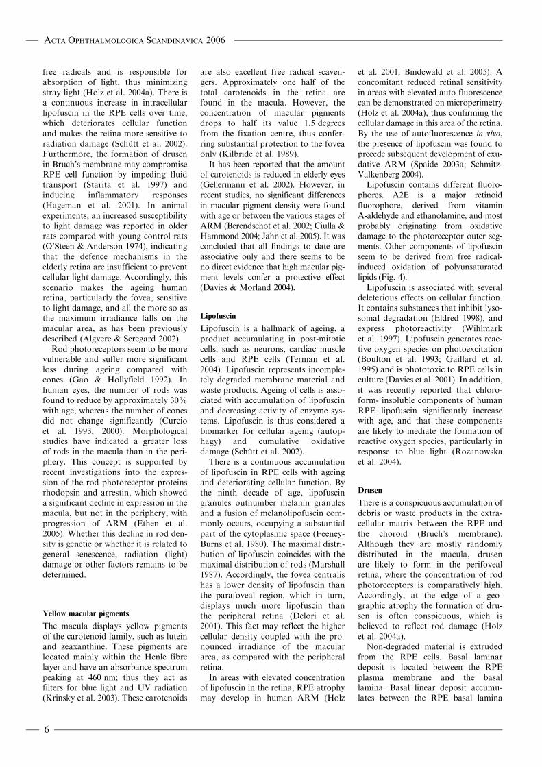
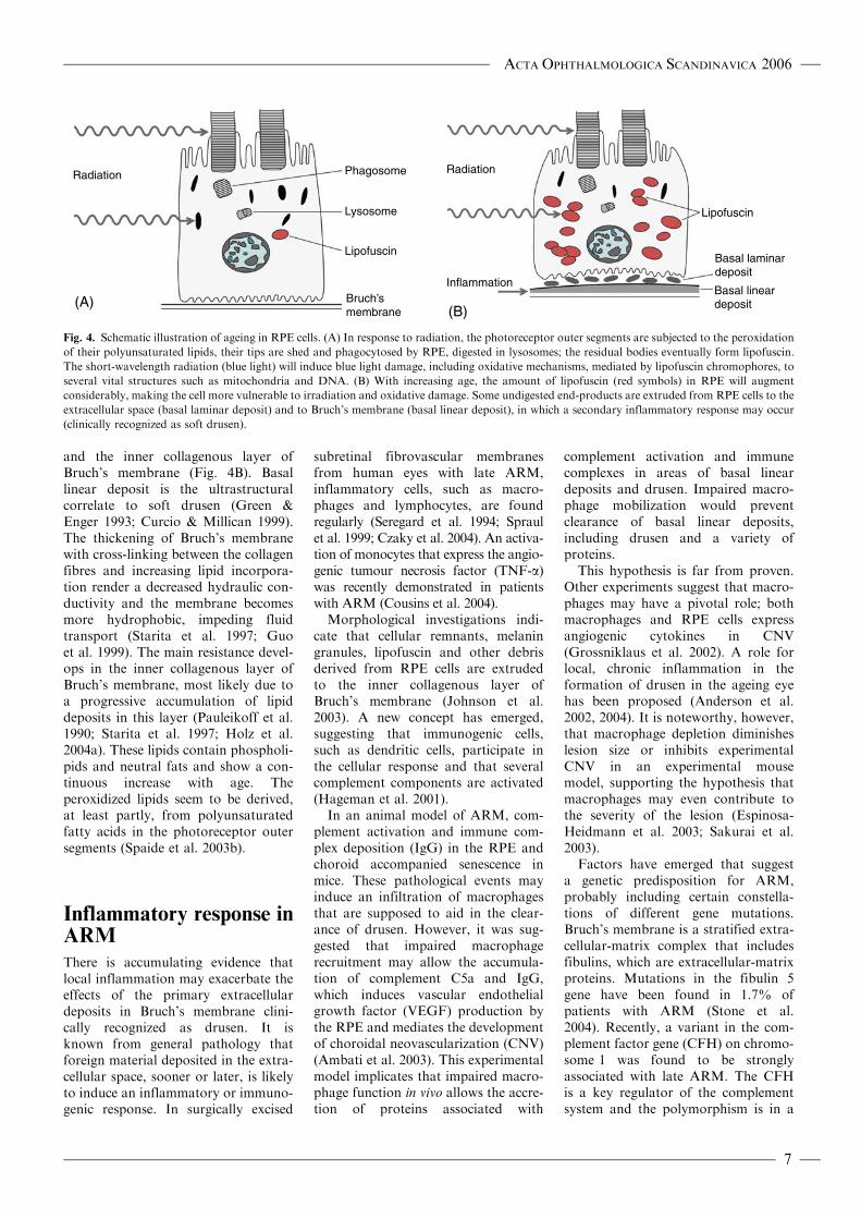
Fig. 4. Schematic illustration of ageing in RPE cells. (A) In response to radiation, the photoreceptor outer segments are subjected to the peroxidation
of their polyunsaturated lipids, their tips are shed and phagocytosed by RPE, digested in lysosomes; the residual bodies eventually form lipofuscin.
The short-wavelength radiation (blue light) will induce blue light damage, including oxidative mechanisms, mediated by lipofuscin chromophores, to
several vital structures such as mitochondria and DNA. (B) With increasing age, the amount of lipofuscin (red symbols) in RPE will augment
considerably, making the cell more vulnerable to irradiation and oxidative damage. Some undigested end-products are extruded from RPE cells to the
extracellular space (basal laminar deposit) and to Bruch’s membrane (basal linear deposit), in which a secondary inflammatory response may occur
(clinically recognized as soft drusen).
ACTA OPHTHALMOLOGICA SCANDINAVICA 2006
7
region of CFH that binds heparin andC-reactive protein (Edwards et al.2005; Haines et al. 2005; Klein et al.2005). In addition, the complementfactor H, the major inhibitor of thealternative complement pathway, accu-mulates within drusen and is synthe-sized by the RPE; multiple HF1variants were reported occurring at afrequency of 50% in ARM and 29% incontrols (Hageman et al. 2005).
To date it seems reasonable toassume that inflammatory and immu-nological responses operate in theextracellular matrix between the RPEand choroid, although the pathogeneticmechanisms are far from clear.
Choroidal neovascularization (CNV)
Inflammatory response is likely toinduce further pathological changes,including vascular alterations. Anincreased expression of angiogenicgrowth factors, such as VEGF, are pre-sent in the RPE and in the outernuclear layer of the maculae withARM (Kvanta et al. 1996; Kliffenet al. 1997). Macrophages, RPE cellsand vascular endothelial cells expressangiogenic cytokines and, in associa-tion with other pathogenetic mechan-isms, induce a neovascular response(Grossniklaus et al. 2002).
The CNV generally grows into theinner portion of Bruch’s membrane,ultrastructurally recognized as the basallinear deposit, an area into which perox-idized lipids have accumulated (Spaide2003b; Holtz et al. 2004). The patho-genesis of CNV is ultimately a conse-quence of a disturbed balance betweenseveral pro- and antiangiogenic factors.The extensive literature on angiogenesisis not included in this survey.
Accordingly, intravitreal triamcino-lone acetonide was found to inhibitexperimental CNV in a laser-treatedrat model (Ciulla et al. 2003), andwhen photodynamic therapy was com-bined with a triamcinolone injection,favourable results were achieved inhuman eyes with CNV (Spaide et al.2005). It was recently reported thatC-reactive protein and interleukin 6,markers of inflammation and indicatorsof the risk of cardiovascular disease, areelevated in neovascular ARM (Seddonet al. 2004, 2005). Furthermore, neo-vascular ARM seems to be associatedwith cardiovascular disease (Age-Related Eye Disease Study 2004).
Free radicals
Accumulating evidence indicates thatoxidative (free radical) damage is apathogenetic factor in ageing and hasa significant influence on the develop-ment of ARM. It has been estimatedthat approximately 3% of the oxygenused produces reactive oxygen species.Reactive oxygen species can be derivedfrom leakage of electrons from mito-chondrial aerobic metabolism, andinclude superoxide, hydrogen peroxideand singlet oxygen.
Choroidal blood flow far supersedesthat required to nourish the neuralretina. Its venous blood flow is oxyge-nated to 90%. In the submacular area,choroidal blood flow is significantlyhigher than in the periphery (Alm &Bill 1973). Approximately 1% of theoxygen in the blood is extracted fromthe choroidal circulation, indicatingthat there is a tremendous excess ofoxygen that is never used.
Due to the high blood perfusion rate,the oxygen tension in the retina is veryhigh. Oxidative damage is likely tooccur in an environment of extremelyhigh oxygen tension, such as the centreof the macula, which has a continuousand lifelong exposure to radiation(photons). Reactive oxygen species(free radicals) are formed, particularlyin response to short-wavelength irra-diation. Photoreceptor outer segmentsare rich in polyunsaturated fatty acids.The outer segments, altered by irradia-tion, are phagocytosed by the RPEcells, which may become damaged bythe engulfed reactive oxygen species.Experiments show that rod outer seg-ments mediate mitochondrial DNAdamage and apoptosis in human RPEcells in tissue culture (Jin et al. 2001).This scenario demonstrates the pivotalinfluence of photoreceptor outer seg-ments on RPE function.
Lipid peroxidation has been definedas ‘the oxidative deterioration of poly-unsaturated lipids’ (i.e. lipids that con-tain more than double carbon� carbondouble bonds). Free radicals deterio-rate lysosome function, giving rise tobyproducts such as lipofuscin. Withage, there is an increasing accumula-tion of lipofuscin in the RPE cells,featuring ARM. Lipofuscin accumu-lates in the lysosomes of RPE cellsand is associated with several adverseeffects on RPE cell function andsurvival.
A2E (N-retinidene-N-retinylethano-leamine) is a major fluorophore of lipo-fuscin and is generated, at least partly,from transretinal. A2E generates freeradicals in response to irradiation, par-ticularly by blue light. The formationof reactive oxygen species will impairthe oxygen-driven activity in the mito-chondria, the ‘power plant’ of the cell,by altering its DNA. A2E specificallytargets cytochrome oxidase (COX)inhibiting mitochondrial respiration(Schutt et al. 2000; Sparrow & Cai2001). Mitochondrial changes may, inturn, lead to apoptosis of elderly RPEcells that have become loaded withlipofuscin.
The body has two lines of defence:
(1) antioxidant enzymes, such assuperoxide dismutase, catalase andperoxidase, and
(2) antioxidant nutrients, such asalpha-tocopherol (vitamin E) andascorbic acid (vitamin C).
However, with ageing the defensivemechanisms of the body seem tobecome insufficient. There is a contin-uous increase in lipofuscin in post-mitotic cells, particularly in the RPE,reflecting the insufficiency of theirdefence to oxidative damage.
It is reasonable to assume that whenthe defensive mechanisms becomeinsufficient with increasing age, degen-erative changes in RPE cells ultimatelyoccur in response to a lifelong exposureto radiation and reactive oxygen spe-cies (Barja 2002). Research on RPEcells in tissue culture has identified sev-eral pathogenetic cellular events, suchas the accumulation of lipofuscin andother chromophores, which are likelyto mediate and aggravate mitochon-drial DNA damage (Liang & Godley2003). Protective effects from a varietyof antioxidants have been demon-strated. Antioxidants capture free radi-cals, thereby to some extent alleviatingthe chain reaction of deleterious effectson other molecules.
Experimental blue lightdamage
Animal experiments show that retinalexposure to excessive levels of whitelight induce apoptotic cell death ofphotoreceptors and, with a shortdelay, do the same in RPE cells(Hafezi et al. 1997; Grimm et al.
ACTA OPHTHALMOLOGICA SCANDINAVICA 2006
8
2001). It has been unequivocallydemonstrated that the blue light hazardis mediated through absorption of bluelight by rhodopsin.
Accumulating evidence indicatesthat light exposure triggers photorecep-tor apoptosis only in the presence ofrhodopsin. The RPE protein RPE65,which is an important determinant forrhodopsin regeneration in mice, isa prerequisite for light damage (Iseliet al. 2002; Wenzel et al. 2005). Nolight damage could be induced inRPE65-deficient mice deprived of rho-dopsin. Light damage occurs onlywhen the retina is supplied with reti-noid metabolites such as 11-cis retinal,the chromophore of rod and coneopsins, or other toxic byproducts(Grimm et al. 2000a). Repetitivephoton absorptions in associationwith rhodopsin regeneration seem tobe required in order to trigger photo-receptor apoptosis. It is conceivable,although not proven, that repetitivelight stimuli in a mesopic level of adap-tation may make the retina particularlysensitive to light damage.
The photoelectric effect, known fromquantum physics, explains many of thebiological events induced by short-wavelength irradiation. The high-energy photons in the spectrum ofblue light and adjacent UVA radiationhave the power to damage the cellularfunction and structure of photorecep-tors and RPE. These photons createreactive oxygen species, which are dele-terious to a variety of cellular orga-nelles, particularly the mitochondrialDNA, and ultimately result in apopto-tic cell death.
It is well established that the forma-tion of reactive oxygen speciesincreases with decreasing wavelength(Boulton et al. 1993; Rozanowskaet al. 1998). On the cellular level,470–490-nm light was reported toinduce oxidant injury in both theinner and outer segments of rod photo-receptors, an event requiring rhodopsinactivation (Demontis et al. 2002).
The increased risk of mitochondrialdamage induced by blue light has beendemonstrated in many experimentalmodels. For example, red light of acertain intensity is insufficient toinduce retinal damage, whereas bluelight of the same intensity will causeretinal injury. Deep blue light hasbeen described as 50–80 times moreefficient at causing photoreceptor
damage than green light (Rapp &Smith 1992). Following exposure toblue light (400–480 nm), photoreceptorapoptosis was induced in the rat, themost pronounced cell death occurring16 hours after irradiation in the dark.Immunoreactivity for caspase-3 wasup-regulated in the outer nuclear layerof the retina and, concurrently, cyto-chrome C was released from the mito-chondria. These events result inapoptotic cell death (Wu et al. 1999,2002).
Using narrow-band blue (403 nm)and green (550 nm) light, adjusted tothe same energy, exposure to blue lightwas found to severely damage rodphotoreceptors, while green light didnot (Grimm et al. 2001; Wenzel et al.2005).
Photoconsumption of oxygen byhuman ocular lipofuscin increaseswith decreasing wavelengths of theexciting irradiation, the effects of bluelight being much more pronouncedthan those of red light (Pawlack et al.2002). Blue light damage is thus likelyto be mediated by endogenous chromo-phores, such as lipofuscin (Boultonet al. 2001). It has been further demon-strated that chloroform-insolublecomponents (ChNS) of lipofuscingranules significantly increase with age-ing, and that ChNS mediate photo-induced oxygen uptake (photogenerat-ing singlet oxygen) with rates thatincrease with decreasing wavelengths(Rozanowska et al. 2004). Thus, theoxidative damage to RPE cells loadedwith lipofuscin is enhanced in responseto blue light, a phenomenon thatoccurs in cells in elderly subjects andin patients with ARM.
Light radiation (near-ultraviolet)was demonstrated to accelerate the for-mation of lipofuscin fluorophores andageing of RPE cells in tissue culture (Liet al. 1999). Autofluorescence fromlipofuscin can be demonstrated inresponse to blue light. In addition,exposure of RPE cells in culture tonear UV light reduced the proliferationof the cells, accompanied by increasedlipofuscin content. In turn, lipofuscinaccumulation in RPE cells inducedenhanced sensitivity to blue light irra-diation (Wihlmark et al. 1997; Nilssonet al. 2003).
Thus, lipofuscin fluorophores seemto play an important role in the bluelight hazard phenomenon. The fluoro-phore A2E mediates blue light-induced
(peak around 430 nm) damage to RPEcells, involving oxidative mechanisms,where DNA is the target of the photo-dynamic effects (Sparrow & Cai 2001;Sparrow et al. 2002, 2003). A2E targetsmitochondria and induces apoptosis(Suter et al. 2000). A2E also acts asan inhibitor of lysomal degradativefunctions (Bermann et al. 2001). It isnotable that when oxygen was removedfrom the culture media of human RPEcells in culture, the blue light damagewas found to be basically blocked(Sparrow et al. 2002).
In an experimental model, transgenicmice with over-expression of APOB100 and increased LDL cholesterolin the blood were more susceptible toblue light-induced formation of sub-RPE deposits than the controls; thushigh-fat diet and blue light exposureaggravate oxidant injury (Espinosa-Heidmann et al. 2004).
As previously mentioned, the forma-tion of lipofuscin in RPE cells requiresa normal visual cycle. The absence ofretinoid metabolites such as retinalprevents RPE apoptosis (Grimm et al.2000a; Katz & Redmond 2001).Accordingly, light damage to the retinais dependent on the amount of rhodop-sin available for bleaching and RPE65-deficient mice, lacking rhodopsin, areprotected against light-induced apopto-sis of RPE cells (Grimm et al. 2001).
These results have been explained onthe basis of rhodopsin photoreversal:blue light regenerates rhodopsin fromits bleaching intermediates and canthus provide increased numbersof bleachable rhodopsin molecules(Grimm et al. 2000b). Blue light pro-motes the photoisomerization of all-trans-retinal , which leads to the regen-eration of rhodopsin and an increasein phototransduction, thus in turnpromoting photoreceptor apoptosis(Margrain et al. 2004). This conceptwas supported by experiments onmutant mice that lack rhodopsin andin these animals no blue light photo-receptor damage could be induced.Accordingly, when both metabolic rho-dopsin regeneration and photoreversalbleaching were inhibited in wild-typemice, blue light exposure induced onlymoderate lesions. Green light had noeffect (Grimm et al. 2001).
It should be emphasized, however,that light-induced retinal degenerationis a complex series of events, involvingcaspase-dependent apoptosis and a
ACTA OPHTHALMOLOGICA SCANDINAVICA 2006
9
number of caspase-independent path-ways to cellular death (Hao et al.2002; Wenzel et al. 2005).
The dramatic change inocular transmittance inpseudophakic eyes
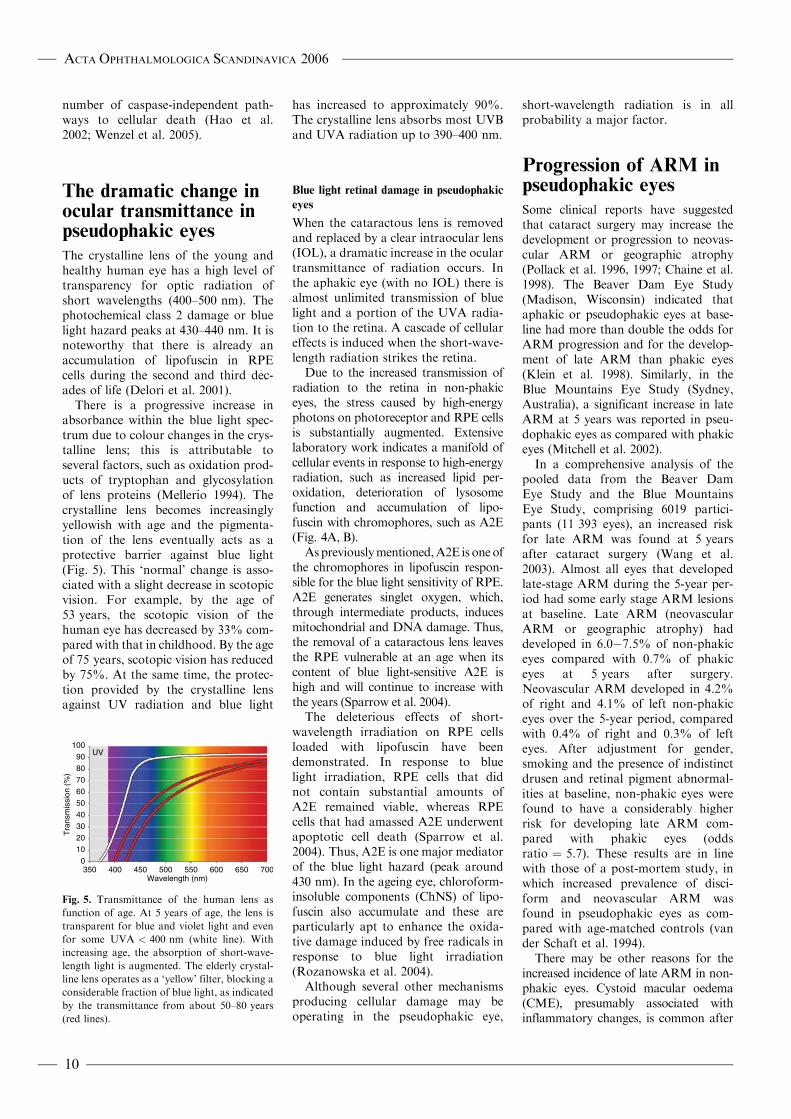
The crystalline lens of the young andhealthy human eye has a high level oftransparency for optic radiation ofshort wavelengths (400–500 nm). Thephotochemical class 2 damage or bluelight hazard peaks at 430–440 nm. It isnoteworthy that there is already anaccumulation of lipofuscin in RPEcells during the second and third dec-ades of life (Delori et al. 2001).
There is a progressive increase inabsorbance within the blue light spec-trum due to colour changes in the crys-talline lens; this is attributable toseveral factors, such as oxidation prod-ucts of tryptophan and glycosylationof lens proteins (Mellerio 1994). Thecrystalline lens becomes increasinglyyellowish with age and the pigmenta-tion of the lens eventually acts as aprotective barrier against blue light(Fig. 5). This ‘normal’ change is asso-ciated with a slight decrease in scotopicvision. For example, by the age of53 years, the scotopic vision of thehuman eye has decreased by 33% com-pared with that in childhood. By the ageof 75 years, scotopic vision has reducedby 75%. At the same time, the protec-tion provided by the crystalline lensagainst UV radiation and blue light
has increased to approximately 90%.The crystalline lens absorbs most UVBand UVA radiation up to 390–400 nm.
Blue light retinal damage in pseudophakic
eyes
When the cataractous lens is removedand replaced by a clear intraocular lens(IOL), a dramatic increase in the oculartransmittance of radiation occurs. Inthe aphakic eye (with no IOL) there isalmost unlimited transmission of bluelight and a portion of the UVA radia-tion to the retina. A cascade of cellulareffects is induced when the short-wave-length radiation strikes the retina.
Due to the increased transmission ofradiation to the retina in non-phakiceyes, the stress caused by high-energyphotons on photoreceptor and RPE cellsis substantially augmented. Extensivelaboratory work indicates a manifold ofcellular events in response to high-energyradiation, such as increased lipid per-oxidation, deterioration of lysosomefunction and accumulation of lipo-fuscin with chromophores, such as A2E(Fig. 4A, B).
Aspreviouslymentioned,A2E is oneofthe chromophores in lipofuscin respon-sible for the blue light sensitivity of RPE.A2E generates singlet oxygen, which,through intermediate products, inducesmitochondrial and DNA damage. Thus,the removal of a cataractous lens leavesthe RPE vulnerable at an age when itscontent of blue light-sensitive A2E ishigh and will continue to increase withthe years (Sparrow et al. 2004).
The deleterious effects of short-wavelength irradiation on RPE cellsloaded with lipofuscin have beendemonstrated. In response to bluelight irradiation, RPE cells that didnot contain substantial amounts ofA2E remained viable, whereas RPEcells that had amassed A2E underwentapoptotic cell death (Sparrow et al.2004). Thus, A2E is one major mediatorof the blue light hazard (peak around430 nm). In the ageing eye, chloroform-insoluble components (ChNS) of lipo-fuscin also accumulate and these areparticularly apt to enhance the oxida-tive damage induced by free radicals inresponse to blue light irradiation(Rozanowska et al. 2004).
Although several other mechanismsproducing cellular damage may beoperating in the pseudophakic eye,
short-wavelength radiation is in allprobability a major factor.
Progression of ARM inpseudophakic eyes
Some clinical reports have suggestedthat cataract surgery may increase thedevelopment or progression to neovas-cular ARM or geographic atrophy(Pollack et al. 1996, 1997; Chaine et al.1998). The Beaver Dam Eye Study(Madison, Wisconsin) indicated thataphakic or pseudophakic eyes at base-line had more than double the odds forARM progression and for the develop-ment of late ARM than phakic eyes(Klein et al. 1998). Similarly, in theBlue Mountains Eye Study (Sydney,Australia), a significant increase in lateARM at 5 years was reported in pseu-dophakic eyes as compared with phakiceyes (Mitchell et al. 2002).
In a comprehensive analysis of thepooled data from the Beaver DamEye Study and the Blue MountainsEye Study, comprising 6019 partici-pants (11 393 eyes), an increased riskfor late ARM was found at 5 yearsafter cataract surgery (Wang et al.2003). Almost all eyes that developedlate-stage ARM during the 5-year per-iod had some early stage ARM lesionsat baseline. Late ARM (neovascularARM or geographic atrophy) haddeveloped in 6.0�7.5% of non-phakiceyes compared with 0.7% of phakiceyes at 5 years after surgery.Neovascular ARM developed in 4.2%of right and 4.1% of left non-phakiceyes over the 5-year period, comparedwith 0.4% of right and 0.3% of lefteyes. After adjustment for gender,smoking and the presence of indistinctdrusen and retinal pigment abnormal-ities at baseline, non-phakic eyes werefound to have a considerably higherrisk for developing late ARM com-pared with phakic eyes (oddsratio ¼ 5.7). These results are in linewith those of a post-mortem study, inwhich increased prevalence of disci-form and neovascular ARM wasfound in pseudophakic eyes as com-pared with age-matched controls (vander Schaft et al. 1994).
There may be other reasons for theincreased incidence of late ARM in non-phakic eyes. Cystoid macular oedema(CME), presumably associated withinflammatory changes, is common after
UV
0
10
20
30
40
50
60
70
80
90
100
350 400 450 500 550 600 650 700Wavelength (nm)
Tra
nsm
issi
on (
%)
Fig. 5. Transmittance of the human lens as
function of age. At 5 years of age, the lens is
transparent for blue and violet light and even
for some UVA < 400 nm (white line). With
increasing age, the absorption of short-wave-
length light is augmented. The elderly crystal-
line lens operates as a ‘yellow’ filter, blocking a
considerable fraction of blue light, as indicated
by the transmittance from about 50–80 years
(red lines).
ACTA OPHTHALMOLOGICA SCANDINAVICA 2006
10
cataract surgery. The greatest incidenceof clinically significant postoperativeCME occurs 1–3 months after surgery.In the majority of cases this conditionresolves within weeks or months. Inapproximately 1% of cataract extrac-tions, chronic CME persists for morethan 6 months (Dick et al. 2001).Notwithstanding this, the impact ofCME on the development of late ARMremains uncertain and ambiguous.
It is reasonable to assume that anincreased susceptibility to phototoxiclight damage after cataract surgery per-sists, making the retina more vulnerableto longlasting radiation, particularly inelderly subjects(Pollack et al. 1996).
Putative prophylacticmeasures
‘Yellow’ intraocular lenses (IOLs)
The IOLs with UV-filter were intro-duced in the 1980s. Ultraviolet filterIOLs are supposed to confer protectionagainst radiation below 400 nm; forlonger wavelengths the transmittanceincreases substantially compared withthat of the normal ageing crystallinelens. However, it was recently reportedthat a number of traditional IOLs actu-ally transmit more than 10% of UVAradiation between 350 nm and 400 nm(Laube et al. 2004); these high-energy
photons have the capacity to inducedeleterious cellular effects (Fig. 3).
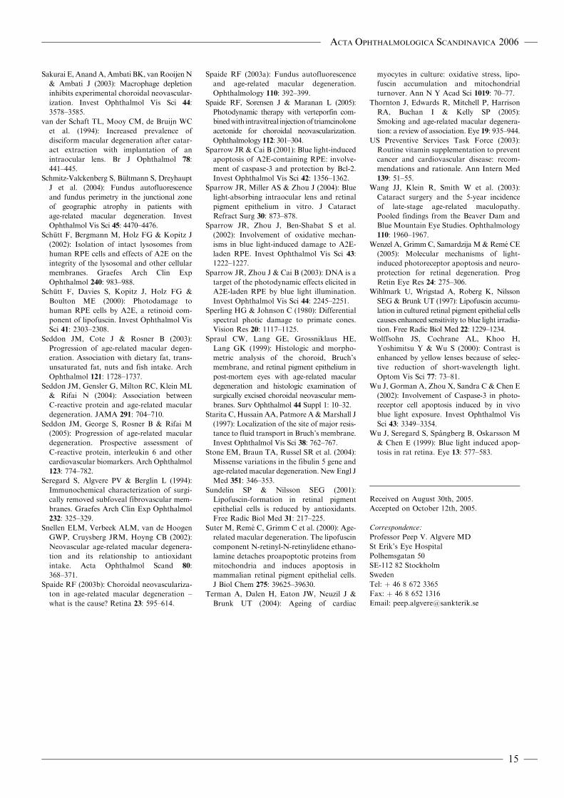
New types of ‘yellow’ IOLs havebeen introduced, which have a filteringeffect that mimics that of the ageinghuman lens by attenuating lightfrom wavelengths between 400 nmand 500 nm (Figs 6 and 7). The yellowIOLs provide a better degree of pro-tection against UV radiation andblue light than traditional IOLs thathave only UV filtering capabilities(Mainster 2005 & Sparrow 2003).
Experimental data indicate that theyellow filters protect RPE cells that areexposed to blue, green and white light,respectively. Pigmented rabbits wereexposed to xenon light; one eye of theanimals was protected by a yellow filterand a clear polymethylmethacrylate
(PMMA) lens was placed in front ofthe fellow eye. The total irradiance ofthe eyes was equalized by neutral den-sity filters in front of the PMMA mate-rial. Electrophysiological recordingsshowed significantly more damage toboth neuroretinal and RPE functionin eyes behind the PMMA materialthan in those behind yellow filters(Nilsson et al. 1990).
Human RPE cells were allowed toaccumulate A2E in tissue culture tolevels that are comparable to theamounts present in vivo. When pro-tected by a yellow IOL, transmissionof blue light (peak 430 nm) was attenu-ated by approximately 50% and apop-totic cell death was reduced by 80%compared with irradiation in theabsence of IOL. In contrast, when fourdifferent traditional IOLs were tested inthe same experimental setting, thereduction of the number of non-viablecells was only 10–20% when exposed toblue light, 40–60% in response to greenlight (peak 550 nm) and 20–40% inresponse to white light (390–750 nm).The experiments thus demonstrate thateven green and white light may havedetrimental effects on A2E-laden RPEcells, such as those in elderly eyes. Theseadverse effects are alleviated by a yellowIOL. In conclusion, the blue light-absorbing IOL was associated with sig-nificant reduction (78�82%) in thedeath of A2E-laden RPE cells thatwere exposed to blue, green or whitelight (Sparrow et al. 2004).
The yellow IOL gives a yellowish hueto the ocular image which, generally,does not seem to be a problem. Theyellow IOLs induce a reduction of thescotopic sensitivity (Mainster 2005 &Sparrow 2003). This moderate loss ofscotopic vision was found to be of thesame magnitude as that of a 53-year-old human lens. Patients with the bluelight-blocking IOL exhibit a significantthreshold elevation at 410 nm (violetlight) and at 450 nm (blue light).However, no significant differences invisual performance were found at500 nm or 560 nm (green light)between patients implanted with theyellow IOL and those implanted withthe clear IOL (Jackson 2005).
The yellow IOLs reduce the opticalchromatic aberration by blockingblue light. In addition, there is anenhancement of contrast sensitivityand a reduction of glare (Wolffsohnet al. 2000). These phenomena are
Spe
ctra
l Ene
rgy
Dis
trib
utio
n
Wavelength (nm)
Noon
300 400 500 600 700 800 900 1000 1100
UV VIS IR1.3
1.1
0.9
0.7
0.5
0.3
0.1
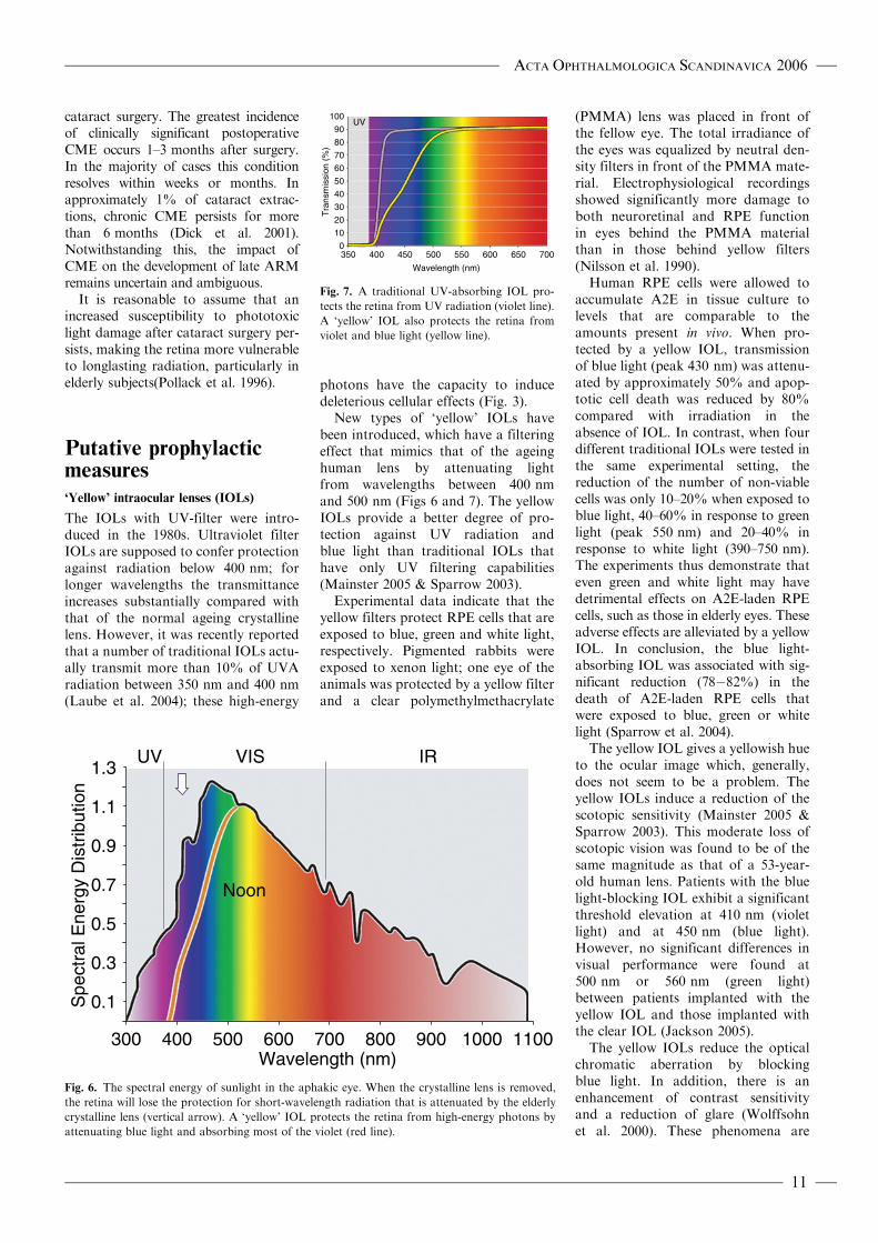
Fig. 6. The spectral energy of sunlight in the aphakic eye. When the crystalline lens is removed,
the retina will lose the protection for short-wavelength radiation that is attenuated by the elderly
crystalline lens (vertical arrow). A ‘yellow’ IOL protects the retina from high-energy photons by
attenuating blue light and absorbing most of the violet (red line).
UV
0
10
20
30
40
50
60
70
80
90
100
350 400 450 500 550 600 650 700
Wavelength (nm)
Tra
nsm
issi
on (
%)
Fig. 7. A traditional UV-absorbing IOL pro-
tects the retina from UV radiation (violet line).
A ‘yellow’ IOL also protects the retina from
violet and blue light (yellow line).
ACTA OPHTHALMOLOGICA SCANDINAVICA 2006
11

definitively advantageous for elderlyeyes or in subjects with ARM.However, perception of the colourblue is certainly affected. On the otherhand, blue cones do not contribute tohigh visual acuity. Clinical experienceindicates that people with a clear IOLin one eye and a yellow IOL in thefellow eye are not disturbed by theslight differences in the images.
Thus, experimental and theoreticaldata indicate that a yellow IOL is likelyto confer protection to retinal cells.The blocking of UVA radiation fromthe retina is important. Although theindications for the use of yellow IOLshave not been elucidated, it is, from atheoretical point of view, most rationalto use a yellow IOL for cataract sur-gery in patients at risk for ARM and inthose with ARM (Margrain et al. 2004;Nilsson 2004; Braunstein & Sparrow2005). A randomized clinical trial iswarranted to assess to what extent yel-low IOLs will reduce the risk for orprogression of ARM.
Prophylactic treatmentwith antioxidants
Another approach to prophylactictreatment for ARM is based on thecapacity of various antioxidants to alle-viate or reduce the detrimental effectsof reactive oxygen species in responseto radiation and other noxious stimuli.Extensive laboratory investigationshave demonstrated the role of oxida-tive stress and the inhibitory effects ofantioxidants on RPE cellular function(Beatty et al. 2000; Sundelin & Nilsson2001).
The Age-related Eye Disease Study(AREDS) (2001), a randomized,placebo-controlled clinical trial, com-prising 3640 participants, using supple-mentation with high-dose antioxidants(average follow-up 6.5 years) showeda significant reduction in rates of atleast moderate visual loss in certaincategories of ARM. Participants wererandomized to daily antioxidants(vitamin C 500 mg, vitamin E 400 IU,b-carotene 15 mg, zinc 80 mg andcopper 2 mg) or placebo. Subjectswith extensive intermediate size drusen,at least one large druse, non-centralgeographic atrophy in one or botheyes, or advanced ARM in one eyehad a statistically significant odds
reduction for the development ofadvanced (late) ARM. At 5 years, theestimated probability of progression toadvanced ARM (neovascular ARM,geographic atrophy) was 28% forthose assigned to placebo, and 20%for those assigned to antioxidants pluszinc.
There was no difference in mortalityin the various subgroups treated withantioxidants or placebo (Age-relatedEye Disease Study 2001). Further ana-lyses indicated that participants ran-domly assigned to receive zinc (zincalone or antioxidants plus zinc) had alower mortality rate than those nottaking zinc (Age-related Eye DiseaseStudy 2004).
It is known from earlier studies thathigh doses of b-carotene may increasethe risk of lung cancer in smokers(Omenn et al. 1996; US PreventiveServices Task Force 2003). Therefore,b-carotene was excluded from the anti-oxidant regimen for smokers in theAREDS. A meta-analysis of antioxi-dant supplements for prevention ofgastrointestinal cancers revealed thatcertain combinations of vitamins, suchas b-carotene and vitamin A, increasedmortality (Bjelakovic et al. 2004).However, trials in which vitamin Cwas given separately or in differentcombinations with b-carotene, vitaminA or vitamin E showed no significanteffect on gastrointestinal cancers ormortality.
It is conceivable that very high dosesof a solitary antioxidant can induceadverse effects. One meta-analysis sug-gested that high-dose vitamin E supple-ments may increase all-cause mortality(Miller et al. 2005). Notwithstanding,the AREDS formulation had a 14%reduction in mortality risk afteran average of 6.5 years (Chew &Clemons 2005).
The Health Professionals Follow-upStudy (Leitzmann et al. 2003) foundthat men who consumed more than100 mg/day of supplemental zinc hada higher risk of advanced prostate can-cer, but not of organ-confined cancers.However, a later study concluded that80 mg/day of supplemental zinc doesnot increase the risk of prostate cancer(Jarrard 2005).
The putative role of nutritional fac-tors (e.g. a low antioxidant intake) inthe incidence of disease has beenapproached. For example, in patientswith low levels of antioxidant intake,
the prevalence rate of neovascularARM was reported to be about twiceas high as that in subjects with highlevels of intake (Snellen et al. 2002). Aprotective role for the intake of fruitand fish has been suggested (Seddonet al. 2003; Cho et al. 2004).
The US National Eye Institute con-cluded that people at risk of developingadvanced ARM lowered this risk byabout 25% when treated according tothe AREDS. It has been estimatedthat if people in the USA at risk ofadvanced ARM receive AREDS-recommended supplements, morethan 300 000 of them would avoiddeveloping advanced ARM during thenext 5 years (Age-related Eye DiseaseStudy 2003). Accordingly, the AmericanAcademy of Ophthalmology hasrecommended the AREDS formulafor antioxidant supplementation.
Notwithstanding, further research iswarranted to elucidate the complicatedbalance between oxidative damage andthe effects of antioxidant and nutri-tional prophylaxis.
ReferencesAge-related Eye Disease Study (AREDS)
(2003): Potential public health impact of
Age-related Eye Disease Study results.
AREDS Report 11. Arch Ophthalmol 121:
1621–1624.
Age-related Eye Disease Study (AREDS)
(2004): Associations of mortality with ocular
disorders and intervention of high-dose anti-
oxidants and zinc in the Age-related Eye
Disease Study. AREDS Report 13. Arch
Ophthalmol 122: 716–726.
Age-related Eye Disease Study (AREDS)
(2001): A randomized, placebo-controlled,
clinical trial of high-dose supplementation
with vitamins C and E, beta carotene and
zinc for age-related macular degeneration
and vision loss. AREDS Report 8. Arch
Ophthalmol 119: 1417–1436.
Algvere PV & Seregard S (2002): Age-related
maculopathy: pathogenetic features and new
treatment modalitites. Acta Ophthalmol
Scand 80: 136–143.
Alm A & Bill A (1973): Ocular and optic nerve
blood flow at normal and increased intrao-
cular pressures in monkeys (Macaca irus): a
study with radioactively labelled micro-
spheres including flow determination of
brain and some other tissues. Exp Eye Res
15: 15–29.
Ambati J, Anand A, Fernandez S et al. (2003):
An animal model of age-related macular
degeneration in senescent Ccl-2- or Ccr-2-
deficient mice. Nature Med 9: 1390–1397.
Anderson DH, Mullins RF, Hageman GS &
Johnson LV (2002): A role for local
ACTA OPHTHALMOLOGICA SCANDINAVICA 2006
12

inflammation in the formation of drusen in
the ageing eye. Am J Ophthalmol 134:
411–431.
Anderson DH, Talaga KC, Rivest AJ, Barron E,
Hageman GS & Johnson LV (2004):
Characterization of beta amyloid assemblies
in drusen: the deposits associated with age-
ing and age-related macular degeneration.
Exp Eye Res 78: 243–256.
Barja G (2002): Endogenous oxidative stress:
relationship to ageing, longevity and caloric
restriction. Ageing Res Rev 1: 397–411.
Beatty S, Koh H-H, Phil M, Henson D &
Boulton M (2000): The role of oxidative
stress in the pathogenesis of age-related
macular degeneration. Surv Ophthalmol
454: 115–134.
Benson H (1991): University Physics. New
York: Wiley and Sons, Inc.; 820–826.
Berendschot TTJM, Willemse-Assink JJM,
Basiaanse M, de Jong PTVM & van
Norren D (2002): Macular pigment and mel-
anin in age-related maculopathy in a general
population. Invest Ophthalmol Vis Sci 43:
1928–1932.
Bermann M, Schutt F, Holz FG & Kopitz J
(2001): Does A2E, a retinoid component of
lipofuscin and inhibitor of lysosomal degra-
dative functions, directly affect the activity
of lysosomal hydrolases? Exp Eye Res 72:
191–195.
Bindewald A, Schmitz-Valckenberg S, Jorzik JJ
et al. (2005): Classification of abnormal
fundus autofluorescence patterns in the
junctional zone of geographic atrophy in
patients with age-related macular degenera-
tion. Br J Ophthalmol 89: 874–878.
Bjelakovic G, Nikolova D, Simonetti RG &
Gluud C (2004): Antioxidant supplements
for prevention of gastrointestinal cancers: a
systematic review and meta-analysis. Lancet
364: 1219–1228.
Boettner EA & Wolter JR (1962):
Transmission of the ocular media. Invest
Ophthalmol 1: 776–783.
Boulton M, Dontsov A, Jarvisevans J,
Ostrovsky M & Svistunenko D (1993):
Lipofuscin is a photoinducible free-radical
generator. J Photochem Photobiol B 19:
201–204.
Boulton M, Rozanowska M & Rozanowska B
(2001): Retinal photodamage. J Photochem
Photobiol B 64: 144–161.
Braunstein RE & Sparrow JR (2005): A blue-
blocking intraocular lens should be used in
cataract surgery. Arch Ophthalmol 123:
547–549.
Chaine G, Hullo A, Sahel J et al (1998):
France-DMLA Study Group. Case-control
study of risk factors for age-related macular
degeneration. Br J Ophthalmol 82:
996–1002.
Chew EY & Clemons T (2005): Vitamin E
and Age-related Eye Disease Study supple-
mentation for age-related macular degenera-
tion. [Editorial.] Arch Ophthalmol 123:
395–396.
Cho E, Seddon JM, Rosner B, Willett WC &
Hankinson SE (2004): Prospective study of
intake of fruits, vegetables, vitamins and
carotenoids and risk of age-related maculo-
pathy. Arch Ophthalmol 122: 883–892.
Cicerone CM (1976): Cones survive rods in the
light-damaged eye of the albino rat. Science
194: 1183–1185.
Ciulla TA, Criswell MH, Danis RP et al.
(2003): Choroidal neovascular membrane
inhibition in a laser-treated rat model with
intraocular sustained release triamcinolone
acetonide microimplants. Br J Ophthalmol
87: 1032–1037.
Ciulla TA & Hammond BR (2004): Macular
pigment density and ageing, assessed in the
normal elderly and those with cataracts and
age-related macular degeneration. Am J
Ophthalmol 138: 582–587.
Cousins SW, Espinosa-Heidmann DG &
Csaky KG (2004): Monocyte activation in
patients with age-related macular degenera-
tion. Arch Ophthalmol 122: 1013–1018.
Cruickshanks KJ, Klein R & Klein BR (1993):
Sunlight and age-related macular degenera-
tion. The Beaver Dam Eye Study. Arch
Ophthalmol 111: 514–518.
Cruickshanks KJ, Klein R, Klein BE &
Nondahl DM (2001): Sunlight and the
5-year incidence of early age-related maculo-
pathy. The Beaver Dam Eye Study. Arch
Ophthalmol 119: 246–250.
Curcio CA, Medeiros NE & Millican CL
(1993): Photoreceptor loss in age-related
macular degeneration. Invest Ophthalmol
Vis Sci 37: 1236–1249.
Curcio CA & Millican CL (1999): Basal linear
deposit and large drusen are specific for early
age-related maculopathy. Arch Ophthalmol
117: 329–339.
Curcio CA, Owsley C & Jackson GR (2000):
Spare the rods, save the cones in ageing and
age-related maculopathy. Invest Ophthalmol
Vis Sci 41: 2015–2018.
Czaky K, Baffi J, Chan C-C & Byrnes GA
(2004): Clinicopathologic correlation of pro-
gressive fibrovascular proliferation associated
with occult choroidal neovascularization in
age-related macular degeneration. Arch
Ophthalmol 122: 650–652.
Davies NP & Morland AB (2004): Macular
pigments: their characteristics and putative
role. Prog Retin Eye Res 23: 533–559.
Davies S, Elliott MH, Floor E (2001):
Photocytotoxicity of lipofuscin in human
retinal pigment epithelial cells. Free Radic
Biol Med 31: 256–265.
Delori FC, Goger DG & Dorey CK (2001):
Age-related accumulation and spatial distribu-
tion of lipofuscin in RPE of normal subjects.
Invest Ophthalmol Vis Sci 42: 1855–1866.
Demontis GC, Longoni B & Marchiafava PL
(2002): Molecular steps in light-induced oxi-
dative damage to retinal rods. Invest
Ophthalmol Vis Sci 43: 2421–2427.
Dick JSB, Jampol LM & Haller JA (2001):
Macular oedema. In: Ryan SJ (ed). Retina.
Vol. 2, 3rd edn. St. Louis, Missouri: Mosby;
987–981.
Edwards AO, Ritter R, Abel KJ, Manning A,
PanhuysenC& Farrer LA (2005): Complement
factor H polymorphism and age-related
macular degeneration. Science 308: 421–424.
Eldred GE (1998): Lipofuscin and other lysoso-
mal storage deposits in the retinal pigment
epithelium. In: Marmor MF & Wolfensberger
TJ (eds). The Retinal Pigment Epithelium:
Function and Disease. New York: Oxford
University Press; 651–668.
Espinosa-Heidmann DG, Sall J & Hernandez
Cousins SW (2004): Basal laminar deposit
formation in APO B100 transgenic mice:
complex interactions between dietary fat,
blue light and vitamin E. Invest
Ophthalmol Vis Sci 45: 260–266.
Espinosa-HeidmannDG,SunerIJ,HernandezEP,
Monroy D, Czaky KG & Cousins SW
(2003): Macrophage depletion diminishes
lesion size and severity in experimental
choroidal neovascularization. Invest
Ophthalmol Vis Sci 44: 3586–3592.
EthenCM, FengX, Olsen TW&FerringtonDA
(2005): Declines in arrestin and rhodopsin in
the macula with progression of age-related
macular degeneration. Invest Ophthalmol
Vis Sci 46: 769–775.
Feeney-Burns L, Berman ER & Rothman H
(1980): Lipofuscin of human retinal pigment
epithelium. Am J Ophthalmol 90: 783–791.
Gaillard ER, Atherton SJ, Eldred G & Dillon J
(1995): Photophysical studies on human
retinal lipofuscin. Photochem Photobiol 61:
448–453.
Gao H & Hollyfield JG (1992): Ageing of the
human retina. Differential loss of neurons
and retinal pigment epithelial cells. Invest
Ophthalmol Vis Sci 33: 1–17.
Gellerman W, Ermakov IV, Ermakova MR,
McClane RW, Zhao DY, Bernstein PS
(2002): In vivo resonant Raman measure-
ment of macular carotenoid pigments in
the young and the ageing human retina.
J Opt Soc Am A 19: 1172–1186.
Green WR & Enger C (1993): Age-related
macular degeneration: histopathological
studies. Ophthalmology 100: 1519–1535.
Grimm C, Reme CE, Rol PO et al. (2000b):
Blue light’s effects on rhodopsin: photore-
versal of bleaching in living rat eyes. Invest
Ophthalmol Vis Sci 41: 3984–3990.
Grimm G, Wentzel A, Hafezi F, Yu S,
Redmond TM & Reme CE (2000a):
Protection of RPE65-deficient mice identi-
fies rhodopsin as a mediator of light-
induced retinal degeneration. Nat Genet
25: 63–66.
Grimm C, Wenzel A, Williams TP, Rol PO,
Hafezi F & Reme CE (2001): Rhodopsin-
mediated blue light damage to the rat retina:
effect of photoreversal bleaching. Invest
Ophthalmol Vis Sci 42: 497–505.
Grossniklaus HE, Ling JX, Wallace TM et al.
(2002): Macrophage and retinal pigment
epithelium expression of angiogenic cyto-
kines in choroidal neovascularization. Mol
Vis 8: 199–126.
Guo L, Hussain AA, Limb A & Marshall J
(1999): Age-dependant variation in metallo-
proteinase activity of isolated human
Bruch’s membrane and choroids. Invest
Ophthalmol Vis Sci 40: 2676–2682.
Hafezi F, Marti A, Munz K & Reme CE
(1997): Light-induced apoptosis: differential
ACTA OPHTHALMOLOGICA SCANDINAVICA 2006
13
timing in the retina and pigment epithelium.
Exp Eye Res 64: 963–970.
Hageman GS, Anderson DH, Johnson LV
et al. (2005): A common haplotype in the
complement regulatory gene factor H
(HF1/CFH) predisposes individuals to age-
related macular degeneration. Proc Natl
Acad Sci U S A 102: 7227–7232.
Hageman GS, Luthert PJ, Victor-Chong NH,
Johnson LV, Anderson DH & Mullins RF
(2001): An integrated hypothesis that con-
siders drusen as biomarkers of immunome-
diated processes at the RPE–Bruch’s
membranes interface in ageing and age-
related macular degeneration. Prog Retin
Eye Res 20: 705–732.
Haines JL, Hauser MA, Schmidt S et al.
(2005): Complement factor H variant
increases the risk of age-related macular
degeneration. Science 308: 419–421.
HamWT, Ruffolo JJ, Mueller HA, Clarke AM
& Moon ME (1978): Histologic analysis of
photochemical lesions produced in rhesus
retina by short-wavelength light. Invest
Ophthalmol Vis Sci 17: 1029–1035.
Hao WS, Wentzel A, Obin MS et al. (2002):
Evidence for two apoptotic pathways in
light-induced retinal degeneration. Nature
Genet 32: 254–260.
Holz FG, Bellman C, Staudt S, Schutt F &
Vocker HE (2001): Fundus autofluorescence
and development of geographic atrophy in
age-related macular degeneration. 42:
1051–1056.
Holz FG, Pauleikhoff D, Klein R & Bird AC
(2004b): Pathogenesis of lesions in late age-
related macular disease. Am J Ophthalmol
137: 504–510.
HolzFG, PaulenikhoffD, SpaideRF&BirdAC
(2004a): Age-Related Macular Degeneration.
Berlin/Heidelberg/New York: Springer-
Verlag; 34–35.
Iseli H-P, Wentzel A, Hafezi F, Reme CE &
Grimm C (2002): Light damage susceptibil-
ity and RPE65 in rats. Exp Eye Res 75:
407–413.
JacksonG(2005): Scotopic visionmaybeaffected
by blue-blocking lens. [Ophthalmology Times.]
www.ophthalmologytimes.com. [Accessed 1
July 2005.]
Jahn C, Wustemeyer H, Brinkman C et al.
(2005): Macular pigment density in age-
related maculopathy. Graefes Arch Clin
Exp Ophthalmol 243: 222–227.
Jarrard DF (2005): Does zinc supplementation
increase the risk of prostate cancer? Arch
Ophthalmol 123: 102–103.
Jin G-F, Hurst JS & Godley BF (2001): Rod
outer segments mediate mitochondrial
DNA damage and apoptosis in human
retinal pigment epithelium. Curr Eye Res
23: 11–19.
Johnson PT, LewisGP, TalagaKC, BrownMH,
Kappel PJ, Fisher SK, Anderson DH &
Johnson LV (2003): Drusen-associated de-
generation in the retina. Invest Ophthalmol
Vis Sci 44: 4481–4488.
Katz ML & Redmond TM (2001): Effect of
RPE65 knockout on accumulation of lipo-
fuscin fluorophores in the retinal pigment
epithelium. Invest Ophthalmol Vis Sci 42:
3023–3030.
Kilbride PE, Alexander KR, Fishman M &
Fishman GA (1989): Human macular pig-
ment assessed by imaging fundus reflecto-
metry. Vision Res 29: 663–674.
KleinR,KleinBE, JensenSC&CruickshanksKJ
(1998): The relationship of ocular factors
to the incidence and progression of age-
related maculopathy. Arch Ophthalmol
116: 506–513.
Klein RJ, Zeiss C, Chew EY et al. (2005):
Complement factor H polymorphism in
age-related macular degeneration. Science
308: 385–389.
Kliffen M, Sharma HS, Mooy CM, Kerkliet S
& de Jong PTVM (1997): Increased expres-
sion of angiogenic growth factors in age-
related maculopathy. Br J Ophthalmol 81:
154–162.
Krinsky NI, Landrum JT & Bone RA (2003):
Biological role of the protective effect of
lutein and zeaxanthin in the eye. Annu Rev
Nutr 23: 171–201.
Kuwabara T & Funahashi M (1976): Light
effect on the synaptic organ of the rat.
Invest Ophthalmol 15: 407–411.
Kvanta A, Algvere PV, Berglin L & Seregard S
(1996): Subfoveal fibrovascular membranes
in age-related macular degeneration express
vascular endothelial growth factor. Invest
Ophthalmol Vis Sci 37: 1929–1934.
Laube T, Apel H & Koch H-R (2004):
Ultraviolet radiation absorption of intrao-
cular lenses. Ophthalmology 111: 880–885.
Leitzmann MF, Stampfer MJ, Wu K, Colditz
GA, Willet WC & Giovannucci EL (2003):
Zinc supplement use and the risk of prostate
cancer. J Natl Cancer Inst 95: 1004–1007.
Li W, Yanoff M, Li Y & He Z (1999): Artificial
senescence of bovine retinal pigment epithe-
lial cells induced by near-ultraviolet in vitro.
Mech Ageing Dev 110: 137–155.
Liang FQ & Godley BF (2003): Oxidative
stress-induced mitochondrial DNA damage
in human retinal pigment epithelial cells: a
possible mechanism for RPE ageing and
age-related macular degeneration. Exp Eye
Res 76: 397–403.
Mainster MA (2005): Intraocular lenses should
block UV radiation and violet but not blue
light. Arch Ophthalmol 123: 550–555.
Mainster MA & Sparrow JR (2003): How
much blue light should an IOL transmit?
Br J Ophthalmol 89: 1523–1529.
Margrain TH, Boulton M, Marshall J et al.
(2004): Do blue light filters confer protec-
tion against age-related macular degenera-
tion? Prog Retin Eye Res 23: 523–531.
Marshall J (1985): Radiation and the ageing
eye. Ophthalmic Physiol Opt 5: 241–263.
Marshall J (1987): The ageing retina: physiol-
ogy or pathology. Eye 1: 282–295.
Mellerio J (1994): Light effects on the retina.
In: Albert DM & Jakobiec FA (eds).
Principles and Practice of Ophthalmology,
Basic Sciences. Philadelphia: Saunders;
1326–1345.
Miller ER, Pastor-Barriuso R, Dalal D et al.
(2005): Meta-analysis: high-dose vitamin E
supplementation may increase all-cause
mortality. Ann Intern Med 142: 37–46.
Mitchell P, Wang JJ, Foran S & Smith W
(2002): Five-year incidence of age-related
maculopathy lesions. The Blue Mountain
Eye Study. Ophthalmology 109: 1092–1097.
Nilsson SE (2004): Are there advantages in
implanting a yellow IOL to reduce the
risk of AMD? Acta Ophthalmol Scand 82:
123–125.
Nilsson SE, Sundelin SP, Wihlmark U &
Brunk UT (2003): Ageing of cultured retinal
pigment epithelial cells: oxidative reactions,
lipofuscin formation and blue light damage.
Doc Ophthalmol 106: 13–16.
Nilsson SEG, Textorius O, Andersson BE &
Swenson B (1990): Does a blue light absorb-
ing IOL material protect the neuro-retina
and pigment epithelium better than cur-
rently used materials? Lasers Light
Ophthalmol 3: 1–10.
Noell WK, Walker W, Kang B & Berman S
(1966): Retinal damage by visible light.
Invest Ophthalmol 5: 450–473.
O’Steen WK & Anderson KV (1974):
Photoreceptor degeneration in albino rats:
dependency on age. Invest Ophthalmol 13:
334–339.
Omenn GS, Goodman GE, Thornquist MD et
al. (1996): Effects of a combination of beta
carotene and vitamin A on lung cancer and
cardiovascular disease. N Engl J Med 334:
1150–1155.
Organisciak DT & Winkler BS (1994): Retinal
light damage: practical and theoretical
considerations. Prog Retinal Eye Res 13:
1–29.
Pauleikoff D, Harper CA, Marshall J & Bird AC
(1990): Ageing changes in Bruch’s membrane:
a histochemical and morphological study.
Ophthalmology 97: 171–178.
Pawlack A, Rozanowska M, Zareba M et al.
(2002): Action spectra for the photo-
consumption of oxygen by human ocular
lipofuscin and lipofuscin extracts. Arch
Biochem Biophys 403: 59–62.
Pollack A, Marcovich A, Bukelman A &
Oliver M (1996): Age-related macular
degeneration after extracapsular cataract
extraction with intraocular lens implanta-
tion. Ophthalmology 103: 1546–1554.
Pollack A, Marcovich A, Bukelman A, ZalishM,
Oliver M (1997): Development of exudative
age-related macular degeneration after cat-
aract surgery. Eye 11: 523–530.
Rapp LM & Smith SC (1992): Morphologic
comparisons between rhodopsin-mediated
and short-wavelength classes of retinal light
damage. Invest Ophthalmol Vis Sci 33:
3367–3377.
RozanowskaM, Pawlak A, Rozanowski B et al.
(2004): Age-related changes in the photo-
reactivity of retinal lipofuscin granules: role
of chloroform-insoluble components. Invest
Ophthalmol Vis Sci 45: 1052–1060.
Rozanowska M, Wessels J, Boulton M et al.
(1998): Blue light induced singlet oxygen
generation by retinal lipofuscin in non-
polar media. Free Radic Biol Med 24:
1107–1112.
ACTA OPHTHALMOLOGICA SCANDINAVICA 2006
14
Sakurai E, AnandA, Ambati BK, vanRooijenN
& Ambati J (2003): Macrophage depletion
inhibits experimental choroidal neovascular-
ization. Invest Ophthalmol Vis Sci 44:
3578–3585.
van der Schaft TL, Mooy CM, de Bruijn WC
et al. (1994): Increased prevalence of
disciform macular degeneration after catar-
act extraction with implantation of an
intraocular lens. Br J Ophthalmol 78:
441–445.
Schmitz-Valckenberg S, Bultmann S, Dreyhaupt
J et al. (2004): Fundus autofluorescence
and fundus perimetry in the junctional zone
of geographic atrophy in patients with
age-related macular degeneration. Invest
Ophthalmol Vis Sci 45: 4470–4476.
Schutt F, Bergmann M, Holz FG & Kopitz J
(2002): Isolation of intact lysosomes from
human RPE cells and effects of A2E on the
integrity of the lysosomal and other cellular
membranes. Graefes Arch Clin Exp
Ophthalmol 240: 983–988.
Schutt F, Davies S, Kopitz J, Holz FG &
Boulton ME (2000): Photodamage to
human RPE cells by A2E, a retinoid com-
ponent of lipofuscin. Invest Ophthalmol Vis
Sci 41: 2303–2308.
Seddon JM, Cote J & Rosner B (2003):
Progression of age-related macular degen-
eration. Association with dietary fat, trans-
unsaturated fat, nuts and fish intake. Arch
Ophthalmol 121: 1728–1737.
Seddon JM, Gensler G, Milton RC, Klein ML
& Rifai N (2004): Association between
C-reactive protein and age-related macular
degeneration. JAMA 291: 704–710.
Seddon JM, George S, Rosner B & Rifai M
(2005): Progression of age-related macular
degeneration. Prospective assessment of
C-reactive protein, interleukin 6 and other
cardiovascular biomarkers. Arch Ophthalmol
123: 774–782.
Seregard S, Algvere PV & Berglin L (1994):
Immunochemical characterization of surgi-
cally removed subfoveal fibrovascular mem-
branes. Graefes Arch Clin Exp Ophthalmol
232: 325–329.
Snellen ELM, Verbeek ALM, van de Hoogen
GWP, Cruysberg JRM, Hoyng CB (2002):
Neovascular age-related macular degenera-
tion and its relationship to antioxidant
intake. Acta Ophthalmol Scand 80:
368–371.
Spaide RF (2003b): Choroidal neovasculariza-
ton in age-related macular degeneration –
what is the cause? Retina 23: 595–614.
Spaide RF (2003a): Fundus autofluorescence
and age-related macular degeneration.
Ophthalmology 110: 392–399.
Spaide RF, Sorensen J & Maranan L (2005):
Photodynamic therapy with verteporfin com-
binedwith intravitreal injectionof triamcinolone
acetonide for choroidal neovascularization.
Ophthalmology 112: 301–304.
Sparrow JR & Cai B (2001): Blue light-induced
apoptosis of A2E-containing RPE: involve-
ment of caspase-3 and protection by Bcl-2.
Invest Ophthalmol Vis Sci 42: 1356–1362.
Sparrow JR, Miller AS & Zhou J (2004): Blue
light-absorbing intraocular lens and retinal
pigment epithelium in vitro. J Cataract
Refract Surg 30: 873–878.
Sparrow JR, Zhou J, Ben-Shabat S et al.
(2002): Involvement of oxidative mechan-
isms in blue light-induced damage to A2E-
laden RPE. Invest Ophthalmol Vis Sci 43:
1222–1227.
Sparrow JR, Zhou J & Cai B (2003): DNA is a
target of the photodynamic effects elicited in
A2E-laden RPE by blue light illumination.
Invest Ophthalmol Vis Sci 44: 2245–2251.
Sperling HG & Johnson C (1980): Differential
spectral photic damage to primate cones.
Vision Res 20: 1117–1125.
Spraul CW, Lang GE, Grossniklaus HE,
Lang GK (1999): Histologic and morpho-
metric analysis of the choroid, Bruch’s
membrane, and retinal pigment epithelium in
post-mortem eyes with age-related macular
degeneration and histologic examination of
surgically excised choroidal neovascular mem-
branes. Surv Ophthalmol 44 Suppl 1: 10–32.
Starita C, Hussain AA, Patmore A&Marshall J
(1997): Localization of the site of major resis-
tance to fluid transport in Bruch’s membrane.
Invest Ophthalmol Vis Sci 38: 762–767.
Stone EM, Braun TA, Russel SR et al. (2004):
Missense variations in the fibulin 5 gene and
age-relatedmacular degeneration. NewEngl J
Med 351: 346–353.
Sundelin SP & Nilsson SEG (2001):
Lipofuscin-formation in retinal pigment
epithelial cells is reduced by antioxidants.
Free Radic Biol Med 31: 217–225.
Suter M, Reme C, Grimm C et al. (2000): Age-
related macular degeneration. The lipofuscin
component N-retinyl-N-retinylidene ethano-
lamine detaches proapoptotic proteins from
mitochondria and induces apoptosis in
mammalian retinal pigment epithelial cells.
J Biol Chem 275: 39625–39630.
Terman A, Dalen H, Eaton JW, Neuzil J &
Brunk UT (2004): Ageing of cardiac
myocytes in culture: oxidative stress, lipo-
fuscin accumulation and mitochondrial
turnover. Ann N Y Acad Sci 1019: 70–77.
Thornton J, Edwards R, Mitchell P, Harrison
RA, Buchan I & Kelly SP (2005):
Smoking and age-related macular degenera-
tion: a review of association. Eye 19: 935–944.
US Preventive Services Task Force (2003):
Routine vitamin supplementation to prevent
cancer and cardiovascular disease: recom-
mendations and rationale. Ann Intern Med
139: 51–55.
Wang JJ, Klein R, Smith W et al. (2003):
Cataract surgery and the 5-year incidence
of late-stage age-related maculopathy.
Pooled findings from the Beaver Dam and
Blue Mountain Eye Studies. Ophthalmology
110: 1960–1967.
Wenzel A, Grimm C, Samardzija M& Reme CE
(2005): Molecular mechanisms of light-
induced photoreceptor apoptosis and neuro-
protection for retinal degeneration. Prog
Retin Eye Res 24: 275–306.
Wihlmark U, Wrigstad A, Roberg K, Nilsson
SEG & Brunk UT (1997): Lipofuscin accumu-
lation in cultured retinal pigment epithelial cells
causes enhanced sensitivity to blue light irradia-
tion. Free Radic Biol Med 22: 1229–1234.
Wolffsohn JS, Cochrane AL, Khoo H,
Yoshimitsu Y & Wu S (2000): Contrast is
enhanced by yellow lenses because of selec-
tive reduction of short-wavelength light.
Optom Vis Sci 77: 73–81.
Wu J, Gorman A, Zhou X, Sandra C & Chen E
(2002): Involvement of Caspase-3 in photo-
receptor cell apoptosis induced by in vivo
blue light exposure. Invest Ophthalmol Vis
Sci 43: 3349–3354.
Wu J, Seregard S, Spangberg B, Oskarsson M
& Chen E (1999): Blue light induced apop-
tosis in rat retina. Eye 13: 577–583.
Received on August 30th, 2005.
Accepted on October 12th, 2005.
Correspondence:
Professor Peep V. Algvere MD
St Erik’s Eye Hospital
Polhemsgatan 50
SE-112 82 Stockholm
Sweden
Tel: þ 46 8 672 3365
Fax: þ 46 8 652 1316
Email: [email protected]
ACTA OPHTHALMOLOGICA SCANDINAVICA 2006
15