Age Dependency of Trauma-Induced Neocortical Epileptogenesis
14
HYPOTHESIS AND THEORY ARTICLE published: 18 September 2013 doi: 10.3389/fncel.2013.00154 Age dependency of trauma-induced neocortical epileptogenesis Igor Timofeev 1,2 *,Terrence J. Sejnowski 3,4 , Maxim Bazhenov 5 , Sylvain Chauvette 2 and Laszlo B. Grand 2 1 Department of Psychiatry and Neuroscience, Université Laval, Québec, QC, Canada 2 Le Centre de Recherche de l’Institut Universitaire en santé Mentale de Québec, Québec, QC, Canada 3 Computational Neurobiology Laboratory, Howard Hughes Medical Institute,The Salk Institute for Biological Studies, La Jolla, CA, USA 4 Division of Biological Sciences, University of California at San Diego, La Jolla, CA, USA 5 Department of Cell Biology and Neuroscience, University of California at Riverside, Riverside, CA, USA Edited by: Roberto Di Maio, University of Pittsburgh, USA Reviewed by: Kunjumon Vadakkan, University of Manitoba, Canada JoelTabak, Florida State University, USA *Correspondence: IgorTimofeev, Le Centre de Recherche de l’Institut Universitaire en santé Mentale de Québec, Local F-6500, 2601 de la Canardière, Québec, QC G1J 2G3, Canada e-mail: [email protected] Trauma and brain infection are the primary sources of acquired epilepsy, which can occur at any age and may account for a high incidence of epilepsy in developing countries. We have explored the hypothesis that penetrating cortical wounds cause deafferentation of the neocortex, which triggers homeostatic plasticity and lead to epileptogenesis (Houweling et al., 2005). In partial deafferentation experiments of adult cats, acute seizures occurred in most preparations and chronic seizures occurred weeks to months after the operation in 65% of the animals (Nita et al., 2006, 2007; Nita and Timofeev, 2007). Similar deafferentation of young cats (age 8–12 months) led to some acute seizures, but we never observed chronic seizure activity even though there was enhanced slow-wave activity in the partially deafferented hemisphere during quiet wakefulness.This suggests that despite a major trauma, the homeostatic plasticity in young animals was able to restore normal levels of cortical excitability, but in fully adult cats the mechanisms underlying homeostatic plasticity may lead to an unstable cortical state. To test this hypothesis we made an undercut in the cortex of an elderly cat. After several weeks this animal developed seizure activity. These observations may lead to an intervention after brain trauma that prevents epileptogenesis from occurring in adults. Keywords: sleep, wake, trauma, excitability, epileptogenesis, seizure, epilepsy INTRODUCTION Epilepsy is used to describe over 40 different types of neurological pathologies resulting from different etiologies. The main com- mon features of epilepsy are the presence of unprovoked seizures and the abnormal local neuronal synchronization (Timofeev et al., 2012). Traumatic brain injury in particular is a major risk factor for epileptogenesis (Feeney and Walker, 1979; Temkin et al., 1995; Annegers et al., 1998). Cortical trauma leads to paroxysmal activity within 24 h in up to 80% of patients with penetrating wounds and stops within a 48 h period (Kollevold, 1976; Dinner, 1993). In Viet- nam and Croatia, post-war epidemiological studies reported that about 50% of patients with penetrating cranial wounds develop recurring seizures 10–15 years after the trauma (Salazar et al., 1985; Marcikic et al., 1998). Trauma-induced epilepsy is poorly controlled by the currently available medication. Early administration of anticonvulsant med- ication decreases the percentage of early posttraumatic seizures but does not prevent chronic epilepsy (Temkin etal., 1990, Abbreviations: cAMP, cyclic adenosine monophosphate; EEG, electro- encephalography; EMG, electromyography; EOG, electrooculography; EPSC, excitatory postsynaptic current; EPSP, excitatory postsynaptic potential; HCN, hyperpolarization-activated cyclic nucleotide-gated; IPSC, inhibitory postsynaptic current; IPSP, inhibitory postsynaptic potential; KCNQ, delayed rectifier voltage- gated potassium channel; LFP, local field potential; PDS, paroxysmal depolarizing shift; REM, rapid eye movement; SWS, slow-wave sleep; TIE, trauma-induced epileptogenesis; W, wake 1999; Chang and Lowenstein, 2003). Thus, understanding the mechanisms of trauma-induced epileptogenesis (TIE) – the set of latent processes caused by the initial insult that lead to the development of epilepsy – may lead to the development of new preventive approaches (Figure 1). Both the cortex and the underlying white matter are damaged in the vast majority of brain-penetrating wounds. We review what is known about the changes that occur in the cortex following brain trauma. Experiments with direct damage of the cortex and the underlying white matter in young and adult cats will be described in detail below. The evidence points toward homeostatic mecha- nisms that may account for the differences between the effects of brain trauma in young and adult cats. CORTICAL ACTIVITY DURING STATES OF VIGILANCE There are three major states of vigilance: waking (W), slow- wave sleep (SWS), and rapid eye movement (REM) sleep. During normal levels of cortical activity, excitation and inhibition are balanced. At the level of neocortex, persistent synaptic activ- ity and neuronal firing characterize both waking state and REM sleep, but in contrast, during SWS the membrane potential oscil- lates between depolarized and hyperpolarized states. Changes in the state of vigilance are controlled by shifts in the levels of neuromodulation. During waking state, electroencephalography (EEG) activities are characterized by low-amplitude, high-frequency oscillations Frontiers in Cellular Neuroscience www.frontiersin.org September 2013 | Volume 7 | Article 154 | 1
Transcript of Age Dependency of Trauma-Induced Neocortical Epileptogenesis
“fncel-07-00154” — 2013/9/16 — 15:39 — page 1 — #1
HYPOTHESIS AND THEORY ARTICLEpublished: 18 September 2013doi: 10.3389/fncel.2013.00154
Age dependency of trauma-induced neocorticalepileptogenesisIgorTimofeev1,2*,Terrence J. Sejnowski 3,4, Maxim Bazhenov 5, Sylvain Chauvette 2 and Laszlo B. Grand 2
1 Department of Psychiatry and Neuroscience, Université Laval, Québec, QC, Canada2 Le Centre de Recherche de l’Institut Universitaire en santé Mentale de Québec, Québec, QC, Canada3 Computational Neurobiology Laboratory, Howard Hughes Medical Institute, The Salk Institute for Biological Studies, La Jolla, CA, USA4 Division of Biological Sciences, University of California at San Diego, La Jolla, CA, USA5 Department of Cell Biology and Neuroscience, University of California at Riverside, Riverside, CA, USA
Edited by:
Roberto Di Maio, University ofPittsburgh, USA
Reviewed by:
Kunjumon Vadakkan, University ofManitoba, CanadaJoel Tabak, Florida State University,USA
*Correspondence:
Igor Timofeev, Le Centre deRecherche de l’Institut Universitaireen santé Mentale de Québec, LocalF-6500, 2601 de la Canardière,Québec, QC G1J 2G3, Canadae-mail: [email protected]
Trauma and brain infection are the primary sources of acquired epilepsy, which can occurat any age and may account for a high incidence of epilepsy in developing countries.We have explored the hypothesis that penetrating cortical wounds cause deafferentationof the neocortex, which triggers homeostatic plasticity and lead to epileptogenesis(Houweling et al., 2005). In partial deafferentation experiments of adult cats, acute seizuresoccurred in most preparations and chronic seizures occurred weeks to months after theoperation in 65% of the animals (Nita et al., 2006, 2007; Nita and Timofeev, 2007). Similardeafferentation of young cats (age 8–12 months) led to some acute seizures, but we neverobserved chronic seizure activity even though there was enhanced slow-wave activity inthe partially deafferented hemisphere during quiet wakefulness.This suggests that despitea major trauma, the homeostatic plasticity in young animals was able to restore normallevels of cortical excitability, but in fully adult cats the mechanisms underlying homeostaticplasticity may lead to an unstable cortical state. To test this hypothesis we made anundercut in the cortex of an elderly cat. After several weeks this animal developed seizureactivity. These observations may lead to an intervention after brain trauma that preventsepileptogenesis from occurring in adults.
Keywords: sleep, wake, trauma, excitability, epileptogenesis, seizure, epilepsy
INTRODUCTIONEpilepsy is used to describe over 40 different types of neurologicalpathologies resulting from different etiologies. The main com-mon features of epilepsy are the presence of unprovoked seizuresand the abnormal local neuronal synchronization (Timofeev et al.,2012). Traumatic brain injury in particular is a major risk factorfor epileptogenesis (Feeney and Walker, 1979; Temkin et al., 1995;Annegers et al., 1998). Cortical trauma leads to paroxysmal activitywithin 24 h in up to 80% of patients with penetrating wounds andstops within a 48 h period (Kollevold, 1976; Dinner, 1993). In Viet-nam and Croatia, post-war epidemiological studies reported thatabout 50% of patients with penetrating cranial wounds developrecurring seizures 10–15 years after the trauma (Salazar et al., 1985;Marcikic et al., 1998).
Trauma-induced epilepsy is poorly controlled by the currentlyavailable medication. Early administration of anticonvulsant med-ication decreases the percentage of early posttraumatic seizuresbut does not prevent chronic epilepsy (Temkin et al., 1990,
Abbreviations: cAMP, cyclic adenosine monophosphate; EEG, electro-encephalography; EMG, electromyography; EOG, electrooculography; EPSC,excitatory postsynaptic current; EPSP, excitatory postsynaptic potential; HCN,hyperpolarization-activated cyclic nucleotide-gated; IPSC, inhibitory postsynapticcurrent; IPSP, inhibitory postsynaptic potential; KCNQ, delayed rectifier voltage-gated potassium channel; LFP, local field potential; PDS, paroxysmal depolarizingshift; REM, rapid eye movement; SWS, slow-wave sleep; TIE, trauma-inducedepileptogenesis; W, wake
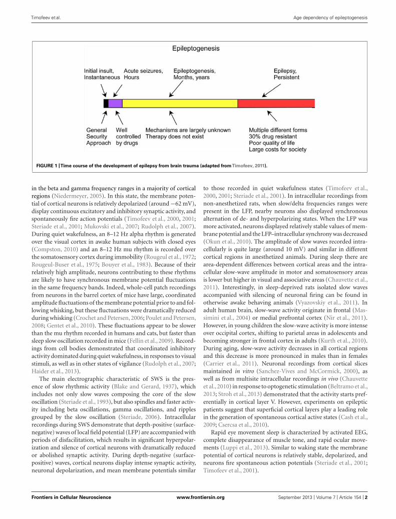
1999; Chang and Lowenstein, 2003). Thus, understanding themechanisms of trauma-induced epileptogenesis (TIE) – the setof latent processes caused by the initial insult that lead to thedevelopment of epilepsy – may lead to the development of newpreventive approaches (Figure 1).
Both the cortex and the underlying white matter are damaged inthe vast majority of brain-penetrating wounds. We review what isknown about the changes that occur in the cortex following braintrauma. Experiments with direct damage of the cortex and theunderlying white matter in young and adult cats will be describedin detail below. The evidence points toward homeostatic mecha-nisms that may account for the differences between the effects ofbrain trauma in young and adult cats.
CORTICAL ACTIVITY DURING STATES OF VIGILANCEThere are three major states of vigilance: waking (W), slow-wave sleep (SWS), and rapid eye movement (REM) sleep. Duringnormal levels of cortical activity, excitation and inhibition arebalanced. At the level of neocortex, persistent synaptic activ-ity and neuronal firing characterize both waking state and REMsleep, but in contrast, during SWS the membrane potential oscil-lates between depolarized and hyperpolarized states. Changesin the state of vigilance are controlled by shifts in the levels ofneuromodulation.
During waking state, electroencephalography (EEG) activitiesare characterized by low-amplitude, high-frequency oscillations
Frontiers in Cellular Neuroscience www.frontiersin.org September 2013 | Volume 7 | Article 154 | 1
“fncel-07-00154” — 2013/9/16 — 15:39 — page 2 — #2
Timofeev et al. Age dependency of epileptogenesis
FIGURE 1 |Time course of the development of epilepsy from brain trauma (adapted fromTimofeev, 2011).
in the beta and gamma frequency ranges in a majority of corticalregions (Niedermeyer, 2005). In this state, the membrane poten-tial of cortical neurons is relatively depolarized (around −62 mV),display continuous excitatory and inhibitory synaptic activity, andspontaneously fire action potentials (Timofeev et al., 2000, 2001;Steriade et al., 2001; Mukovski et al., 2007; Rudolph et al., 2007).During quiet wakefulness, an 8–12 Hz alpha rhythm is generatedover the visual cortex in awake human subjects with closed eyes(Compston, 2010) and an 8–12 Hz mu rhythm is recorded overthe somatosensory cortex during immobility (Rougeul et al., 1972;Rougeul-Buser et al., 1975; Bouyer et al., 1983). Because of theirrelatively high amplitude, neurons contributing to these rhythmsare likely to have synchronous membrane potential fluctuationsin the same frequency bands. Indeed, whole-cell patch recordingsfrom neurons in the barrel cortex of mice have large, coordinatedamplitude fluctuations of the membrane potential prior to and fol-lowing whisking, but these fluctuations were dramatically reducedduring whisking (Crochet and Petersen, 2006; Poulet and Petersen,2008; Gentet et al., 2010). These fluctuations appear to be slowerthan the mu rhythm recorded in humans and cats, but faster thansleep slow oscillation recorded in mice (Fellin et al., 2009). Record-ings from cell bodies demonstrated that coordinated inhibitoryactivity dominated during quiet wakefulness, in responses to visualstimuli, as well as in other states of vigilance (Rudolph et al., 2007;Haider et al., 2013).
The main electrographic characteristic of SWS is the pres-ence of slow rhythmic activity (Blake and Gerard, 1937), whichincludes not only slow waves composing the core of the slowoscillation (Steriade et al., 1993), but also spindles and faster activ-ity including beta oscillations, gamma oscillations, and ripplesgrouped by the slow oscillation (Steriade, 2006). Intracellularrecordings during SWS demonstrate that depth-positive (surface-negative) waves of local field potential (LFP) are accompanied withperiods of disfacilitation, which results in significant hyperpolar-ization and silence of cortical neurons with dramatically reducedor abolished synaptic activity. During depth-negative (surface-positive) waves, cortical neurons display intense synaptic activity,neuronal depolarization, and mean membrane potentials similar
to those recorded in quiet wakefulness states (Timofeev et al.,2000, 2001; Steriade et al., 2001). In intracellular recordings fromnon-anesthetized rats, when slow/delta frequencies ranges werepresent in the LFP, nearby neurons also displayed synchronousalternation of de- and hyperpolarizing states. When the LFP wasmore activated, neurons displayed relatively stable values of mem-brane potential and the LFP–intracellular synchrony was decreased(Okun et al., 2010). The amplitude of slow waves recorded intra-cellularly is quite large (around 10 mV) and similar in differentcortical regions in anesthetized animals. During sleep there arearea-dependent differences between cortical areas and the intra-cellular slow-wave amplitude in motor and somatosensory areasis lower but higher in visual and associative areas (Chauvette et al.,2011). Interestingly, in sleep-deprived rats isolated slow wavesaccompanied with silencing of neuronal firing can be found inotherwise awake behaving animals (Vyazovskiy et al., 2011). Inadult human brain, slow-wave activity originate in frontal (Mas-simini et al., 2004) or medial prefrontal cortex (Nir et al., 2011).However, in young children the slow-wave activity is more intenseover occipital cortex, shifting to parietal areas in adolescents andbecoming stronger in frontal cortex in adults (Kurth et al., 2010).During aging, slow-wave activity decreases in all cortical regionsand this decrease is more pronounced in males than in females(Carrier et al., 2011). Neuronal recordings from cortical slicesmaintained in vitro (Sanchez-Vives and McCormick, 2000), aswell as from multisite intracellular recordings in vivo (Chauvetteet al., 2010) in response to optogenetic stimulation (Beltramo et al.,2013; Stroh et al., 2013) demonstrated that the activity starts pref-erentially in cortical layer V. However, experiments on epilepticpatients suggest that superficial cortical layers play a leading rolein the generation of spontaneous cortical active states (Cash et al.,2009; Csercsa et al., 2010).
Rapid eye movement sleep is characterized by activated EEG,complete disappearance of muscle tone, and rapid ocular move-ments (Luppi et al., 2013). Similar to waking state the membranepotential of cortical neurons is relatively stable, depolarized, andneurons fire spontaneous action potentials (Steriade et al., 2001;Timofeev et al., 2001).
Frontiers in Cellular Neuroscience www.frontiersin.org September 2013 | Volume 7 | Article 154 | 2
“fncel-07-00154” — 2013/9/16 — 15:39 — page 3 — #3
Timofeev et al. Age dependency of epileptogenesis
CORTICAL ACTIVITY DURING STATES OF EPILEPSYMultiple brain structures are involved in seizure generation. Inneocortical epilepsy the neocortex is a primary source of epilepticactivity (Timofeev, 2010). Neocortical seizures that are primarilyfocal often become secondarily generalized tonic–clonic seizures(Crunelli and Leresche, 2002). Electrographically, these seizuresare commonly composed of spike-wave/polyspike-wave EEG dis-charges at 1.0–2.5 Hz and runs of fast spikes at 7–16 Hz (Timofeevand Steriade, 2004). LFP recordings revealed the presence of rip-ple activity during spike components of spike-wave complexes, inparticular at the onset of electrographic seizures (Grenier et al.,2003). During the spike component of spike-wave complexes,both excitatory and inhibitory cortical neurons are implicated inthe generation of paroxysmal depolarizing shift (PDS). WithinPDS, a majority of regular-spiking (mainly pyramidal) neuronsgenerates only one or a few spikes, while fast-spiking (mainlyinhibitory) interneurons fire throughout PDS with very high fre-quencies (Timofeev et al., 2002). Therefore, inhibitory activitydominates synaptic components of spikes of spike-wave com-plexes. Given that during seizure activity the reversal potential forGABAA inhibitory postsynaptic potential (IPSP) is shifted towardmore depolarized values (Cohen et al., 2002; Timofeev et al., 2002),the reversed IPSPs contribute to the generation of PDS.
Chemical synaptic interactions might not be the mostimportant mechanism that generates a PDS. During seizures theextracellular concentrations of Ca2+ decreases and K+ increases(Heinemann et al., 1977; Pumain et al., 1983; Somjen, 2002).A reduction in the extracellular Ca2+ by itself impairs thepresynaptic release of neurotransmitter (Katz, 1969). However,simultaneous reduction in Ca2+ and increase in K+ in theextracellular milieu to the levels attained during seizure activityalso prevents action potential propagation dramatically impair-ing chemical synaptic interactions (Seigneur and Timofeev, 2010).Therefore mechanisms other than chemical synaptic interactionsmay be responsible for short-range synchronization, such aselectrical coupling via gap junctions, ephaptic interactions, andextracellular communication via activity-dependent ionic changes(Jefferys, 1995; Timofeev et al., 2012). Several research groups havedemonstrated on several models of neocortical epilepsy that thala-mocortical neurons are not a major contributor to the generationof these cortical seizures (Steriade and Contreras, 1995; Pinaultet al., 1998; Timofeev et al., 1998; Steriade and Timofeev, 2001;Meeren et al., 2002; Polack and Charpier, 2006; Polack et al., 2007;Nita et al., 2008b). The runs of paroxysmal fast EEG spikes alsohave a purely cortical origin since thalamocortical neurons do notshow significant oscillatory activity when fast runs occur (Tim-ofeev et al., 1998). At the level of neocortex, fast runs start andterminate almost simultaneously over large distances, suggestingthe presence of a common input responsible for turning on and offthese fast runs. However, within fast runs, the synchrony is loose;neighboring sites of neocortex (<1 mm inter-electrode distance)can oscillate with different frequencies (Boucetta et al., 2008). Thefast-spiking neurons usually oscillate at double the frequency ofnearby recorded LFP (Timofeev et al., 1998). These observationssuggest that during seizure, long-range (mainly chemical) synapticinteractions do not have a leading role in the synchronization ofneuronal activity (Timofeev et al., 2012).
Neocortical seizures are nocturnal, occurring more oftenduring SWS (Timofeev, 2011), and when they occur during SWS,the secondary generalized seizures last much longer than duringwake (Bazil and Walczak, 1997). Why should SWS be a factor in theonset of cortical seizures? One of the major differences betweenSWS and other states of vigilance is the low activity of neuro-modulatory systems and, as a result, the network cannot maintainpermanent active states. Therefore, the main difference betweenSWS and other states of vigilance in the cortex is the presence ofhyperpolarized silent states.
Several types of anesthesia also create alternating silent andactive states. If the anesthetic used does not increase GABAergicprocesses and does not decrease gap-junction communications,it is often a seizure-triggering factor. In particular, ketamine–xylazine anesthesia in cats induces slow oscillation (alternationof active and silent states). The duration of silent states inanesthetized animals was 150–200% longer than during sleep,depending on the cortical area (Chauvette et al., 2011). As a result,75% of cats maintained under ketamine–xylazine anesthesia forseveral hours exhibited electrographic seizures (Boucetta et al.,2008). An increase in network silence may be a factor contribut-ing to seizure onsets because prolonged network silence increasesneuronal excitability (reviewed in Timofeev, 2011).
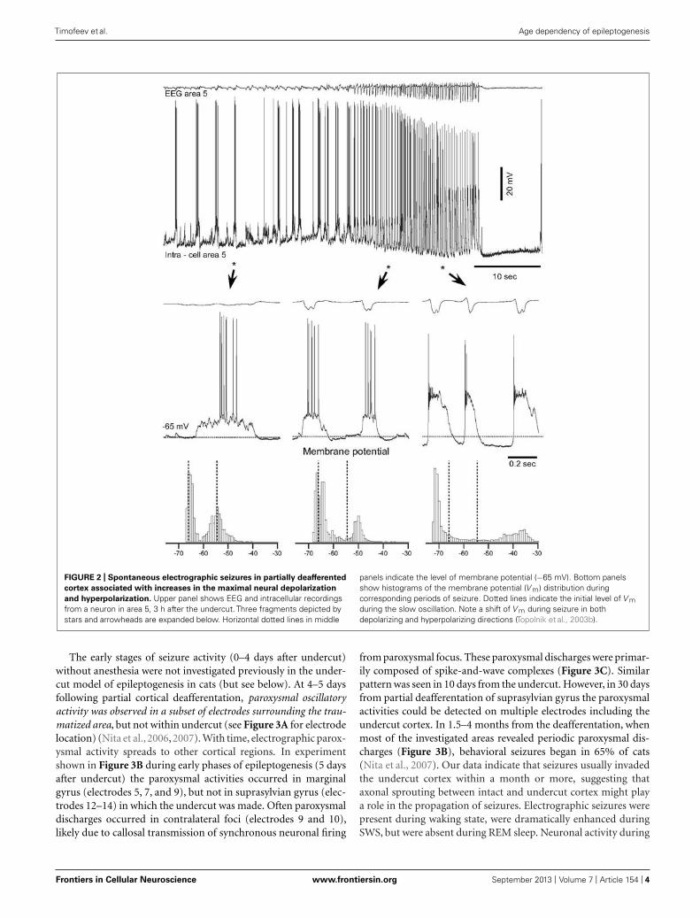
PARTIAL CORTICAL DEAFFERENTATION IS A MODEL FORTRAUMA-INDUCED EPILEPTOGENESISEpilepsy induced by a penetrating wound progresses through thesame stages of epileptogenesis as other forms of acquired epilepsy.Multiple forms of TIE has been described, but much less atten-tion was paid to epileptogenesis triggered by penetrating wounds(Hunt et al., 2013). In this experimental model a large part ofaxons connecting a given cortical area with other brain regions issevered (as example see Figure 2 in Timofeev et al., 2010). Our pre-vious experiments on cats demonstrated that immediate reactionto brain penetration, in which only slight cortical but large whitematter damage was produced, resulted in a dramatic reduction ofLFP amplitudes in areas above the damaged white matter. About3–4 h after the cortical undercut was produced, there were twomajor outcomes. In 30% of anesthetized cats slow-wave activitywas fully or partially recovered. However, in the remaining 70%of animals, slow oscillatory activity was periodically transformedinto paroxysmal discharges (Topolnik et al., 2003a,b). An exam-ple of electrographic seizure in ketamine–xylazine-anesthetizedcat with an undercut cortex is shown in Figure 2. The slow oscil-lation in the undercut cortex is different from the normal slowoscillation: in acute conditions, above the undercut area, silentstates last longer than usual (Figure 2, left, see also Figure 4 inTopolnik et al., 2003b). The electrographic seizure evolves contin-uously from the slow oscillation. Seizure onset is characterized bya shortening of both active and silence states and a slight increasein the amplitude of depolarization during active states (Figure 2,middle). The body of seizure was associated with a slight, steadyhyperpolarization and a dramatic increase in the amplitude dur-ing PDS (Figure 2, right). Under anesthesia, the seizures usuallyterminated with postictal depression characterized by EEG flatten-ing and neuronal hyperpolarization (Figure 2). This paroxysmalactivity usually lasted for 8–10 h and then spontaneously stopped.
Frontiers in Cellular Neuroscience www.frontiersin.org September 2013 | Volume 7 | Article 154 | 3
“fncel-07-00154” — 2013/9/16 — 15:39 — page 4 — #4
Timofeev et al. Age dependency of epileptogenesis
FIGURE 2 | Spontaneous electrographic seizures in partially deafferented
cortex associated with increases in the maximal neural depolarization
and hyperpolarization. Upper panel shows EEG and intracellular recordingsfrom a neuron in area 5, 3 h after the undercut. Three fragments depicted bystars and arrowheads are expanded below. Horizontal dotted lines in middle
panels indicate the level of membrane potential (−65 mV). Bottom panelsshow histograms of the membrane potential (V m) distribution duringcorresponding periods of seizure. Dotted lines indicate the initial level of V mduring the slow oscillation. Note a shift of V m during seizure in bothdepolarizing and hyperpolarizing directions (Topolnik et al., 2003b).
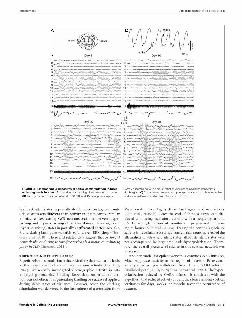
The early stages of seizure activity (0–4 days after undercut)without anesthesia were not investigated previously in the under-cut model of epileptogenesis in cats (but see below). At 4–5 daysfollowing partial cortical deafferentation, paroxysmal oscillatoryactivity was observed in a subset of electrodes surrounding the trau-matized area, but not within undercut (see Figure 3A for electrodelocation) (Nita et al., 2006, 2007). With time, electrographic parox-ysmal activity spreads to other cortical regions. In experimentshown in Figure 3B during early phases of epileptogenesis (5 daysafter undercut) the paroxysmal activities occurred in marginalgyrus (electrodes 5, 7, and 9), but not in suprasylvian gyrus (elec-trodes 12–14) in which the undercut was made. Often paroxysmaldischarges occurred in contralateral foci (electrodes 9 and 10),likely due to callosal transmission of synchronous neuronal firing
from paroxysmal focus. These paroxysmal discharges were primar-ily composed of spike-and-wave complexes (Figure 3C). Similarpattern was seen in 10 days from the undercut. However, in 30 daysfrom partial deafferentation of suprasylvian gyrus the paroxysmalactivities could be detected on multiple electrodes including theundercut cortex. In 1.5–4 months from the deafferentation, whenmost of the investigated areas revealed periodic paroxysmal dis-charges (Figure 3B), behavioral seizures began in 65% of cats(Nita et al., 2007). Our data indicate that seizures usually invadedthe undercut cortex within a month or more, suggesting thataxonal sprouting between intact and undercut cortex might playa role in the propagation of seizures. Electrographic seizures werepresent during waking state, were dramatically enhanced duringSWS, but were absent during REM sleep. Neuronal activity during
Frontiers in Cellular Neuroscience www.frontiersin.org September 2013 | Volume 7 | Article 154 | 4
“fncel-07-00154” — 2013/9/16 — 15:39 — page 5 — #5
Timofeev et al. Age dependency of epileptogenesis
FIGURE 3 | Electrographic signatures of partial deafferentation-induced
epileptogenesis in a cat. (A) Location of recording electrodes in cats brain.(B) Paroxysmal activities recorded at 5, 10, 30, and 45 days post-surgery.
Note an increasing with time number of electrodes revealing paroxysmaldischarges. (C) An expanded segment of paroxysmal discharge showing spikeand wave pattern (modified from Nita et al., 2007).
brain activated states in partially deafferented cortex, even out-side seizures was different than activity in intact cortex. Similarto intact cortex, during SWS, neurons oscillated between depo-larizing and hyperpolarizing states (see above). However, silent(hyperpolarizing) states in partially deafferented cortex were alsofound during both quiet wakefulness and even REM sleep (Tim-ofeev et al., 2010). These and related data suggest that prolongednetwork silence during seizure-free periods is a major contributingfactor in TIE (Timofeev, 2011).
OTHER MODELS OF EPILEPTOGENESISRepetitive brain stimulation induces kindling that eventually leadsto the development of spontaneous seizure activity (Goddard,1967). We recently investigated electrographic activity in catsundergoing neocortical kindling. Repetitive neocortical stimula-tion was not efficient in generating kindling or seizures if appliedduring stable states of vigilance. However, when the kindlingstimulation was delivered in the first minute of a transition from
SWS to wake, it was highly efficient in triggering seizure activity(Nita et al., 2008a,b). After the end of these seizures, cats dis-played continuing oscillatory activity with a frequency around1.5 Hz lasting from tens of minutes and progressively increas-ing to hours (Nita et al., 2008a). During the continuing seizureactivity intracellular recordings from cortical neurons revealed thealternation of active and silent states, although silent states werenot accompanied by large amplitude hyperpolarization. There-fore, the overall presence of silence in this cortical network wasincreased.
Another model for epileptogenesis is chronic GABA infusion,which suppresses activity in the region of infusion. Paroxysmalactivity emerges upon withdrawal from chronic GABA infusion(Brailowsky et al., 1988, 1989; Silva-Barrat et al., 1992). The hyper-polarization induced by GABA infusion is consistent with thehypothesis that reduced activity or periodic silence in some corticalterritories for days, weeks, or months favor the occurrence ofseizures.
Frontiers in Cellular Neuroscience www.frontiersin.org September 2013 | Volume 7 | Article 154 | 5

“fncel-07-00154” — 2013/9/16 — 15:39 — page 6 — #6
Timofeev et al. Age dependency of epileptogenesis
STRUCTURAL CHANGES ACCOMPANYING PARTIAL CORTICALDEAFFERENTATIONCutting the majority of fibers arriving to a given region triggers aset of morphological and functional changes. Axonal transectioncauses some neurons to degenerate and others that survive mayexhibit axonal sprouting.
Neuronal degenerationPartial cortical deafferentation produced a dramatic reduction(≈25%) in cortical thickness (Avramescu et al., 2009) suggest-ing a reduction in the number of cortical cells in the affectedregion. Detailed examination of the cortex above the white mat-ter transection has demonstrated the presence of delamination,reduction in the number of neurons, shrinkage as well as changein neuronal orientation in cortical depth (Avramescu et al., 2009).These features are similar to those described in the developmen-tal condition called shaken infant syndrome (Marin-Padilla et al.,2002) although mechanisms leading to these changes might bedifferent. The neuronal loss was not equally distributed over thecortical depth profile. There was a major loss of excitatory neu-rons in deep cortical layers (below 1.5 mm) with a moderate lossin superficial layers. By contrast, there was a highly significant lossof GABAergic neurons in both superficial and deep, but not inmiddle cortical layers. Why GABAergic neurons were reduced innumber is not clear. These cells in the neocortex possess shortaxons that normally do not reach white matter. It is generallyagreed that recurrent seizures may induce neuronal loss in affectedbrain regions (Cendes, 2005). However, in the cortical undercutmodel, seizures always started and generally dominated in areassurrounding the undercut cortex (Topolnik et al., 2003b; Nita et al.,2006, 2007) but not within the undercut cortex. In the region sur-rounding the undercut cortex, there was no significant alterationin the distribution of neuron types as observed within the undercutregion (Avramescu et al., 2009). The cause of interneuronal lossremains to be investigated. One of the causes might be a higherdependency of interneurons upon aerobic metabolism than othertypes of cortical neurons (Sloper et al., 1980; Ribak et al., 1982).Another cause would be a high kainate neurotoxicity of interneu-rons due to the presence of AMPA/kainate receptors with highcalcium permeability (Iino et al., 1990; Weiss et al., 1990). Theseare possibly the main two factors responsible for preferential lossof GABAergic cells in the undercut model. In layer V pyramidalneurons from the undercut cortex of rats, there was a tendencytoward reduction of basal and apical dendritic branches, but thesedifferences did not reach the level of significance (Salin et al., 1995;Avramescu et al., 2009). The preservation of layer V neurons inthe undercut model might look surprising, because the axons ofthese neurons were severed. However, layer V neurons possess veryextensive local intracortical connectivity (Markram et al., 1997),which helped them to survive cutting of extracortically going axon.In corticospinal neurons axotomy induced a reduction in the sizeof cell bodies (Tseng and Prince, 1996).
Axonal sproutingSometimes severed axons grow to re-innervate the targeted tissue.Axonal damage of corticospinal neurons at the level of the cer-vical spinal cord, however, did not induce noticeable sprouting
within neocortex (Tseng and Prince, 1996). In contrast, a corti-cal undercut produced local sprouting characterized by increasedtotal axonal length, increase in the number of axonal collater-als and number of axonal swellings (Salin et al., 1995) suggestingthat local factors and not the damage of axon per se may triggerthe axonal sprouting process. Glutamate uncaging experimentsrevealed that cortical undercut increased the number of “hot spots”on layer V pyramidal neurons accompanied with a decrease in theamplitude of individual excitatory postsynaptic current (EPSCs;Jin et al., 2006); the increased number of “hot spots” was alsofound in fast-spiking interneurons (Jin et al., 2011). The levelof “inhibitory hot spots” was decreased for both pyramidal cellsand interneurons (Jin et al., 2011). Direct investigation of con-nectivity patterns performed in vivo revealed that in partiallydeafferented cortex the connection probability between neuronswas increased starting at 2 weeks after the undercut (Avramescuand Timofeev, 2008). The amplitude of EPSPs in chronic stageswas significantly increased compared to controls at 2 and 6 weeksfrom the undercut, but it was significantly lower at 4 weeks. Thecoefficient of variation of responses was decreased with time sug-gesting a more reliable functioning of implicated synapses. Itshould be noted that synchronous network activity controls axonalsprouting after cortical trauma (Carmichael and Chesselet, 2002).Therefore, cortical trauma induces a pathological loop: axonalsprouting contributes to increased network synchronization lead-ing to seizure and synchronous cortical paroxysmal activity in thepartially deafferented cortex directly contributes to the reinforce-ment of sprouting. Altogether, a partial cortical isolation increasesthe number and the duration of silent states in the cortical net-work, which boosts neuronal connectivity and synaptic (network)excitability.
CHANGES IN THE INTRINSIC EXCITABILITY IN THE PARTIAL CORTICALDEAFFERENTATION MODELFollowing cortical trauma, intrinsic currents also undergo changesthat increase neuronal excitability. In acute conditions, the rela-tive number of intrinsically bursting neurons doubles both in theundercut cortex and in surrounding areas (Topolnik et al., 2003a),which was likely induced by local changes of K+ concentrationdue to direct neuronal damage (Jensen et al., 1994; Jensen andYaari, 1997). The membrane potential of neurons is more hyper-polarized within the undercut cortex compared to surroundingareas (Topolnik et al., 2003a). The neuronal excitability and theoverall firing, particularly in deep layers, is decreased within theundercut cortex (Topolnik et al., 2003a). Overall this results in amisbalance in excitability in the undercut and surrounding areas,creating conditions for the generation of acute seizures. Indeed,we found, using detailed conductance based models of the tha-lamocortical network including Na+ and K+ ion concentrationdynamics, that change in the normal balance of ionic concentra-tions (particularly an increase in extracellular K+ concentrations)may promote epileptiform discharges and lead to PDS (Frohlichet al., 2010; Krishnan and Bazhenov, 2011).
In chronic conditions the signs of changes of intrinsic excitabil-ity are opposite. Starting from 2 weeks after isolation in in vivoconditions the input resistance of neurons as well as their intrinsicexcitability, which is measured as the number of spikes elicited by
Frontiers in Cellular Neuroscience www.frontiersin.org September 2013 | Volume 7 | Article 154 | 6
“fncel-07-00154” — 2013/9/16 — 15:39 — page 7 — #7
Timofeev et al. Age dependency of epileptogenesis
a given current pulse or as instantaneous firing rate, is increased(Avramescu and Timofeev, 2008). Similar finding was obtained invitro (Prince and Tseng, 1993), suggesting that it is the networkexcitability and not necessarily the network properties of the trau-matized tissue that is affected. Altogether the changes in intrinsicand synaptic excitability produce an increase in the duration ofthe silent state and a compensatory increase in the instantaneousspontaneous firing rates (R = 0.87, p < 0.01), suggesting thata homeostatic regulation of the neuronal excitability took place(Avramescu and Timofeev, 2008).
HOMEOSTATIC PLASTICITY IN BRAIN TRAUMA RECOVERYAND EPILEPTOGENESISBrain excitability is maintained at a level via homeostatic mech-anisms that is neither too low nor too high. Silencing a corticalculture network for 2 days upregulates synaptic excitability andan increase in network activity down-regulates excitatory synap-tic efficacy (Turrigiano et al., 1998; Watt et al., 2000; Murthy et al.,2001), but not all connections (Kim and Tsien, 2008). Conversely,prolonged levels of enhanced activity induced by the blockade ofsynaptic inhibition or elevated [K+]o, reduces the size of mEPSCs(Lissin et al., 1998; Turrigiano et al., 1998; Leslie et al., 2001). Simi-lar activity-dependent changes in mEPSC size have been observedin spinal cell cultures (O’Brien et al., 1998). Synaptic scaling occurspost-synaptically in part by changes in the number of open chan-nels (Turrigiano et al., 1998; Watt et al., 2000), although all synapticcomponents may increase (Murthy et al., 2001) including numbersof postsynaptic glutamate receptors (Rao and Craig, 1997; Lissinet al., 1998; O’Brien et al., 1998; Liao et al., 1999). There is a simi-lar regulation of NMDA currents by activity (Watt et al., 2000; seehowever Lissin et al., 1998). Interestingly, mIPSCs are scaled downwith activity blockade, opposite in direction to changes in excita-tory currents. This effect is reversible (Rutherford et al., 1997) andis accompanied by a reduction in the number of opened GABAA
channels and GABAA receptors clustered at synaptic sites (Kilmanet al., 2002). In addition, intrinsic excitability is regulated by activ-ity. After chronic blockade of activity, Na+ currents increase andK+ currents decrease in size, resulting in an enhanced responsive-ness of pyramidal cells to current injection (Desai et al., 1999).Some of these processes may also occur in vivo (Desai et al., 2002).Thus, homeostatic plasticity also controls the levels of neuronalactivity through intrinsic mechanisms (Turrigiano et al., 1998;Murthy et al., 2001). In recent studies, we have demonstrated that(a) during TIE, cortical neurons undergo long-lasting silent peri-ods during all states of vigilance (Nita et al., 2007), (b) in a neocor-tical kindling model of epilepsy, seizures are followed by continu-ing outlasting activity (Nita et al., 2008a,b). This outlasting activitycan last for up to 2 h and consist of silent and active states. There-fore, silent periods are increased in both models of epileptogenesis.
Based on the experimental data, we developed network com-putational models in which partial cortical deafferentation led toup-regulation of the neuronal excitability and the development ofseizure-like activity (Houweling et al., 2005; Frohlich et al., 2006,2008; Volman et al., 2011a,b, 2012, 2013). First, we found thatonly sufficiently strong deafferentation leads to the pathologicalnetwork synchronization; after a weak deafferentation homeo-static plasticity was able to recover the normal asynchronous
network activity (Houweling et al., 2005). Therefore, we predictedthe existence of a critical degree of deafferentation (a thresh-old) for pathological network reorganization. Second, we foundthat both spatially defined (Houweling et al., 2005) and randomlydeafferented group of neurons may lead to pathological burst-ing (Frohlich et al., 2008). Third, we found that the network,to be prone to paroxysmal bursting should include a popula-tion of cells with relatively high density of intact neurons anda population of cells with high levels of deafferentation and lowspontaneous activity (Volman et al., 2011a,b). This suggests that,in the heterogeneous networks, epileptic activity should arise nearthe boundary of intact and deafferented areas and propagate tothe deafferented population as observed experimentally (Topolniket al., 2003b; Nita et al., 2006, 2007). Fourth, our studies predicteda critical role of interaction between neurons and glial cells in TIE(Volman et al., 2012, 2013). More recently we developed a sophis-ticated network model implementing both homeostatic plasticityand ion concentration dynamics (Gonzalez et al., 2013). This studyrevealed that the threshold between normal and pathological net-work activity (Frohlich et al., 2010) is reduced after deafferentationfollowed by homeostatic scaling. Therefore, after deafferentationeven physiological level fluctuations of the input to the networkmay trigger transition to recurrent epileptiform activity that wouldbe impossible in the normal (healthy) network.
Importantly, our modeling studies suggest an existence of bista-bility between normal and pathological (paroxysmal) activity inthe same network depending on its initial connectivity structure.Homeostatic scaling can lead to the different dynamical networkstates depending on the initial connectivity: either to recoveryof normal activity (when the damage was small) or pathologicalparoxysmal activity (when the damage was large).
WHY DO NOT ALL ANIMALS DISPLAY DEAFFERENTATION-INDUCEDEPILEPTOGENESIS?We have conducted many experiments in which cortical undercutwas used to trigger epileptogenesis. Most of the anesthetized catspresented acute seizure activity that stopped after several hours(Topolnik et al., 2003a,b), but only 65% of cats developed seizuresin chronic conditions (Nita et al., 2006, 2007; Nita and Timofeev,2007). These animals weighted more than 2.5 kg and their age wasunknown. Since 2008, new regulations of the Canadian Councilon Animal Care recommended that all experiments be performedon animals bred for research. Breeders sell only young cats (8–14month), weighing only 2.0–2.5 kg. Since 2008 experiments on thepartial deafferentation model of epileptogenesis were performedonly on young cats. Given recent technological advances we nowrecord electrographic activity immediately after the end of surgery,using a wireless system (Grand et al., 2013).
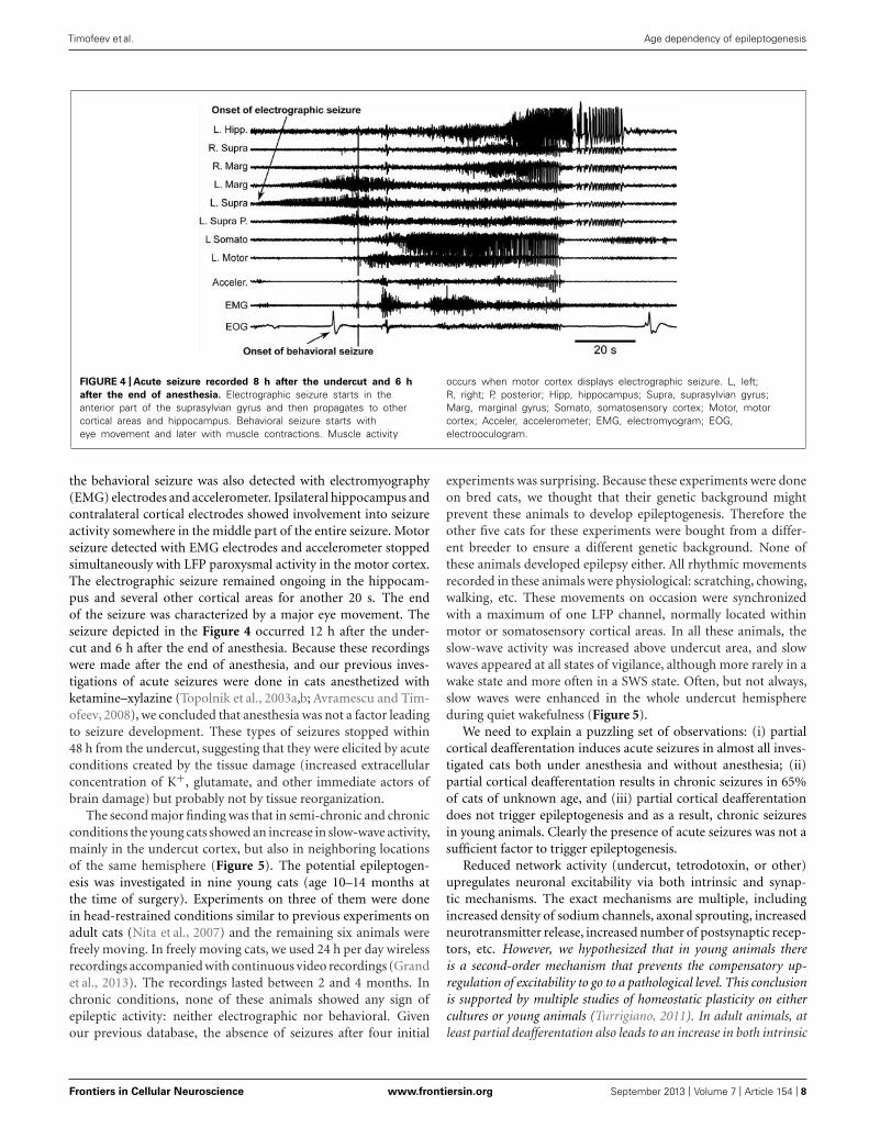
The first major finding was that, as in previous experiments,cats developed acute seizures (Figure 4). These seizures started atthe border between the undercut and intact cortex [anterior partof the left suprasylvian gyrus (L. Supra) in Figure 4] and thenother cortical areas became involved. Behavioral seizures startedtens of seconds later. In the example shown in Figure 4, the behav-ioral seizure started with eye deflection, which happened 25 s afterthe onset of electrographic seizure. About 5 s later, the motorcortex got involved in paroxysmal activity and at the same time
Frontiers in Cellular Neuroscience www.frontiersin.org September 2013 | Volume 7 | Article 154 | 7
“fncel-07-00154” — 2013/9/16 — 15:39 — page 8 — #8
Timofeev et al. Age dependency of epileptogenesis
FIGURE 4 | Acute seizure recorded 8 h after the undercut and 6 h
after the end of anesthesia. Electrographic seizure starts in theanterior part of the suprasylvian gyrus and then propagates to othercortical areas and hippocampus. Behavioral seizure starts witheye movement and later with muscle contractions. Muscle activity
occurs when motor cortex displays electrographic seizure. L, left;R, right; P, posterior; Hipp, hippocampus; Supra, suprasylvian gyrus;Marg, marginal gyrus; Somato, somatosensory cortex; Motor, motorcortex; Acceler, accelerometer; EMG, electromyogram; EOG,electrooculogram.
the behavioral seizure was also detected with electromyography(EMG) electrodes and accelerometer. Ipsilateral hippocampus andcontralateral cortical electrodes showed involvement into seizureactivity somewhere in the middle part of the entire seizure. Motorseizure detected with EMG electrodes and accelerometer stoppedsimultaneously with LFP paroxysmal activity in the motor cortex.The electrographic seizure remained ongoing in the hippocam-pus and several other cortical areas for another 20 s. The endof the seizure was characterized by a major eye movement. Theseizure depicted in the Figure 4 occurred 12 h after the under-cut and 6 h after the end of anesthesia. Because these recordingswere made after the end of anesthesia, and our previous inves-tigations of acute seizures were done in cats anesthetized withketamine–xylazine (Topolnik et al., 2003a,b; Avramescu and Tim-ofeev, 2008), we concluded that anesthesia was not a factor leadingto seizure development. These types of seizures stopped within48 h from the undercut, suggesting that they were elicited by acuteconditions created by the tissue damage (increased extracellularconcentration of K+, glutamate, and other immediate actors ofbrain damage) but probably not by tissue reorganization.
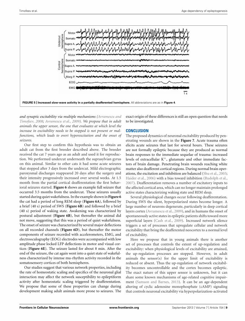
The second major finding was that in semi-chronic and chronicconditions the young cats showed an increase in slow-wave activity,mainly in the undercut cortex, but also in neighboring locationsof the same hemisphere (Figure 5). The potential epileptogen-esis was investigated in nine young cats (age 10–14 months atthe time of surgery). Experiments on three of them were donein head-restrained conditions similar to previous experiments onadult cats (Nita et al., 2007) and the remaining six animals werefreely moving. In freely moving cats, we used 24 h per day wirelessrecordings accompanied with continuous video recordings (Grandet al., 2013). The recordings lasted between 2 and 4 months. Inchronic conditions, none of these animals showed any sign ofepileptic activity: neither electrographic nor behavioral. Givenour previous database, the absence of seizures after four initial
experiments was surprising. Because these experiments were doneon bred cats, we thought that their genetic background mightprevent these animals to develop epileptogenesis. Therefore theother five cats for these experiments were bought from a differ-ent breeder to ensure a different genetic background. None ofthese animals developed epilepsy either. All rhythmic movementsrecorded in these animals were physiological: scratching, chowing,walking, etc. These movements on occasion were synchronizedwith a maximum of one LFP channel, normally located withinmotor or somatosensory cortical areas. In all these animals, theslow-wave activity was increased above undercut area, and slowwaves appeared at all states of vigilance, although more rarely in awake state and more often in a SWS state. Often, but not always,slow waves were enhanced in the whole undercut hemisphereduring quiet wakefulness (Figure 5).
We need to explain a puzzling set of observations: (i) partialcortical deafferentation induces acute seizures in almost all inves-tigated cats both under anesthesia and without anesthesia; (ii)partial cortical deafferentation results in chronic seizures in 65%of cats of unknown age, and (iii) partial cortical deafferentationdoes not trigger epileptogenesis and as a result, chronic seizuresin young animals. Clearly the presence of acute seizures was not asufficient factor to trigger epileptogenesis.
Reduced network activity (undercut, tetrodotoxin, or other)upregulates neuronal excitability via both intrinsic and synap-tic mechanisms. The exact mechanisms are multiple, includingincreased density of sodium channels, axonal sprouting, increasedneurotransmitter release, increased number of postsynaptic recep-tors, etc. However, we hypothesized that in young animals thereis a second-order mechanism that prevents the compensatory up-regulation of excitability to go to a pathological level. This conclusionis supported by multiple studies of homeostatic plasticity on eithercultures or young animals (Turrigiano, 2011). In adult animals, atleast partial deafferentation also leads to an increase in both intrinsic
Frontiers in Cellular Neuroscience www.frontiersin.org September 2013 | Volume 7 | Article 154 | 8
“fncel-07-00154” — 2013/9/16 — 15:39 — page 9 — #9
Timofeev et al. Age dependency of epileptogenesis
FIGURE 5 | Increased slow-wave activity in a partially deafferented hemisphere. All abbreviations are as in Figure 4.
and synaptic excitability via multiple mechanisms (Avramescu andTimofeev, 2008; Avramescu et al., 2009). We propose that in adultanimals the upper sensor, the one that evaluates at which level theincrease in excitability needs to be stopped is not present or mal-functions, which leads to overt hyperexcitation and the onset ofseizures.
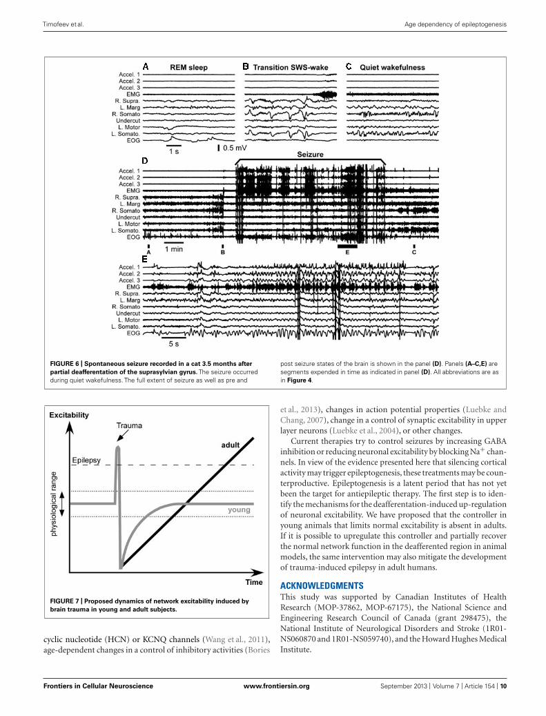
Our first step to confirm this hypothesis was to obtain anadult cat from the first breeder described above. The breederreceived the cat 7 years ago as an adult and used it for reproduc-tion. We performed undercut underneath the suprasylvian gyruson this animal. Similar to other cats it had some acute seizuresthat stopped after 3 days from the undercut. Mild electrographicparoxysmal discharges reappeared 20 days after the surgery andtheir intensity progressively increased over several weeks. At 1.5month from the partial cortical deafferentation the first behav-ioral seizures started. Figure 6 shows an example full seizure thatoccurred 3.5 months from the undercut. These seizures usuallystarted during quiet wakefulness. In the example shown in Figure 6the cat had a period of long REM sleep (Figure 6A), followed bya brief (40 s) period of SWS (Figure 6B) and followed by a brief(40 s) period of waking state. Awakening was characterized bypostural adjustment (Figure 6B), but thereafter the animal didnot move, suggesting that this was a period of quiet wakefulness.The onset of seizure was characterized by several major deflectionson all recorded channels (Figure 6D), but thereafter the motorcomponents of seizure recorded with accelerometers, EMG, andelectrooculography (EOG) electrodes were accompanied with lowamplitude phase locked LFP deflections in motor and visual cor-tices (Figure 6E). The seizure lasted for about 8 min. After theend of the seizure, the cat again went into a quiet state of wakeful-ness characterized by intense mu-rhythm activity recorded in thesomatosensory cortices of both hemispheres.
Our studies suggest that various network properties, includingthe rate of homeostatic scaling and specifics of the neuronal glialinteraction may affect the network susceptibility to epileptiformactivity after homeostatic scaling triggered by deafferentation.We propose that some of these properties can change duringdevelopment making adult animals more prone to seizures. The
exact origin of these differences is still an open question that needsto be investigated.
CONCLUSIONThe proposed dynamics of neuronal excitability produced by pen-etrating wounds are shown in the Figure 7. Acute trauma oftenelicits acute seizures that last for several hours. These seizuresare not formally epileptic because they are produced as normalbrain responses to the immediate sequelae of trauma: increasedlevels of extracellular K+, glutamate and other immediate fac-tors of brain damage. Penetrating brain wounds reaching whitematter also deafferent cortical regions. During normal brain oper-ations, the excitation and inhibition are balanced (Shu et al., 2003;Haider et al., 2006) with a bias toward inhibition (Rudolph et al.,2007). Deafferentation removes a number of excitatory inputs tothe affected cortical area, which can no longer maintain prolongedactive states characterizing waking state and REM sleep.
Several physiological changes occur following deafferentation.During SWS the silent, hyperpolarized states become longer. Alarge number of neurons degenerate, particularly in deep corticallayers cortex (Avramescu et al., 2009), and in humans the onset ofspontaneously active states in epileptic patients shifts toward moresuperficial layers (Cash et al., 2009). Increased network silencetriggers a set of processes that upregulate cellular and networkexcitability that bring the deafferented neocortex to a normal levelof excitability.
Here we propose that in young animals there is anotherset of processes that controls the extent of up-regulation andexcitability: when physiological levels of excitability are attained,the up-regulation processes are stopped. However, in adultanimals the sensor(s) for the upper limit of excitability isreduced or absent. Thus the up-regulation of network excitabil-ity becomes uncontrollable and the cortex becomes epileptic.The exact nature of this upper sensor is unknown, but it canshare some known mechanisms of age-related cognitive impair-ment (Samson and Barnes, 2013). It can be an age-dependentaltering of cyclic adenosine monophosphate (cAMP) signalingthat controls neuronal excitability via hyperpolarization-activated
Frontiers in Cellular Neuroscience www.frontiersin.org September 2013 | Volume 7 | Article 154 | 9
“fncel-07-00154” — 2013/9/16 — 15:39 — page 10 — #10
Timofeev et al. Age dependency of epileptogenesis
FIGURE 6 | Spontaneous seizure recorded in a cat 3.5 months after
partial deafferentation of the suprasylvian gyrus. The seizure occurredduring quiet wakefulness. The full extent of seizure as well as pre and
post seizure states of the brain is shown in the panel (D). Panels (A–C,E) aresegments expended in time as indicated in panel (D). All abbreviations are asin Figure 4.
FIGURE 7 | Proposed dynamics of network excitability induced by
brain trauma in young and adult subjects.
cyclic nucleotide (HCN) or KCNQ channels (Wang et al., 2011),age-dependent changes in a control of inhibitory activities (Bories
et al., 2013), changes in action potential properties (Luebke andChang, 2007), change in a control of synaptic excitability in upperlayer neurons (Luebke et al., 2004), or other changes.
Current therapies try to control seizures by increasing GABAinhibition or reducing neuronal excitability by blocking Na+ chan-nels. In view of the evidence presented here that silencing corticalactivity may trigger epileptogenesis, these treatments may be coun-terproductive. Epileptogenesis is a latent period that has not yetbeen the target for antiepileptic therapy. The first step is to iden-tify the mechanisms for the deafferentation-induced up-regulationof neuronal excitability. We have proposed that the controller inyoung animals that limits normal excitability is absent in adults.If it is possible to upregulate this controller and partially recoverthe normal network function in the deafferented region in animalmodels, the same intervention may also mitigate the developmentof trauma-induced epilepsy in adult humans.
ACKNOWLEDGMENTSThis study was supported by Canadian Institutes of HealthResearch (MOP-37862, MOP-67175), the National Science andEngineering Research Council of Canada (grant 298475), theNational Institute of Neurological Disorders and Stroke (1R01-NS060870 and 1R01-NS059740), and the Howard Hughes MedicalInstitute.
Frontiers in Cellular Neuroscience www.frontiersin.org September 2013 | Volume 7 | Article 154 | 10
“fncel-07-00154” — 2013/9/16 — 15:39 — page 11 — #11
Timofeev et al. Age dependency of epileptogenesis
REFERENCESAnnegers, J. F., Hauser, W. A., Coan,
S. P., and Rocca, W. A. (1998). Apopulation-based study of seizuresafter traumatic brain injuries. N.Engl. J. Med. 338, 20–24. doi:10.1056/NEJM199801013380104
Avramescu, S., Nita, D., and Timofeev,I. (2009). Neocortical post-traumaticepileptogenesis is associated withthe loss of GABAergic neurons.J. Neurotrauma 26, 799–812. doi:10.1089/neu.2008.0739
Avramescu, S., and Timofeev, I.(2008). Synaptic strength modu-lation following cortical trauma:a role in epileptogenesis. J.Neurosci. 28, 6760–6772. doi:10.1523/JNEUROSCI.0643-08.2008
Bazil, C. W., and Walczak, T. S.(1997). Effects of sleep and sleepstage on epileptic and nonepilepticseizures. Epilepsia 38, 56–62. doi:10.1111/j.1528-1157.1997.tb01077.x
Beltramo, R., D’Urso, G., Dal Maschio,M., Farisello, P., Bovetti, S., Clo-vis, Y., et al. (2013). Layer-specificexcitatory circuits differentially con-trol recurrent network dynamics inthe neocortex. Nat. Neurosci. 16,227–234. doi: 10.1038/nn.3306
Blake, H., and Gerard, R. W. (1937).Brain potentials during sleep. Am. J.Physiol. 119, 692–703.
Bories, C., Husson, Z., Guitton, M.J., and De Koninck, Y. (2013).Differential balance of prefrontalsynaptic activity in successful ver-sus unsuccessful cognitive aging.J. Neurosci. 33, 1344–1356. doi:10.1523/JNEUROSCI.3258-12.2013
Boucetta, S., Chauvette, S., Bazhenov,M., and Timofeev, I. (2008). Focalgeneration of paroxysmal fast runsduring electrographic seizures.Epilepsia 49, 1925–1940. doi:10.1111/j.1528-1167.2008.01707.x
Bouyer, J. J., Tilquin, C., and Rougeul,A. (1983). Thalamic rhythms incat during quiet wakefulness andimmobility. Electroencephalogr. Clin.Neurophysiol. 55, 180–187. doi:10.1016/0013-4694(83)90186-4
Brailowsky, S., Kunimoto, M., Menini,C., Silva-Barrat, C., Riche, D., andNaquet, R. (1988). The GABA-withdrawal syndrome: a new modelof focal epileptogenesis. Brain Res.442, 175–179. doi: 10.1016/0006-8993(88)91448-5
Brailowsky, S., Silva-Barrat, C., Menini,C., Riche, D., and Naquet, R. (1989).Effects of localized, chronic GABAinfusions into different corticalareas of the photosensitive baboon,Papio papio. Electroencephalogr. Clin.Neurophysiol. 72, 147–156. doi:10.1016/0013-4694(89)90176-4
Carmichael, S. T., and Chesselet, M. F.(2002). Synchronous neuronal activ-ity is a signal for axonal sproutingafter cortical lesions in the adult. J.Neurosci. 22, 6062–6070.
Carrier, J., Viens, I., Poirier, G., Robil-lard, R., Lafortune, M., Vande-walle, G., et al. (2011). Sleep slowwave changes during the middle yearsof life. Eur. J. Neurosci. 33, 758–766. doi: 10.1111/j.1460-9568.2010.07543.x
Cash, S. S., Halgren, E., Dehghani, N.,Rossetti, A. O., Thesen, T., Wang, C.,et al. (2009). The human K-complexrepresents an isolated cortical down-state. Science 324, 1084–1087. doi:10.1126/science.1169626
Cendes, F. (2005). Progressivehippocampal and extrahippocampalatrophy in drug resistant epilepsy.Curr. Opin. Neurol. 18, 173–177.doi: 10.1097/01.wco.0000162860.49842.90
Chang, B. S., and Lowenstein, D.H. (2003). Practice parameter:antiepileptic drug prophylaxis insevere traumatic brain injury: reportof the Quality Standards Subcom-mittee of the American Academyof Neurology. Neurology 60, 10–16.doi: 10.1212/01.WNL.0000031432.05543.14
Chauvette, S., Crochet, S., Volgushev,M., and Timofeev, I. (2011). Proper-ties of slow oscillation during slow-wave sleep and anesthesia in cats.J. Neurosci. 31, 14998–15008. doi:10.1523/JNEUROSCI.2339-11.2011
Chauvette, S., Volgushev, M., and Timo-feev, I. (2010). Origin of active statesin local neocortical networks duringslow sleep oscillation. Cereb. Cor-tex 20, 2660–2674. doi: 10.1093/cer-cor/bhq009
Cohen, I., Navarro, V., Clemenceau, S.,Baulac, M., and Miles, R. (2002).On the origin of interictal activityin human temporal lobe epilepsy invitro. Science 298, 1418–1421. doi:10.1126/science.1076510
Compston, A. (2010). The Bergerrhythm: potential changes from theoccipital lobes in man, by E.D.Adrian and B.H.C. Matthews (fromthe Physiological Laboratory, Cam-bridge). Brain 1934: 57; 355–385.Brain 133, 3–6. doi: 10.1093/brain/awp324
Crochet, S., and Petersen, C. C.(2006). Correlating whisker behaviorwith membrane potential in barrelcortex of awake mice. Nat. Neu-rosci. 9, 608–610. doi: 10.1038/nn1690
Crunelli, V., and Leresche, N. (2002).Childhood absence epilepsy: genes,channels, neurons and networks. Nat.
Rev. Neurosci. 3, 371–382. doi:10.1038/nrn811
Csercsa, R., Dombovári, B., Fabó, D.,Wittner, L., Eross, L., Entz, L., et al.(2010). Laminar analysis of slow waveactivity in humans. Brain 133, 2814–2829. doi: 10.1093/brain/awq169
Desai, N. S., Cudmore, R. H., Nel-son, S. B., and Turrigiano, G.G. (2002). Critical periods forexperience-dependent synaptic scal-ing in visual cortex. Nat. Neurosci. 5,783–789. doi: 10.1038/nn878
Desai, N. S., Rutherford, L. C., andTurrigiano, G. G. (1999). Plasticityin the intrinsic excitability of corticalpyramidal neurons. Nat. Neurosci. 2,515–520. doi: 10.1038/9165
Dinner, D. (1993). “Posttraumaticepilepsy,” in The Treatment ofEpilepsy: Principles, ed. E. Wyllie(Philadelphia: Lea & Fibinger), 654–658.
Feeney, D. M., and Walker, A. E.(1979). The prediction of post-traumatic epilepsy. A mathemati-cal approach. Arch. Neurol. 36,8–12. doi: 10.1001/archneur.1979.00500370038005
Fellin, T., Halassa, M. M., Terunuma,M., Succol, F., Takano, H.,Frank, M., et al. (2009). Endoge-nous nonneuronal modulators ofsynaptic transmission control corti-cal slow oscillations in vivo. Proc.Natl. Acad. Sci. U.S.A. 106,15037–15042. doi: 10.1073/pnas.0906419106
Frohlich, F., Bazhenov, M., andSejnowski, T. J. (2008). Pathologicaleffect of homeostatic synaptic scalingon network dynamics in diseases ofthe cortex. J. Neurosci. 28, 1709–1720.doi: 10.1523/JNEUROSCI.4263-07.2008
Frohlich, F., Bazhenov, M., Timofeev,I., Steriade, M., and Sejnowski, T. J.(2006). Slow state transitions of sus-tained neural oscillations by activity-dependent modulation of intrinsicexcitability. J. Neurosci. 26, 6153–6162. doi: 10.1523/JNEUROSCI.5509-05.2006
Frohlich, F., Sejnowski, T. J.,and Bazhenov, M. (2010).Network bistability mediatesspontaneous transitions betweennormal and pathological brainstates. J. Neurosci. 30, 10734–10743. doi: 10.1523/JNEUROSCI.1239-10.2010
Gentet, L. J., Avermann, M., Matyas,F., Staiger, J. F., and Petersen, C.C. H. (2010). Membrane potentialdynamics of GABAergic neuronsin the barrel cortex of behavingmice. Neuron 65, 422–435. doi:10.1016/j.neuron.2010.01.006
Goddard, G. V. (1967). Development ofepileptic seizures through brain stim-ulation at low intensity. Nature 214,1020–1021. doi: 10.1038/2141020a0
Gonzalez, O., Krishnan, G. P.,Sejnowski, T., Timofeev, I., andBazhenov, M. (2013). “Homeo-static synaptic scaling mediatesdistinct types of paroxysmal activityfollowing brain trauma,” SFN meet-ing (San Diego: SFN), programnumber 248.12/X10. Available at:http://www.abstractsonline.com/Plan/ViewAbstract.aspx?sKey=4affa068-caad-4f8c-8ba7-0b1b19ee178d&cKey=6bd4220a-2fa2-4850-a32f-ae636ca743b1&mKey=8D2A5BEC-4825-4CD6-9439-B42BB151D1CF
Grand, L., Ftomov, S., and Tim-ofeev, I. (2013). Long-term syn-chronized electrophysiological andbehavioral wireless monitoring offreely moving animals. J. Neu-rosci. Methods 212, 237–241. doi:10.1016/j.jneumeth.2012.10.008
Grenier, F., Timofeev, I., and Steri-ade, M. (2003). Neocortical very fastoscillations (ripples, 80–200 Hz) dur-ing seizures: intracellular correlates.J. Neurophysiol. 89, 841–852. doi:10.1152/jn.00420.2002
Haider, B., Duque, A., Hasenstaub, A.R., and McCormick, D. A. (2006).Neocortical network activity in vivois generated through a dynamic bal-ance of excitation and inhibition.J. Neurosci. 26, 4535–4545. doi:10.1523/JNEUROSCI.5297-05.2006
Haider, B., Hausser, M., and Caran-dini, M. (2013). Inhibition domi-nates sensory responses in the awakecortex. Nature 493, 97–100. doi:10.1038/nature11665
Heinemann, U., Lux, H. D., and Gut-nick, M. J. (1977). Extracellular freecalcium and potassium during parox-ysmal activity in the cerebral cortex ofthe cat. Exp. Brain Res. 27, 237–243.doi: 10.1007/BF00235500
Houweling, A. R., Bazhenov, M.,Timofeev, I., Steriade, M., andSejnowski, T. J. (2005). Homeostaticsynaptic plasticity can explain post-traumatic epileptogenesis in chroni-cally isolated neocortex. Cereb. Cortex15, 834–845. doi: 10.1093/cercor/bhh184
Hunt, R. F., Boychuk, J. A., andSmith, B. N. (2013). Neural cir-cuit mechanisms of posttraumaticepilepsy. Front. Cell. Neurosci. 7:89.doi: 10.3389/fncel.2013.00089
Iino, M., Ozawa, S., and Tsuzuki,K. (1990). Permeation of calciumthrough excitatory amino acid recep-tor channels in cultured rat hip-pocampal neurones. J. Physiol. 424,151–165.
Frontiers in Cellular Neuroscience www.frontiersin.org September 2013 | Volume 7 | Article 154 | 11
“fncel-07-00154” — 2013/9/16 — 15:39 — page 12 — #12
Timofeev et al. Age dependency of epileptogenesis
Jefferys, J. G. (1995). Nonsynap-tic modulation of neuronal activityin the brain: electric currents andextracellular ions. Physiol. Rev. 75,689–723.
Jensen, M. S., Azouz, R., and Yaari, Y.(1994). Variant firing patterns in rathippocampal pyramidal cells modu-lated by extracellular potassium. J.Neurophysiol. 71, 831–839.
Jensen, M. S., and Yaari, Y. (1997).Role of intrinsic burst firing, potas-sium accumulation, and electricalcoupling in the elevated potassiummodel of hippocampal epilepsy. J.Neurophysiol. 77, 1224–1233.
Jin, X., Huguenard, J. R., andPrince, D. A. (2011). Reorganiza-tion of inhibitory synaptic circuitsin rodent chronically injured epilep-togenic neocortex. Cereb. Cortex21, 1094–1104. doi: 10.1093/cer-cor/bhq181
Jin, X., Prince, D. A., and Huguenard,J. R. (2006). Enhanced excitatorysynaptic connectivity in layer Vpyramidal neurons of chronicallyinjured epileptogenic neocortexin rats. J. Neurosci. 26, 4891–4900. doi: 10.1523/JNEUROSCI.4361-05.2006
Katz, B. (1969). The Release of NeuronalTransmitter Substances. Springfield,IL: Thomas.
Kilman, V., van Rossum, M. C., andTurrigiano, G. G. (2002). Activitydeprivation reduces miniature IPSCamplitude by decreasing the numberof postsynaptic GABA(A) receptorsclustered at neocortical synapses. J.Neurosci. 22, 1328–1337.
Kim, J., and Tsien, R. W. (2008).Synapse-specific adaptations toinactivity in hippocampal cir-cuits achieve homeostatic gaincontrol while dampening networkreverberation. Neuron 58, 925–937. doi: 10.1016/j.neuron.2008.05.009
Kollevold, T. (1976). Immediate andearly cerebral seizures after headinjuries. Part I. J. Oslo City Hosp. 26,99–114.
Krishnan, G. P., and Bazhenov,M. (2011). Ionic dynamics medi-ate spontaneous termination ofseizures and postictal depressionstate. J. Neurosci. 31, 8870–8882. doi: 10.1523/JNEUROSCI.6200-10.2011
Kurth, S., Ringli, M., Geiger, A.,LeBourgeois, M., Jenni, O. G.,and Huber, R. (2010). Mappingof cortical activity in the firsttwo decades of life: a high-densitysleep electroencephalogram study. J.Neurosci. 30, 13211–13219. doi:10.1523/JNEUROSCI.2532-10.2010
Leslie, K. R., Nelson, S. B., and Tur-rigiano, G. G. (2001). Postsynapticdepolarization scales quantal ampli-tude in cortical pyramidal neurons. J.Neurosci. 21, RC170.
Liao, D., Zhang, X., O’Brien, R., Ehlers,M. D., and Huganir, R. L. (1999).Regulation of morphological postsy-naptic silent synapses in developinghippocampal neurons. Nat. Neurosci.2, 37–43. doi: 10.1038/4540
Lissin, D. V., Gomperts, S. N., Carroll,R. C., Christine, C. W., Kalman, D.,Kitamura, M., et al. (1998). Activ-ity differentially regulates the sur-face expression of synaptic AMPAand NMDA glutamate receptors.Proc. Natl. Acad. Sci. U.S.A. 95,7097–7102. doi: 10.1073/pnas.95.12.7097
Luebke, J. I., and Chang, Y. M.(2007). Effects of aging on the elec-trophysiological properties of layer 5pyramidal cells in the monkey pre-frontal cortex. Neuroscience 150, 556–562. doi: 10.1016/j.neuroscience.2007.09.042
Luebke, J. I., Chang, Y. M., Moore,T. L., and Rosene, D. L. (2004).Normal aging results in decreasedsynaptic excitation and increasedsynaptic inhibition of layer 2/3 pyra-midal cells in the monkey prefrontalcortex. Neuroscience 125, 277–288.doi: 10.1016/j.neuroscience.2004.01.035
Luppi, P.-H., Clement, O., and Fort,P. (2013). Paradoxical (REM)sleep genesis by the brainstemis under hypothalamic con-trol. Curr. Opin. Neurobiol. doi:10.1016/j.conb.2013.02.006 [Epubahead of print].
Marcikic, M., Melada, A., and Kovace-vic, R. (1998). Management of warpenetrating craniocerebral injuriesduring the war in Croatia. Injury29, 613–618. doi: 10.1016/S0020-1383(98)00146-6
Marin-Padilla, M., Parisi, J. E.,Armstrong, D. L., Sargent, S.K., and Kaplan, J. A. (2002).Shaken infant syndrome: devel-opmental neuropathology, progres-sive cortical dysplasia, and epilepsy.Acta Neuropathol. (Berl.) 103,321–332. doi: 10.1007/s00401-001-0470-z
Markram, H., Lubke, J., Frotscher, M.,Roth, A., and Sakmann, B. (1997).Physiology and anatomy of synap-tic connections between thick tuftedpyramidal neurones in the develop-ing rat neocortex. J. Physiol. 500,409–440.
Massimini, M., Huber, R., Ferrarelli,F., Hill, S., and Tononi, G. (2004).The sleep slow oscillation as a
traveling wave. J. Neurosci. 24, 6862–6870. doi: 10.1523/JNEUROSCI.1318-04.2004
Meeren, H. K., Pijn, J. P., Van Lui-jtelaar, E. L., Coenen, A. M., andLopes da Silva, F. H. (2002). Corticalfocus drives widespread corticotha-lamic networks during spontaneousabsence seizures in rats. J. Neurosci.22, 1480–1495.
Mukovski, M., Chauvette, S., Timo-feev, I., and Volgushev, M. (2007).Detection of active and silent statesin neocortical neurons from the fieldpotential signal during slow-wavesleep. Cereb. Cortex 17, 400–414. doi:10.1093/cercor/bhj157
Murthy, V. N., Schikorski, T., Stevens,C. F., and Zhu, Y. (2001). Inactivityproduces increases in neurotransmit-ter release and synapse size. Neuron32, 673–682. doi: 10.1016/S0896-6273(01)00500-1
Niedermeyer, E. (2005). “The nor-mal EEG of the waking adult,” inElectroencephalography: Basic Princi-ples, Clinical Applications and RelatedFields. eds E. Niedermeyer and F.Lopes da Silva (Philadelphia: Lippin-cott Williams & Wilkins), pp 167–192.
Nir, Y., Staba, R. J., Andrillon,T., Vyazovskiy Vladyslav, V.,Cirelli, C., Fried, I., et al. (2011).Regional slow waves and spindlesin human sleep. Neuron 70, 153–169. doi: 10.1016/j.neuron.2011.02.043
Nita, D., Cisse, Y., and Timofeev, I.(2008a). State-dependent slow out-lasting activities following neocorti-cal kindling in cats. Exp. Neurol. 211,456–468. doi: 10.1016/j.expneurol.2008.02.010
Nita, D., Cisse, Y., Frohlich, F., andTimofeev, I. (2008b). Cortical andthalamic components of neocorticalkindling-induced epileptogenesis inbehaving cats. Exp. Neurol. 211, 518–528. doi: 10.1016/j.expneurol.2008.02.028
Nita, D., Cissé, Y., Timofeev, I.,and Steriade, M. (2006). Increasedpropensity to seizures after chroniccortical deafferentation in vivo. J.Neurophysiol. 95, 902–913. doi:10.1152/jn.00742.2005
Nita, D., and Timofeev, I. (2007).“Incessant transitions between activeand silent states in thalamocorticalcircuits lead to epilepsy,” in Mech-anisms of Spontaneous Active Statesin the Neocortex, ed. I. Timofeev(Trivandrum: Research Signpost),135–168.
Nita, D. A., Cisse, Y., Timofeev, I.,and Steriade, M. (2007). Waking–sleep modulation of paroxysmal
activities induced by partial corti-cal deafferentation. Cereb. Cortex17, 272–283. doi: 10.1093/cercor/bhj145
O’Brien, R. J., Kamboj, S., Ehlers,M. D., Rosen, K. R., Fischbach,G. D., and Huganir, R. L. (1998).Activity-dependent modulation ofsynaptic AMPA receptor accumula-tion. Neuron 21, 1067–1078. doi:10.1016/S0896-6273(00)80624-8
Okun, M., Naim, A., and Lampl, I.(2010). The subthreshold relationbetween cortical local field poten-tial and neuronal firing unveiledby intracellular recordings inawake rats. J. Neurosci. 30, 4440–4448. doi: 10.1523/JNEUROSCI.5062-09.2010
Pinault, D., Leresche, N., Charpier,S., Deniau, J. M., Marescaux, C.,Vergnes, M., et al. (1998). Intra-cellular recordings in thalamic neu-rones during spontaneous spike andwave discharges in rats with absenceepilepsy. J. Physiol. 509, 449–456. doi: 10.1111/j.1469-7793.1998.449bn.x
Polack, P.-O., and Charpier, S. (2006).Intracellular activity of cortical andthalamic neurones during high-voltage rhythmic spike discharge inLong–Evans rats in vivo. J. Physiol.571, 461–476. doi: 10.1113/jphys-iol.2005.100925
Polack, P.-O., Guillemain, I., Hu,E., Deransart, C., Depaulis, A.,and Charpier, S. (2007). Deeplayer somatosensory cortical neu-rons initiate spike-and-wave dis-charges in a genetic model of absenceseizures. J. Neurosci. 27, 6590–6599. doi: 10.1523/JNEUROSCI.0753-07.2007
Poulet, J. F. A., and Petersen, C.C. H. (2008). Internal brain stateregulates membrane potential syn-chrony in barrel cortex of behavingmice. Nature 454, 881–885. doi:10.1038/nature07150
Prince, D. A., and Tseng, G. F. (1993).Epileptogenesis in chronically injuredcortex: in vitro studies. J. Neurophys-iol. 69, 1276–1291.
Pumain, R., Kurcewicz, I., andLouvel, J. (1983). Fast extracel-lular calcium transients: involve-ment in epileptic processes. Science222, 177–179. doi: 10.1126/science.6623068
Rao, A., and Craig, A. M. (1997).Activity regulates the synaptic local-ization of the NMDA receptorin hippocampal neurons. Neuron19, 801–812. doi: 10.1016/S0896-6273(00)80962-9
Ribak, C. E., Bradurne, R. M., and Har-ris, A. B. (1982). A preferential loss
Frontiers in Cellular Neuroscience www.frontiersin.org September 2013 | Volume 7 | Article 154 | 12
“fncel-07-00154” — 2013/9/16 — 15:39 — page 13 — #13
Timofeev et al. Age dependency of epileptogenesis
of GABAergic, symmetric synapsesin epileptic foci: a quantitative ultra-structural analysis of monkey neo-cortex. J. Neurosci. 2, 1725–1735.
Rougeul, A., Letalle, A., and Corvisier,J. (1972). Activite rythmique ducortex somesthesique primaire enrelation avec l’immobilite chezle chat libre eveille. Electroen-cephalogr. Clin. Neurophysiol. 33,23–39. doi: 10.1016/0013-4694(72)90022-3
Rougeul-Buser, A., Bouyer, J. J., andBuser, P. (1975). From attentivenessto sleep. A topographical analysis oflocalized “synchronized” activities onthe cortex of normal cat and mon-key. Acta Neurobiol. Exp. (Wars.) 35,805–819.
Rudolph, M., Pospischil, M., Timofeev,I., and Destexhe, A. (2007). Inhibi-tion determines membrane potentialdynamics and controls action poten-tial generation in awake and sleepingcat cortex. J. Neurosci. 27, 5280–5290. doi: 10.1523/JNEUROSCI.4652-06.2007
Rutherford, L. C., DeWan, A., Lauer,H. M., and Turrigiano, G. G. (1997).Brain-derived neurotrophic factormediates the activity-dependent reg-ulation of inhibition in neocorticalcultures. J. Neurosci. 17, 4527–4535.
Salazar, A., Jabbari, B., Vance, S.,Grafman, J., Amin, D., and Dillon,J. (1985). Epilepsy after penetrat-ing head injury. I. Clinical corre-lates: a report of the Vietnam HeadInjury Study. Neurology 35, 1406–1414. doi: 10.1212/WNL.35.10.1406
Salin, P., Tseng, G.-F., Hoffman, S.,Parada, I., and Prince, D. A. (1995).Axonal sprouting in layer V pyra-midal neurons of chronically injuredcerebral cortex. J. Neurosci. 15, 8234–8245.
Samson, R. D., and Barnes, C. A.(2013). Impact of aging brain cir-cuits on cognition. Eur. J. Neurosci.37, 1903–1915. doi: 10.1111/ejn.12183
Sanchez-Vives, M. V., and McCormick,D. A. (2000). Cellular and networkmechanisms of rhythmic recurrentactivity in neocortex. Nat. Neu-rosci. 3, 1027–1034. doi: 10.1038/79848
Seigneur, J., and Timofeev, I. (2010).Synaptic impairment induced byparoxysmal ionic conditions in neo-cortex. Epilepsia 52, 132–139. doi:10.1111/j.1528-1167.2010.02784.x
Shu, Y., Hasenstaub, A., andMcCormick, D. A. (2003). Turningon and off recurrent balanced cor-tical activity. Nature 423, 288–293.doi: 10.1038/nature01616
Silva-Barrat, C., Araneda, S., Menini,C., Champagnat, J., and Naquet, R.(1992). Burst generation in neocor-tical neurons after GABA withdrawalin the rat. J. Neurophysiol. 67, 715–727.
Sloper, J. J., Johnson, P., and Pow-ell, T. P. (1980). Selective degener-ation of interneurons in the motorcortex of infant monkeys followingcontrolled hypoxia: a possible causeof epilepsy. Brain Res. 198, 204–209. doi: 10.1016/0006-8993(80)90356-X
Somjen, G. G. (2002). Ion regu-lation in the brain: implicationsfor pathophysiology. Neuroscien-tist 8, 254–267. doi: 10.1177/1073858402008003011
Steriade, M. (2006). Grouping of brainrhythms in corticothalamic sys-tems. Neuroscience 137, 1087–1106.doi: 10.1016/j.neuroscience.2005.10.029
Steriade, M., and Contreras, D.(1995). Relations between corticaland thalamic cellular events dur-ing transition from sleep patterns toparoxysmal activity. J. Neurosci. 15,623–642.
Steriade, M., and Timofeev, I.(2001). Corticothalamic operationsthrough prevalent inhibition ofthalamocortical neurons. ThalamusRelat. Syst. 1, 225–236. doi:10.1017/S147292880100022X
Steriade, M., Nuñez, A., and Amz-ica, F. (1993). A novel slow (<1 Hz)oscillation of neocortical neurons invivo: depolarizing and hyperpolar-izing components. J. Neurosci. 13,3252–3265.
Steriade, M., Timofeev, I., and Gre-nier, F. (2001). Natural waking andsleep states: a view from inside neo-cortical neurons. J. Neurophysiol. 85,1969–1985.
Stroh, A., Adelsberger, H., Groh,A., Rühlmann, C., Fischer, S.,Schierloh, A., et al. (2013). Mak-ing waves: initiation and prop-agation of corticothalamic Ca2+waves in vivo. Neuron 77, 1136–1150. doi: 10.1016/j.neuron.2013.01.031
Temkin, N. R., Dikmen, S. S., Ander-son, G. D., Wilensky, A. J., Holmes,M. D., Cohen, W., et al. (1999).Valproate therapy for preventionof posttraumatic seizures: a ran-domized trial. J. Neurosurg. 91,593–600. doi: 10.3171/jns.1999.91.4.0593
Temkin, N. R., Dikmen, S. S., Wilen-sky, A. J., Keihm, J., Chabal,S., and Winn, H. R. (1990). Arandomized, double-blind study ofphenytoin for the prevention of
post-traumatic seizures. N. Engl. J.Med. 323, 497–502. doi: 10.1056/NEJM199008233230801
Temkin, N. R., Haglund, M. M., andWinn, H. R. (1995). Causes, preven-tion, and treatment of post-traumaticepilepsy. New Horizons (Baltimore,MD) 3, 518–522.
Timofeev, I. (2010). “Pathophysiol-ogy of neocortical seizures,” inThe Atlas of Epilepsies, ed. C. P.Panayiotopoulos (London: Springer-Verlag), 203–212.
Timofeev, I. (2011). “Injury inducedepileptogenesis: contribution ofactive inhibition, disfacilitation anddeafferentation to seizure induc-tion in thalamocortical system,” inInhibitory Synaptic Plasticity, eds M.A. Woodin, and A. Maffei (New York:Springer), 107–122.
Timofeev, I., Bazhenov, M., Avramescu,S., and Nita, D. A. (2010).Posttraumatic epilepsy: the rolesof synaptic plasticity. Neuroscien-tist 16, 19–27. doi: 10.1177/1073858409333545
Timofeev, I., Bazhenov, M., Seigneur,J., and Sejnowski, T. (2012). “Neu-ronal synchronization and thalamo-cortical rhythms in sleep, wake, andepilepsy,” in Jasper’s Basic Mechanismsof the Epilepsies, eds J. L. Noebels,M. Avoli, M. A. Rogawski, R. W.Olsen, and A. V. Delgado-Escueta(New York: Oxford University Press),157–175.
Timofeev, I., Grenier, F., and Steriade,M. (1998). Spike-wave complexesand fast components of corticallygenerated seizures. IV. Paroxysmalfast runs in cortical and thala-mic neurons. J. Neurophysiol. 80,1495–1513.
Timofeev, I., Grenier, F., and Steri-ade, M. (2000). Impact of intrinsicproperties and synaptic factors onthe activity of neocortical networksin vivo. J. Physiol. (Paris) 94, 343–355. doi: 10.1016/S0928-4257(00)01097-4
Timofeev, I., Grenier, F., and Ste-riade, M. (2001). Disfacilitationand active inhibition in the neo-cortex during the natural sleep–wake cycle: an intracellular study.Proc. Natl. Acad. Sci. U.S.A. 98,1924–1929. doi: 10.1073/pnas.98.4.1924
Timofeev, I., Grenier, F., and Steriade,M. (2002). The role of chloride-dependent inhibition and the activityof fast-spiking neurons during corti-cal spike-wave seizures. Neuroscience114, 1115–1132. doi: 10.1016/S0306-4522(02)00300-7
Timofeev, I., and Steriade, M.(2004). Neocortical seizures:
initiation, development and ces-sation. Neuroscience 123, 299–336.doi: 10.1016/j.neuroscience.2003.08.051
Topolnik, L., Steriade, M., and Tim-ofeev, I. (2003a). Hyperexcitabil-ity of intact neurons underliesacute development of trauma-relatedelectrographic seizures in cats invivo. Eur. J. Neurosci. 18, 486–496. doi: 10.1046/j.1460-9568.2003.02742.x
Topolnik, L., Steriade, M., and Timo-feev, I. (2003b). Partial cortical deaf-ferentation promotes developmentof paroxysmal activity. Cereb. Cor-tex 13, 883–893. doi: 10.1093/cer-cor/13.8.883
Tseng, G-F., and Prince, D. A. (1996).Structural and functional alter-ations in rat corticospinal neuronsafter axotomy. J. Neurophysiol. 75,248–267.
Turrigiano, G. (2011). Too manycooks? Intrinsic and synaptichomeostatic mechanisms in cor-tical circuit refinement. Annu.Rev. Neurosci. 34, 89–103. doi:10.1146/annurev-neuro-060909-153238
Turrigiano, G. G., Leslie, K. R.,Desai, N. S., Rutherford, L.C., and Nelson, S. B. (1998).Activity-dependent scaling of quan-tal amplitude in neocortical neurons.Nature 391, 892–896. doi: 10.1038/36103
Volman, V., Bazhenov, M., andSejnowski, T. J. (2011a). Patternof trauma determines the thresh-old for epileptic activity in amodel of cortical deafferentation.Proc. Natl. Acad. Sci. U.S.A. 108,15402–15407. doi: 10.1073/pnas.1112066108
Volman, V., Sejnowski, T. J., andBazhenov, M. (2011b). Topo-logical basis of epileptogenesisin a model of severe corticaltrauma. J. Neurophysiol. 106,1933–1942. doi: 10.1152/jn.00458.2011
Volman, V., Bazhenov, M.,and Sejnowski, T. J. (2012).Computational models ofneuron–astrocyte interaction inepilepsy. Front. Comput. Neurosci.6:58. doi: 10.3389/fncom.2012.00058
Volman, V., Bazhenov, M., andSejnowski, T. J. (2013). Divideand conquer: functional segrega-tion of synaptic inputs by astro-cytic microdomains could alleviateparoxysmal activity followingbrain trauma. PLoS Comput. Biol.9:e1002856. doi: 10.1371/journal.pcbi.1002856
Frontiers in Cellular Neuroscience www.frontiersin.org September 2013 | Volume 7 | Article 154 | 13
“fncel-07-00154” — 2013/9/16 — 15:39 — page 14 — #14
Timofeev et al. Age dependency of epileptogenesis
Vyazovskiy, V. V., Olcese, U., Hanlon,E. C., Nir, Y., Cirelli, C., and Tononi,G. (2011). Local sleep in awake rats.Nature 472, 443–447. doi: 10.1038/nature10009
Wang, M., Gamo, N. J., Yang, Y.,Jin, L. E., Wang, X-J., Laubach,M., et al. (2011). Neuronal basis ofage-related working memory decline.Nature 476, 210–214. doi: 10.1038/nature10243
Watt, A. J., van Rossum, M.C., MacLeod, K. M., Nelson,S. B., and Turrigiano, G. G.(2000). Activity coregulates quantal
AMPA and NMDA currents atneocortical synapses. Neuron 26,659–670. doi: 10.1016/S0896-6273(00)81202-7
Weiss, J. H., Koh, J., Baimbridge, K.G., and Choi, D. W. (1990). Corticalneurons containing somatostatin- orparvalbumin-like immunoreactivityare atypically vulnerable to excito-toxic injury in vitro. Neurology 40,1288–1292. doi: 10.1212/WNL.40.8.1288
Conflict of Interest Statement: Theauthors declare that the research was
conducted in the absence of anycommercial or financial relationshipsthat could be construed as a potentialconflict of interest.
Received: 07 June 2013; accepted: 26August 2013; published online: 18September 2013.Citation: Timofeev I, SejnowskiTJ, Bazhenov M, Chauvette Sand Grand LB (2013) Age depen-dency of trauma-induced neocorticalepileptogenesis. Front. Cell. Neu-rosci. 7:154. doi: 10.3389/fncel.2013.00154
This article was submitted to the journalFrontiers in Cellular Neuroscience.Copyright © 2013 Timofeev, Sejnowski,Bazhenov, Chauvette and Grand. This isan open-access article distributed underthe terms of the Creative CommonsAttribution License (CC BY). The use,distribution or reproduction in otherforums is permitted, provided the origi-nal author(s) or licensor are credited andthat the original publication in this jour-nal is cited, in accordance with acceptedacademic practice. No use, distribution orreproduction is permitted which does notcomply with these terms.
Frontiers in Cellular Neuroscience www.frontiersin.org September 2013 | Volume 7 | Article 154 | 14