AFM file 5 Table of Contents Page Welcome Message 6 Organizing Committee 7 Program 8 Bruker Workshop...
127
6 th AFM BioMED Conference San Diego 2014 La Jolla, California, USA December 13-17, 2014 AFM BioMED 2014 Conference Program
Transcript of AFM file 5 Table of Contents Page Welcome Message 6 Organizing Committee 7 Program 8 Bruker Workshop...

http://afmbiomed.org 1
6th AFM BioMED Conference
San Diego 2014
La Jolla, California, USA
December 13-17, 2014
AFM BioMED
2014 Conference
Program
http://afmbiomed.org 2
http://afmbiomed.org 3
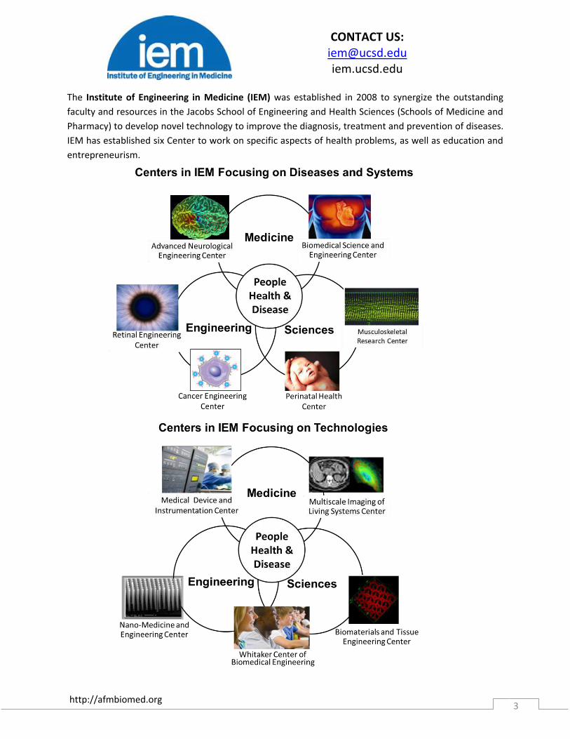
The Institute of Engineering in Medicine (IEM) was established in 2008 to synergize the outstanding
faculty and resources in the Jacobs School of Engineering and Health Sciences (Schools of Medicine and
Pharmacy) to develop novel technology to improve the diagnosis, treatment and prevention of diseases.
IEM has established six Center to work on specific aspects of health problems, as well as education and
entrepreneurism.
CONTACT US: [email protected] iem.ucsd.edu

http://afmbiomed.org 4
Thank You to Our
Sponsors!

http://afmbiomed.org 5
Table of Contents
Page Welcome Message 6 Organizing Committee 7 Program 8
Bruker Workshop 9 Day 1-December 14,2014 10 Day 2-December 15,2014 13 Day 3-December 16,2014 16 Day 4-December 17,2014 19 Poster List 21 Abstracts
Day 1 26 Day 2 46 Day 3 70 Day 4 89
Poster Abstracts 102
http://afmbiomed.org 6
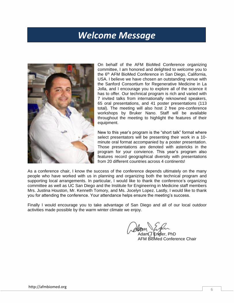
On behalf of the AFM BioMed Conference organizing committee, I am honored and delighted to welcome you to the 6th AFM BioMed Conference in San Diego, California, USA. I believe we have chosen an outstanding venue with the Sanford Consortium for Regenerative Medicine in La Jolla, and I encourage you to explore all of the science it has to offer. Our technical program is rich and varied with 7 invited talks from internationally reknowned speakers, 65 oral presentations, and 41 poster presentations (113 total). The meeting will also host 2 free pre-conference workshops by Bruker Nano. Staff will be available throughout the meeting to highlight the features of their equipment. New to this year’s program is the “short talk” format where select presentators will be presenting their work in a 10-minute oral format accompanied by a poster presentation. Those presentations are denoted with astericks in the program for your convience. This year’s program also features record geographical diversity with presentations from 20 different countries across 4 continents!
As a conference chair, I know the success of the conference depends ultimately on the many people who have worked with us in planning and organizing both the technical program and supporting local arrangements. In particular, I would like to thank the conference’s organizing committee as well as UC San Diego and the Institute for Engineering in Medicine staff members Mrs. Justina Houston, Mr. Kenneth Tomory, and Ms. Jocelyn Lopez. Lastly, I would like to thank you for attending the conference. Your attendance helps ensure the meeting’s success. Finally I would encourage you to take advantage of San Diego and all of our local outdoor activities made possible by the warm winter climate we enjoy.
Adam J Engler, PhD AFM BioMed Conference Chair
Welcome Message

http://afmbiomed.org 7
Pierre PAROT Professor CEA Marcoule, France Jean-Luc PELLEQUER Professor CEA Marcoule, France Daniel NAVAJAS Professor University of Barcelona, Spain Sanjay KUMAR Professor UC Berkeley, USA Vesna SVETLICIC Professor Rudjer Boskovic Institute, Zagreb, Croatia Simon SCHEURING Research Director Inserm, Marseille, France Jun HU Professor Shanghai Institute of Applied Physics, CAS, China Adam ENGLER Associate Professor UC San Diego, USA (Local Chair)
Organizing Committee

http://afmbiomed.org 8
Toble
Topics Chairs Invited Speakers Imaging Clemens Franz
IT, Karlsruhe, Germany
James J. De Yoreo
PNNL, Richland, USA
Integrative AFM Developments Robert Ros
ASU, Tempe, USA
Tilman Schäffer
University of Tübingen, Germany
Forces and Biomechanics Hermann Gaub
LMU Munich, Germany
Hongbin Li
UBC, Vancouver, Canada
Biomedical Applications James Gimzewski
UC Los Angeles, USA
Hans Oberleithner
Münster University, Germany
AFM BioMed Conference San Diego, December (13)14AFM BioMed Conference San Diego, December (13)14--17, 201417, 2014
After Barcelona 2007 (Spain), Monterey 2008 (USA), Red Island 2010 (Croatia), Paris 2011 (France) and Shanghai 2013 (China), AFM BioMed Conference has the pleasure to announce the 6th conference on AFM for Life Sciences and Nanomedicine, on December (13)14-17, 2014 (including training) in San Diego, California, USA.
The conference is hosted by the University of California, San Diego (UCSD). The venue is the Auditorium of Sanford Consortium for Regenerative Medicine.
The Conference is chaired by Professor Adam Engler, UCSD.
PROGRAMPROGRAM
http://afmbiomed.org 9
Saturday December 13th 2014
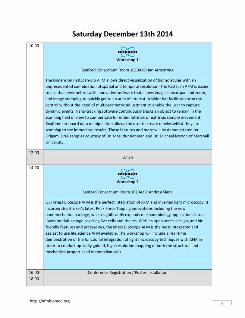
10:00
Workshop 1
Sanford Consortium Room 1013A/B: Ian Armstrong
The Dimension FastScan-Bio AFM allows direct visualization of biomolecules with an
unprecedented combination of spatial and temporal resolution. The FastScan AFM is easier
to use than ever before with innovative software that allows image canvas pan and zoom,
and image stamping to quickly get to an area of interest. A slider bar facilitates scan rate
control without the need of multiparametric adjustment to enable the user to capture
dynamic events. Nano-tracking software continuously tracks an object to remain in the
scanning field of view to compensate for either intrinsic or extrinsic sample movement.
Realtime on-board data manipulation allows the user to create movies whilst they are
scanning to see immediate results. These features and more will be demonstrated on
Origami DNA samples courtesy of Dr. Masudur Rahman and Dr. Michael Norton of Marshall
University.
12:00
Lunch
14:00
Workshop 2
Sanford Consortium Room 1013A/B: Andrea Slade
Our latest BioScope AFM is the perfect integration of AFM and inverted light microscopy. It
incorporates Bruker’s latest Peak Force Tapping innovations including the new
nanomechanics package, which significantly expands mechanobiology applications into a
lower modulus range covering live cells and tissues. With its open access design, and bio
friendly features and accessories, the latest BioScope AFM is the most integrated and
easiest to use life science AFM available. The workshop will include a real-time
demonstration of the functional integration of light microscopy techniques with AFM in
order to conduct optically guided, high-resolution mapping of both the structural and
mechanical properties of mammalian cells.
16:00-
18:00
Conference Registration / Poster Installation

http://afmbiomed.org 10
All the scientific sessions will happen in the
Roth Auditorium of Sanford Consortium for Regenerative Medicine.
Sunday Dec 14th 2014 - DAY 1 8:00 Registration / Poster Installation Abstract #
8:45 Welcome Address and Conference Introduction
Adam J. Engler, Conference Chair
Shu Chien, Director of IEM
9:00 Invited Lecture: “Using in situ AFM to understand how proteins assemble into ordered structures that direct the formation of mineralized tissues”
James J. De Yoreo, Pacific Northwest National Laboratory, Richland, WA, USA
1-1
9:30 “Marine polysaccharide networks: self-assembly vs. self-organization revealed by atomic force microscopy”
Vesna Svetličić, Ruđer Bošković Institute, Zagreb, Croatia
1-2
9:50 “Protein-protein and Protein-membrane interaction of Annexin-A5”
Atsushi Miyagi, INSERM, Université Aix-Marseille, Marseille, France
1-3
10:10 “Imaging electrostatic charge distribution in biomembranes using low oscillation Dynamic Atomic Force Microscopy”
Jaime Colchero, Universidad de Murcia, Madrid, Spain
1-4
10:30
Coffee Break
10:50 “Probing the compressibility of tumor cell nuclei by combined atomic force-confocal microscopy”
Marina Krause, Radboud University Nijmegen Medical Centre, Nijmegen, The Netherlands
1-5
11:10 “Probing of antigens on malaria infected erythrocytes using protein-antibody affinity based molecular force spectroscopy”
Himanshu Singh, National University of Singapore, Singapore
1-6
11:20* “High-Speed Atomic Force Microscopy of ESCRT protein assembly”
Lorena Redondo-Morata, INSERM, Aix-Marseille Université, Marseille, France
1-7
http://afmbiomed.org 11
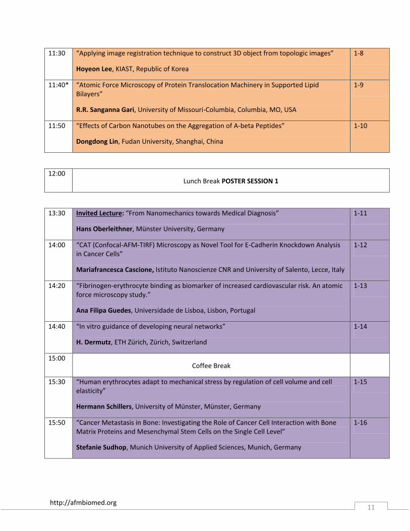
11:30 “Applying image registration technique to construct 3D object from topologic images”
Hoyeon Lee, KIAST, Republic of Korea
1-8
11:40* “Atomic Force Microscopy of Protein Translocation Machinery in Supported Lipid Bilayers”
R.R. Sanganna Gari, University of Missouri-Columbia, Columbia, MO, USA
1-9
11:50 “Effects of Carbon Nanotubes on the Aggregation of A-beta Peptides”
Dongdong Lin, Fudan University, Shanghai, China
1-10
12:00
Lunch Break POSTER SESSION 1
13:30
Invited Lecture: “From Nanomechanics towards Medical Diagnosis”
Hans Oberleithner, Münster University, Germany
1-11
14:00 “CAT (Confocal-AFM-TIRF) Microscopy as Novel Tool for E-Cadherin Knockdown Analysis in Cancer Cells”
Mariafrancesca Cascione, Istituto Nanoscienze CNR and University of Salento, Lecce, Italy
1-12
14:20 “Fibrinogen-erythrocyte binding as biomarker of increased cardiovascular risk. An atomic force microscopy study.”
Ana Filipa Guedes, Universidade de Lisboa, Lisbon, Portugal
1-13
14:40 “In vitro guidance of developing neural networks”
H. Dermutz, ETH Zürich, Zürich, Switzerland
1-14
15:00
Coffee Break
15:30 “Human erythrocytes adapt to mechanical stress by regulation of cell volume and cell elasticity”
Hermann Schillers, University of Münster, Münster, Germany
1-15
15:50 “Cancer Metastasis in Bone: Investigating the Role of Cancer Cell Interaction with Bone Matrix Proteins and Mesenchymal Stem Cells on the Single Cell Level”
Stefanie Sudhop, Munich University of Applied Sciences, Munich, Germany
1-16
http://afmbiomed.org 12
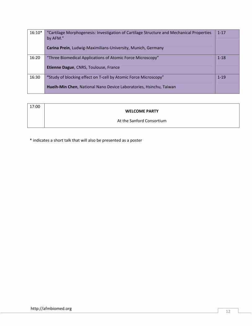
16:10* “Cartilage Morphogenesis: Investigation of Cartilage Structure and Mechanical Properties by AFM.”
Carina Prein, Ludwig-Maximilians-University, Munich, Germany
1-17
16:20 “Three Biomedical Applications of Atomic Force Microscopy”
Etienne Dague, CNRS, Toulouse, France
1-18
16:30 “Study of blocking effect on T-cell by Atomic Force Microscopy”
Hueih-Min Chen, National Nano Device Laboratories, Hsinchu, Taiwan
1-19
17:00
WELCOME PARTY
At the Sanford Consortium
* indicates a short talk that will also be presented as a poster
http://afmbiomed.org 13
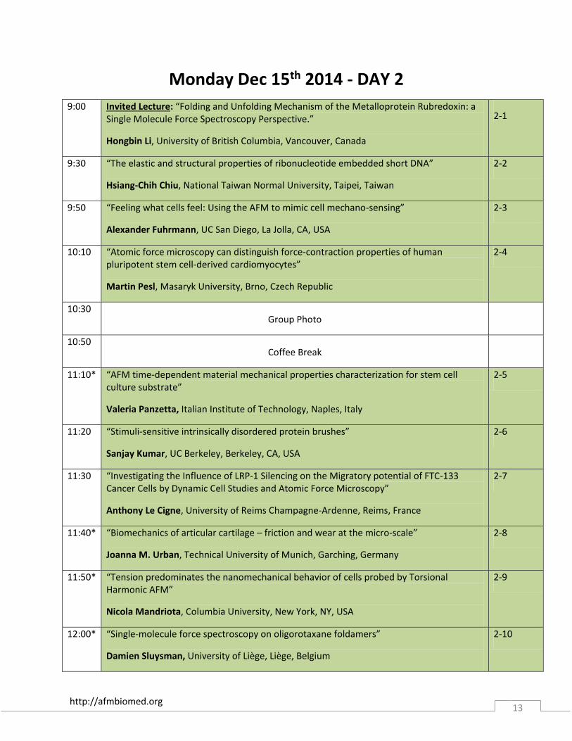
Monday Dec 15th 2014 - DAY 2
9:00
Invited Lecture: “Folding and Unfolding Mechanism of the Metalloprotein Rubredoxin: a Single Molecule Force Spectroscopy Perspective.”
Hongbin Li, University of British Columbia, Vancouver, Canada
2-1
9:30 “The elastic and structural properties of ribonucleotide embedded short DNA”
Hsiang-Chih Chiu, National Taiwan Normal University, Taipei, Taiwan
2-2
9:50 “Feeling what cells feel: Using the AFM to mimic cell mechano-sensing”
Alexander Fuhrmann, UC San Diego, La Jolla, CA, USA
2-3
10:10 “Atomic force microscopy can distinguish force-contraction properties of human pluripotent stem cell-derived cardiomyocytes”
Martin Pesl, Masaryk University, Brno, Czech Republic
2-4
10:30
Group Photo
10:50
Coffee Break
11:10* “AFM time-dependent material mechanical properties characterization for stem cell culture substrate”
Valeria Panzetta, Italian Institute of Technology, Naples, Italy
2-5
11:20 “Stimuli-sensitive intrinsically disordered protein brushes”
Sanjay Kumar, UC Berkeley, Berkeley, CA, USA
2-6
11:30 “Investigating the Influence of LRP-1 Silencing on the Migratory potential of FTC-133 Cancer Cells by Dynamic Cell Studies and Atomic Force Microscopy”
Anthony Le Cigne, University of Reims Champagne-Ardenne, Reims, France
2-7
11:40* “Biomechanics of articular cartilage – friction and wear at the micro-scale”
Joanna M. Urban, Technical University of Munich, Garching, Germany
2-8
11:50* “Tension predominates the nanomechanical behavior of cells probed by Torsional Harmonic AFM”
Nicola Mandriota, Columbia University, New York, NY, USA
2-9
12:00* “Single-molecule force spectroscopy on oligorotaxane foldamers”
Damien Sluysman, University of Liège, Liège, Belgium
2-10
http://afmbiomed.org 14
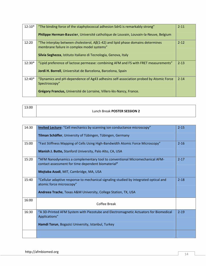
12:10* “The binding force of the staphylococcal adhesion SdrG is remarkably strong”
Philippe Herman-Bausier, Université catholique de Louvain, Louvain-la-Neuve, Belgium
2-11
12:20 “The interplay between cholesterol, Aβ(1-42) and lipid phase domains determines membrane failure in complex model systems”
Silvia Seghezza, Istituto Italiano di Tecnologia, Genova, Italy
2-12
12:30* “Lipid preference of lactose permease: combining AFM and FS with FRET measurements”
Jordi H. Borrell, Universitat de Barcelona, Barcelona, Spain
2-13
12:40* “Dynamics and pH-dependence of Ag43 adhesins self-association probed by Atomic Force Spectroscopy”
Grégory Francius, Université de Lorraine, Villers-lès-Nancy, France.
2-14
13:00
Lunch Break POSTER SESSION 2
14:30 Invited Lecture: “Cell mechanics by scanning ion conductance microscopy”
Tilman Schäffer, University of Tübingen, Tübingen, Germany
2-15
15:00 “Fast Stiffness Mapping of Cells Using High-Bandwidth Atomic Force Microscopy”
Manish J. Butte, Stanford University, Palo Alto, CA, USA
2-16
15:20 “AFM Nanodynamics a complementary tool to conventional Micromechanical AFM-contact assessment for time dependent biomaterial”
Mojtaba Azadi, MIT, Cambridge, MA, USA
2-17
15:40 “Cellular adaptive response to mechanical signaling studied by integrated optical and atomic force microscopy”
Andreea Trache, Texas A&M University, College Station, TX, USA
2-18
16:00
Coffee Break
16:30 “A 3D-Printed AFM System with Piezotube and Electromagnetic Actuators for Biomedical Applications”
Hamdi Torun, Bogazici University, Istanbul, Turkey
2-19
http://afmbiomed.org 15
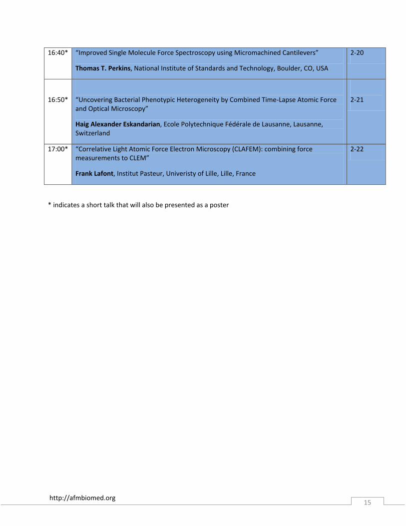
16:40* “Improved Single Molecule Force Spectroscopy using Micromachined Cantilevers”
Thomas T. Perkins, National Institute of Standards and Technology, Boulder, CO, USA
2-20
16:50*
“Uncovering Bacterial Phenotypic Heterogeneity by Combined Time-Lapse Atomic Force and Optical Microscopy”
Haig Alexander Eskandarian, Ecole Polytechnique Fédérale de Lausanne, Lausanne, Switzerland
2-21
17:00* “Correlative Light Atomic Force Electron Microscopy (CLAFEM): combining force measurements to CLEM”
Frank Lafont, Institut Pasteur, Univeristy of Lille, Lille, France
2-22
* indicates a short talk that will also be presented as a poster
http://afmbiomed.org 16
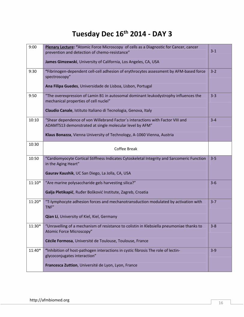
Tuesday Dec 16th 2014 - DAY 3
9:00 Plenary Lecture: “Atomic Force Microscopy of cells as a Diagnostic for Cancer, cancer prevention and detection of chemo-resistance”
James Gimzewski, University of California, Los Angeles, CA, USA
3-1
9:30 “Fibrinogen-dependent cell-cell adhesion of erythrocytes assessment by AFM-based force spectroscopy”
Ana Filipa Guedes, Universidade de Lisboa, Lisbon, Portugal
3-2
9:50 “The overexpression of Lamin B1 in autosomal dominant leukodystrophy influences the mechanical properties of cell nuclei”
Claudio Canale, Istituto Italiano di Tecnologia, Genova, Italy
3-3
10:10 “Shear dependence of von Willebrand Factor´s interactions with Factor VIII and ADAMTS13 demonstrated at single molecular level by AFM”
Klaus Bonazza, Vienna University of Technology, A-1060 Vienna, Austria
3-4
10:30
Coffee Break
10:50 “Cardiomyocyte Cortical Stiffness Indicates Cytoskeletal Integrity and Sarcomeric Function in the Aging Heart”
Gaurav Kaushik, UC San Diego, La Jolla, CA, USA
3-5
11:10* “Are marine polysaccharide gels harvesting silica?”
Galja Pletikapić, Ruđer Bošković Institute, Zagreb, Croatia
3-6
11:20* “T-lymphocyte adhesion forces and mechanotransduction modulated by activation with TNF”
Qian Li, University of Kiel, Kiel, Germany
3-7
11:30* “Unravelling of a mechanism of resistance to colistin in Klebsiella pneumoniae thanks to Atomic Force Microscopy”
Cécile Formosa, Université de Toulouse, Toulouse, France
3-8
11:40* “Inhibition of host-pathogen interactions in cystic fibrosis The role of lectin-glycoconjugates interaction”
Francesca Zuttion, Université de Lyon, Lyon, France
3-9
http://afmbiomed.org 17
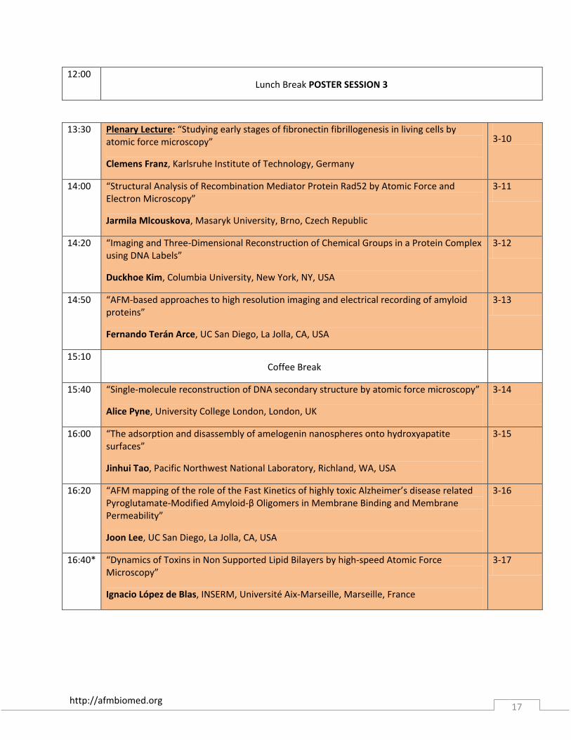
12:00
Lunch Break POSTER SESSION 3
13:30
Plenary Lecture: “Studying early stages of fibronectin fibrillogenesis in living cells by atomic force microscopy”
Clemens Franz, Karlsruhe Institute of Technology, Germany
3-10
14:00 “Structural Analysis of Recombination Mediator Protein Rad52 by Atomic Force and Electron Microscopy”
Jarmila Mlcouskova, Masaryk University, Brno, Czech Republic
3-11
14:20 “Imaging and Three-Dimensional Reconstruction of Chemical Groups in a Protein Complex using DNA Labels”
Duckhoe Kim, Columbia University, New York, NY, USA
3-12
14:50 “AFM-based approaches to high resolution imaging and electrical recording of amyloid proteins”
Fernando Terán Arce, UC San Diego, La Jolla, CA, USA
3-13
15:10
Coffee Break
15:40 “Single-molecule reconstruction of DNA secondary structure by atomic force microscopy”
Alice Pyne, University College London, London, UK
3-14
16:00 “The adsorption and disassembly of amelogenin nanospheres onto hydroxyapatite surfaces”
Jinhui Tao, Pacific Northwest National Laboratory, Richland, WA, USA
3-15
16:20 “AFM mapping of the role of the Fast Kinetics of highly toxic Alzheimer’s disease related Pyroglutamate-Modified Amyloid-β Oligomers in Membrane Binding and Membrane Permeability”
Joon Lee, UC San Diego, La Jolla, CA, USA
3-16
16:40* “Dynamics of Toxins in Non Supported Lipid Bilayers by high-speed Atomic Force Microscopy”
Ignacio López de Blas, INSERM, Université Aix-Marseille, Marseille, France
3-17

http://afmbiomed.org 18
18:00
GALA BANQUET
Birch Aquarium at Scripps
* indicates a short talk that will also be presented as a poster
http://afmbiomed.org 19
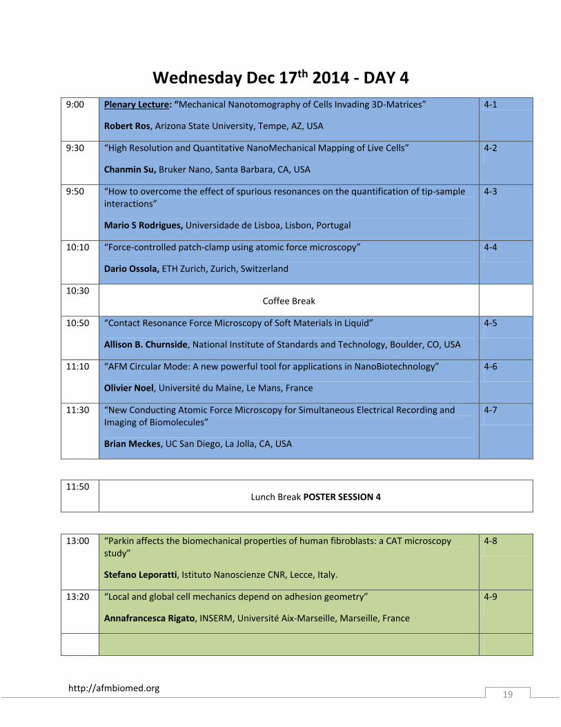
Wednesday Dec 17th 2014 - DAY 4
9:00
Plenary Lecture: “Mechanical Nanotomography of Cells Invading 3D-Matrices”
Robert Ros, Arizona State University, Tempe, AZ, USA
4-1
9:30 “High Resolution and Quantitative NanoMechanical Mapping of Live Cells”
Chanmin Su, Bruker Nano, Santa Barbara, CA, USA
4-2
9:50 “How to overcome the effect of spurious resonances on the quantification of tip-sample interactions”
Mario S Rodrigues, Universidade de Lisboa, Lisbon, Portugal
4-3
10:10 “Force-controlled patch-clamp using atomic force microscopy”
Dario Ossola, ETH Zurich, Zurich, Switzerland
4-4
10:30
Coffee Break
10:50 “Contact Resonance Force Microscopy of Soft Materials in Liquid”
Allison B. Churnside, National Institute of Standards and Technology, Boulder, CO, USA
4-5
11:10 “AFM Circular Mode: A new powerful tool for applications in NanoBiotechnology”
Olivier Noel, Université du Maine, Le Mans, France
4-6
11:30 “New Conducting Atomic Force Microscopy for Simultaneous Electrical Recording and Imaging of Biomolecules”
Brian Meckes, UC San Diego, La Jolla, CA, USA
4-7
11:50
Lunch Break POSTER SESSION 4
13:00 “Parkin affects the biomechanical properties of human fibroblasts: a CAT microscopy study”
Stefano Leporatti, Istituto Nanoscienze CNR, Lecce, Italy.
4-8
13:20 “Local and global cell mechanics depend on adhesion geometry”
Annafrancesca Rigato, INSERM, Université Aix-Marseille, Marseille, France
4-9

http://afmbiomed.org 20
13:40 “Influence of cellular adhesiveness on the formation of cell boundaries”
Steve Pawlizak, University of Leipzig, Leipzig, Germany
4-10
14:00 “Impact of the Actin Cytoskeleton on the Mechanical Properties of Cells and Tissues”
Celine Heu, University of New South Wales, Sydney, Australia
4-11
14:20 “Dynamic coupling of ALCAM to the actin cortex strengthens cell adhesion to CD6”
Joost te Riet, Radboud UMC, Nijmegen, The Netherlands
4-12
14:40 “Nanomechanical and topographical imaging of living cells by Atomic Force Microscopy with colloidal probes”
Alessandro Podestà, Università degli Studi di Milano, Milano, Italy
4-13
15:30
Awards Ceremony
Announcement of AFM BioMed Conference 2016

http://afmbiomed.org 21
Poster Presentation List
Board # Title Presenter Abstract #
1* “Uncovering Bacterial Phenotypic Heterogeneity by Combined Time-Lapse Atomic Force and Optical Microscopy”
Haig Alexander Eskandarian
Ecole Polytechnique Fédérale de Lausanne, Lausanne, Switzerland
2-20
2 “High-speed force mapping on living cells with a small-cantilever AFM”
Tilman Schäffer
University of Tübingen, Tübingen, Germany
P-1
3* “Correlative Light Atomic Force Electron Microscopy (CLAFEM): combining force measurements to CLEM”
Frank Lafont
Institut Pasteur, Univeristy of Lille, Lille, France
2-21
4 “Non-contact imaging combining the Scanning Ion Conductance Microscopy and the Atomic Force Microscope”
Livie Dorwling-Carter
ETH Zürich, Zürich, Switzerland
P-2
5* “Improved Single Molecule Force Spectroscopy using Micromachined Cantilevers”
Thomas T. Perkins
National Institute of Standards and Technology, Boulder, CO, USA
2-19
6 “FluidFM for local electroplating and electrografting in liquid”
Luca Hirt
Institute for Biomedical Engineering, ETH Zurich, Switzerland
P-3
7 “Photoplastic AFM Cantilevers with integrated microchannel for Single Cell Experiments”
Vincent Martinez
Institute for Biomedical Engineering, ETH Zurich, Switzerland
P-4
8 “AFM Surface Analysis of Nanosilver Toxicity on Marine Heterotrophic Bacteria”
Nirav Patel
UC San Diego, La Jolla, CA
P-5
9 “A sticky tale: Sample preparation technique determines cell surface receptor mobility and adhesion”
Thomas Mueller
University of Birmingham
P-6
10* “Dynamics of Toxins in Non Supported Lipid Bilayers by high-speed Atomic Force Microscopy”
Ignacio López de Blas,
INSERM, Université Aix-Marseille, Marseille, France
3-18
http://afmbiomed.org 22
11* “Atomic Force Microscopy of Protein Translocation Machinery in Supported Lipid Bilayers”
R.R. Sanganna Gari
University of Missouri-Columbia, Columbia, MO, USA
1-9
12 “Atomic Force Microscopy Study on Photosystem II-rich Region of Thylakoid Membrane in Chlamydomonas reinhardtii during State Transition”
Witchukorn Phuthong
Stanford University
P-7
13 “Estimating water content of individual fibrin fibers”
Emilios Dimitriadis
NIBIB, NIH
P-8
14 “Exploring the structure of exosomes with nanofilaments using atomic force microscopy”
Jung-Reem Woo
UC Los Angeles
P-9
15 “Stable imaging of heterogeneus samples in liquids with atomic force microscopy”
Jaime Colchero
Universidad de Murcia
P-10
16 “New emerging technique of Electrostatic force MIcroscopy AFM gives insight into the DNA-protein interaction path”
Parminder Kaur
North Carolina state University
P-11
17* “High-Speed Atomic Force Microscopy of ESCRT protein assembly”
Lorena Redondo-Morata
INSERM, Aix-Marseille Université, Marseille, France
1-7
18* AFM time-dependent material
mechanical properties characterization
for stem cell culture substrate
Valeria Panzetta
Italian Institute of Technology, Naples, Italy
2-5
19 “Hydrophobic interaction governs unspecific adhesion of staphylococci”
Nicolas Thewes
Saarland University, Saarbrücken, Germany
P-12
20* “Tension predominates the nanomechanical behavior of cells probed by Torsional Harmonic AFM”
Nicola Mandriota
Columbia University, New York, NY, USA
2-8
21* “Single-molecule force spectroscopy on oligorotaxane foldamers”
Damien Sluysmans
University of Liège, Liège, Belgium
2-9
22 “Polymeric Ultrasound Contrast Agents Baptiste Sarrazin P-13
http://afmbiomed.org 23
Mechanical Properties” CEA Saclay, Saclay, France
23* “The binding force of the staphylococcal adhesion SdrG is remarkably strong”
Philippe Herman-Bausier
Université catholique de Louvain, Louvain-la-Neuve, Belgium
2-10
24 “Elasticity of pulmonary arteries within human lung tissue: Application of AFM to study pulmonary arterial hypertension”
Delphine Sicard
Mayo Clinic, Rochester, MN, USA
P-14
25* “Dynamics and pH-dependence of Ag43 adhesins self-association probed by Atomic Force Spectroscopy”
Grégory Francius
Université de Lorraine, Villers-lès-Nancy, France
2-13
26* “Biomechanics of articular cartilage – friction and wear at the micro-scale”
Joanna M. Urban, Technical University of Munich, Garching, Germany
2-7
27 “Characterisation of the mechanical properties of blood and artificial clots”
Celine Heu
MechBio Team, Graduate School of Biomedical Engineering, UNSW Australia
P-15
28 “Studying in situ protein adsorption and bacterial adhesion via fast scanning AFM and force spectroscopy”
Christian Spengler
Saarland University
P-16
29 “Atomic force microscopy reveals distinct regional nanomechanical properties of the extracellular matrix in healthy, aged, and osteoarthritic native human menisci”
Jeanie Kwok
UC San Diego
P-17
30* “Lipid preference of lactose permease: combining AFM and FS with FRET measurements”
Jordi H. Borrell
Universitat de Barcelona, Barcelona, Spain
2-12
31 “Investigating the Impact of Antigen Density on Antigen-antibody Binding Efficiency with AFM”
Bin Li
Shanghai Institute Of Applied Physics, Shanghai, China
P-18
32* “Are marine polysaccharide gels harvesting silica?”
Galja Pletikapić
Ruđer Bošković Institute, Zagreb, Croatia
3-6

http://afmbiomed.org 24
33* “Unravelling of a mechanism of resistance to colistin in Klebsiella pneumoniae thanks to Atomic Force Microscopy”
Cécile Formosa
Université de Toulouse, Toulouse, France
3-9
34* “Inhibition of host-pathogen interactions in cystic fibrosis The role of lectin-glycoconjugates interaction”
Francesca Zuttion
Université de Lyon, Lyon, France
3-10
35 “CFTR is involved in polyphenol-induced swelling of the endothelial glycocalyx”
Hermann Schillers
University of Münster, Münster, Germany
P-19
36* “Cartilage Morphogenesis: Investigation of Cartilage Structure and Mechanical Properties by AFM.”
Carina Prein
Ludwig-Maximilians-University, Munich, Germany
1-17
37 “AFM-based sarcolemmal surface analysis of living cardiomyocytes unveils unexpected mitochondrial shift in heart failure”
Véronique Lachaize
CNRS, LAAS, Toulouse, France
P-20
38* “T-lymphocyte adhesion forces and mechanotransduction modulated by activation with TNF”
Qian Li
University of Kiel, Kiel, Germany
3-8
39 “Using the FluidFM to connect neurons, a step towards building engineered networks”
Mathias J. Aebersold
Laboratory of Biosensors and Bioelectronics, Institute for Biomedical Engineering, ETH Zurich
P-21
40 “The role of glycocalyx in cellular interactions between lung carcinoma cells and the endothelium”
Katarzyna Malek-Zietek
Center for Nanometer-scale Science and Advanced Materials, Jagiellonian University
P-22
41 “Diagnostic of vascular dysfunction in endothelium nano-mechanics”
Magdalena Jaglarz
Center for Nanometer-scale Science and Advanced Materials, Jagiellonian University
P-23
* indicates a short talk that will also be presented as a poster

http://afmbiomed.org 25
http://afmbiomed.org 26
Abstracts Day 1
Sunday, December 14, 2014 1-1 Using in situ AFM to understand how proteins assemble into ordered
structures that direct the formation of mineralized tissues
Jim De Yoreo [1,2], Jinhui Tao [1]
[1] Pacific Northwest National Laboratory, [2] University of Washington Self-assembly and subsequent mineralization of protein matrices is a widespread paradigm in the biological production of hard tissues. The architecture of the underlying matrix imposes order on the nucleating mineral phase. Correct assembly and mineralization are critical to biological function, while the structural complexity and mechanical properties are unparalleled in synthetic materials. To understand the physical controls governing these processes, we have used in situ AFM to investigate the assembly dynamics and subsequent mineralization for both microbial and human-derived proteins. We have correlated the observed behavior with the results of in situ dynamic force (DFS) and infrared spectroscopy to determine the underlying energies and chemical interactions that drive assembly. Results from microbial membrane systems reveal the key role played by conformational transformations in controlling the pathways and kinetics of protein self-assembly. Moreover, the pathway to the final ordered state often passes through transient, less-ordered conformational states. Thus the concept of a folding funnel with kinetic traps used to describe protein folding is also applicable to protein matrix self-assembly. Results from assembly of collagen show that the equilibrium morphology is highly dependent on the concentration of background K+ ions (Fig. 1), which modify collagen-collagen binding free energies through their impact on the absorption of water to the collagen molecules, as well as the strength of collagen binding to the underlying mica surface through a shift in the surface charge towards less negative values. These results highlight the delicate balance in the triad of solvent-protein-surface interaction energies required to achieve the correct assembly architecture. AFM analysis of calcium phosphate nucleation on collagen matrices shows that mineral nucleation is promoted through a reduction in the interfacial free energy. However, hydroxyapatite formation via an initial amorphous precursor is observed at supersaturations that are too low to be easily explained by classical nucleation theory (Fig. 2). Though correlations with cryoTEM data, we show that the existence of metastable clusters provides a potential low-barrier pathway to crystallization that circumvents the large classical barriers to nucleation. Finally, DFS analysis in the calcium carbonate system shows that binding free energy is directly related to the nucleation barrier, providing an energetic rationale for the conventional wisdom that good binders are good nucleators. Our results provide new insights into mechanisms of biomineralization, from matric assembly to the maturation of final crystalline structures.
http://afmbiomed.org 27
1-2
Marine polysaccharide networks: self-assembly vs. self-organization revealed by atomic force microscopy
Vesna Svetličić
Division for Marine and Environmental Research, Ruđer Bošković Institute, Zagreb, Croatia
Polysaccharides in marine environment tend to organize under influence of molecular and interfacial forces into complex gel structures possessing both the cohesive properties of solids and the diffusive properties of liquids. Such self-assembled networks are centers of microbial activities regulating cycling of matter and energy. Do self-organized polysaccharide networks exist out there?

http://afmbiomed.org 28
1-3
Protein-protein and Protein-membrane interaction of Annexin-A5
Atsushi Miyagi, Simon Scheuring
U1006 INSERM, Université Aix-Marseille, Campus Luminy, 13009 Marseille, France
"Annexins are members of a protein family with highly conserved sequence among animal and plant kingdoms. Its main feature is the ability of binding to negatively charged phospholipids in a calcium ion-dependent manner. In the presence of Ca2+, annexin-A5 binds to phospholipids that contain dioleoyl-sn-glycero-3-phospho-L-serine (DOPS) and is able to form trimers, which then organize in highly ordered structures, two-dimensional crystals with p6 symmetry. The p6 symmetry crystal is formed by annexin-A5 trimers arranged in hexagons resembling honeycombs. In the honeycomb-like holes (p6 axis), annexin trimers are loosely assembled in two equally favorable orientations. The trapped trimers are weakly bound to the hexagonal annexin frames and they can therefore rotate. We were able to observe, using high-speed atomic force microscope, the rotational movement of this annexin trimer, its collapse into a dimer or monomer, as well as its re-assembly into trimer with a temporal resolution of 20-650ms. Furthermore by flushing the fuid cell with EDTA and caged calcium, we show repeated disassembly and assembly of annexin 2D crystals triggered by the release of caged calcium ions by UV laser flash. Rotational freedom of the “weak” non-p6-lattice annexins. a) Representative high-resolution frames from a longer HS-AFM movie at 650ms frame rate. b) Due to the relative membrane interaction weakness of the non-p6-lattice annexins, trimers (left), dimers (middle) and monomers (right) annexins can be found at the p6 axis position. All these assemblies reveal high-degree of motional freedom within the honey-comb delimited by the other Annexin-A5 trimers. c) Angular motion analysis of Annexin-A5 trimers. The graph shows the result of rotation analysis of the molecule marked “Mol 1” in the right panel."
http://afmbiomed.org 29
1-4 Imaging electrostatic charge distribution in biomembranes using low
oscillation Dynamic Atomic Force Microscopy
Jaime Colchero [1], Lisa Almonte [1,2], A M. Baro [2]
[1] Dep. Física. CIOyN. Universidad de Murcia, Campus Espinardo, E-30100 Murcia [2] Instituto de Ciencia de Materiales de Madrid-CSIC, Campus de Cantoblanco E-28049 Madrid]
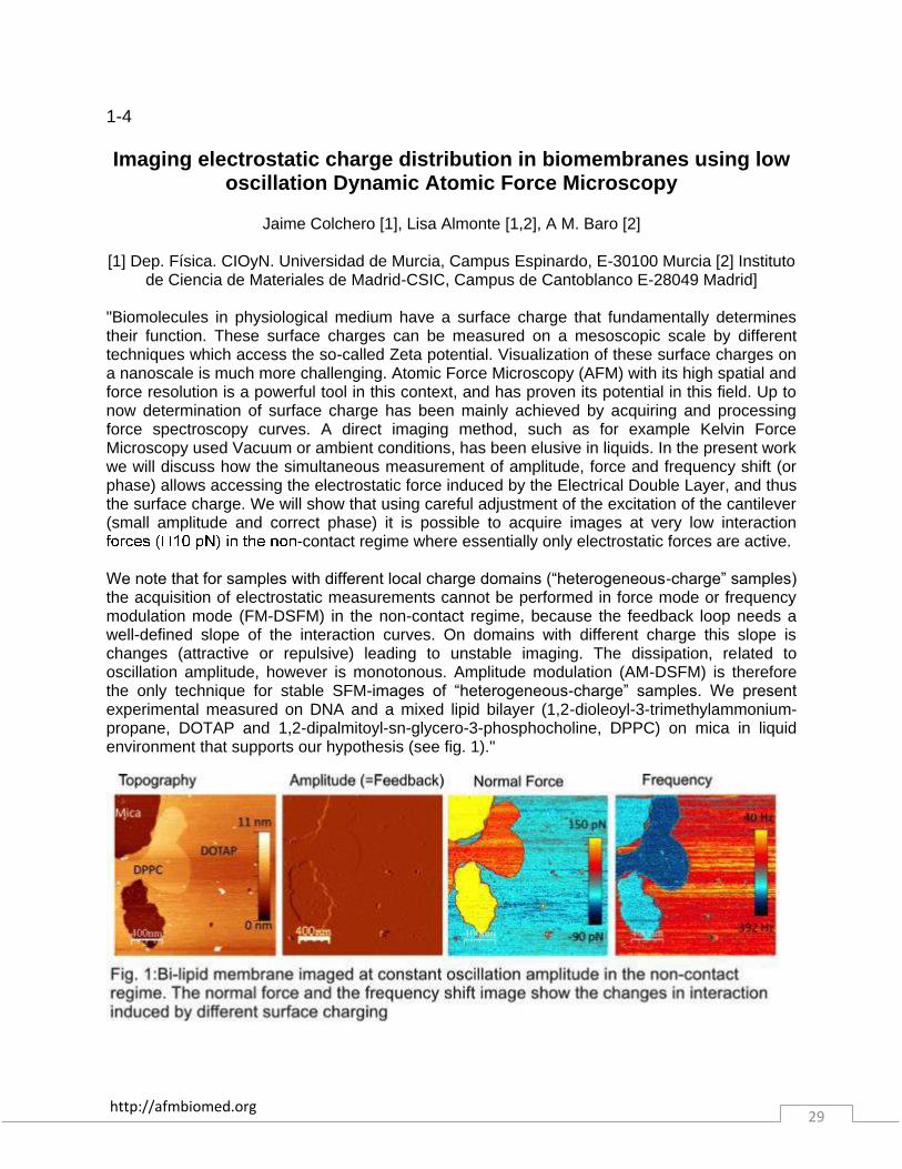
"Biomolecules in physiological medium have a surface charge that fundamentally determines their function. These surface charges can be measured on a mesoscopic scale by different techniques which access the so-called Zeta potential. Visualization of these surface charges on a nanoscale is much more challenging. Atomic Force Microscopy (AFM) with its high spatial and force resolution is a powerful tool in this context, and has proven its potential in this field. Up to now determination of surface charge has been mainly achieved by acquiring and processing force spectroscopy curves. A direct imaging method, such as for example Kelvin Force Microscopy used Vacuum or ambient conditions, has been elusive in liquids. In the present work we will discuss how the simultaneous measurement of amplitude, force and frequency shift (or phase) allows accessing the electrostatic force induced by the Electrical Double Layer, and thus the surface charge. We will show that using careful adjustment of the excitation of the cantilever (small amplitude and correct phase) it is possible to acquire images at very low interaction
-contact regime where essentially only electrostatic forces are active. We note that for samples with different local charge domains (“heterogeneous-charge” samples) the acquisition of electrostatic measurements cannot be performed in force mode or frequency modulation mode (FM-DSFM) in the non-contact regime, because the feedback loop needs a well-defined slope of the interaction curves. On domains with different charge this slope is changes (attractive or repulsive) leading to unstable imaging. The dissipation, related to oscillation amplitude, however is monotonous. Amplitude modulation (AM-DSFM) is therefore the only technique for stable SFM-images of “heterogeneous-charge” samples. We present experimental measured on DNA and a mixed lipid bilayer (1,2-dioleoyl-3-trimethylammonium-propane, DOTAP and 1,2-dipalmitoyl-sn-glycero-3-phosphocholine, DPPC) on mica in liquid environment that supports our hypothesis (see fig. 1)."
http://afmbiomed.org 30
1-5 Probing the compressibility of tumor cell nuclei by combined atomic
force-confocal microscopy
Marina Krause [1], Joost te Riet [2], Katarina Wolf [1]
[1] Department of Cell Biology and [2] Department of Tumor Immunology, Nijmegen Centre for Molecular Life Sciences, Radboud University Nijmegen Medical Centre, Nijmegen, The
Netherlands
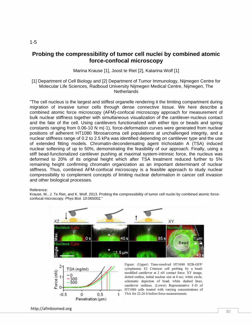
"The cell nucleus is the largest and stiffest organelle rendering it the limiting compartment during migration of invasive tumor cells through dense connective tissue. We here describe a combined atomic force microscopy (AFM)-confocal microscopy approach for measurement of bulk nuclear stiffness together with simultaneous visualization of the cantilever-nucleus contact and the fate of the cell. Using cantilevers functionalized with either tips or beads and spring constants ranging from 0.06-10 N m(-1), force-deformation curves were generated from nuclear positions of adherent HT1080 fibrosarcoma cell populations at unchallenged integrity, and a nuclear stiffness range of 0.2 to 2.5 kPa was identified depending on cantilever type and the use of extended fitting models. Chromatin-decondensating agent trichostatin A (TSA) induced nuclear softening of up to 50%, demonstrating the feasibility of our approach. Finally, using a stiff bead-functionalized cantilever pushing at maximal system-intrinsic force, the nucleus was deformed to 20% of its original height which after TSA treatment reduced further to 5% remaining height confirming chromatin organization as an important determinant of nuclear stiffness. Thus, combined AFM-confocal microscopy is a feasible approach to study nuclear compressibility to complement concepts of limiting nuclear deformation in cancer cell invasion and other biological processes. Reference: Krause, M., J. Te Riet, and K. Wolf. 2013. Probing the compressibility of tumor cell nuclei by combined atomic force-confocal microscopy. Phys Biol. 10:065002."
http://afmbiomed.org 31
1-6
Probing of antigens on malaria infected erythrocytes using protein-antibody affinity based molecular force spectroscopy
Himanshu Singh [1,2], Kripa Madnani [4], Jianshu Cao [2,5], Peter R. Preiser [2,4], Chwee Teck
Lim [1,2,3]
[1]:Department of Biomedical Engineering, National University of Singapore, Singapore, 119615; [2]:Infectious Diseases IRG, Singapore-MIT Alliance for Research & Technology,
Singapore, 138602; [3] : Mechanobiology Institute, Singapore, 117411,[4]:School of Biological Sciences, Nanyang Technological University, Singapore, 639798; [5]: Department of
Chemistry, Massachusetts Institute of Technology, Cambridge, MA, 02139 "Plasmodium (P.) falciparum, the deadliest of all malaria parasites, causes almost 90% of the total malaria infection cases around the world and approximately 650K deaths per year. The unique ability to evade the antibody mediated immune recognition, while maturing inside the infected Red Blood Cell (iRBC) during the infection cycle is one of the most crucial survival strategies developed by P. falciparum parasite and is mainly attributed to a number of variant surface antigen families like PfEMP1, RIFIN, STEVOR which the parasite exports and expresses at different time points in an infection cycle. PfEMP1 has been well demonstrated to be involved in virulence and immune evasion and RIFIN has also been located on an iRBC surface. However, very little is known yet about the biological significance of STEVOR proteins. Some recent studies, using fluorescence microscopy, have provided indirect visual evidence about location of STEVOR proteins. But the exact locations and distribution of these proteins is not yet confirmed. In this study, using molecular force mapping technique, we provide, for the first time, direct visual-quantitative evidences of variant STEVOR proteins being expressed on the extracellular surface of the RBC membrane during the late asexual stages of infection. The technique used involves the probing of iRBC surface using a bare tip and an anti-STEVOR antibody functionalized tip. The tip-substrate interactions were quantified and compared in terms of binding frequency (percentage of specific interactions based molecular events). Specific interactions were classified as molecular events filtered with rupture or detachment force threshold obtained from control experiments. The adhesion maps obtained from experiments clearly show strong adhesion spots from probing of iRBCs with functionalized tips as compared to those from bare tip-iRBCs interactions. Also, the quantitative estimates show significant difference in binding frequency from 4% (fresh tip vs. iRBCs) to 61% (functionalized tip vs. iRBCs) in the filtered regime thus demonstrating the extracellular surface expression of STEVOR proteins. Moreover, the binding frequency was found to increase with parasite maturation (39% in trophozoite stage to 91% in mature schizont stage), showing an increase in the surface STEVOR expression. A number of control experiments have also been performed to ensure the specificity and accuracy of our results. Taken together, our data provides first direct evidence for the extracellular expression of STEVOR proteins in late asexual stages which opens new opportunities to further explore the complex antigenic properties of the parasite and obtain more insights into multi-ligands mediated pathology of the parasite with different receptors."

http://afmbiomed.org 32
1-7
High-Speed Atomic Force Microscopy of ESCRT protein assembly
Lorena Redondo-Morata[1], Nicolas Chiaruttini[2], Atsushi Miyagi[1], Adai Colom-Diego[1], Aurélien Roux[2] and Simon Scheuring[1]
[1] U1006 INSERM, Aix-Marseille Université, Parc Scientifique et Technologique de Luminy, 163 avenue de Luminy, 13009 Marseille, France ; [2], University of Geneva, Department of
Biochemistry, 30 quai Ernest Ansermet, CH-1211 Geneva 4, Switzerland "The endosomal sorting complex required for transport (ESCRT) mediate one of the most unusual membrane remodelling in cells. When oligomerize, they are able to bud the membrane forming constriction necks that will be cleaved afterwards to form multivesicular bodies or the viral envelope, among others. Since the implementation of High-Speed Atomic Force Microscopy (HS-AFM), efforts have been addressed to achieve simultaneous observation of structure, dynamics and function of biological assemblies. The high spatial and temporal resolution of HS-AFM reveals itself as a key feature for the observation of biological assemblies. The underlying question is: what dynamic processes can HS-AFM help to be better understood? Among all dynamic biological processes, studying endocytic processes is challenging because it involved different molecular mechanisms. In the case of the ESCRT endocytic pathway, the choreography of the ESCRT complexes, how they sort the cargo and finally how they deform and cleave the membrane. In this work, we use HS-AFM to study the ESCRT membrane sculpture machinery. In particular, we are interested in one of the proteins of the ESCRT-III complex, Snf7. We provide evidence of the Snf7 assembling into circular filaments that coil around the membrane constriction site. We observe interfilament dynamics that provide a basis for a mechanistic explanation how the machinery creates tension for membrane fission."

http://afmbiomed.org 33
1-8
Applying image registration technique to construct 3D object from topologic images
Hoyeon Lee [1], Sangmo Shin [2], Seungryong Cho [1]
[1] KAIST, [2] Hanbat National University
"Atomic force microscope is widely used to acquire topologic images of nanoscale structures. Acquiring multiple topologic images at various view angles of an object, one may want to synthesize the volumetric image of the object. Since a partial overlap may exist between two topologic images, the overlap information can be used for image registration or fusion. We developed an appropriate registration technique for the partial topologic images in this work. We implemented an AFM-simulating device that is composed of a laser source and a distance sensor as shown in Fig. 1 (a). Although the scanning principles are quite different, this simulating system provides topologic images of an object under interest very efficiently. We have used a 3D-printed model of a simple protein as a scanned object. For image registration, we used spin-image and iterative closest point algorithm. Spin-image reduces 3D surface image to 2D image via calculating vertical and horizontal distances of the entire vertices from one vertex point of interest. A high similarity between the two spin images would imply that the vertices of interests in those spin images may be near coincidence. After finding several candidates of high similarity, we used an iterative closest point algorithm to derive the best-matching transformation. Fig. 1 (b) shows images of the objects before registration and Fig. 1 (c) shows the registration result. A successful image registration has been achieved through the proposed method. In conclusion, we applied an image registration technique to topologic images acquired from an experimental equipment mimicking the AFM. Image registration was successfully performed, and 3D image synthesis from multiple topologic images at various angles would be accordingly possible."

http://afmbiomed.org 34
1-9
Atomic Force Microscopy of Protein Translocation Machinery in Supported Lipid Bilayers
R.R. Sanganna Gari, N.C. Frey, B. P. Marsh, C. Mao, L.L. Randall, and G.M. King
University of Missouri-Columbia
More than 30% of proteins in any organism are transported from the site of synthesis into or through cell membranes to properly localize and function. The general secretory or Sec system is the major route of export for proteins from the cytosol of Escherichia coli and all eubacteria. The pathway through the membrane – the translocon – is provided by SecYEG, a protein complex that is highly conserved having homologs across the kingdoms of life. SecA is the ATPase of the general secretory system and it binds SecYEG to perform translocation. In so doing, SecA makes large surface area contact with the unstructured cytoplasmic loops spanning transmembrane helices 6-7 and 8-9 of SecY. Despite their broad functional significance, measurements of flexible and disordered protein domains remain a significant experimental challenge. Recently, atomic force microscopy (AFM) has emerged as an important complementary tool in biophysics and is well suited for studying membrane protein dynamics in near-native conditions. However, AFM has not been widely applied to non-crystalline membrane proteins. Here we studied purified SecYEG that was reconstituted into liposomes at low concentration. After confirming activity, changes in the structure of SecYEG as a function of time were directly visualized [1]. The dynamics observed were significant in magnitude and were attributed to the aforementioned loops of SecY. In addition, we identified a distribution between monomers and dimers of SecYEG as well as a smaller population of higher order oligomers. We have also imaged SecA engaged on SecYEG and related the structural states observed to the activity of the translocase [2]. Currently we are working towards determining the oligomeric state of SecA during active protein translocation, and the interesting dependency which we uncovered of SecYEG oligomeric state on the protein species being transported. Taken together, this work provides a novel and near-native vista of central components of the protein translocation machinery. Figure 1: AFM imaging reveals the oligomeric state of the translocon SecYEG in a native lipid environment at physiologically relevant temperature and ionic strength ((a): upper left panels, monomer, dimer and tetramer), structural dynamics of individual SecYEG monomers ((a): lower left panels), and rapid fluctuations of cytoplasmic loops of SecYEG that are involved with protein-protein interactions ((a): right). Panel (b) shows a visualization of native protein-protein interactions in membrane. The first image (t = 0 s) shows SecA binding to SecYEG in a supported lipid bilayer (confirmed via comparison of AFM data with available crystal structures). Subsequent imaging reveals the unbinding of SecA (t = 16 s) and finally rebinding of SecA (t = 48 s). A cartoon depiction of these events is shown below the AFM data.

http://afmbiomed.org 35
1-10
Effects of Carbon Nanotubes on the Aggregation of A-beta Peptides
Dongdong Lin, Luogang Xie, Yin Luo, Guanghong Wei, and Xinju Yang
State Key Laboratory of Surface Physics and Department of Physics, Fudan University
The pathogenesis of Alzheimer’s disease (AD) is associated with the aggregation of the amyloid-β (Aβ) peptides into toxic aggregates with β-sheet character. Thus how to inhibit the aggregation of Aβ peptide has been an urgent problem, and intensive efforts have been made to solve it. Here the influence of hydroxylated single walled carbon nanotubes (SWCNTs) on the aggregation of Aβ(16-22) and Aβ(1-40) peptides are investigated by atomic force microscopy (AFM) and ThT fluorescence measurements, together with the all-atom explicit-water replica exchange molecular-dynamics (REMD) simulations. The results show that hydroxylated SWCNTs can significantly inhibit the β-sheet formation and shift the conformations of Aβ(16-22) oligomers from ordered β-sheet-rich structures toward disordered coil aggregates. As Aβ(16-22) is only the central hydrophobic core fragment of Aβ peptides, it is essential to extend such studies to the full length Aβ peptides. Recently, the effects of SWCNTs on the aggregations of Aβ(1-40) are investigated by the same methods. It is found that SWCNTs has similar inhibitory action on Aβ(1-40). The present of CNTs can effectively adsorb the oligomers of Aβ(1-40) on CNTs’ surface, and hence inhibit the formation of fibrils. Furthermore, the adding of CNTs to the Aβ(1-40) solution which contains long fibrils can significantly break the long fibrils to short ones or other species, indicating CNTs can efficiently destroy the formed aggregates of Aβ(1-40). The results provide further insight into targets for the design of β-sheet breakers to modulate the aggregation and inhibit the aggregation of full-length Aβ peptides.

http://afmbiomed.org 36
1-11 From Nanomechanics towards Medical Diagnosis
Hans Oberleithner
University of Munster, Germany
Excessive sodium chloride (salt) intake, as common in most populations worldwide, damages kidney, heart and blood vessels. Although this has been known for decades, the mechanisms behind are still obscure. With the advent of atomic force microscopy (AFM) in basic medical sciences novel approaches addressing this topic became feasible. One of them is the combination of AFM imaging and mechanical measurements at the level of single living cells originating from the vascular system. I will describe the road that we have been following over the past 17 years using AFM as a tool to observe and sometimes interfere with vascular endothelium, the inner cell layer of blood vessels. Using AFM for cell imaging in 1997, it was observed that aldosterone, the crucial steroid hormone in charge of sodium retention in the body, swells vascular endothelium. Cell swelling could be blocked by amiloride, a diuretic drug thought to act only on kidney. This unexpected paradox, at that time critically viewed by the medical community, was solved some 10 years later by using AFM as a nanoindenter. The renal sodium channel, a plasma membrane protein mediating sodium transport in kidney, could be detected in vascular endothelium. This channel, under the control of aldosterone, mediates sodium entry into cells and can be blocked by amiloride. Moreover, force measurements proved that endothelial cells stiffen in ambient high sodium (range: 2pN/nm) and that a channel block can prevent this response. With the improvement of AFM force measurements in living cells a very soft and negatively charged gel-like layer called endothelial glycocalyx (thickness range: 500nm, stiffness range: 0.2 pN/nm) at the blood-facing surface of endothelial cells could be mechanically characterized and functionally related to ambient sodium. A similar negatively charged gel could be also detected on the surface of red blood cells (RBC) by applying AFM imaging and nanoindentation. It turned out that sodium ions in the blood determine the magnitude of the repulsive forces between RBC and the inner blood vessel wall. In fact, the RBC surface (erythrocyte glycocalyx) “mirrors” the surface of the vascular endothelium (endothelial glycocalyx). These conclusions were drawn on the basis of AFM “differential” surface imaging and force measurements in the pN- range. At about this time, the idea came up that RBC could serve as in vitro test models reporting the functional conditions (sodium binding to the glycocalyx) of the endothelial surface. This would have considerable medical impact since blood vessel function (e.g. regulation of blood pressure) strongly depends of a negatively charged surface (glycocalyx). In the meantime a simple so-called salt blood test was developed that quantifies salt sensitivity of an individual and, in particular, gives insight into the functional condition of the blood vessel wall (i.e. vascular endothelium). In conclusion, AFM has been spanning the bridge between basic vascular research and medical diagnosis.

http://afmbiomed.org 37
1-12
CAT (Confocal-AFM-TIRF) Microscopy as Novel Tool for E-Cadherin Knockdown Analysis in Cancer Cells
Maria Francesca Cascione [1, 2], Daniele Vergara [3], Giuseppe Maruccio [1, 2], Pasquale
Simeone [4] Michele Maffia [3], Ross Rinaldi [1, 2], and Stefano Leporatti [1]
[1] NNL-Istituto Nanoscienze CNR, Lecce, Italy; [2] Mathematics and Physics Department, Università del Salento, Lecce, Italy; [3] Department of Biological and Environmental Sciences and Technologies (DiSteBa), Università del Salento, Lecce, Italy; [4] Unit of Cancer Pathology
and Department of Neuroscience and Imaging, Research Centre on Aging (Ce.S.I), G. d’Annunzio, University Foundation, Chieti-Pescara, Italy
"The epithelial-mesenchymal transition (EMT) is a biological process that allows an epithelial cell to undergo numerous biochemical changes assuming a mesenchymal phenotype characterized by increased migratory capacity, invasiveness and high resistance to apoptosis. Loss of E-cadherin expression is a crucial step of EMT and is involved in cancer invasion and metastatization. In human tumours, down-regulation of E-cadherin is frequently associated with poor prognosis. We investigated biophysical changes of epithelial breast cancer cell lines upon shRNA-mediated stable knockdown of E-cadherin expression (Ecadsh) through CAT (Confocal-AFM-TIRF) microscopy, a combination of an advanced scanning probe microscope (Bioscope Catalyst, Bruker Inc. USA), a confocal microscope (LSM 700, Zeiss GERMANY), and a total internal reflection fluorescence microscope (Laser TIRF 3, Zeiss GERMANY). The CAT microscopy is a novel powerful tool for cancer cell investigation: AFM allows topographical single cell membrane characterization; confocal microscopy permits volume cell investigation whereas TIRF gives information about cell-substrate interface. Their combined use provides topographic and spectroscopic imaging and nano-scale adhesion forces. Therefore simultaneous combination of all three microscopies produce a complete three-dimensional visualisation of the cell.In particular, AFM analysis of Ctr and Ecadsh cells revealed significant differences in the stiffness of the two cell lines. The change in elasticity is also accompanied by alterations in the cytoskeletal architecture of cells that are correlated with a different organization of actin filaments. Advanced integration of complementary techniques allowed us to extend our knowledge on the cellular modifications associated with E-cadherin down-regulation in breast cancer cells."
http://afmbiomed.org 38
1-13
Fibrinogen-erythrocyte binding as biomarker of increased cardiovascular risk. An atomic force microscopy study.
Ana Filipa Guedes [1], Filomena A. Carvalho [1], Luís Sargento [2], Nuno Lousada [2], Carlos
Moreira [3], José Braz Nogueira [3], and Nuno C. Santos [1]
[1] Instituto de Medicina Molecular, Faculdade de Medicina, Universidade de Lisboa, Lisbon, Portugal. [2] Hospital Pulido Valente, Centro Hospitalar Lisboa Norte, Lisbon, Portugal. [3]
Hospital Santa Maria, Centro Hospitalar Lisboa Norte, Lisbon, Portugal.
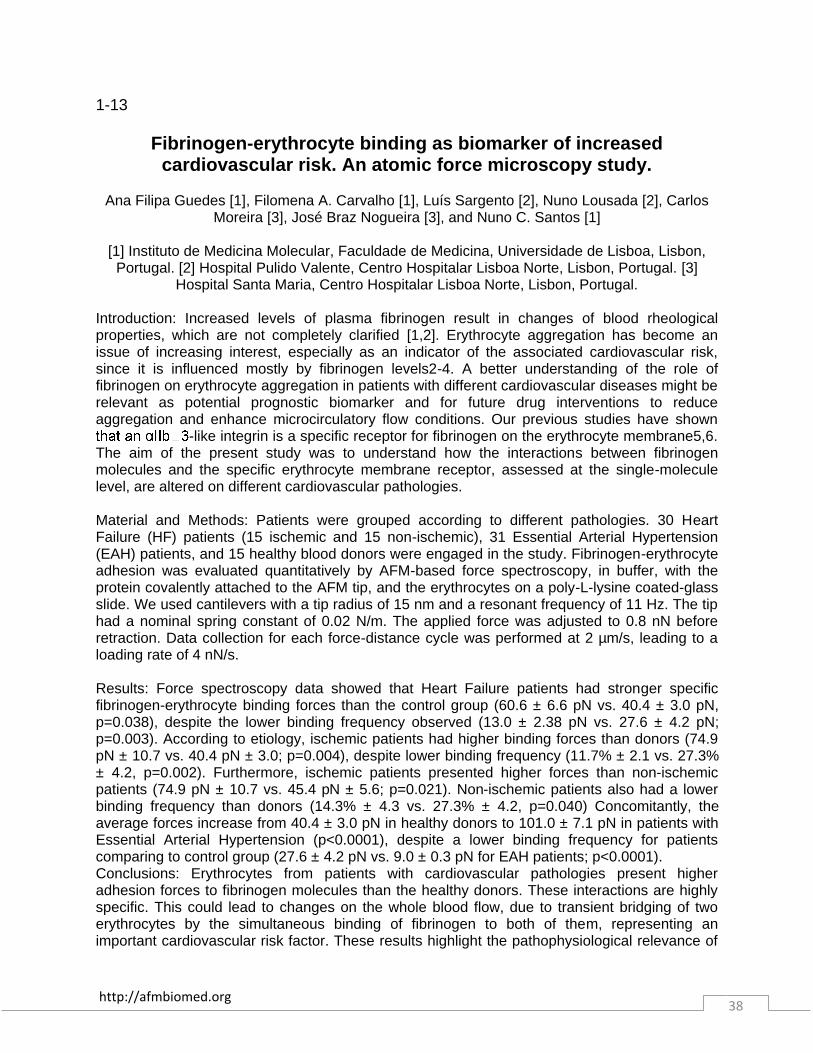
Introduction: Increased levels of plasma fibrinogen result in changes of blood rheological properties, which are not completely clarified [1,2]. Erythrocyte aggregation has become an issue of increasing interest, especially as an indicator of the associated cardiovascular risk, since it is influenced mostly by fibrinogen levels2-4. A better understanding of the role of fibrinogen on erythrocyte aggregation in patients with different cardiovascular diseases might be relevant as potential prognostic biomarker and for future drug interventions to reduce aggregation and enhance microcirculatory flow conditions. Our previous studies have shown
-like integrin is a specific receptor for fibrinogen on the erythrocyte membrane5,6. The aim of the present study was to understand how the interactions between fibrinogen molecules and the specific erythrocyte membrane receptor, assessed at the single-molecule level, are altered on different cardiovascular pathologies. Material and Methods: Patients were grouped according to different pathologies. 30 Heart Failure (HF) patients (15 ischemic and 15 non-ischemic), 31 Essential Arterial Hypertension (EAH) patients, and 15 healthy blood donors were engaged in the study. Fibrinogen-erythrocyte adhesion was evaluated quantitatively by AFM-based force spectroscopy, in buffer, with the protein covalently attached to the AFM tip, and the erythrocytes on a poly-L-lysine coated-glass slide. We used cantilevers with a tip radius of 15 nm and a resonant frequency of 11 Hz. The tip had a nominal spring constant of 0.02 N/m. The applied force was adjusted to 0.8 nN before retraction. Data collection for each force-distance cycle was performed at 2 µm/s, leading to a loading rate of 4 nN/s. Results: Force spectroscopy data showed that Heart Failure patients had stronger specific fibrinogen-erythrocyte binding forces than the control group (60.6 ± 6.6 pN vs. 40.4 ± 3.0 pN, p=0.038), despite the lower binding frequency observed (13.0 ± 2.38 pN vs. 27.6 ± 4.2 pN; p=0.003). According to etiology, ischemic patients had higher binding forces than donors (74.9 pN ± 10.7 vs. 40.4 pN ± 3.0; p=0.004), despite lower binding frequency (11.7% ± 2.1 vs. 27.3% ± 4.2, p=0.002). Furthermore, ischemic patients presented higher forces than non-ischemic patients (74.9 pN ± 10.7 vs. 45.4 pN ± 5.6; p=0.021). Non-ischemic patients also had a lower binding frequency than donors (14.3% ± 4.3 vs. 27.3% ± 4.2, p=0.040) Concomitantly, the average forces increase from 40.4 ± 3.0 pN in healthy donors to 101.0 ± 7.1 pN in patients with Essential Arterial Hypertension (p<0.0001), despite a lower binding frequency for patients comparing to control group (27.6 ± 4.2 pN vs. 9.0 ± 0.3 pN for EAH patients; p<0.0001). Conclusions: Erythrocytes from patients with cardiovascular pathologies present higher adhesion forces to fibrinogen molecules than the healthy donors. These interactions are highly specific. This could lead to changes on the whole blood flow, due to transient bridging of two erythrocytes by the simultaneous binding of fibrinogen to both of them, representing an important cardiovascular risk factor. These results highlight the pathophysiological relevance of
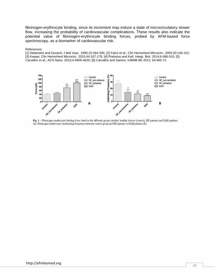
http://afmbiomed.org 39
fibrinogen-erythrocyte binding, since its increment may induce a state of microcirculatory slower flow, increasing the probability of cardiovascular complications. These results also indicate the potential value of fibrinogen-erythrocyte binding forces, probed by AFM-based force spectroscopy, as a biomarker of cardiovascular risk. References: [1] Delamaire and Durand; J Mal Vasc. 1990;15:344-345; [2] Falcó et al.; Clin Hemorheol Microcirc. 2005;33:145-151; [3] Kwaan; Clin Hemorheol Microcirc. 2010;44:167-176; [4] Pretorius and Kell; Integr. Biol. 2014;6:486-510; [5] Carvalho et al.; ACS Nano. 2010;4:4609-4620; [6] Carvalho and Santos; IUBMB life 2012; 64:465-72

http://afmbiomed.org 40
1-14
In vitro guidance of developing neural networks
H.Dermutz, R. Grüter, E. Lilli, A. Truong, L. Demkó, T. Zambelli, J. Vörös
Laboratory of Biosensors and Bioelectronics, ETH Zürich Gloriastrasse 35, ETZ 8092 Zürich "Neuron cultures in neuroscience are still a very powerful and essential tool when it comes to basic questions about network behavior and learning. In vivo, the tremendous complexity of the brain topology results in too many variable parameters influencing the basic network of interest. Many groups have started to use chemically patterned surfaces to create defined networks. These small networks are a very useful tool for investigating the basic functionalities and parameters of neuron networks. We developed a method to control the location and the time of neurite outgrowth by locally switching the surface from repulsive to adhesive1. We use a AFM-cantilever with a microchannel inside (FluidFM, Cytosurge, Switzerland) to locally deposit an adhesion promoter onto a polymer coated glass substrate. The Poly-l-Lysine with grafted Polyethylenglycol (Pll-g-PEG, SuSoS, Switzerland) monolayer is known to be non-fouling and prevents neurites from adhering to the substrate. By showering the Poly-l-Lysine (PLL) onto the Pll-g-PEG monolayer with the FluidFM, the non-adhesive Pll-g-PEG is exchanged with adhesion promoting PLL. After depositing PLL spots into the non-fouling Pll-g-PEG background, primary hippocampal neurons from E17 Wistar rats are dispensed over the whole substrate. The non-adherent cells are later rinsed off resulting in patterned cell spots. Furthermore it is possible to write adhesive cues into the non-adhesive substrate with micrometer precision. This results in neurite outgrowth only in the defined direction. Control over polarity of the patterned network can be achieved with correct timing of writing the connection path between two cell spots. We have shown that the patterned network has spontaneous activity after 15 days in culture. The system presented has flexible control over the topology of a neuron network. The possibility to change the surface from non-adhesive to attractive, even when the cells are already present, makes it a very powerful tool for investigating network development and network plasticity. In addition, we have started to adapt the system to use also biological more relevant axon guidance cues. This will allow us to tune the efficiency of axon guidance and the design of even more complex networks. References: [1] Dermutz, H.; Grüter, R. R.; Truong, A. M.; Demkó, L.; Vörös, J.; Zambelli, T. Local Polymer Replacement for Neuron Patterning and in Situ Neurite Guidance. Langmuir 2014, 30, 7037–7046."

http://afmbiomed.org 41
1-15 Human erythrocytes adapt to mechanical stress by regulation of cell
volume and cell elasticity
Hermann Schillers, Gloria von der Haar, Mike Waelte
Institute of Physiology II, University of Münster, Robert-Koch-Str. 27b 48149 Münster, Germany
Blood exhibit non-Newtonian flow behaviour allowing an unimpeded perfusion of small blood vessels. The shear rate dependency of apparent blood viscosity is based on the deformability of erythrocytes. Due to the lack of intracellular organelles erythrocytes could control their volume over a large scale by regulatory volume decrease/increase. Cell deformation upon shear stress triggers release of adenosine triphosphate followed by autocrine purinergic signalling which activates cell volume changes. We used atomic force microscopy to deform erythrocytes with a defined load and to measure cell elasticity and cell height simultaneously with high time resolution. Repetitive application of 1 nN mechanical load with a spherical indenter (diameter 10 µm) results in a decrease of MCV (by 40%) and elasticity (by 15%) within 5 minutes. The shear stress in small capillaries induces the release of ATP, which is degraded rapidly to ADP by ectonucleotidases. ADP induces synchronized reduction of cell volume and elasticity which allows erythrocytes to pass small capillaries easily. Leaving capillaries will reduce shear stress induced ATP release causing an increase of local AMP and adenosine concentration due to degradation of ATP and ADP and erythrocytes recover their normal volume. It was shown that elevating intracellular cyclic adenosine monophosphate (cAMP) level facilitates ATP release in human erythrocytes. Stimulation with membrane permeable cAMP is followed by a fast but transient decrease of cell volume and elasticity. We conclude that erythrocytes volume regulation upon shear stress by autocrine purinergic signalling is an important mechanism to maintain microvascular perfusion.

http://afmbiomed.org 42
1-16
Cancer Metastasis in Bone: Investigating the Role of Cancer Cell Interaction with Bone Matrix Proteins and Mesenchymal Stem Cells
on the Single Cell Level
Stefanie Sudhop [1, 2], Ediz Sariisik [3], Domenik Zistl [1, 2], Cvetan Popov [3], Cem Saracel [1,5], Denitsa Docheva [1,4], Arndt Schilling [1,5], Martin Benoit [3], and Hauke Clausen-
Schaumann [1-3]
[1] Center for Applied Tissue Engineering and Regenerative Medicine – CANTER and [2] Department of Applied Sciences and Mechatronics, Munich University of Applied Sciences,
Munich, Germany; [3] Center for NanoScience and [4] Experimental Surgery and Regenerative Medicine, Department of Surgery, Ludwig-Maximilians-University, Munich, Germany; [5]
Experimental Plastic Surgery, Clinic for Plastic and Hand Surgery, Klinikum Rechts der Isar, Technical University of Munich. Munich Germany
For some of the most common cancer types, including prostate cancer and breast cancer, the formation of bone metastases is a frequent complication. The spread of the cancer cells into the skeleton is associated with a poor prognosis for the patient. Although the molecular mechanisms of cancer cell growth in the bone microenvironment have been an area of active investigation, the initial steps of tumor cell-to-bone interaction, that lead to cancer cell colonization, remain to be elucidated. In any case, a complex, bidirectional interplay between cancer cells with bone matrix proteins and with cell types residing in the bone tissue is supposed to be involved: Cancer cells express adhesion molecules (e. g. Integrins) that may facilitate their interaction with bone matrix proteins and therefore contribute to their invasive capability and progression into the bone. These adhesion molecules may also play a role in cross-talk between tumor cells with mesenchymal stem cells (MSC) residing in the bone marrow. It is assumed, that MSCs stimulate the invasion of tumor cells into the bone by remodeling the bone microenvironment and thus creating a physical space where the cancer cells can enter. In this study we investigated the adhesive capacity of the two prostate carcinoma cell lines PC3 (bone marrow specific) and LnCAP (lymph node specific) as well as the invasive and non-invasive breast cancer cell lines MDA-MB-231 and MCF-7. Using atomic force microscopy (AFM) based force spectroscopy, the adhesion patterns on bone-marrow derived stem cells (SCP1) and collagen type I, the major bone matrix protein, for both cell line have been analyzed. PC3 cells have a higher affinity to SCP1 cells as well as to collagen type I compared to LnCAP cells. By β1-Integrin-antibody-blocking the adhesion events were reduced, indication a role of these adhesion molecules in cancer cell-to-bone interaction. A similar behavior could be observed for two breast cancer cell lines. An additional factor that may have influence on metastasis development might be the mineralization state of the bone tissue. In case of breast cancer, epidemiological studies have correlated calcium and/or vitamin deficiencies in patients with increased tumor metastatic growth. On the other hand, Vitamin D treatment increases survival rates and prolongs disease-free intervals of the patients. The biological mechanisms underlying the effect of Vitamin D in cancer therapy is not well understood. Recent studies mainly focused on the response of cancer cells on Vitamin D treatment. In contrast, we put the effect of Vitamin D on bone mineralization to the fore and investigated the adhesion of the invasive and non-invasive prostate and breast cancer cell lines on poorly mineralized matrix compared to mineralized matrix.

http://afmbiomed.org 43
1-17 Cartilage Morphogenesis: Investigation of Cartilage Structure and Mechanical Properties by AFM.
Carina Prein [1-3], Attila Aszodi [1, 3], and Hauke Clausen-Schaumann [2, 3]
[1] Experimental Surgery and Regenerative Medicine, Department of Sugery, Ludwig-
Maximilians-University, Munich, Germany, [2] Department of Applied Sciences and Mechatronics, Munich University of Applied Sciences, Munich, Germany, [3] Center for Applied
Tissue Engineering and Regenerative Medicine – CANTER, Munich University of Applied Sciences, Munich, Germany
Cartilage, a connective tissue which can be found in many areas of the human body, is composed of water and a specialized extracellular matrix (ECM) produced by chondrocytes. The normal function of cartilage depends on the composition and structural architecture of the ECM which is composed of a dense collagen network in which proteoglycans (PGs) and non-collagenous glycoproteins are entrapped. The tension of the collagen fibrils and the hydrated nature of the PGs are responsible for the specific mechanical properties of the cartilage that endure tensile, compressive and shear stresses. Disturbed turnover of ECM molecules or structural alterations in the ECM are associated with age-dependent and/or pathophysiological changes of the cartilage. Damage of the ECM, especially degradation of the collagen network, may initiate mechanical weakening of the tissue. Therefore, the detection of mechanical changes and the possibility to image the ECM structure of cartilage provide important information about cartilage development, the influence of cartilage components on the structural and mechanical properties of cartilage or the diagnosis of osteoarthritis (OA). Although the standard clinical diagnosis of OA still relies on radiography or macroscopic examination through arthroscopy, recently, atomic force microscopy (AFM) has been applied to analyze the functional properties of native cartilage at the nanometer scale, where biomechanical alterations and pathological changes begin. AFM allows both the imaging of collagen fibrils within the ECM and measurement of the elastic properties of the cartilage. Here we applied AFM-based imaging and nano-indentation techniques on native cartilage sections to correlate structural and biomechanical properties in wild type mice at different developmental stages. Furthermore, we determined the importance of several matrix and cellular components on cartilage biomechanical properties using genetically modified mice. Our data demonstrate that cartilage structural architecture can be correlated with the compressive stiffness of various matrix compartments opening a new way to diagnose the differences between physiological and pathophysiological connective tissues and investigate its influence on cartilage morphogenesis and biomechanical properties.

http://afmbiomed.org 44
1-18 Three Biomedical Applications of Atomic Force Microscopy
Etienne DAGUE [1-3]
[1] CNRS, LAAS, 7 avenue du colonel Roche, F-31400 Toulouse, France, [2] CNRS, ITAV-USR 3505, F-31100 Toulouse, France, [3] Univ de Toulouse, LAAS, ITAV, F-31000 Toulouse, France Atomic Force Microscopy is now widely used to explore biological questions [1]. In this presentation, we will focus on 3 applications of AFM in life science and medicine. The first one is related to yeast cells. C. albicans is a human opportunistic pathogen responsible for benign to dreadful infections. Using AFM, we have explored its’ adhesive properties and discovered the formation of adhesive nanodomains made of aggregated proteins [2] (see left panel of figure 1). We also tested the effect of caspofungin (a last chance drug against C. albicans) on the cells nanomechanical and adhesive properties [3]. In the second application, Pseudomonas aeruginosa cells were treated with 2 major antibiotics: ticarcillin (figure 1 center panel) and tobramycin. We have demonstrated that treated cells present an altered shape, roughness and elasticity [4]. Moreover, we took advantage of force spectroscopy to study the cell wall of a multi resistant strain, and we unravelled the mechanism of action of an innovative molecule: CX1, efficient against this multi resistant strain [5]. Finally, I will deal with exciting results obtained on living cardiomyocytes (CM). The cells were extracted from mice heart, adhered to laminin coated petri dish and kept alive during the AFM experiments using the perfusing cell from Brucker (figure 1 right panel). Combining AFM and electron microscopy, we have demonstrated a dramatic morphological modification of the CM after heart failure that is correlated with the modification of the nanomechanical properties of the cells. We have also studied the role of the protein ephrin B1 in CM elasticity and shape [6]. REFERENCES [1] Pillet F., Chopinet L., Formosa C., Dague E., 2014 Atomic Force Microscopy and pharmacology; from microbiology to cancerology, Biochimica and Biopysica Acta General Subjects 1840 1028-1050; [2] Formosa C., Schiavone M., Boisramé A., Lavie Richard M., Duval R.E., Dague E., Revision submitted. Multiparametric imaging of adhesive nanodomains at the surface of Candida albicans by Atomic Force Microscopy Nanomedicine NBM; [3] Formosa C., Schiavone M., Martin-Yken H., François J.M., Duval R. E., Dague E., 2013 Nanoscale effects of caspofungin against two yeast species; Saccharomyces cerevisiae and Candida albicans, Antimicrobial Agents and Chemotherapy 57. 3498-3506; [4] Formosa C., Grare M., Duval R.E., Dague E. 2012. Nanoscale effects of antibiotics on P. aeruginosa 8. 14-16 Nanomedecine NBM; [5] Formosa C., Grare M., Coutable A., Jauvert E., Regnouf de Vains J.-B., Mourer M., Duval R.E., Dague E. 2012. Nanoscale analysis of the effects of antibiotics and Cx1 on Pseudomonas aeruginosa multidrug-resistant, Scientific Reports (Nature Publishing Group) 2. 575; [6] Genet G., Guilbeau-Frugier C., Honton B., Dague E., Schneider M.D., Coatrieux C., Calise D., Cardin C., Nieto C., Payré B., Dubroca C., Marck P., Heymes C., Dubrac A., Avranitis C., Despas F., Altié M-F.; Seguelas M-H., Delisle M-B., Davy A., Senard J-M., Pathak A., Gales C. 2012. Ephrin-B1 is a novel component of the lateral membrane of the cardiomyocyte and is essential for the stability of cardiac tissue architecture cohesion Circulation Research 110. 688-700
Figure 1: from left to right: AFM adhesion image obtained on Candida albicans, AFM deflection image of
Pseudomonas aeruginosa treated by ticarcillin, AFM 3D image of a living cardiomyocyte.

http://afmbiomed.org 45
1-19
Study of blocking effect on T-cell by Atomic Force Microscopy
Hueih-Min Chen, Feng-Sheng Kao and Wei-jen Wu
National Applied Research Laboratories, National Nano Device Laboratories, Hsinchu, Taiwan 300
"Drug screening based on the binding of drug targets to on-chip capture proteins using surface plasma resonance (SPR) has previously been studied in our lab. However, measurement of binding strength reduction between proteins after the introduction of the target drug was indirect. In this report, a technique for accurate direct measurement of protein to protein interactions before and after the introduction of a drug candidate is developed using AFM. The method is applied to known immuno- suppressant drug candidate Echinacea purpurea derived cynarin. T-cell/CD28 is on-chip immobilized and B-cell/CD80 is immobilized on an AFM tip. The difference in unbinding force between these two proteins before and after the introduction of cynarin is measured. The method is described in detail including determination of the loading rates, maximum probability of bindings, and average unbinding forces. At an AFM loading rate of 1.44 x10⁴ pN/s, binding events were largely reduced from 61 to 47 after cynarin introduction. Similarly, maximum probability of bindings reduced from 70% to 35% with a blocking effect of about 35% for a fixed contact time of 0.5 s or greater. Furthermore, average unbinding forces were reduced from 61.4 pN to 38.9 pN with a blocking effect of ~37% as compared with ~9% by SPR. AFM, which can provide accurate quantitative measures, is shown to be a good method for drug screening. The method could be applied to a wider variety of drug candidates with advances in bio-chip technology and a more comprehensive AFM database of protein to protein interactions."
http://afmbiomed.org 46
Abstracts Day 2
Monday, December 15, 2014
2-1 Folding and Unfolding Mechanism of the Metalloprotein Rubredoxin:
a Single Molecule Force Spectroscopy Perspective.
Hongbin Li
Department of Chemistry, University of British Columbia, Vancouver, Canada
Metal ions play important roles in a range of biological processes. In metalloproteins, metal centers serve both as the active site for a range of functional purposes, as well as important structural elements to facilitate protein folding and assembly. The release and incorporation of metal ions from/into metalloproteins have important biological consequences and are intimately connected to the unfolding and folding of metalloproteins. However, it is challenging to directly observe the reversible unfolding and folding of metalloproteins due to loss or decomposition of the metal center. Here, we combine single molecule atomic force microscopy and protein engineering techniques to investigate the unfolding and refolding of a small iron sulfur protein rubredoxin at the single molecule level. Our results reveal that the unfolding of rubredoxin and its subsequent mechanical rupture of the iron center is stochastic and follows multiple, complex pathways that include concurrent rupture of multiple ferric-thiolate bonds as well as sequential rupture of ferric-thiolate bonds that leads to the formation of intermediate species. After unfolding, iron can remain attached to the CXXC iron chelation motif when rubredoxin is completely unfolded. Upon relaxation, the unfolded rubredoxin can refold into its holo-native state with the reconstituted iron-sulfur center, demonstrating the reversible nature of the unfolding-refolding rubredoxin. The loss of iron from the unfolded rubredoxin prevents the protein from refolding into its holo-native state. Our results clearly demonstrate the unfolding and refolding mechanism of rubredoxin, and provide supporting evidence for the iron-priming mechanism for the folding of rubredoxin. References: [1] Zheng, P., Li, H. Angew. Chem. Internat. Ed. in press, DOI: 10.1002/anie.201408105. [2] Zheng, P., Chou, C., Guo, Y., Wang, Y., Li, H. J. Amer. Chem. Soc. 135, 17783, (2013). [3] Zheng, P., Takayama, S. J., A. Mauk, G., Li, H. J. Amer. Chem. Soc. 135, 7992, (2013).
http://afmbiomed.org 47
2-2
The elastic and structural properties of ribonucleotide embedded short DNA
Hsiang-Chih Chiu [1,2], Kyung Duk Koh [3], Marina Evich [5], Annie L. Lesiak [4], Markus W.
Germann [5], Angelo Bongiorno [4], Elisa Riedo [2] and Francesca Storici [3]
[1] Department of Physics, National Taiwan Normal University, Taipei, Taiwan, [2] School of Physics, Georgia Institute of Technology, Atlanta, GA, USA, [3] School of Biology, Georgia
Institute of Technology, Atlanta, GA, USA,[4] School of Chemistry, Georgia Institute of Technology, Atlanta, GA, USA, [5] Department of Chemistry, Georgia State University, Atlanta,
GA, USA. Ribonucleoside monophosphates (rNMPs), the fundamental units of RNA, have been recently found as the most abundant non-canonical nucleotides present in the genome of cells. By means of the atomic force microscopy (AFM) based single molecule force spectroscopy, we measured the stretch moduli of rNMPs embedded DNA of two different sequences. Our results show that the stretch moduli of short DNA with rNMP intrusions can either be decreased by 32% or slightly increased. Furthermore, molecular dynamics simulations and nuclear magnetic resonance spectroscopy both indicate that, in the rNMPs embedded DNA, there exists substantial local structural distortions in the sugar-phosphate backbone of DNA molecules at the rNMPs sites and the nucleotide next to them along the 3’ direction. Our finding shows that the rNMPs in DNAs are capable of inducing local structural alterations, leading to different elastic properties, which might have impact on the biological functions of DNA [1]. Reference: [1] Chiu et al., Nanoscale, 6, 10009 (2014)

http://afmbiomed.org 48
2-3
Feeling what cells feel: Using the AFM to mimic cell mechano-sensing
Alexander Fuhrmann, Jessica H. Wen, Ludovic G. Vincent and Adam J. Engler
Department of Bioengineering, UC San Diego, La Jolla, CA USA
Cells use actin-myosin based contractions to mechanically sense, or “feel”, their extracellular environment. For example, adult stem cell fate is regulated by cell mechano-sensing. Biomaterials that more closely approximate the chemical and mechanical properties of soft tissues are of particular interest to elucidate cell-ECM interactions and to serve as platforms for regenerative medicine applications, e.g. differentiation of cells toward a specific linage. The mechanical properties of these materials are commonly characterized by rheometry or less frequently by AFM. Due to local variations in porosity and the nature of the measurement technique, macro-scale rheology may provide mechanical data different from what cells actually sense on the nanoscale. Here, we use an AFM tip to simulate how a cell pulls via nano-scale focal adhesions on different formulations of both polydimethylsiloxane (PDMS) and polyacrylamide (PA) substrates. We show that, despite being very soft and viscous, cells adhere and spread to 100:1 PDMS as this material appears stiff when mechanically probed over the length and frequency ranges typical of actin-myosin cell contractions. These results contrast minimally spread cells on soft, 1 kPa PA substrates whose local mechanical properties are independent of probing parameters. On stiffer 50:1 PDMS and 34 kPa PA substrates, AFM measurements over the same range of probing parameters produced similar results for individual samples. Further, we varied ligand concentration and ligand anchoring density, i.e. tethering, on PA substrates given a recent report that proposed differences in surface protein attachment to explain cell commitment. We were able to measure small differences in tethering but concluded by differentiation assays that these had little influence on cells’ stiffness sensing. Our findings imply that the stiffness of planar matrices regulates stem cell differentiation independently of protein tethering and porosity and that bulk mechanical measurements may not accurately describe local cell-relevant mechanical properties.
http://afmbiomed.org 49
2-4
Atomic force microscopy can distinguish force-contraction properties of human pluripotent stem cell-derived cardiomyocytes
Jan Pribyl [1,2] and Martin Pesl [1], Ivana Acimovic [1], Alexandra Vilotic [1], Petr Dvorak [1]
Albano C. Meli [1,3] Petr Skladal [1,4], Vladimir Rotrekl [1]
[1] Department of Biology, Faculty of Medicine, [2] CEITEC, and [4] Department of Biochemistry, Faculty of Science, Masaryk University, Brno, Czech Republic
[3] INSERM U1046, University of Montpellier I, Montpellier, France Introduction: Stem cell-derived cardiomyocytes (SC-CMs) can be differentiated in vitro from human embryonic stem cells (hESCs) and human induced pluripotent stem cells (hiPSCs). Traditional suspension method for 3D cluster formation, so called embryoid bodies (EBs) results in heterogeneous EB population. Formation of EBs through forced aggregation of the cells allows controlling initial number of SCs and obtaining highly uniform EBs in size and shape. The expression of cardiac markers was checked by qRT-PCR and immuno-cytochemistry in spontaneously beating embryoid bodies (EBs) and enzymatically-dissociated CMs. Electrophysiological methods serve well for conduction and excitation studies, but cannot reflex the mechano-biological properties. The contractions of cell clusters resemble functionality, but remain to be studied in detail. In this study [1], we present a method to understand the mechano-biological properties of spontaneously beating homogenous EBs containing SC-CMs. Using a cantilever of atomic force microscopy (AFM) with defined stiffens in direct contact with contracting EBs and a surface reflecting the laser beam, the excursions are recorded on photodiode in real time, creating equivalent of mechanocardiogram (MCG) and thus allowing detail description of mechano-biological properties, as previously published [2]. Methods: Using a defined number (2000) of undifferentiated single cells in AggreWell plates, we formed homogeneous EBs in size and shape. The differentiation process was achieved using defined growth factors at different stages to enhance mesodermal differentiation and production of cardiac progenitors. Molecular and functional characterization was then achieved to confirm the cardiac identity of the resulting cells (MYH6, MYH7 and RYR2 genes as well as sarcomeric pattern for cardiac troponin T and alpha-actinin). Atomic force microscopy was performed in Tyrode’s solution and drugs modulating the beta-adrenergic receptors were applied as well as caffeine. Results: Contrary to suspension method, the forced aggregation using AggreWell plates allows production of highly uniform-sized EBs with high percentage of contracting clusters. Produced hESC-CMs and hiPSC-CMs expressed cardiac specific markers. Using atomic force microscopy-based technique, we measured the beating properties of hESC-EBs and hiPSC-EBs (fig A). A slower beat rate in hESC-CMs was obtained when compared to hiPSC-CMs (51 ± 5 vs 74 ± 7 bpm), while contracting force remained similar (31 ± 7 vs 39 ± 9 nN). Both cell types respond comparably upon stimulation or inhibition (fig. B) of beta-adrenergic pathway and caffeine. Conclusion: Our results [1] indicate that the mechano-biological properties of homogenous beating EBs, containing SC-CMs can be investigated by atomic force microscopy and be used to test drugs on 3D homogeneous syncytium containing beating human cardiomyocytes.

http://afmbiomed.org 50
REFERENCES: 1 Pesl M, et al. Forced aggregation and defined factors allow highly uniform-sized embryoid bodies and functional cardiomyocytes from human embryonic and induced pluripotent stem cells. Heart Vessels. 2013 2 Liu J, et al. (2012) Atomic Force Mechanobiology of Pluripotent Stem Cell-Derived Cardiomyocytes. PLoS ONE 7(5): e37559.

http://afmbiomed.org 51
2-5
AFM time-dependent material mechanical properties characterization for stem cell culture substrate
Valeria Panzetta [1], Sabato Fusco [1], Marta De Menna [2], Paolo Antonio Netti [1,3]
[1] Italian Institute of Technology, Naples, Italy; [2] Interdisciplinary Research Centre on Biomaterials, Naples, Italy; [3] University of Naples Federico II, Italy
Nowadays the importance of extracellular matrix (ECM) mechanical properties in controlling cell functions and responses it’s well established. The matrix is not merely considered a structural
support, but a key player in the cells/niche crosstalk. Indeed, previous studies demonstrated the existence of a strong correlation between cytoskeleton organization, spreading, differentiation and matrix stiffness, when cells were cultured on viscoelastic materials like polyacrylamide hydrogels (PAAm) [1-2]. Nevertheless, some recent reports showed that some cell parameters, as spreading and differentiation, were unaffected by the stiffness of elastomeric substrates as, for example, polydimethylsiloxane (PDMS) [3]. Then, in order to design new materials able to act as guidance for cell behaviors, a more fine characterization of time dependent mechanical properties is necessary.
In this context, we explored the role of time-dependent behavior of ECM and, then, the interplay between ECM mechanics, spreading and focal adhesions (FAs) dynamics. We used PAAm and PDMS flat matrices, having equal local elastic modulus (30 kPa) measured by AFM indentation. In particular, the nanoindentation creep behavior of the two materials was investigated by using AFM at a constant loading rate comparable with velocity of cellular processes (500 nm/s). Vertical deflection is presented as a function of hold period. It is maintained constant in the case of PDMS, demonstrating its predominantly elastic nature; while it decreased very rapidly when PAAm was loaded, indicating its viscoelastic nature.
We analyzed spreading area of human mesenchymal stem cells (hMSC) on PAAm and PDMS matrices and tried a reduced cellular spreading on PAAm substrate. Also FAs formation and maturation seemed to be affected by the time-dependent behavior of ECM, indeed we observed the fraction of smaller, immature is higher on PAAm than on PDMS, even if the mean length of mature FAs is the same on the two materials. We retained that the low spreading and the higher proportion of punctuate and small FAs on PAAm matrix could be associated to the time dependent dissipation of energy (creep) and to the consequent loss of tractional force in cells.
References:
[1] Pelham RJ, Jr. & Wang Y (1997) Cell locomotion and focal adhesions are regulated by substrate flexibility. (Translated from eng) Proc Natl Acad Sci U S A 94(25):13661-13665 (in eng) [2] Engler AJ et al. (2006) Matrix elasticity directs stem cell lineage specification. Cell 126(4): 677-89 [3] Trappmann B et al. (2013) Extracellular-matrix tethering regulates stem-cell fate. Nature Materials 11, 642-649.
http://afmbiomed.org 52
2-6
Stimuli-sensitive intrinsically disordered protein brushes
Sanjay Kumar
Department of Bioengineering, UC Berkeley, Berkeley, CA USA Grafting polymers onto surfaces at high density to yield polymer brush coatings is a widely employed strategy to reduce biofouling and interfacial friction. These brushes almost universally feature synthetic polymers, which are often heterogeneous and do not readily allow incorporation of chemical functionalities at precise sites along the constituent chains. To complement these synthetic systems, we introduce a biomimetic, recombinant intrinsically disordered protein that can assemble into an environment-sensitive brush. This macromolecule adopts an extended conformation and can be grafted to solid supports to form oriented protein brushes that swell and collapse dramatically with changes in solution pH and ionic strength. We illustrate the value of sequence specificity by using proteases with mutually orthogonal recognition sites to modulate brush height in situ to predictable values. This study demonstrates that stimuli-responsive brushes can be fabricated from proteins and introduces them as a new class of smart biomaterial building blocks.
http://afmbiomed.org 53
2-7
Investigating the Influence of LRP-1 Silencing on the Migratory potential of FTC-133 Cancer Cells by Dynamic Cell Studies and
Atomic Force Microscopy
Le Cigne A.[1], Chieze L.[1], Almagro S.[2] , Dedieu S.[2], Martiny L.[2], Devy J.[2], and Molinari M.[1]
[1] Nanoscience Research Laboratory (LRN), University of Reims Champagne-Ardenne, 21 rue Clément Ader, 51685 Reims Cedex 2, France; [2] Laboratory of Signaling and Matrix Receptors
(SiRMa), University of Reims Champagne-Ardenne, Moulin de la Housse, BP 1039, 51687 Reims Cedex 2, France
The scavenger receptor low-density lipoprotein receptor-related protein 1 (LRP-1) mediates the clearance of a variety of biological molecules from the pericellular environment, including proteinases which degrade the extracellular matrix in cancer progression. As a potential target against cell invasion, LRP-1-dependent endocytosis has been widely studied and was found to have prognostic value. In addition to this role, it has been shown that LRP-1 is able to interact with other membrane-anchored proteins, including some integrins like β1, to regulate their trafficking and the associated intracellular signals. Altering the function of LRP-1 in order to modulate extracellular proteolysis could thus affect cell-to-matrix adhesion-deadhesion balance, cytoskeleton structure, cell polarization, directional migration, and could lead to contradictory effects depending on the cellular context. Recent data has indeed provided evidence that cell migration can be decreased by LRP-1 silencing, despite an increase in pericellular proteolytic activities. In this work, we correlated cellular dynamics studies and atomic force microscopy (AFM) experiments carried out on a human thyroid carcinoma cell line silenced for LRP-1 through a short hairpin-RNA (shRNA) strategy. Results show that LRP-1 silencing, confirmed at the transcriptional and translational levels, induces changes in cell morphology such as increased cell area and circularity, and in the number of focal adhesions, both on gelatin and fibronectin. These changes are accompanied by a decrease in cell velocity, as shown by time-lapse videomicroscopy, and by an altered directional persistence on both substrates. Young's modulus determination by AFM shows that the morphological and behavioural following LRP-1 silencing are correlated with a significant increase in Young's modulus on gelatin, but not on fibronectin. In order to confirm that these changes were not due to differences in the cytoskeleton distribution, an alternate, thin-film model have been used to probe the cell periphery, after confirming the pertinence of the model on agarose. The influence of LRP-1 silencing on integrin clustering dynamics have also been investigated through adhesion experiments using beads functionalized with gelatin. Further investigations involving tip functionalization with antibodies directed against integrins subunits will allow us to fine-tune our knowledge of the effects of LRP-1 silencing.
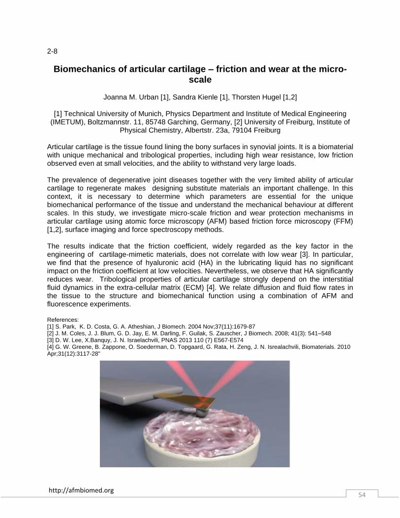
http://afmbiomed.org 54
2-8
Biomechanics of articular cartilage – friction and wear at the micro-scale
Joanna M. Urban [1], Sandra Kienle [1], Thorsten Hugel [1,2]
[1] Technical University of Munich, Physics Department and Institute of Medical Engineering
(IMETUM), Boltzmannstr. 11, 85748 Garching, Germany, [2] University of Freiburg, Institute of Physical Chemistry, Albertstr. 23a, 79104 Freiburg
Articular cartilage is the tissue found lining the bony surfaces in synovial joints. It is a biomaterial with unique mechanical and tribological properties, including high wear resistance, low friction observed even at small velocities, and the ability to withstand very large loads. The prevalence of degenerative joint diseases together with the very limited ability of articular cartilage to regenerate makes designing substitute materials an important challenge. In this context, it is necessary to determine which parameters are essential for the unique biomechanical performance of the tissue and understand the mechanical behaviour at different scales. In this study, we investigate micro-scale friction and wear protection mechanisms in articular cartilage using atomic force microscopy (AFM) based friction force microscopy (FFM) [1,2], surface imaging and force spectroscopy methods. The results indicate that the friction coefficient, widely regarded as the key factor in the engineering of cartilage-mimetic materials, does not correlate with low wear [3]. In particular, we find that the presence of hyaluronic acid (HA) in the lubricating liquid has no significant impact on the friction coefficient at low velocities. Nevertheless, we observe that HA significantly reduces wear. Tribological properties of articular cartilage strongly depend on the interstitial fluid dynamics in the extra-cellular matrix (ECM) [4]. We relate diffusion and fluid flow rates in the tissue to the structure and biomechanical function using a combination of AFM and fluorescence experiments. References: [1] S. Park, K. D. Costa, G. A. Atheshian, J Biomech. 2004 Nov;37(11):1679-87 [2] J. M. Coles, J. J. Blum, G. D. Jay, E. M. Darling, F. Guilak, S. Zauscher, J Biomech. 2008; 41(3): 541–548 [3] D. W. Lee, X.Banquy, J. N. Israelachvili, PNAS 2013 110 (7) E567-E574 [4] G. W. Greene, B. Zappone, O. Soederman, D. Topgaard, G. Rata, H. Zeng, J. N. Isrealachvili, Biomaterials. 2010 Apr;31(12):3117-28"
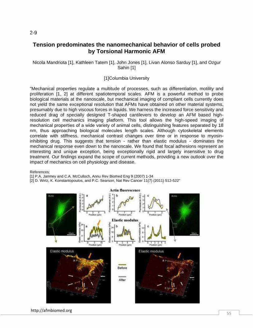
http://afmbiomed.org 55
2-9 Tension predominates the nanomechanical behavior of cells probed
by Torsional Harmonic AFM
Nicola Mandriota [1], Kathleen Tatem [1], John Jones [1], Livan Alonso Sarduy [1], and Ozgur Sahin [1]
[1]Columbia University
"Mechanical properties regulate a multitude of processes, such as differentiation, motility and proliferation [1, 2] at different spatiotemporal scales. AFM is a powerful method to probe biological materials at the nanoscale, but mechanical imaging of compliant cells currently does not yield the same exceptional resolution that AFMs have obtained on other material systems, presumably due to high viscous forces in liquids. We harness the increased force sensitivity and reduced drag of specially designed T-shaped cantilevers to develop an AFM based high-resolution cell mechanics imaging platform. This tool allows the high-speed imaging of mechanical properties of a wide variety of animal cells, distinguishing features separated by 18 nm, thus approaching biological molecules length scales. Although cytoskeletal elements correlate with stiffness, mechanical contrast changes over time or in response to myosin-inhibiting drug. This suggests that tension - rather than elastic modulus - dominates the mechanical response even down to the nanoscale. We found that focal adhesions represent an interesting and unique exception, being exceptionally rigid and largely insensitive to drug treatment. Our findings expand the scope of current methods, providing a new outlook over the impact of mechanics on cell physiology and disease. References: [1] P.A. Janmey and C.A. McCulloch, Annu Rev Biomed Eng 9 (2007) 1-34 [2] D. Wirtz, K. Konstantopoulos, and P.C. Searson, Nat Rev Cancer 11(7) (2011) 512-522"

http://afmbiomed.org 56
2-10 Single-molecule force spectroscopy on oligorotaxane foldamers
Damien SLUYSMANS [1], Carson BRUNS[2], Zhixue ZHU[2], J. Fraser STODDART[2] and Anne-
Sophie DUWEZ[1]
[1] Department of Chemistry, University of Liège (Belgium), [2] Department of Chemistry, Northwestern University, Evanston, Illinois (USA)
The diversity of molecular architectures found in nature is still a vast source of inspiration for the design of well-defined artificial molecules. In this context, foldamers, which are synthetic molecules mimicking the folded conformations of biomolecules such as proteins, have gained more attention recently [1]. They have enormous potential regarding the understanding of noncovalent bonding interactions relating the structure of a biomolecule to its particular function. Mechanically interlocked molecules (MIMs) are complex structures whose components are linked mechanically and bound through weak interactions such as hydrogen bonding and π-stacking. MIMs whose components can move with respect to another one have been synthesized in order to design artificial molecular motors and switches [2]. Foldamers in which interactions between mechanically interlocked components dictate the single-molecule assembly into a folded secondary structure have been described recently. The unique folding motif is based on a flexible thread-bearing component containing 1,5-dioxynaphthalene (DNP) donors, which folds its way through a series of cyclobis(paraquat-p-phenylene) (CBPQT4+) acceptor rings in a serpentine fashion to enable extended donor–acceptor (D–A) stacking between DNP units and the electron-poor 4,4’-bipyridinium (BIPY2+) units of the rings [3]. Here, we investigate the mechanochemical properties of those oligorotaxane foldamers by AFM-based single-molecule force spectroscopy. The molecules were grafted on a surface and stretched by an AFM tip, revealing a specific saw-tooth profile upon retraction. The distance between successive peaks appears to be a bimodal distribution, corresponding to the opening of one or two folded motifs present on each side of a ring. The rupture forces measured are also consistent with this hypothesis, i.e. with the breaking of hydrogen bonds and π-π stacking interactions. In order to modulate the interactions responsible for the folded conformation, the force spectroscopy experiments were performed in various solvents. In toluene, the π-π stacking competition with the solvent leads to a decrease in the force required to unfold the molecule, compared to experiments in dimethylformamide and acetonitrile. Interestingly, at lower loading rates, a hopping phenomenon was observed, which corresponds to fluctuations of the molecule between partially folded and unfolded conformations against the pulling force [4] (see figure). Pulling-relaxing experiments were instrumental in understanding the dynamics and the reversibility of the unfolding-refolding process. References. [1] G. Guichard and I. Huc, Chem. Commun. 2011, 47, 5933-5941; [2] C. J. Bruns and J. F. Stoddart, Acc. Chem. Res. 2014, 47, 2186-2199; [3] Z. Zhu, J. F. Stoddart et al., Chem. Sci. 2013, 4, 1470; [4] J. P. Junke r, F. Ziegler and M. Rief, Science 2009, 323, 633-637."
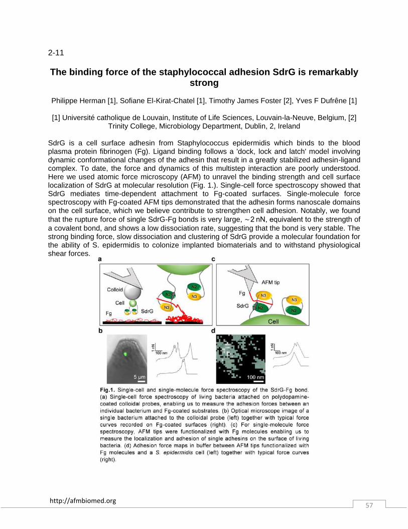
http://afmbiomed.org 57
2-11 The binding force of the staphylococcal adhesion SdrG is remarkably
strong
Philippe Herman [1], Sofiane El-Kirat-Chatel [1], Timothy James Foster [2], Yves F Dufrêne [1]
[1] Université catholique de Louvain, Institute of Life Sciences, Louvain-la-Neuve, Belgium, [2] Trinity College, Microbiology Department, Dublin, 2, Ireland
SdrG is a cell surface adhesin from Staphylococcus epidermidis which binds to the blood plasma protein fibrinogen (Fg). Ligand binding follows a 'dock, lock and latch' model involving dynamic conformational changes of the adhesin that result in a greatly stabilized adhesin-ligand complex. To date, the force and dynamics of this multistep interaction are poorly understood. Here we used atomic force microscopy (AFM) to unravel the binding strength and cell surface localization of SdrG at molecular resolution (Fig. 1.). Single-cell force spectroscopy showed that SdrG mediates time-dependent attachment to Fg-coated surfaces. Single-molecule force spectroscopy with Fg-coated AFM tips demonstrated that the adhesin forms nanoscale domains on the cell surface, which we believe contribute to strengthen cell adhesion. Notably, we found that the rupture force of single SdrG-Fg bonds is very large, ∼ 2 nN, equivalent to the strength of a covalent bond, and shows a low dissociation rate, suggesting that the bond is very stable. The strong binding force, slow dissociation and clustering of SdrG provide a molecular foundation for the ability of S. epidermidis to colonize implanted biomaterials and to withstand physiological shear forces.
http://afmbiomed.org 58
2-12 The interplay between cholesterol, Aβ (1-42) and lipid phase domains
determines membrane failure in complex model systems
Silvia Seghezza, Alberto Diaspro, Claudio Canale, Silvia Dante
Nanophysics, Istituto Italiano di Tecnologia, Via Morego 30, 16163 Genova, Italy The interaction between amyloid peptides and plasma membrane has been identified as one of the molecular mechanisms at the basis of many neurodegenerative disorders, such as Alzheimer’s disease (AD). Both peptides aggregation level and local membrane composition are fundamental in determining the nature of this interaction. In the case of AD, the role of cholesterol as a modulator of the toxicity of amyloid-β peptides Aβ(1-40) and Aβ(1-42) has been thoroughly investigated both on cells and on model membrane systems, but results are still controversial. In our study we employed AFM technique to investigate the effect of Aβ(1-42) peptide at the earliest stage of aggregation on phase segregating model lipid systems containing different percentages of cholesterol. As model membranes we used POPC/POPS/SM/Chol silicon supported lipid bilayers. Cholesterol content was varied in a range spanning different regions of the lipid system phase diagram. For zero or low cholesterol percentages we observed a membrane destabilization consisting in phase reorganization, bilayer thinning and decrease in the force required by AFM tip to penetrate inside the membrane; no significant effects were detected when we increased cholesterol content up to 20%. An accurate interpretation of our results in the light of system phase diagram suggested that cholesterol role in preventing or triggering Aβ toxic effect is strongly related to the specific lipid phases which are present in the system at a given lipid composition.
http://afmbiomed.org 59
2-13 Lipid preference of lactose permease: combining AFM and FS with FRET measurements
Òscar Domènech, M. Teresa Montero, Jordi H.Borrell
Departament de Fisicoquímica, Facultat de Farmàcia, UB, Barcelona. Catalonia, Spain.
We have exploited Förster resonance energy transfer (FRET) tools in solution to study lipid selectivity between single tryptophan mutants of lactose permease (LacY) and different phospholipids labeled with pyrene. Our results support the selective affinity of LacY for phosphatidylethanolamine (PE) over phosphatidylglycerol (PG). Based on AFM observations, in this communication we will present evidences, on: (i) LacY preference for fluid domains in PE:PG biomimetic bilayer models; (ii) the existence of a lipid boundary region around LacY (at low lipid-to-protein ratio); and based on AFM-Force Spectroscopy (FS) mode, (iii) on the influence of the lipid environment on the unfolding force (Fu) required to extract LacY form the bilayers. The composition of the lipid boundary region around LacY is inferred by means of a binary phase (PE:PG) diagram constructed based on AFM observations of supported lipid bilayers responding to temperature changes. References: [1] Suárez-Germà, C.; Hernández-Borrell, J.; Prieto, M.; Loura, L.M. Modeling FRET to investigate the selectivity of lactose permease of Escherichia coli for lipids (2014) Molecular Membrane Biology 3, 1382, 842-852 [2] Suàrez-Germà, C.; Morros, A.; Montero, M.; Hernández-Borrell, J.; Domènech, O. Combined Force Spectroscopy, AFM and calorimetric studies to reveal the nanostructural organization of biomimetic membranes (2014) Chemistry and Physics of Lipids 183, 208-2014. [3] Suárez-Germà, C.; Domènech, O.; Montero, M.T.; Hernández-Borrell,J. Effect of lactose permesase presence on the strucure and nanomechanics of two-component supported lipid bilayers. (2014) Biochimica et Biophysica Acta-Biomembranes, 1382, 3, 842-852."

http://afmbiomed.org 60
2-14 Dynamics and pH-dependence of Ag43 adhesins self-association
probed by Atomic Force Spectroscopy
Adrien Jacquot [1,2], Chizuko Sakamoto [3,4], Angélina Razafitianamaharavo [5,6], Céline Caillet [5,6], Jenny Merlin [5,6], Ahmad Fahs [1,2], Jean-Marc Ghigo [3], Jérôme F.L. Duval
[5,6], Christophe Beloin [3] and Gregory Francius [1,2]
E.g. [1] Université de Lorraine, Laboratoire de Chimie Physique et Microbiologie pour l’Environnement, UMR 7564, Villers-lès-Nancy, F-54601, France. [2] CNRS, Laboratoire de Chimie Physique et Microbiologie pour l’Environnement, UMR 7564, Villers-lès-Nancy, F-54601, France. [3] Institut Pasteur, Unité de Génétique des Biofilms, 25-28 rue du Dr Roux, Paris cedex 15, F-75724, France. [4] Université Paris Diderot, Sorbonne Paris Cité, Cellule Pasteur, rue du Dr Roux, Paris, F-75015, France. [5] Université de Lorraine, Laboratoire Interdisciplinaire des Environnements Continentaux, UMR 7360, Vandœuvre-lès-Nancy, F-54501, France. [6] CNRS, Laboratoire Interdisciplinaire des Environnements Continentaux, UMR 7360, Vandœuvre-lès-Nancy, F-54501, France. Self-associating auto-transporter (SAAT) adhesins are two-domains cell surface proteins involved in bacteria auto-aggregation and biofilm formation. Antigen 43 (Ag43) is a SAAT adhesin commonly found in Escherichia coli whose variant Ag43a has been shown to promote persistence of uropathogenic E. coli within the bladder. The recent resolution of the tri-dimensional structure of the 499 amino- -domain in Ag43a has shed light on the possible mechanism governing the self-recognition of SAATs adhesins, in particular the importance of trans-interactions between the L shape beta-helical scaffold of two beta-domains of neighboring adhesins. In this study, we use Single-Molecule Force Spectroscopy (SMFS) and Dynamic Force Spectroscopy (DFS) to unravel the dynamics of Ag43-self association under various pH and molecular elongation rates conditions that mimic the situations encountered by E. coli in its natural environment. Results evidenced an important stretchability of Ag43a with unfolding of sub-domains leading to molecular extension as long as 150 nm. Nanomechanical analysis of molecular stretching data suggested that self-association of Ag43 can lead to the formation of dimers and tetramers driven by rapid and weak cis- as well as slow but strong trans-interaction forces with magnitude as large as 100-250 pN. The dynamics of cis- and trans- interactions were demonstrated to be strongly influenced by pH and applied shear force, thus suggesting that environmental conditions can modulate Ag43-mediated aggregation of bacteria at the molecular level.
http://afmbiomed.org 61
2-15 Cell mechanics by scanning ion conductance microscopy
Tilman E. Schäffer
Institute of Applied Physics, University of Tübingen, Auf der Morgenstelle 10, 72076 Tübingen,
Germany Scanning ion conductance microscopy (SICM) allows for fast and quantitative elasticity mapping of live cells with sub-micrometer lateral resolution. This is achieved by inducing a microfluidic flow through the pipette, which indents the sample surface below the pipette tip without direct mechanical contact. The local Young’s modulus is obtained from the measured ionic current in combination with a finite-element model. Using SICM-based elasticity mapping, the dynamics of individual cytoskeleton fibers was visualized over several hours. Furthermore, the Young’s modulus of live human platelets was visualized during spreading, during thrombin-induced activation, and during cytochalasin D-induced cytoskeleton depolymerization, thereby gaining new insights into the mechanics of hemostasis.

http://afmbiomed.org 62
2-16 Fast Stiffness Mapping of Cells Using High-Bandwidth Atomic Force
Microscopy
Andrew Wang [1], Karthik Vijayraghavan [2], Olav Solgaard [2], and Manish J. Butte [2]
[1] Department of Pediatrics, Stanford University, [2] Department of Electrical Engineering, Stanford University
The cytoskeleton controls cellular morphology and mediates the mechanical interactions between a cell and its environment. Atomic force microscopy (AFM) has the unique capability to map cytoskeletal mechanics and structures with nanometer resolution. However, imaging dynamic cytomechanical processes with conventional AFM techniques is impossible due to low temporal resolution. Here we present fast nanomechanical mapping of living cells using high-bandwidth AFM (HB-AFM), where >10^6 nanoindentation measurements were acquired in ~10 min— a task that would take weeks to finish using conventional AFM. For the first time, high bandwidth measurements enabled capture of the entire tip-sample interaction for each tap on cells, engendering a new measurement (“force phase”) that exceeds the contrast of conventional tapping mode phase, and enabling spectral visualization of >10 harmonics. The abundance of measurements allowed discovery of subtle cytomechanical features, including the stiffness of fibers of the cellular spectrin network in situ. This approach bridges HB-AFM and high-harmonic imaging, and allows investigation of dynamic material and mechanical properties of living cells.

http://afmbiomed.org 63
2-17
AFM Nanodynamics a complementary tool to conventional Micromechanical AFM-contact assessment for time dependent
biomaterial
Mojtaba Azadi [1], Hadi Tavakoli Nia [2], Eliot Frank [1], Christine Ortiz [1], Alan Grodzinsky [1]
[1] MIT, Cambridge, MA, [2] Massachusetts General Hospital, Boston, MA
Articular cartilage is the tissue that covers the ends of bones in human and animal joints. Articular cartilage is composed of a gel of highly charged molecules called aggrecan enmeshed within a fibrous network of collagen. Together with many other extracellular proteins, the fiber-reinforced mesh of aggrecan and collagen enables cartilage to withstand compressive static loads (time-independent loading) due to standing as well as high frequency content dynamic loads (time-dependent loading) caused by walking, running and jumping. Macroscopic tissue mechanics has been used as a tool to study cartilage structure and material behavior associated with flow-independent intrinsic macromolecular viscoelasticity and fluid flow–dependent poroelasticity. These studies illuminated the importance of intra-tissue fluid flow and pressurization to the poro-viscoelastic properties of cartilage. Of course, these macro scale mechanical measurement techniques used to study cartilage from large animal tissue samples cannot be used for small animal samples such as those obtained from promising genetically modified mice (cartilage thickness < 50 µm). Micro-indentation techniques and AFM-based indentation capture valuable information on micromechanics of cartilage tissues, cell extracellular matrix of cells and molecules. Micromechanics in these AFM-based studies, however, is captured mainly at quasi-static or slow loading rates (i.e. force-displacement, creep, stress relaxation) in which fluid flow gives little or no contribution or little to the measured micromechanical properties of cartilage. As a result, these conventional (low frequency) methods mainly capture matrix structural information but do not capture fluid-solid interactions of cartilage tissue that are dominant at higher dynamic loading rates corresponding to running, jumping and traumatic joint injury. Other AFM techniques such as Asylum “Contact Resonance Viscoelastic Mapping” and “AM-FM Viscoelastic Mapping” detect viscous time dependent characteristics such as viscoelastic loss modulus but at very high frequencies not relevant to biomaterials (> 100 KHz) and also for harder materials (> 1 Gpa) such as polymers. We developed a nanodynamics rheology system to capture time dependent nano-scale mechanics of the cartilage tissue, ECM and aggrecan molecules in wide range of frequencies (1-10 kHz) pertinent to dynamic loadings such as walking, running and injury. We have conducted several studies with mouse tissues and cartilage matrix molecules in which we measured both micromechanics and nanodynamics of the tissue. Our studies shows that the conventional micromechanical indentation outcome (equilibrium elastic modulus) is useful for detecting structural function but is not a sensitive indicator of alterations in the extracellular matrix of mouse cartilage. In contrast, we discovered that our nanodynamics method that captures nanomechanical behavior over a wide range of frequencies, provides several indicators related to solid-fluid interaction within cartilage that are much more sensitive to changes within the extracellular matrix and tissue. These indicators include low and high frequency limits of the dynamic modulus (EL, EH), the peak angle, peak frequency (fp), the self-
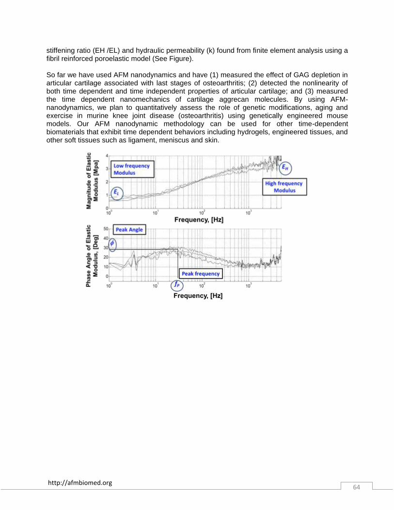
http://afmbiomed.org 64
stiffening ratio (EH /EL) and hydraulic permeability (k) found from finite element analysis using a fibril reinforced poroelastic model (See Figure). So far we have used AFM nanodynamics and have (1) measured the effect of GAG depletion in articular cartilage associated with last stages of osteoarthritis; (2) detected the nonlinearity of both time dependent and time independent properties of articular cartilage; and (3) measured the time dependent nanomechanics of cartilage aggrecan molecules. By using AFM-nanodynamics, we plan to quantitatively assess the role of genetic modifications, aging and exercise in murine knee joint disease (osteoarthritis) using genetically engineered mouse models. Our AFM nanodynamic methodology can be used for other time-dependent biomaterials that exhibit time dependent behaviors including hydrogels, engineered tissues, and other soft tissues such as ligament, meniscus and skin.
http://afmbiomed.org 65
2-18
Cellular adaptive response to mechanical signaling studied by integrated optical and atomic force microscopy
Andreea Trache [1, 2]
[1] Texas A&M Health Science Center, Department of Medical Physiology, College Station, TX;
[2] Texas A&M University, Department of Biomedical Engineering, College Station, TX Objective: Real-time cellular adaptive responses to discrete mechanical stimulation were studied by analyzing the force balance between the pre-existing cytoskeletal tension and external tensile stress. Results: The ability to measure real-time mechanosensitive events at the sub-cellular level in response to discrete mechanical stimulation is a critical component in understanding mechanically-induced cellular remodeling. Integration of mechanical stimulation with simultaneous fluorescence imaging by spinning-disk confocal and total internal reflection fluorescence microscopy enabled visualization and quantification of molecular dynamic events at the sub-cellular level in real-time. The atomic force microscope (AFM) probe consisted of a 2 micron glass bead functionalized with fibronectin. The functionalized probe sustained long-lived interactions with the vascular smooth muscle cells by inducing a functional focal adhesion. Thus, mechanical stimulation (i.e. tensile stress) of live cells was induced by direct manipulation of cortical actin through an active matrix-integrin-actin linkage. Fluorescence imaging experiments performed simultaneously with AFM mechanical stimulation enabled the investigation of molecular interactions between sub-cellular structures involved in transducing mechanical signaling and inducing cellular remodeling. Our results show that stressing integrin-matrix bond activates intracellular biochemical pathways that eventually trigger downstream cytoskeletal and focal adhesions remodeling. Moreover, intracellular pre-stress was induced by expressing RhoA variants (wild type, dominant negative, constitutively active) or treatment with ML-7 (a specific myosin light chain kinase inhibitor) to generate specific cytoskeletal tension characteristics prior to mechanical stimulation. For cells expressing RhoA-dominant negative, the AFM probe detached from the cell surface at higher forces. Due to the absence of strong cortical actin fibers, these cells are soft and the focal adhesion formed between the cell and the fibronectin coated bead is weak. The result is poor cell contractility and the loss of contact between the probe and the cell. RhoA-constitutively active expressing cells were found to exhibit high contractile activity due to strong stress fiber formation, but at the same time they were also elastic and able to pull back after initial tensile force application. In contrast, cells treated with ML-7 were significantly softer than RhoA-constitutively active expressing cells and were not able to oppose the tensile stress. These cells presented an intact actin network, but myosin blocking induced a decrease in cellular contractility. Our findings show that cells sense and adapt to physical microenvironmental changes through a coordinated response of the actomyosin apparatus necessary to establish a new homeostatic state. Conclusions: Using a non-classical integrated microscopy approach we have shown that cellular adaptive response to the external force is modulated by the pre-existing cytoskeletal tension, and that cellular adaptation to the applied tensile stress is a characteristic of the integrated cell system as a whole.

http://afmbiomed.org 66
2-19
A 3D-printed AFM system with piezotube and electromagnetic actuators for biomedical applications
Semih Sevim [1], Sevil Özer [2], Luying Feng [2], Kate Crawford [2], Hamdi Torun [2]
[1] Department of Mechanical Engineering, Bogazici University, Istanbul, Turkey [2] Department
of Electrical and Electronics Engineering, Bogazici University, Istanbul, Turkey An atomic force microscope (AFM) has been developed for biomolecular force spectroscopy. Design, implementation and characterization of the AFM head are described here. The head is portable and was manufactured at low cost using stereolithography. A flexible software-based controller was implemented that can be adapted easily for different applications. The AFM head, made of a rigid polymer material (Rigid Opaque, Stratasys, Ltd., MN, USA) is shown in Fig. 1(a). The head houses a piezotube actuator, a laser diode, a quadrature photodetector and an AFM cantilever. The cantilever is mounted to the piezotube using a customized holder. In addition, an electromagnet was employed with the head for direct cantilever actuation and for other magnetic applications. A one-dimensional actuation coil is integrated to the head as shown in Fig. 1(b). The system allows cantilever actuation using the piezotube actuator and the electromagnet. Biomolecular force spectroscopy experiments to probe interactions between FGF-2 and heparin, was performed using the new system. Fig. 1(c) shows a typical force curve, exhibiting an unbinding event with a force strength of 500 pN, whereas in Fig. 3(d) shows a force curve indicating no adhesion/rupture events.
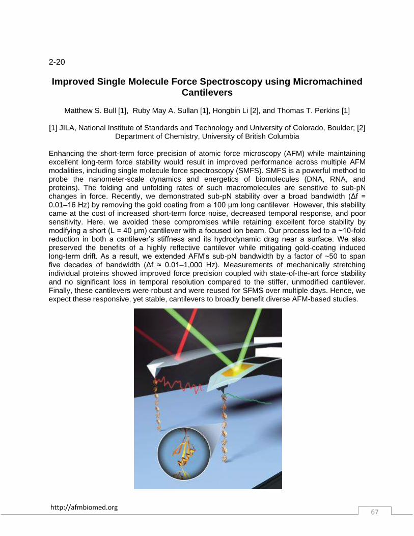
http://afmbiomed.org 67
2-20 Improved Single Molecule Force Spectroscopy using Micromachined
Cantilevers
Matthew S. Bull [1], Ruby May A. Sullan [1], Hongbin Li [2], and Thomas T. Perkins [1]
[1] JILA, National Institute of Standards and Technology and University of Colorado, Boulder; [2] Department of Chemistry, University of British Columbia
Enhancing the short-term force precision of atomic force microscopy (AFM) while maintaining excellent long-term force stability would result in improved performance across multiple AFM modalities, including single molecule force spectroscopy (SMFS). SMFS is a powerful method to probe the nanometer-scale dynamics and energetics of biomolecules (DNA, RNA, and proteins). The folding and unfolding rates of such macromolecules are sensitive to sub-pN changes in force. Recently, we demonstrated sub-pN stability over a broad bandwidth (Δf = 0.01–16 Hz) by removing the gold coating from a 100 μm long cantilever. However, this stability came at the cost of increased short-term force noise, decreased temporal response, and poor sensitivity. Here, we avoided these compromises while retaining excellent force stability by modifying a short (L = 40 μm) cantilever with a focused ion beam. Our process led to a ~10-fold reduction in both a cantilever’s stiffness and its hydrodynamic drag near a surface. We also preserved the benefits of a highly reflective cantilever while mitigating gold-coating induced long-term drift. As a result, we extended AFM’s sub-pN bandwidth by a factor of ~50 to span five decades of bandwidth (Δf ≈ 0.01–1,000 Hz). Measurements of mechanically stretching individual proteins showed improved force precision coupled with state-of-the-art force stability and no significant loss in temporal resolution compared to the stiffer, unmodified cantilever. Finally, these cantilevers were robust and were reused for SFMS over multiple days. Hence, we expect these responsive, yet stable, cantilevers to broadly benefit diverse AFM-based studies.

http://afmbiomed.org 68
2-21
Uncovering Bacterial Phenotypic Heterogeneity by Combined Time-Lapse Atomic Force and Optical Microscopy
Haig Alexander ESKANDARIAN [1,2], Joelle Ven [1,2], Adrian Nievergelt [2], Shinji Kondo [3],
Chen Yang [2], John McKinney [1], Georg Fantner [2]
[1] School of Life Sciences, Laboratory of Living Matter, Swiss Federal Institute of Technology (EPFL), [2] School of Engineering, Laboratory for Bio- & Nano- Instrumentation, Swiss Federal
Institute of Technology (EPFL), [3] Department of Bio-Engineering, University of Tokyo Characterizing phenotypic individuality in an isogenic bacterial population is a burgeoning field of research. Single-cell studies have traditionally been driven by established tools like optical microscopy or flow cytometry. However, these tools are restrictive in the number of phenotypes one can simultaneously monitor. This suggests that using more revealing tools and techniques could enhance our current understanding of the physiological conditions underlying bacterial individuality. We have adapted Atomic Force Microscopy (AFM) to long-term time-lapse imaging of the bacterium, Mycobacterium smegmatis, providing a unique opportunity to visualize and quantitate numerous physiological components relevant to such basic processes as cellular growth and division, or persistence to antibiotic stress. We are characterizing bacterial phenotypic heterogeneity with unprecedented detail by simultaneously combining surface mechanical measurements and cell surface morphology revealed by AFM at the nano-scale, with internal biological sensors monitored by optical fluorescence microscopy. We have revealed for the first time by time-lapse AFM the dynamic organization of the surface morphology of Mycobacterium smegmatis during growth, quantifying the rate of elongation, volume accumulation, the dynamics of polar growth, and the ability to predict the site of division. Our study provides an unprecedented level of complexity to describing simultaneously a combination of bacterial phenotypes, which may underlie behavioral differences under selective environmental conditions.

http://afmbiomed.org 69
2-22
Correlative Light Atomic Force Electron Microscopy (CLAFEM): combining force measurements to CLEM
Frank Lafont
Institute Pasteur, CNRS, INSERM, Univ. Lille
We developed different types of correlative methods in order to take benefit from the three microscopy modes: biophotonic, electron and near-field. First, we achieved to perform atomic force microscopy (AFM)-nanoguided photonic measurements. The AFM allowed to nanomanipulate objects at the cell surface and photonic microscopy was used to follow the recruitment at the plasma membrane of cellular proteins tagged with a fluorescent probe. This was also applied using Photoactivated Localization Microscopy (PALM). In a second approach while fluorescent-based super-resolution microscopy methods were used to follow cellular proteins, AFM was used to explore the biophysical properties of the cell (e.g. Young modulus) in a correlative manner. After AFM measurements, samples were processed for electron microscopy allowing to add another layer of information. We will discuss the pros/cons of these methods and their future based on results obtained studying host-pathogen interactions."

http://afmbiomed.org 70
Abstracts Day 3
Tuesday, December 16, 2014
3-1 Atomic Force Microscopy of cells as a Diagnostic for Cancer, cancer
prevention and detection of chemo-resistance
James Gimzewski
Department of Chemistry and Biochemistry, California NanoSystems Institute, Jonsson Comprehensive Cancer Center, University of California, Los Angeles, CA 90095, USA .
International Center for Materials Nanoarchitectonics Satellite (MANA), National Institute for Materials Science (NIMS), Tsukuba, Japan Centre for Nanoscience and Quantum Information,
University of Bristol, Bristol, UK Since our first paper on nanomechanics of cancer cells from human patients in 2007, the relationship between cell mechanics and physiological function has developed into a field of great interest to the AFM and medical community. In this talk I will present some examples on the use of AFM to study the effects of green tea extract, cisplatin resitance in ovarian cancer cells as well as coreelative methods such as STED to understand better the role of actin in their mechanical properties. We also will discuss recent biophysical and morphological effects of nanodiamond/nanoplatinum solution (DPV576) on metastatic murine breast cancer cells tracking in vitro changes in cell stiffness and early structural alterations of metastatic breast cancer cells post treatment with DPV576, which may have important implications in the role of nanodiamond/nanoplatinum based cancer cell therapy and sensitization to chemotherapy drugs.

http://afmbiomed.org 71
3-2 Fibrinogen-dependent cell-cell adhesion of erythrocytes assessment
by AFM-based force spectroscopy
Filomena A. Carvalho, Ana Filipa Guedes, and Nuno C. Santos
Instituto de Medicina Molecular, Faculdade de Medicina, Universidade de Lisboa, Portugal Increased erythrocyte aggregation has been related to the presence of high levels of fibrinogen molecules on human blood plasma. Erythrocyte hyperaggregation may lead to disturbances on the normal blood flow, thrombus formation and high risk of developing cardiovascular pathologies. By Atomic Force Microscopy (AFM)-based force spectroscopy, we measured erythrocyte-erythrocyte adhesion forces in the absence and in the presence of increasing fibrinogen concentrations. For that, AFM cell-cell adhesion experiments were done using tipless cantilevers, functionalized with concanavalin A. Erythrocytes were isolated from human healthy donors’ blood, using standard procedures, and adhered to poly-L-lysine coated glass-slides. After the capture of one cell by the functionalized cantilever, using the AFM contact mode, it was moved to the top of another erythrocyte and the cell-cell contact was established with an applied force of 300 pN, in constant height and closed-loop mode. The AFM tip resonant frequency was maintained at 2 Hz and a cell-cell contact time of 5s was maintained before cantilever retraction, with a z-range displacement of 50 µm. Cell-cell adhesion curves were obtained in the absence or in the presence of increasing fibrinogen concentrations on the medium (from 0 to 1 mg/ml). The analysis of these curves yielded the quantification of the detachment force and the work necessary to break the interaction. The work necessary to separate two erythrocytes significantly increase with increasing fibrinogen concentrations, from 0.45 ± 0.04 fJ without fibrinogen to 12.0 ± 0.13 fJ at 1 mg/ml fibrinogen (p<0.001). Additionally, the maximum cell-cell detachment force increased from 71.9 ± 2.9 pN (without fibrinogen) to 250.4 ± 3.2 pN at 1 mg/ml fibrinogen (p<0.0001). We also observed a 3.5-fold increase on the number of membrane tethers per curve on the cell-cell detachment in the presence of fibrinogen 1 mg/ml comparing with the experiments without fibrinogen. AFM data allow, for the first time, the quantification of the adhesion force necessary to detach two erythrocytes in the presence of different concentrations of fibrinogen molecules. Our in vitro study tried to mimic what happens in vivo on the human blood flow. The results confirm that increasing fibrinogen plasma levels are associated with the higher tendency of erythrocytes to aggregate, probably by transient simultaneous binding of the protein to two cells, bridging them. This transient aggregation impairs blood flow and is associated with a higher risk of cardiovascular diseases."
http://afmbiomed.org 72
3-3
The overexpression of Lamin B1 in autosomal dominant leukodystrophy influences the mechanical properties of cell nuclei.
Claudio Canale [1], Silvia Seghezza [1], Denise Ferrera [2], Sabina Cappellari [3], Pietro Cortelli
[3], and Laura Gasparini [2]
[1] Dept. Nanophysics, Istituto Italiano di Tecnologia, [2] Dept.of Neuroscience and Brain Technologies, Istituto Italiano di Tecnologia, [3] Dept. of Biomedical and Neuromotor Sciences,
University of Bologna Mutations in proteins that constitute the nuclear lamina are associated to a series of pathologies, generally termed laminopathies, that include Emery-Dreifuss and limb-girdle muscular dystrophies, dilated cardiomyopathy, Dunningan-type familial partial lipodystrophy, Charcot-Marie tooth disorder type 2, and Hutchison-Gilford progeria syndrome (1). Recently, gene duplication and protein overexpression of lamin B1 (LB1) has been linked to autosomal dominant leukodystrophy (ADLD). However, the mechanisms by which LB1 overexpression causes the disease are still unclear. Alterations in lamins affect the nuclear envelope, compromising the structural integrity of the nucleus and affecting its mechanical properties (2). However, it remains unclear whether the pathological overexpression of LB1 that is associated with ADLD alters nuclear mechanics and function in human cells. To address this central question, we performed AFM indentation to understand how LB1 impacts nuclear rigidity. The experiments were carried out on primary cultures of human skin fibroblasts derived from patients with ADLD and the results were compared with that obtained on control (CTR) cells derived from age matched healthy subjects. We performed the analysis on ADLD and CTR nuclei isolated by osmotic lysis from quiescent fibroblasts within 16 passages. We demonstrated that LB1 overexpression increases nuclear rigidity in ADLD human skin fibroblasts. To investigate whether the nuclei display a similar behavior also in living cell, we performed AFM indentation on adherent living CTR and ADLD fibroblasts in quiescent conditions. The stiffness of living fibroblasts nuclei was 6-fold higher than that of isolated nuclei, likely due to the significant contributions from adhesive forces, the underlying cytoskeleton and cytosolic factors to the measurement. Transient LB1 overexpression in HEK293 cells mimics the pathological phenotype of ADLD fibroblasts. Similar effects were obtained by LB1 overexpression in neural cells that are the target cells in the disease. Patch-clamp experiments on isolated nuclei demonstrated that LB1 overexpression in ADLD fibroblasts has functional consequences and is associated with altered nuclear ionic permeability. In conclusion, by employing AFM on samples derived from ADLD patients we revealed the unknown role of LB1 in controlling the mechanical properties of cell nuclei. LB1 overexpression affects nuclear rigidity in human cells, possibly leading to changes in ionic signaling in ADLD cells (3). References: [1] Butin-Israeli et al. (2012) Trends Genet. 28, 464–471 [2] Lammerding et al. (2006) J. Biol. Chem. 281, 25768–25780 [3] Ferrera, Canale et al. (2014) Faseb J. 28, 3906-3918"

http://afmbiomed.org 73
3-4 Shear dependence of von Willebrand Factor´s interactions with Factor
VIII and ADAMTS13 demonstrated at single molecular level by AFM
K. Bonazza[1], H. Rottensteiner[2], G.Schrenk[2], J.Frank[1], G. Allmaier[1], P.L. Turecek[2] and G. Friedbacher[1]
[1] Institute of Chemical Technologies and Analytics, Vienna University of Technology, A-1060
Vienna, Austria [2] Baxter Innovations, A-1221 Vienna, Austria Von Willebrand factor (VWF) is a multimeric adhesive glycoprotein which has a dual functionin hemostasis. It mediates platelet adhesion at sites of vascular injury, which is necessary for primary hemostasis, and stabilizes factor VIII in the circulation. The pathophysiological significance of these different biological functions is evident in von Willebrand's disease (VWD), the most common hemorrhagic disorder, affecting 1% of the population. More than with other blood coagulation proteins structure of VWF determines its function. At high blood vessel wall shear rates the molecule is stretched which mediate adhesion and aggregation of blood platelets [1]. This understanding can now be complemented by new data showing that shear-stretching also affects other functions of VWF. The availability of a recombinant (r) VWF product for the first time enables detailed biophysical investigations with this molecule- Using a customized device to simulate shear conditions and conserve the rVWF molecules in their unstable, elongated conformation, we visualize at single molecular level on one hand the elongation of rVWF and the other hand proteolytic cleavage of rVWF by rADAMTS13. Following a recently published procedure to visualize single protein binding events [2], here the binding of both rADAMTS13 and rFVIII to rVWF was studied in a shear rate dependent manner. Taken together, these large multimeric molecule rVWF shows an interesting behavior at the borderline between biomechanics and biochemistry. As a consequence of a shear force induced stretching they react with each other forming fibers and they radically change their affinity to collagen, to platelets, to the cleaving protease ADAMTS13, and surprisingly also towards coagulation Factor VIII as it was thought previously that binding of FVIII to VWF is mainly controlled by limited proteolysis during the blood clotting process. References: [1] Schneider et.al. 2007. PNAS. [2] Bonazza et.al 2013. ABC. Figure: The AFM image shows a fully stretched, 2 µm long, single rVWF molecule marked by a nanoscopic scratch (dark brown)."
http://afmbiomed.org 74
3-5 Cardiomyocyte Cortical Stiffness Indicates Cytoskeletal Integrity and
Sarcomeric Function in the Aging Heart
Gaurav Kaushik [1], Alice Spenlehauer [1], Ayla O. Sessions [2], Adriana S. Trujillo [3], Alexander Fuhrmann [1], Zongming Fu [4], Vidya Venkatraman [5], Danielle Pohl [1], Jeremy Tuler [1], Mingyi Wang [6], Edward G. Lakatta [6], Karen Ocorr [7], Rolf Bodmer [7], Sanford I. Bernstein [3], Jennifer E. Van Eyk [4,5], Anthony Cammarato [4], and Adam J. Engler [1, 2, 8]
[1] Department of Bioengineering and [2] Biomedical Sciences Program, University of California,
San Diego; La Jolla, CA USA; [3] Department of Biology and Molecular Biology Institute, San Diego State University; San Diego, CA USA; [4] Department of Medicine, Division of Cardiology, Johns Hopkins University; Baltimore, MD; [5] Advanced Clinical Biosystems Research Institute;
Barbra Streisand Women's Heart Center; The Heart Institute and Department of Medicine, Cedars-Sinai Heart Institute; Los Angeles, CA USA; [6] Laboratory of Cardiovascular Science, National Institute on Aging, National Institutes of Health, Baltimore, MD USA; [7] Development and Aging Program, Sanford-Burnham Institute for Medical Research; La Jolla, CA USA; [8]
Sanford Consortium for Regenerative Medicine; La Jolla, CA USA
Cardiac myocytes are capable of functioning for decades despite minimal cell turnover or regeneration, suggesting that molecular alterations help sustain heart function with age. Identification of compensatory remodeling events in the aging heart remains elusive. Proteomics-based analysis of rhesus monkeys, rats, and Drosophila reveal that reinforcement of the cardiomyocyte cytoskeleton is a highly-conserved aging event. Vinculin acts a unique regulator of cytoskeletal mechanics during cardiac aging: increased cardiac vinculin expression reinforces the cortical cytoskeleton, as measured by cortical stiffness, enhances myofilament organization, improves cellular contractility, elevates hemodynamic stress tolerance, and leads to prolonged lifespan in fly. Though alterations in cortical mechanics are often associated with impaired function, these findings suggest that cytoskeletal reinforcement may compensate for dysfunction in the aging heart.
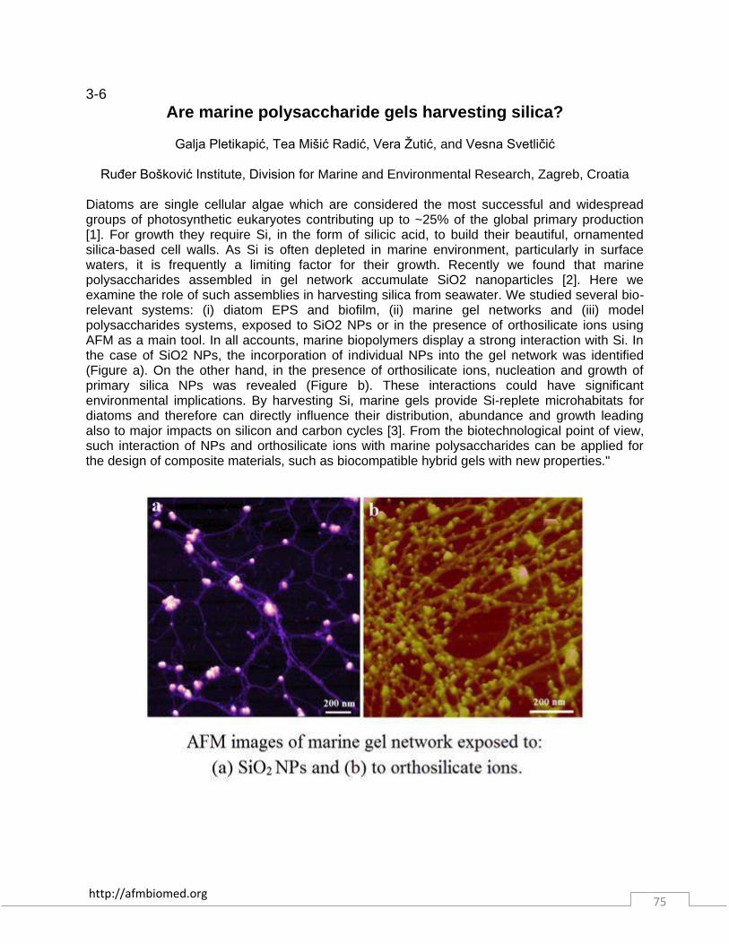
http://afmbiomed.org 75
3-6 Are marine polysaccharide gels harvesting silica?
Galja Pletikapić, Tea Mišić Radić, Vera Žutić, and Vesna Svetličić
Ruđer Bošković Institute, Division for Marine and Environmental Research, Zagreb, Croatia
Diatoms are single cellular algae which are considered the most successful and widespread groups of photosynthetic eukaryotes contributing up to ~25% of the global primary production [1]. For growth they require Si, in the form of silicic acid, to build their beautiful, ornamented silica-based cell walls. As Si is often depleted in marine environment, particularly in surface waters, it is frequently a limiting factor for their growth. Recently we found that marine polysaccharides assembled in gel network accumulate SiO2 nanoparticles [2]. Here we examine the role of such assemblies in harvesting silica from seawater. We studied several bio-relevant systems: (i) diatom EPS and biofilm, (ii) marine gel networks and (iii) model polysaccharides systems, exposed to SiO2 NPs or in the presence of orthosilicate ions using AFM as a main tool. In all accounts, marine biopolymers display a strong interaction with Si. In the case of SiO2 NPs, the incorporation of individual NPs into the gel network was identified (Figure a). On the other hand, in the presence of orthosilicate ions, nucleation and growth of primary silica NPs was revealed (Figure b). These interactions could have significant environmental implications. By harvesting Si, marine gels provide Si-replete microhabitats for diatoms and therefore can directly influence their distribution, abundance and growth leading also to major impacts on silicon and carbon cycles [3]. From the biotechnological point of view, such interaction of NPs and orthosilicate ions with marine polysaccharides can be applied for the design of composite materials, such as biocompatible hybrid gels with new properties."

http://afmbiomed.org 76
3-7 T-lymphocyte adhesion forces and mechanotransduction modulated
by activation with TNF
Qian Li [1], Dieter Adam [2], and Christine Selhuber-Unkel [1]
[1] Biocompatible Nanomaterials, Institute for Materials Science, University of Kiel, Germany; [2] Institute of Immunology, University of Kiel, Germany
Integrin-mediated T-lymphocyte adhesion to endothelial cells is a crucial step in the mammalian inflammatory response and for the elimination of pathogens. In recent years, the cytokine tumor necrosis factor (TNF) stimulated outside-in signalling pathway of integrins in response to proinflammatory event was thoroughly studied. Common knowledge is that TNF stimulates the endothelial cell surface molecules, which in turn activate integrins to facilitate the adhesion of T-lymphocytes. In addition, also an inside-out signalling pathway of integrins in lymphocyte activation by TNF has been reported. However, how this activation modulates T-lymphocyte adhesion strength and dynamics is still not understood. We have applied single-cell force spectroscopy (SCFS) as a biophysical approach to address this question and to investigate T-lymphocyte (Jurkat E6-1) cell adhesion to fibronectin, which is naturally present on top of endothelial cell layers. In detail, we approached single Jurkat E6-1 cells to fibronectin-coated surfaces and analyzed cell detachment forces. We found that TNF significantly increased cell adhesion forces and detachment energies, as well as the number of molecular ruptures and single molecule rupture forces, even at sub-second timescales. Thus our data show that the TNF-stimulated inside-out-signalling pathway directly enhances T-lymphocyte adhesion.
http://afmbiomed.org 77
3-8
Unravelling of a mechanism of resistance to colistin in Klebsiella pneumoniae thanks to Atomic Force Microscopy
Cécile Formosa [1,2], Raphaël E. Duval [2], and Etienne Dague [1]
[1] LAAS-CNRS, Université de Toulouse, France [2] SRSMC-CNRS, Université de Lorraine,
France Infections caused by the bacteria Klebsiella pneumoniae have now became common in healthcare units. Among numerous virulence factors described for this bacteria, are extracellular capsule, which form thick bundles of fibrillous structures covering the bacterial surface. Another important feature of Klebsiella pneumoniae is its ability to resist to a large number of antibiotics. Therefore, management and treatment of infections caused by multidrug resistant K. pneumoniae can be challenging [1]. In this context, colistin, an old antibiotic of the polymyxin class, has been reintroduced and is now a last resort antibiotic for the treatment of such infections. In this study, we have used Atomic Force Microscopy (AFM) in liquid conditions [2, 3] to study the effects on the morphology and the nanomechanical properties on both a multiresistant but colistin-sensitive strain of Klebsiella pneumoniae (i.e. ATCC 700603), and its colistin-resistant derivate, called Kpm [4]. Our results allowed us to image for the first time the capsular polysaccharides of K. pneumoniae in liquid conditions. We could observe that the two different strains had a different capsular morphology; the capsule of the resistant strain is tightly bound to the bacterial cell whereas that of the sensitive strain seems leaky and is spread on a large surface around the cell. Upon treatment with colistin, the capsule was removed from the sensitive strain, whereas for Kpm, colistin interacted with the capsule and increased its Young modulus, without removing it from the bacterial surface. Force curves recorded on top of cells, using a high applied force, revealed the nanoarchitecture of the capsule of the two strains: Kp ATCC 700603 has a soft capsule (3.6 ± 0.8 kPa) in one layer whereas Kpm capsule is harder (21.3 ± 4.7 kPa) and organized in superposed layers. In the case of Kpm, this particular capsular organization remains upon treatment with colistin, while it is lost in the sensitive strain. To conclude, we show in this study for the first time the effects of colistin at the nanoscale on the cell wall of K. pneumoniae using AFM. This led us to formulate a hypothesis regarding the mechanism of resistance to colistin of the resistant strain: the multilayered organization of its capsular polysaccharides interacts with colistin, without letting it through the capsule to reach its target, i.e. the lipopolysaccharide present on the outer membrane. References: [1] Keynan, Y. & Rubinstein, E. The changing face of Klebsiella pneumoniae infections in the community. Int. J. Antimicrob. Agents 30, 385–389 (2007); [2] Chopinet, L., Formosa, C., Rols, M. P., Duval, R. E. & Dague, E. Imaging living cells surface and quantifying its properties at high resolution using AFM in QITM mode. Micron 48, 26–33 (2013); [3] Pillet, F., Chopinet, L., Formosa, C. & Dague, É. Atomic Force Microscopy and pharmacology: From microbiology to cancerology. Biochim. Biophys. Acta BBA - Gen. Subj. 1840, 1028–1050 (2014); [4] Vidaillac, C., Benichou, L. & Duval, R. E. In Vitro Synergy of Colistin Combinations against Colistin-Resistant Acinetobacter baumannii, Pseudomonas aeruginosa, and Klebsiella pneumoniae Isolates. Antimicrob. Agents Chemother. 56, 4856–4861 (2012).
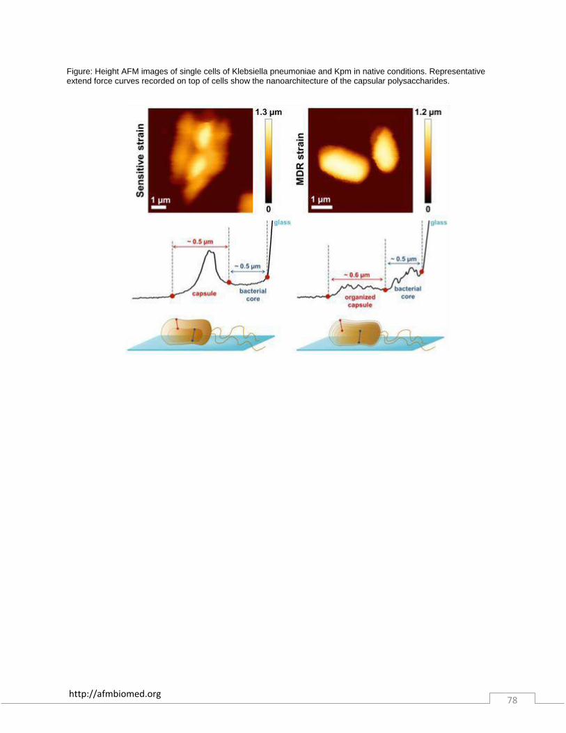
http://afmbiomed.org 78
Figure: Height AFM images of single cells of Klebsiella pneumoniae and Kpm in native conditions. Representative extend force curves recorded on top of cells show the nanoarchitecture of the capsular polysaccharides.
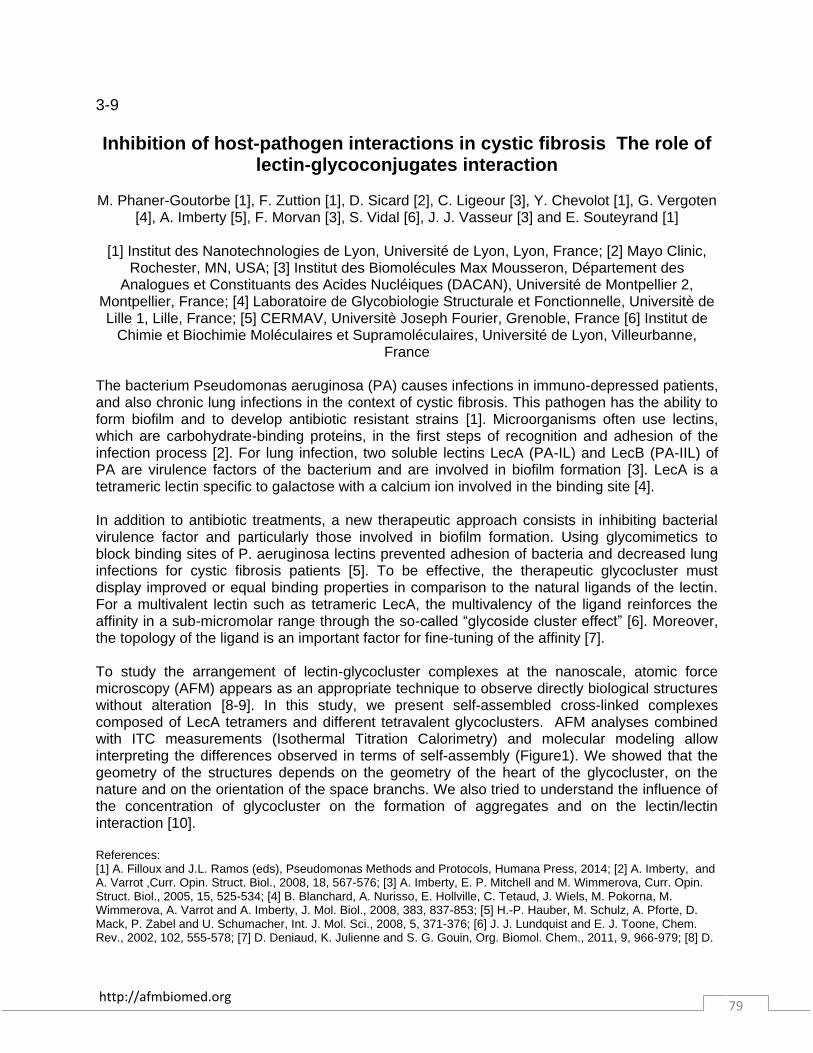
http://afmbiomed.org 79
3-9 Inhibition of host-pathogen interactions in cystic fibrosis The role of
lectin-glycoconjugates interaction
M. Phaner-Goutorbe [1], F. Zuttion [1], D. Sicard [2], C. Ligeour [3], Y. Chevolot [1], G. Vergoten [4], A. Imberty [5], F. Morvan [3], S. Vidal [6], J. J. Vasseur [3] and E. Souteyrand [1]
[1] Institut des Nanotechnologies de Lyon, Université de Lyon, Lyon, France; [2] Mayo Clinic,
Rochester, MN, USA; [3] Institut des Biomolécules Max Mousseron, Département des Analogues et Constituants des Acides Nucléiques (DACAN), Université de Montpellier 2,
Montpellier, France; [4] Laboratoire de Glycobiologie Structurale et Fonctionnelle, Universitè de Lille 1, Lille, France; [5] CERMAV, Universitè Joseph Fourier, Grenoble, France [6] Institut de
Chimie et Biochimie Moléculaires et Supramoléculaires, Université de Lyon, Villeurbanne, France
The bacterium Pseudomonas aeruginosa (PA) causes infections in immuno-depressed patients, and also chronic lung infections in the context of cystic fibrosis. This pathogen has the ability to form biofilm and to develop antibiotic resistant strains [1]. Microorganisms often use lectins, which are carbohydrate-binding proteins, in the first steps of recognition and adhesion of the infection process [2]. For lung infection, two soluble lectins LecA (PA-IL) and LecB (PA-IIL) of PA are virulence factors of the bacterium and are involved in biofilm formation [3]. LecA is a tetrameric lectin specific to galactose with a calcium ion involved in the binding site [4]. In addition to antibiotic treatments, a new therapeutic approach consists in inhibiting bacterial virulence factor and particularly those involved in biofilm formation. Using glycomimetics to block binding sites of P. aeruginosa lectins prevented adhesion of bacteria and decreased lung infections for cystic fibrosis patients [5]. To be effective, the therapeutic glycocluster must display improved or equal binding properties in comparison to the natural ligands of the lectin. For a multivalent lectin such as tetrameric LecA, the multivalency of the ligand reinforces the affinity in a sub-micromolar range through the so-called “glycoside cluster effect” [6]. Moreover, the topology of the ligand is an important factor for fine-tuning of the affinity [7]. To study the arrangement of lectin-glycocluster complexes at the nanoscale, atomic force microscopy (AFM) appears as an appropriate technique to observe directly biological structures without alteration [8-9]. In this study, we present self-assembled cross-linked complexes composed of LecA tetramers and different tetravalent glycoclusters. AFM analyses combined with ITC measurements (Isothermal Titration Calorimetry) and molecular modeling allow interpreting the differences observed in terms of self-assembly (Figure1). We showed that the geometry of the structures depends on the geometry of the heart of the glycocluster, on the nature and on the orientation of the space branchs. We also tried to understand the influence of the concentration of glycocluster on the formation of aggregates and on the lectin/lectin interaction [10]. References: [1] A. Filloux and J.L. Ramos (eds), Pseudomonas Methods and Protocols, Humana Press, 2014; [2] A. Imberty, and A. Varrot ,Curr. Opin. Struct. Biol., 2008, 18, 567-576; [3] A. Imberty, E. P. Mitchell and M. Wimmerova, Curr. Opin. Struct. Biol., 2005, 15, 525-534; [4] B. Blanchard, A. Nurisso, E. Hollville, C. Tetaud, J. Wiels, M. Pokorna, M. Wimmerova, A. Varrot and A. Imberty, J. Mol. Biol., 2008, 383, 837-853; [5] H.-P. Hauber, M. Schulz, A. Pforte, D. Mack, P. Zabel and U. Schumacher, Int. J. Mol. Sci., 2008, 5, 371-376; [6] J. J. Lundquist and E. J. Toone, Chem. Rev., 2002, 102, 555-578; [7] D. Deniaud, K. Julienne and S. G. Gouin, Org. Biomol. Chem., 2011, 9, 966-979; [8] D.
http://afmbiomed.org 80
Sicard, S. Cecioni, M. Iazykov, Y. Chevolot, S. E. Matthews, J.-P. Praly, E. Souteyrand, A. Imberty, S. Vidal and M. Phaner-Goutorbe, Chem. Commun., 2011, 47, 9483-9485; [9] M. Phaner-Goutorbe, M. Iazykov,R. Villey, D. Sicard, Y. Robach, Materials Science and Engineering C 33, 2311-2316 (2013); [10] D. Sicard, Y. Chevolot, E. Souteyrand, A. Imberty, S. Vidal and M. Phaner-Goutorbe, J. Mol. Recognit., 2013, 26, 694-699. Figure: (a)AFM image of the arrangement between the LectinPA-IL and a tetra-galactosylated glycocluster. The cobblestone represent a lectin.(b) corresponding model of the arrangement 1 glycocluster for 3 lectins."
http://afmbiomed.org 81
3-10
Studying early stages of fibronectin fibrillogenesis in living cells by atomic force microscopy
Tetyana Gudzenko and Clemens M. Franz
DFG-Center for Functional Nanostructures, Karlsruhe Institute of Technology (KIT), Wolfgang-
Gaede-Strasse 1a, 76131 Karlsruhe, Germany Fibronectin (FN) is an extracellular matrix protein that is assembled by cells into large fibrillar networks, but the dynamics of FN remodeling and the transition through intermediate fibrillar stages are still incompletely understood. Here we have used fluorescence microscopy and time-lapse atomic force microscopy (AFM) to visualize initial stages of FN fibrillogenesis in living fibroblasts at high resolution and an image acquisition time of 2 to 5 min. Initial FN nanofibrils form within 5 min of cell-matrix contact and subsequently extend at a rate of 0.25±0.02 µm/min at sites of active cell membrane retraction. FN nanofibrils display a complex linear array of globular features spaced at varying distances, indicating the coexistence of different conformational states within the fibril. In some case initial fibrils extended in increments of 0.5 to 1.5 µm in a series of cyclic membrane retractions, suggesting a stepwise fibrillar extension mechanism. In presence of Mn2+, a known activator of integrin adhesion to FN, fibrillogenesis was accelerated almost threefold to 0.68±0.14 µm/min and fibrillar dimensions were increased, underlining the importance of integrin activation for early FN fibrillogenesis. FN fibrillogenesis visualized by time-lapse AFM thus provides new structural and temporal insight into initial steps of cell-driven FN fibrillogenesis.

http://afmbiomed.org 82
3-11
Structural Analysis of Recombination Mediator Protein Rad52 by Atomic Force and Electron Microscopy
Jarmila Mlcouskova [1], Daniel Nemecek [2], Barbora Stefanovie [1, 3], Lumir Krejci [1, 3, 4]
[1] Department of Biology, Faculty of Medicine and [2] Central European Institute of Technology,
Masaryk University, Kamenice 5, Brno; [3] International Clinical Research Center, St. Anne’s University Hospital in Brno, Brno; [4] National Center for Biomolecular Research, Masaryk
University, Kamenice 5, Brno, Czech Republic Homologous recombination (HR) plays a fundamental role in the repair of DNA damage, including double-strand breaks, DNA gaps, and interstrand cross-links. Moreover, HR supports the recovery of stalled or broken replication forks and ensures faithful chromosome segregation during meiosis. In many cases, mutations of proteins involved in the mechanism of HR are associated with cancer and cancer-prone syndromes. Thus, understanding their function could play an important role in development of better tools for cancer diagnosis and therapy. In our studies, we have focused on structural and functional characterization of the human and yeast Rad52 protein. While yeast Rad52 is essential for loading of Rad51 onto RPA-coated single-stranded DNA, referred to as the recombination mediator activity, in humans this role has been adopted by the tumor suppressor protein BRCA2, with RAD52 being involved in an alternative pathway. Importantly, loss of RAD52 function is synthetically lethal in BRCA2-deficient human cells indicating potential applications in cancer therapy. AFM imaging of the yeast Rad52 has revealed the presence of coiled-coil domains similar to those observed for Rad50 protein (de Jager, 2001; van Noort, 2003). AFM experiments have also demonstrated strong binding preference of the yeast Rad52 to the blunt ends of dsDNA in the absence of Mg, which is in agreement with electron microscopy data of human Rad52 (Van Dyck, 1999), however has not been observed in previous AFM studies (Ristic 2003, Wu 2006). Recently, our group has shown that human RAD52 protein is capable of binding small evolutionarily conserved acidic protein DSS1, which is known to stabilize BRCA2 (Pavletich paper). Thus, we decided to structurally characterize the RAD52-DSS1 complexes alone or bound to single-stranded DNA by electron microscopy. The 3-D reconstruction using C7 symmetry confirmed a funnel-like structure of the full-length Rad52 observed previously (Stasiak, 2000). The reconstruction of the RAD 52-DSS1 complexes revealed significant conformational changes induced by DSS1. The RAD 52 ring becomes wider and flatter, while the bottom of the central channel is sealed with solid density. The lobes at the ring circumference are more pronounced and likely include additional density of the 8-kDa DSS1 subunits. Our data suggest possible regulatory role of DSS1 as a molecular switch modulating RAD52 activities.
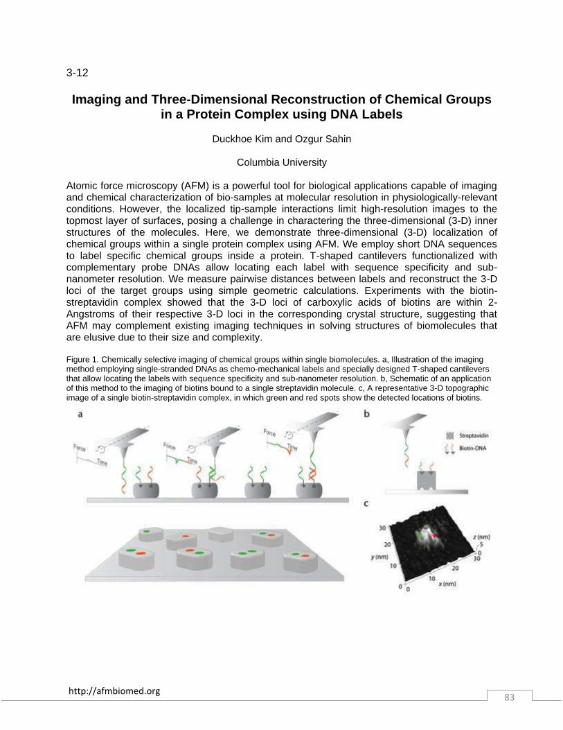
http://afmbiomed.org 83
3-12 Imaging and Three-Dimensional Reconstruction of Chemical Groups
in a Protein Complex using DNA Labels
Duckhoe Kim and Ozgur Sahin
Columbia University
Atomic force microscopy (AFM) is a powerful tool for biological applications capable of imaging and chemical characterization of bio-samples at molecular resolution in physiologically-relevant conditions. However, the localized tip-sample interactions limit high-resolution images to the topmost layer of surfaces, posing a challenge in charactering the three-dimensional (3-D) inner structures of the molecules. Here, we demonstrate three-dimensional (3-D) localization of chemical groups within a single protein complex using AFM. We employ short DNA sequences to label specific chemical groups inside a protein. T-shaped cantilevers functionalized with complementary probe DNAs allow locating each label with sequence specificity and sub-nanometer resolution. We measure pairwise distances between labels and reconstruct the 3-D loci of the target groups using simple geometric calculations. Experiments with the biotin-streptavidin complex showed that the 3-D loci of carboxylic acids of biotins are within 2-Angstroms of their respective 3-D loci in the corresponding crystal structure, suggesting that AFM may complement existing imaging techniques in solving structures of biomolecules that are elusive due to their size and complexity. Figure 1. Chemically selective imaging of chemical groups within single biomolecules. a, Illustration of the imaging method employing single-stranded DNAs as chemo-mechanical labels and specially designed T-shaped cantilevers that allow locating the labels with sequence specificity and sub-nanometer resolution. b, Schematic of an application of this method to the imaging of biotins bound to a single streptavidin molecule. c, A representative 3-D topographic image of a single biotin-streptavidin complex, in which green and red spots show the detected locations of biotins.

http://afmbiomed.org 84
3-13
AFM-based approaches to high resolution imaging and electrical recording of amyloid proteins
Fernando Terán Arce [1], Joon Lee [1], Alan Gillman [1], Hyunbum Jang [2], Bruce Kagan [3],
Ruth Nussinov [2, 4], Christian Griesinger [5] and Ratnesh Lal [1]
[1] Departments of Bioengineering and of Mechanical and Aerospace Engineering, and Materials Science Program, University of California, San Diego, La Jolla, CA, USA; [2] Basic Science Program, SAIC-Frederick, Inc., Center for Cancer Research Nanobiology Program, National Cancer Institute, Frederick, MD, USA; [3] Department of Psychiatry, David Geffen School of Medicine, Semel Institute for Neuroscience and Human Behavior, University of
California, Los Angeles, CA, USA; [4] Department of Human Molecular Genetics and Biochemistry, Sackler School of Medicine, Tel Aviv University, Tel Aviv, Israel; [5] Department of
NMR-Based Structural Biology, Max Planck Institute for Biophysical Chemistry, Göttingen, Germany"
Amyloid proteins are characterized by the formation of fibrils possessing β-sheets. They are involved in several neurodegenerative diseases, including Alzheimer's Disease. Although their exact mechanism of toxicity is not yet known, considerable experimental evidence, including planar lipid bilayer (BLM), calcium imaging, atomic force microscopy (AFM) and electron microscopy (EM), links oligomeric pore structures to membrane permeabilization, leading to cytotoxicity by loss of ionic homeostasis. These pores produce stepwise increases of current across lipid bilayers in BLM data and can be recognized as small protrusions in AFM images. Unlike classical ion channels, these pores exhibit multiple ionic conductances and possess a variable number of subunits. A key translational significance of these ion channels would require designing small bioactive compounds to control channel activity and to understand their mechanism of channel conformational changes. We will describe our new findings from AFM imaging of defined bioactive compounds that alter ion channel conformations and its membrane interactions (aggregation of amyloid oligomers on different membrane surfaces and in the membrane), consistent with electrophysiological recording of the channel open and closed conductances.

http://afmbiomed.org 85
3-14
Single-molecule reconstruction of DNA secondary structure by atomic force microscopy
Alice Pyne [1,2], Ruth Thompson [1], Carl Leung [1], Debdulal Roy [2], Bart Hoogenboom [1]
[1] London Centre for Nanotechnology, University College London, [2] National Physical
Laboratory,London In the living cell, proteins combine multiple read-out mechanisms to achieve DNA-binding specificity and thus regulate gene expression. Besides base-pair recognition, these include a sensitivity to local variations in DNA secondary structure, as are expected to result - among others - from the torsional stress in supercoiled DNA. However, such local structural variations have thus far been hard to determine experimentally. Here we have enhanced the performance of atomic force microscopy (AFM) to resolve the secondary structure and its intramolecular variations for individual nucleic acid molecules in physiological buffer solution. By precise control of the AFM load force and by considering the geometry of both probe and sample, we visualise and reconstruct the double-helix of plasmid DNA. Its average dimensions are reproduced to ~10% accuracy, as verified by comparison with the B-DNA crystal structure. Importantly, we observe local deviations from the average structure, and in particular variations in the depth of the grooves in the double-stranded DNA. We also demonstrate how the DNA double helix can be used as an easily accessible, in-situ and nano-scale ruler for probe size, force sensitivity and spatial resolution of AFM experiments in liquid.

http://afmbiomed.org 86
3-15
The adsorption and disassembly of amelogenin nanospheres onto hydroxyapatite surfaces
Jinhui Tao, Barbara J. Tarasevich, Garry W. Buchko, Wendy J. Shaw, Jim J. De Yoreo
Pacific Northwest National Laboratory
Amelogenin is the dominant matrix protein expressed in enamel-forming tissues and is believed to regulate hydroxyapatite (HAP) mineralization extracellularly. This regulation leads to highly intricate and hierarchical enamel microstructures and unusually hard mechanical properties. The fundamental knowledge of amelogenin’s behavior on HAP surfaces may lead to applications in treatments for caries, periodontal disease, and enamel regeneration. In this study we used in-situ atomic force microscopy (AFM) to understand how amelogenin interacts with the HAP (100) face. Solutions of mouse amelogenins including rp M179 and rp(H)M180, which is a modification of rp M179 with an 11 amino acid histidine tag, in pH 8.0 Tris buffer were introduced into the AFM liquid cell and images of protein adsorption onto HAP (100) were obtained in tapping mode with a minimal tapping force of 50 pN. Prior to injection, the amelogenin in all solutions was in the form of nanospheres, which are aggregates of amelogenin oligomers 20-25 nm in diameter. Our results showed that when the rp(H)M180 nanospheres interacted with the crystal surface, they were destabilized due to weak oligomer-oligomer binding. The nanospheres disassembled on the surface, breaking down into oligomeric subunits 6-8 nm in height and thereby reversing the process of nanosphere self-assembly. The oligomers became more monodisperse in height through post-adsorption disassembly followed by re-aggregation. Above a critical protein concentration of 63 µg/mL, rp(H)M180 formed two-layers of oligomers. In contrast, rpM179 adsorbed in the form of a single monolayer of oligomers over a wide range of protein concentrations, but its adsorption rate was 1.9 times than that of rp(H)M180. From the fit of coverage vs concentration using the Hill equation, the single molecule binding free energies of rp(H)M180-HAP (100), rp(H)M180-rp(H)M180, rpM179-HAP (100), and rpM179-rpM179 were calculated to be 13.3kBT, 12.1kBT, 13.6kBT, and 3.9kBT, respectively. These results indicate rpM179-HAP (100) binding is slightly stronger than rp(H)M180-HAP while rpM179-rpM179 binding is much weaker than rp(H)M180-rp(H)M180. Thus we conclude the histidine tag alters the binding of amelogenin to itself rather than to HAP. Moreover, the different behaviors of rp(H)M180 and rp M179 on HAP surfaces may provide a good model for malformation of enamel under control of mutant proteins due to the ultrahigh sensitivity of amelogenin-HAP interactions to errors in protein sequence.
http://afmbiomed.org 87
3-16
AFM mapping of the role of the Fast Kinetics of highly toxic Alzheimer’s disease related Pyroglutamate-Modified Amyloid‑β
Oligomers in Membrane Binding and Membrane Permeability
Joon Lee [1], Alan L. Gillman [2], Hyunbum Jang [3], Srinivasan Ramachandran [2], Bruce L. Kagan [4], Ruth Nussinov [3,5], Fernando Teran Arce [2], Ratnesh Lal [1,2,3]
[1] Material Science and Engineering, University of California at San Diego, La Jolla, CA, USA; [2] Department of Bioengineering, University of California at San Diego, La Jolla, CA, USA; [3]
Cancer and Inflammation Program, National Cancer Institute at Frederick, Frederick, MD, USA; [4] Department of Psychiatry, David Geffen School of Medicine, University of California, Los Angeles, CA, USA; [5]Department of Human Molecular Genetics and Biochemistry, Sackler
School of Medicine, Tel Aviv University, Tel Aviv, Israel
Alzheimer’s disease (AD) is one of the most leading causes of death in the world. AD is
characterized by the accumulation of amyloid-β peptides (Aβ) and interactions of small
oligomers formed by Aβ peptides with the plasma cell membrane are believed to play a
fundamental role in the processes leading to neuronal cell death. Among the family of Aβs,
pyroglutamate (pE)-modified Aβ peptides constitute the most abundant oligomeric species in the brains of AD patients. Although many biophysical studies have been done for full-length Aβ1−40/42 peptides, these have not been sufficiently characterized for the more abundant
AβpE3−42 fragment. Here we have compared the adsorbed and membrane-inserted oligomeric species of AβpE3−42 and Aβ1−42 peptides using AFM. We find lower concentrations and larger
dimensions for both species of membrane-associated AβpE3−42 oligomers. The larger
dimensions are attributed to the faster self-assembly kinetics of AβpE3−42, and the lower
concentrations are attributed to weaker interactions with zwitterionic lipid headgroups. While adsorbed oligomers produced little or no significant membrane structural damage, increased membrane permeabilization to ionic species is understood in terms of enlarged membrane-inserted oligomers. Using Force spectroscopy technique, we also found that membrane-inserted AβpE3−42 oligomers modify the mechanical properties of the membrane. Taken together, our
results suggest that membrane-inserted oligomers are the primary species responsible for membrane permeability that leads to disruption of cellular ionic homeostasis and related cellular pathology.

http://afmbiomed.org 88
3-17
DYNAMICS OF TOXINS IN NON SUPPORTED LIPID BILAYERS BY HIGH-SPEED ATOMIC FORCE MICROSCOPY
Ignacio López de Blas, Ignacio Casuso, Hirohide Takahashi and Simon Scheuring
U1006 INSERM, Université Aix-Marseille, Campus Luminy, 13009 Marseille, France
Lysenin1 is a Pore-forming Toxin present in the coelomic fluid of the earthworm Eisenia fetida. This toxin is a soluble protein that interacts with sphyngomyelin present in membranes, forms oligomers and inserts into the membrane. These oligomers permeate the membrane conferring immunity against pathogens. The dynamics of Lysenin oligomers has been studied on supported lipid bilayers (SLB) by high-speed atomic force microscopy (HS-AFM) 2,3. With this approach the motion and assembly of the proteins might be affected by the interactions between the substrate and the proteins, and the complete pore formation might be hampered. Here, to avoid these potential pitfalls and open novel experimental possibilities, we measured on non-supported lipid bilayers (NSLBs)4. In order to obtain a non-supported membrane we mount lipid bilayers on patterned silicon substrates with nano-wells (see Figure 1). This setup enables a complete insertion of the proteins in the bilayer suspended on the hole without any steric restriction, eradicate the substrate-protein interaction on the area of the hole and allow us to reproduce an asymmetry of the solutes on both sides of the membrane. The first protein used to test this setup was the Lysenin. Motion of assembly and insertion of Lysenin oligomers in the non-supported membranes appears different from supported bilayers. The use of NSLBs, that avoid protein-substrate interactions, allows studies of protein dynamics under more native conditions not accessible with others approaches, and may allow the development of novel biotechnological systems. References: [1] Sekizawa, Y., et al. (1996). A novel protein, Lysenin, that causes contraction of the isolated rat aorta: its purification from the coleomic fluid of the earthworm, Eisenia fetida. Biomed. Res., 17(3), 197-203; [2] Yilmaz, N., et al. (2013). Real-Time Visualization of Assembling of a Sphingomyelin-Specific Toxin on Planar Lipid Membranes. Biophysical journal, 105(6), 1397-1405; [3] Ando, T., et al. (2001). A high-speed atomic force microscope for studying biological macromolecules. PNAS, 98(22), 12468-12472; [4] Gonçalves, R. P. et al. (2006). Two-chamber AFM: probing membrane proteins separating two aqueous compartments. Nature methods, 3(12), 1007-1012.

http://afmbiomed.org 89
Abstracts Day 4
Wednesday, December 17, 2014
4-1 MECHANICAL NANOTOMOGRAPHY OF CELLS INVADING 3D-
MATRICES
Jack Staunton [1,2], Bryant Doss [1,2], Stuart Lindsay [1,2,3,4], Robert Ros [1,2]
[1] Department of Physics, [2] Center for Biological Physics, [3] Biodesign Institute, [4] Department of Chemistry and Biochemistry, Arizona State University, Tempe, AZ 85287
Mechanical interactions between cells and the extracellular matrix (ECM) are critical to the metastasis of cancer cells. To investigate the mechanical interplay between the cells and ECM during invasion, we created a model using 80-200 µm thick bovine collagen I hydrogels ranging from 0.1-5 kPa in Young’s modulus that were seeded with highly metastatic MDA-MB-231 breast cancer cells. Significant population fractions invaded the matrices either partially or fully within 24 h. We then combined confocal fluorescence microscopy and AFM indentation to determine the Young’s moduli of individual embedded cells and the pericellular matrix using novel analysis methods for heterogeneous samples. In partially embedded cells, we observe a statistically significant correlation between the degree of invasion and the Young’s modulus, which was up to an order of magnitude greater than that of the same cells measured in 2D. ROCK inhibition returned the cells’ Young’s moduli to values similar to 2D and diminished but did not abrogate invasion. This provides evidence that Rho/ROCK-dependent acto-myosin contractility is employed for matrix reorganization during initial invasion, and suggests the observed cell stiffening is due to an attendant increase in actin stress fibers.
http://afmbiomed.org 90
4-2
High Resolution and Quantitative NanoMechanical Mapping of Live Cells
Shuiqing Hu, James Shaw, Andrea Slade and Chanmin Su
Bruker Nano, Santa Barbara, CA USA
A technology based on PeakForce tapping has been developed for quantitative nanomechanical property measurement and high resolution imaging of live cells. By addressing probe design and imaging control we were able to map elastic modulus of various live cells and compared the values with traditional force spectroscopy method. The technique was also able to eliminate artifacts of the large cells and provide unprecedented resolution on live cells. Experiments explored a range of critical values for the tip-cell interaction control to achieve both quantitative nanomechanical properties and high-resolution imaging. In contrast to most live cell AFM imaging practice where microvilli were absent, we were able to resolve microvilli in high resolution imaging on multiple types of the live cells. Detail study of the microvilli structure and quantitative analyses of mechanical data will be presented.
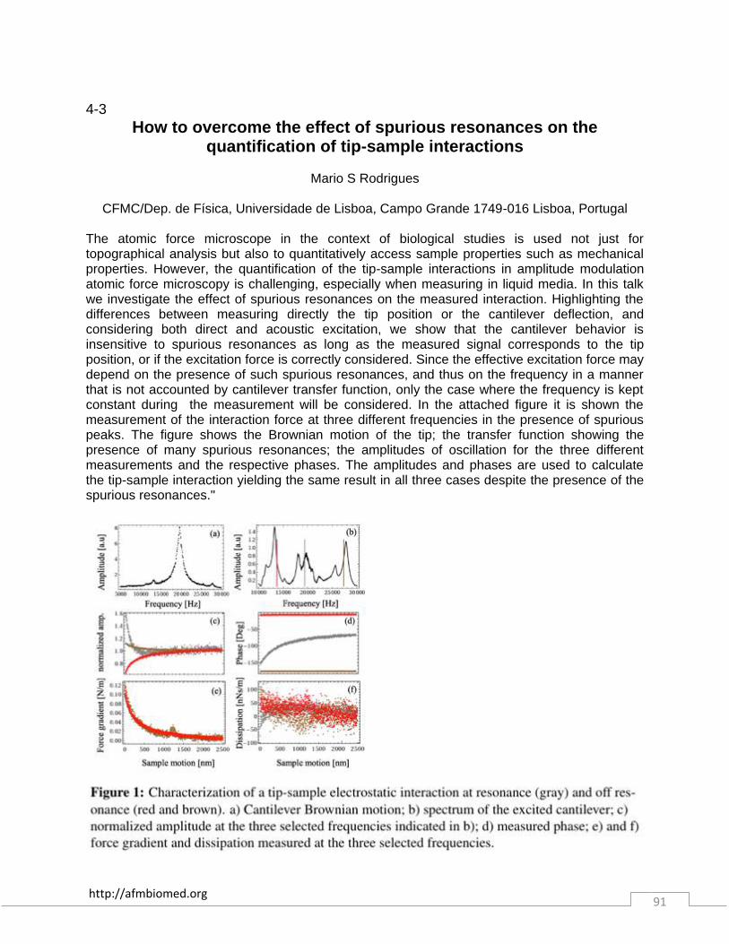
http://afmbiomed.org 91
4-3
How to overcome the effect of spurious resonances on the quantification of tip-sample interactions
Mario S Rodrigues
CFMC/Dep. de Física, Universidade de Lisboa, Campo Grande 1749-016 Lisboa, Portugal
The atomic force microscope in the context of biological studies is used not just for topographical analysis but also to quantitatively access sample properties such as mechanical properties. However, the quantification of the tip-sample interactions in amplitude modulation atomic force microscopy is challenging, especially when measuring in liquid media. In this talk we investigate the effect of spurious resonances on the measured interaction. Highlighting the differences between measuring directly the tip position or the cantilever deflection, and considering both direct and acoustic excitation, we show that the cantilever behavior is insensitive to spurious resonances as long as the measured signal corresponds to the tip position, or if the excitation force is correctly considered. Since the effective excitation force may depend on the presence of such spurious resonances, and thus on the frequency in a manner that is not accounted by cantilever transfer function, only the case where the frequency is kept constant during the measurement will be considered. In the attached figure it is shown the measurement of the interaction force at three different frequencies in the presence of spurious peaks. The figure shows the Brownian motion of the tip; the transfer function showing the presence of many spurious resonances; the amplitudes of oscillation for the three different measurements and the respective phases. The amplitudes and phases are used to calculate the tip-sample interaction yielding the same result in all three cases despite the presence of the spurious resonances."

http://afmbiomed.org 92
4-4 Force-controlled patch-clamp using atomic force microscopy
Dario Ossola [1], Mohamed Yassine Amarouch [2], Pascal Behr [1], János Vörös [1], Hugues
Abriel [2], and Tomaso Zambelli [1]
[1] Laboratory of Biosensors and Bioelectronics, ETH Zurich; [2] Department of Clinical Research, University of Bern
The Fluid Force Microscope (FluidFM) is a recently developed technology based on Atomic Force microscopy (AFM). The uniqueness of this technology consists in hollow cantilevers connected to an external liquid reservoir. The FluidFM allows for precise control on both the force exerted on a sample and on the hydrodynamic pressure at the cantilever’s aperture. Here we present the FluidFM adapted for electrophysiology experiments, in particular for whole-cell patch-clamp and for scanning ion conductance microscopy (SICM). From its invention in the 1970s, the patch clamp technique is the gold standard in electrophysiology research and drug screening since it is the only tool enabling accurate investigation of voltage-gated ion channels, which are responsible for action potentials. Innovation efforts in the field of drug screening are being made to reduce its complexity towards more automated systems, but conventional patch-clamp still remains unmatched in fundamental research due to its versatility. Here we report about a novel technique that merges the patch-clamp and the Atomic Force Microscope (AFM), made possible using the FluidFM. We investigated on the formation of a force-controlled high electric resistance seal between FluidFM aperture and cell membrane (gigaseal). Exploiting the force control capabilities of the FluidFM, the probe is positioned on the cell membrane in a controlled fashion simplifying the most critical step of traditional patch-clamp. We show the compatibility of the system with whole-cell patch-clamp recording of sodium (NaV1.5) voltage-gated ion channels in whole-cell configuration. In addition, we show the feasibility of simultaneous recording of membrane current and force development during cell contraction on freshly isolated adult mouse cardiomyocytes. The same setup is suitable for SICM operated in far contact using the ion conductivity between substrate and tip as feedback signal. SICM adapts very well on soft living tissues where the AFM fails to preserve the integrity of the imaged sample. Our system enables simultaneous AFM and SICM."
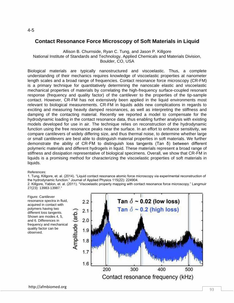
http://afmbiomed.org 93
4-5
Contact Resonance Force Microscopy of Soft Materials in Liquid
Allison B. Churnside, Ryan C. Tung, and Jason P. Killgore National Institute of Standards and Technology, Applied Chemicals and Materials Division,
Boulder, CO, USA
Biological materials are typically nanostructured and viscoelastic. Thus, a complete understanding of their mechanics requires knowledge of viscoelastic properties at nanometer length scales and a broad range of frequencies. Contact resonance force microscopy (CR-FM) is a primary technique for quantitatively determining the nanoscale elastic and viscoelastic mechanical properties of materials by correlating the high-frequency surface-coupled resonant response (frequency and quality factor) of the cantilever to the properties of the tip-sample contact. However, CR-FM has not extensively been applied in the liquid environments most relevant to biological measurements. CR-FM in liquids adds new complications in regards to exciting and measuring heavily damped resonances, as well as interpreting the stiffness and damping of the contacting material. Recently we reported a model to compensate for the hydrodynamic loading in the contact resonance data, thus enabling further analysis with existing models developed for use in air. The technique relies on reconstruction of the hydrodynamic function using the free resonance peaks near the surface. In an effort to enhance sensitivity, we compare cantilevers of widely differing size, and thus thermal noise, to determine whether large or small cantilevers are best able to distinguish material properties in soft materials. We further demonstrate the ability of CR-FM to distinguish loss tangents (Tan δ) between different polymeric materials and different hydrogels in liquid. These materials represent a broad range of stiffness and dissipation representative of biological specimens. Overall, we show that CR-FM in liquids is a promising method for characterizing the viscoelastic properties of soft materials in liquids. References: 1. Tung, Killgore, et. al. (2014). “Liquid contact resonance atomic force microscopy via experimental reconstruction of the hydrodynamic function.” Journal of Applied Physics 115(22): 224904. 2. Killgore, Yablon, et. al. (2011). “Viscoelastic property mapping with contact resonance force microscopy.” Langmuir 27(23): 13983-13987." Figure: Cantilever resonance spectra in fluid, acquired in contact with polymers having two different loss tangents. Shown are modes 4, 5, and 6. Differences in frequency and mechanical quality factor can be observed.

http://afmbiomed.org 94
4-6
AFM Circular Mode: A new powerful tool for applications in NanoBiotechnology
O.Noel [1], K. El-Kirat [2], and P-E Mazeran [3]
[1] Institut des Molécules et des Matériaux du Mans (IMMM), UMR CNRS 6283, Université du
Maine, Le Mans – France; [2] Laboratoire de BioMécanique et BioIngénierie (BMBI), UMR CNRS 7338, Université Technologique de Compiègne (UTC), Compiègne - France; [3]
Laboratoire Roberval, Unité de Recherche en Mécanique, Acoustique et Matériaux, UMR 7337, UTC – France
"The AFM Circular Mode(1) is a new technology based on the Atomic Force Microscopy (AFM). It was first developed to probe nano-tribological properties (2). Its main advantage is that it allows measuring instantaneously and simultaneously friction and others physical properties, which are not all provided by conventional AFM modes. Recently, the AFM Circular Mode was implemented and improved to probe properties of complex biological systems in liquids. In the first part of this talk, I will describe the principle of the AFM Circular Mode(3) and I will show its interest compared to conventional AFM Modes for characterizing biological objects. In the second part of this talk, I will present our preliminary results obtained with the AFM circular mode on supported biological membranes such as phospholipids (pure DOPC (or DPPC) and a 1:1 molar mixture of DOPC and DPPC) immerged in a Tris buffered saline (TBS) solution. First, our measurements show that with the AFM Circular mode, we can clearly dissociate the physical and tribological properties of the substrate and of the phospholipid bilayer. Second, regarding the experimental friction law (friction force vs. the normal load), we show that viscous friction could be evidenced for biological systems. Indeed, a component of the friction force is proportional to the sliding velocity and is independent of the normal load. Therefore, the total friction force may be expressed by two terms: one corresponds to the classical solid/solid friction (Coulomb friction); and the other one is the viscous friction. References: [1] O. Noël, P.E. Mazeran et H. Nasrallah, Methods of surface measurement and modification by local-probe microscopy operating in continuous curvilinear mode, Local-probe microscope and device enabling the implementation of said methods, Patented by the CNRS, PCT/FR2011/051024, 5 mai 2011. [2] O. Noël, P.-E. Mazeran, H. Nasrallah, Sliding velocity dependence of adhesion in a nanometer sized-contact, Phys. Rev. Lett., 108 (2012) 015503. [3] H. Nasrallah, P.-E. Mazeran, O. Noël, Circular mode: A new AFM mode for investigating surface properties, Rev. Scie. Inst. 82 (2011) 113703."
http://afmbiomed.org 95
4-7
New Conducting Atomic Force Microscopy for Simultaneous Electrical Recording and Imaging of Biomolecules
Brian Meckes, Laura Connelly, Lital Alfonta, Meni Wanunu, and Ratnesh Lal
Department of Bioengineering, UC San Diego, La Jolla, CA USA
Cells and tissue in our body are regulated by biological processes that occur at the nanoscale. These processes are determined by the coordinated and cooperative activity of biomolecules that respond to a variety of stimuli including chemical, mechanical, and electrical. The electrical activity of proteins plays an important role in maintaining cellular homeostasis, signaling, and metabolism, and has novel applications in diverse fields including healthcare, chemical processing, and energy production. The activity of the biological macromolecules is determined by their 3D structures. Current techniques do not allow simultaneous recording of electrical activity of protein macromolecules while imaging their molecular structures. Here, we present two new approaches for simultaneous imaging and electrical recording of biological structures using integrated multimodal AFM: 1) supported nanopore substrates for measuring ionic currents while imaging nano-structures and 2) novel probes for single cell analysis of electrochemical activity. We have designed a novel graphene nanopore system that interfaces with a custom-made two-chamber AFM. This system enables simultaneous imaging of biological macromolecules such as lipids and proteins while recording electrical properties such as conductance and capacitance of structures deposited over the nanopore. We demonstrated the device by imaging biological lipid bilayer membranes deposited over the nanopore. This technique can be used to study the structure-activity relationship of ion channels in lipid bilayers by imaging the channel and measuring the conductance state simultaneously. For probe based electrochemical AFM, we will present a novel approach for single cell characterization of E. coli surface expressing alcohol dehydrogenase II (ADHII). We used our newly developed conducting probe AFM to measure the electrochemical activity of a single cell. We measured the electron transfer efficiency of different ADHII mutants containing electron transfer (ET) mediators. Results showed that placing the mediators closer to the NADH binding pocket increased ET efficiency, which could ultimately result in more efficient biofuel cells.
http://afmbiomed.org 96
4-8
Parkin affects the biomechanical properties of human fibroblasts: a CAT microscopy study
Mariafrancesca Cascione [1,2], Daniele Vergara [3], Marzia M. Ferraro [1,3], Loretta L. del
Mercato [1], Tiziana Cocco [4], Ross Rinaldi [1,2], Antonio Gaballo [1] and Stefano Leporatti [1]
[1] NNL-Istituto Nanoscienze CNR, Lecce, Italy. [2] Department of Mathematics and Physics “Ennio De Giorgi”, University of Salento, Lecce, Italy. [3] Department of Biological and
Environmental Sciences and Technologies, University of Salento, Lecce, Italy. [4] Department of Basic Medical Sciences, Neurosciences and Organs of Senses, University of Bari „A. Moro‟,
Bari, Italy.
The actin cytoskeletal architecture affects several cellular processes essential for normal development as well for many pathological conditions including Parkinson’s disease (PD), one of the most common neurodegenerative diseases. Recent studies have provided evidence about the molecular interactions between parkin and cytoplasmic proteins. In particular, it was demonstrated that parkin mutations affect the organization of cytoskeleton proteins. Therefore, we have investigated the contribution of parkin on the mechanical properties of human cells. In details, to investigate the role of parkin in regulating actin filament organization and cellular stiffness, parkin-null fibroblasts isolated from a Parkinson’s disease patient were compared to normal fibroblasts. We have analysed the variation in Young’s modulus of parkin-null fibroblasts by using a CAT (Confocal-AFM-TIRF) microscope. This instrument combines an advanced scanning probe microscope (Bioscope Catalyst, Bruker Inc. USA), a confocal laser scanning microscope (LSM 700, Zeiss GERMANY), and a total internal reflection fluorescence microscope (Laser TIRF 3, Zeiss GERMANY). By using CAT microscope we have acquired force mapping curves on living parkin-null and control cells. The force-distance curves were then analysed using a LabView-based home-made software in order to measure the Young’s moduli of the investigated cells. Our measurements indicate that absence of parkin results in a significant increase of stiffness, in agreement with the different expression of actin regulators in control and parkin-null fibroblasts. All together, these results suggest a link between parkin-associated cellular architecture and the mechanical properties of fibroblast cells.
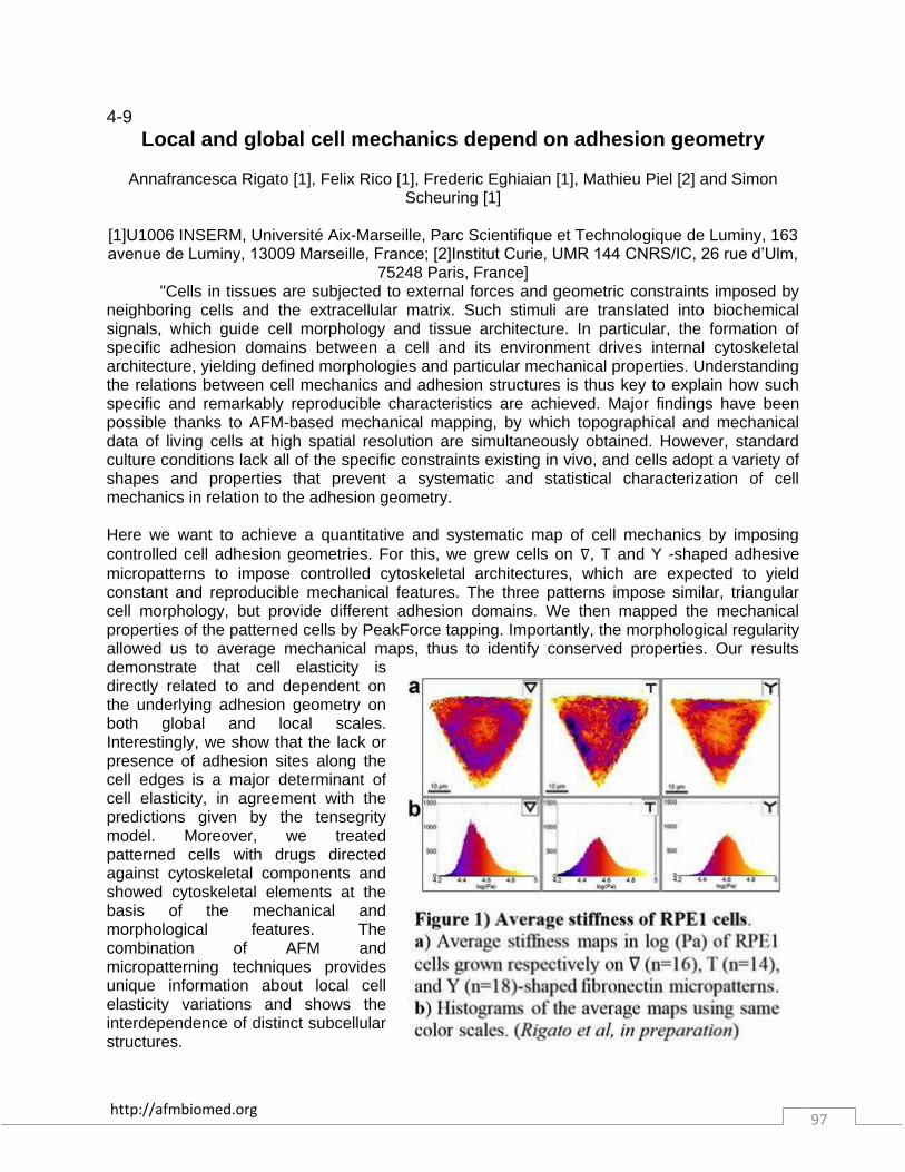
http://afmbiomed.org 97
4-9 Local and global cell mechanics depend on adhesion geometry
Annafrancesca Rigato [1], Felix Rico [1], Frederic Eghiaian [1], Mathieu Piel [2] and Simon
Scheuring [1]
[1]U1006 INSERM, Université Aix-Marseille, Parc Scientifique et Technologique de Luminy, 163 avenue de Luminy, 13009 Marseille, France; [2]Institut Curie, UMR 144 CNRS/IC, 26 rue d’Ulm,
75248 Paris, France] "Cells in tissues are subjected to external forces and geometric constraints imposed by neighboring cells and the extracellular matrix. Such stimuli are translated into biochemical signals, which guide cell morphology and tissue architecture. In particular, the formation of specific adhesion domains between a cell and its environment drives internal cytoskeletal architecture, yielding defined morphologies and particular mechanical properties. Understanding the relations between cell mechanics and adhesion structures is thus key to explain how such specific and remarkably reproducible characteristics are achieved. Major findings have been possible thanks to AFM-based mechanical mapping, by which topographical and mechanical data of living cells at high spatial resolution are simultaneously obtained. However, standard culture conditions lack all of the specific constraints existing in vivo, and cells adopt a variety of shapes and properties that prevent a systematic and statistical characterization of cell mechanics in relation to the adhesion geometry. Here we want to achieve a quantitative and systematic map of cell mechanics by imposing controlled cell adhesion geometries. For this, we grew cells on ∇, T and Y -shaped adhesive micropatterns to impose controlled cytoskeletal architectures, which are expected to yield constant and reproducible mechanical features. The three patterns impose similar, triangular cell morphology, but provide different adhesion domains. We then mapped the mechanical properties of the patterned cells by PeakForce tapping. Importantly, the morphological regularity allowed us to average mechanical maps, thus to identify conserved properties. Our results demonstrate that cell elasticity is directly related to and dependent on the underlying adhesion geometry on both global and local scales. Interestingly, we show that the lack or presence of adhesion sites along the cell edges is a major determinant of cell elasticity, in agreement with the predictions given by the tensegrity model. Moreover, we treated patterned cells with drugs directed against cytoskeletal components and showed cytoskeletal elements at the basis of the mechanical and morphological features. The combination of AFM and micropatterning techniques provides unique information about local cell elasticity variations and shows the interdependence of distinct subcellular structures.

http://afmbiomed.org 98
4-10 Influence of cellular adhesiveness on the formation of cell boundaries
Steve Pawlizak, Anatol Fritsch, Dave Ahrens, Tobias Thalheim, Josef A. Käs
University of Leipzig, Faculty of Physics and Earth Sciences, Institute of Experimental Physics I,
Soft Matter Physics Division, Linnéstr. 5, 04103 Leipzig, Germany The segregation of different cell populations and the formation of cellular boundaries is a fundamental process in every higher organism. During the embryonic development, the basis for cellular organization is established when the first cells differentiate into several populations which eventually segregate into different compartments. These compartments are ""subunits"" of the further development and will persist for life. As a general rule, cells do not cross the well-defined lineage boundaries between different compartments. However, some cells may break ranks: At some point in their development, malignant cells become able to invade adjacent tissues. This raises the question, what physical properties may have changed in such cells to let them eventually overcome compartment boundaries, or in other words, what mechanism drives cellular segregation? The differential adhesion hypothesis (DAH) gives a first explanation by differences in surface tension and adhesiveness of the interacting cells. In this context, we are investigating whether cellular adhesion is in fact a necessary or even sufficient factor to characterize compartmentalization and tumor spreading: Firstly, we apply a common model system for cellular segregation which involves mixing together two different populations of suspended cells. After a certain time, this mixture will eventually segregate into two phases. According to the DAH, the cell type with the higher adhesiveness should be enveloped by the cell type with the lower adhesiveness. Secondly, we apply atomic force microscopy using a modified BioAFM to directly measure the adhesion forces between the different cell types (see Figure). However, comparing the AFM results to the observed segregation behavior is difficult because of the different time scales of both experiments. While segregation experiments usually run over several hours, AFM adhesion measurements are done on much shorter time scales due to technical limitations and to gain acceptable statistics. We tried to address this common issue by varying the AFM contact times at least within a certain range. Moreover, we used another approach to characterize cellular adhesiveness by determining the adhesion molecule density at the cell-substrate interface using high-resolution STED microscopy. Additionally, cellular elasticity is probed using an ""Optical Stretcher"" as well as AFM.”

http://afmbiomed.org 99
4-11 Impact of the actin of the cytoskeleton on the mechanical properties
of cells and tissues
Celine Heu [2,3], Iman Jalilian [1], Anthony Kee [1], Melissa Knothe Tate [3], Galina Schevzov [1], Renee Whan [2] and Peter Gunning [1]
[1] Oncology Research Unit, School of Medical Sciences, UNSW Australia; [2] Biomedical
Imaging Facility (BMIF) - Mark Wainwright Analytical Centre, Lowy Cancer Research Centre, UNSW Australia [3] MechBio team, Graduate School of Biomedical Engineering, UNSW
Australia "The role of the actin cytoskeleton in the architecture and the mechanical properties of the cell has been the subject of numerous studies. Tropomyosins (Tms) are a family of coiled-coil proteins that are the core components of most actin filaments and play a pivotal role in regulating actin stability and function. However, the exact impact of these proteins on the mechanical properties of tissues is still unknown. In this study, we addressed the role of different tropomyosin isoform, Tm5NM1, specifically, in the actin cytoskeleton network and their influence on both cell and tissue mechanical properties. Nano-indentations were performed on different Tm isoform overexpressing rat neuroblastoma cells. Mechanical measurements carried out just above the cell’s nucleus, demonstrated that Tm5NM1-overexpressing cells are significantly stiffer than control cells. This result correlated with the observed increase in the filamentous to globular actin ratio, in the length and number of actin cables per cell compared to control cells. In mouse tissue, Tm5NM1 overexpression increased cell proliferation impacting on the volume of the White Adipose Tissue and increased the amount of actin[1]. We implemented a new protocol to measure the mechanical properties of harvested mouse WAT by Peak Force QNM with a 1 µm colloidal probe mounted on a 0.06N/m cantilever. Six mice for each condition were used to perform elasticity measurements on epididymal fat pads. The Young’s moduli were compared based on around 1000 nano indentation force curves for each condition. We demonstrated that tissue overexpressing Tm5NM1 was significantly stiffer than the control tissue. In conclusion, the actin cytoskeleton has a significant role in regulating the mechanical properties of cells and tissue. In addition, we found that the tropomyosin isoform composition of the actin filaments have an isoform specific, rather than generic, impact on the mechanical properties of cells and tissues. References: [1] A. J. Kee, L. Yang, C. A. Lucas, M. J. Greenberg, W. E. Hughes, G J. Cooney, D. E. James, E. M. Ostap, W. Han, P. W. Gunning and E. C. Hardeman (submitted for publication)

http://afmbiomed.org 100
4-12 Dynamic coupling of ALCAM to the actin cortex strengthens cell
adhesion to CD6
Joost te Riet [1,2], Jonne Helenius [2], Nico Strohmeyer [2], Alessandra Cambi [1], Carl G. Figdor [1], Daniel J. Müller [2]
[1] Department of Tumor Immunology, Radboud Institute for Molecular Life Sciences, Radboud UMC, Nijmegen, The Netherlands [2] Department of Biosystems Science and Engineering, ETH
Zürich, Basel, Switzerland At the immunological synapse, activated leukocyte cell adhesion molecules (ALCAM) on the dendritic cell (DC) and CD6 molecules on the T cell contribute to sustained DC-T cell contacts. However, little is known about how ALCAM-CD6 bonds resist and adapt to mechanical stress. Here, we combine single-cell force spectroscopy (SCFS) with total-internal fluorescence microscopy (TIRFM) to examine ALCAM-CD6-mediated cell adhesion. The combination of cells expressing ALCAM-constructs with specific cytoplasmic tail mutations and improved SCFS analysis routines reveal that the affinity of ALCAM-CD6 bonds is not influenced by linking of the intracellular domains of ALCAM to the actin cortex. In contrast, the recruitment of ALCAM to adhesion sites and the propensity of ALCAM to anchor plasma membrane tethers depend on actin cytoskeletal interactions. Furthermore, linking ALCAM to the actin cortex by adaptor proteins stiffens the cortex and strengthens cell adhesion. We propose a framework of how ALCAMs contribute to DC-T cell adhesion, stabilize DC-T cell contacts, and form a mechanical link between CD6 and the actin cortex to strengthen cell adhesion at the immunological synapse. References: [1] te Riet, et al., J. Cell Sci., 2014;127: p1595-1606
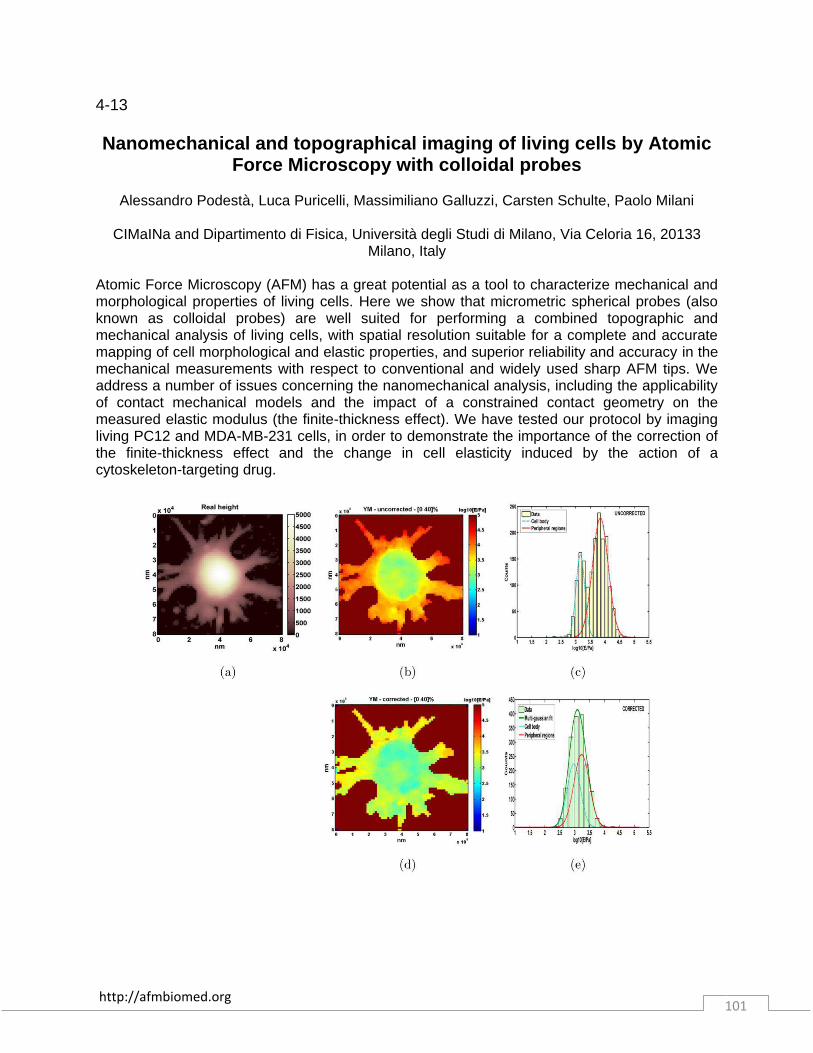
http://afmbiomed.org 101
4-13 Nanomechanical and topographical imaging of living cells by Atomic
Force Microscopy with colloidal probes
Alessandro Podestà, Luca Puricelli, Massimiliano Galluzzi, Carsten Schulte, Paolo Milani
CIMaINa and Dipartimento di Fisica, Università degli Studi di Milano, Via Celoria 16, 20133 Milano, Italy
Atomic Force Microscopy (AFM) has a great potential as a tool to characterize mechanical and morphological properties of living cells. Here we show that micrometric spherical probes (also known as colloidal probes) are well suited for performing a combined topographic and mechanical analysis of living cells, with spatial resolution suitable for a complete and accurate mapping of cell morphological and elastic properties, and superior reliability and accuracy in the mechanical measurements with respect to conventional and widely used sharp AFM tips. We address a number of issues concerning the nanomechanical analysis, including the applicability of contact mechanical models and the impact of a constrained contact geometry on the measured elastic modulus (the finite-thickness effect). We have tested our protocol by imaging living PC12 and MDA-MB-231 cells, in order to demonstrate the importance of the correction of the finite-thickness effect and the change in cell elasticity induced by the action of a cytoskeleton-targeting drug.
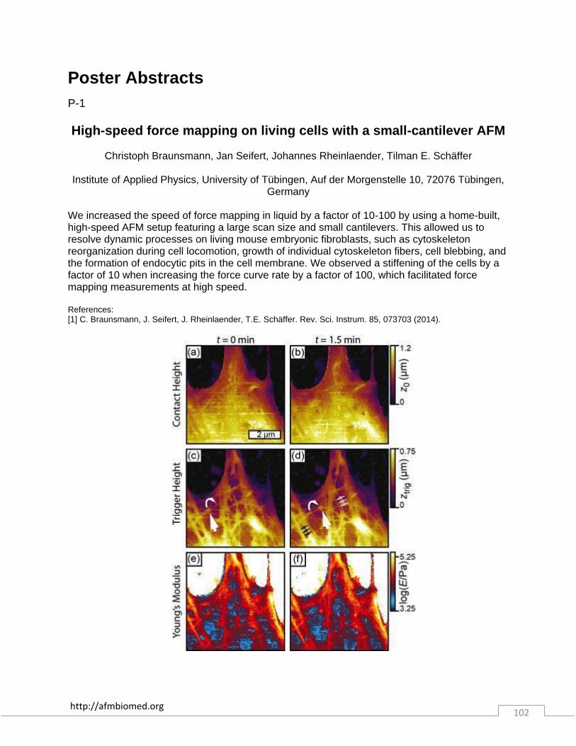
http://afmbiomed.org 102
Poster Abstracts P-1 High-speed force mapping on living cells with a small-cantilever AFM
Christoph Braunsmann, Jan Seifert, Johannes Rheinlaender, Tilman E. Schäffer
Institute of Applied Physics, University of Tübingen, Auf der Morgenstelle 10, 72076 Tübingen,
Germany We increased the speed of force mapping in liquid by a factor of 10-100 by using a home-built, high-speed AFM setup featuring a large scan size and small cantilevers. This allowed us to resolve dynamic processes on living mouse embryonic fibroblasts, such as cytoskeleton reorganization during cell locomotion, growth of individual cytoskeleton fibers, cell blebbing, and the formation of endocytic pits in the cell membrane. We observed a stiffening of the cells by a factor of 10 when increasing the force curve rate by a factor of 100, which facilitated force mapping measurements at high speed. References: [1] C. Braunsmann, J. Seifert, J. Rheinlaender, T.E. Schäffer. Rev. Sci. Instrum. 85, 073703 (2014).
http://afmbiomed.org 103
P-2
Non-contact imaging combining the Scanning Ion Conductance Microscopy and the Atomic Force Microscope
Livie Dorwling-Carter, Dario Ossola, János Vörös, Tomaso Zambelli
Laboratory of Biosensors and Bioelectronics, ETH Zürich Gloriastrasse 35, ETZ 8092 Zürich
The Scanning Ion Conductance Microscope (SICM) was invented in 1989 by Hansma et al. [1] to scan biological surfaces without damaging them. Based on a non-contact ionic current feedback, the SICM allows non-invasive topographical analysis of soft surfaces, becoming a very valuable tool for imaging of living cells. Nevertheless, contact-free scanning with SICM does not ensure force-free scanning [2,3]. Different approaches were presented to assess the forces interacting between the probe and the sample when carrying out ion sensing, from bended micropipettes [4] used as optical levers for optical beam detection to the most recently developed dual flexure optical lever of Drake et al. [5] Here we present a new strategy for direct and systematic recording of the forces acting on the sample in SICM mode by combining the SICM with an Atomic Force Microscope (AFM). The setup was developed thanks to the Fluidic Force Microscope (FluidFM) [6], a recently emerged scanning probe microscopy technique based on an integrated microchannel in an AFM cantilever which allows to deliver liquid from an external reservoir embedded in a plastic chip to the tip (sub-) micrometer aperture. The ability of the microchanneled cantilever of the FluidFM to act both as a scanning probe and as a nanopipette was exploited to build up a tool capable of both AFM and SICM. More than only offering a simultaneous force recording during SICM scans, this new versatile tool also allows a quick switch between the AFM and SICM microscopies when complementary or comparative information is needed. Also, while imaging in AFM mode, the setup of the SICM enables a simultaneous parallel ion mapping. Moreover, combining the FluidFM and SICM allows to use the high-resolution topographical information available with SICM to carry out the usual manipulations achieved with the FluidFM tip (microinjection and micromanipulation of cells [7], patch-clamp) at a precise position. The experimental results are compared to COMSOL simulations in order to optimize the aperture geometry. References: [1] Hansma PK, Drake B, Marti O, Gould SA., Prater C. The Scanning Ion-Conductance Microscope. Science (80- ). 1989;243(4891):641-643. [2] Pellegrino M, Orsini P, Pellegrini M, et al. Weak hydrostatic forces in far-scanning ion conductance microscopy used to guide neuronal growth cones. Neurosci Res. 2011;69(3):234-40. doi:10.1016/j.neures.2010.11.009. [3] Clarke RW, Zhukov A, Richards O, Johnson N, Ostanin V, Klenerman D. Pipette-surface interaction: current enhancement and intrinsic force. J Am Chem Soc. 2013;135(1):322-9. doi:10.1021/ja3094586. [4] Proksch R, Lal R, Hansma PK, Morse D, Stucky G. Imaging the internal and external pore structure of membranes in fluid: Tapping Mode scanning ion conductance microscopy. Biophys J. 1996;71(October):2155-2157. [5] Drake B, Randall C, Bridges D, Hansma PK. A new ion sensing deep atomic force microscope. Rev Sci Instrum. 2014;85(8):083706. doi:10.1063/1.4893640. [6] Meister A, Gabi M, Behr P, et al. FluidFM: combining atomic force microscopy and nanofluidics in a universal liquid delivery system for single cell applications and beyond. Nano Lett. 2009;9(6):2501-7. doi:10.1021/nl901384x. [7] Guillaume-Gentil O, Potthoff E, Ossola D, Franz CM, Zambelli T, Vorholt J a. Force-controlled manipulation of single cells: from AFM to FluidFM. Trends Biotechnol. 2014;32(7):381-8. doi:10.1016/j.tibtech.2014.04.008.

http://afmbiomed.org 104
P-3
FluidFM for local electroplating and electrografting in liquid
Luca Hirt, Janos Vörös, and Tomaso Zambelli
Laboratory of Biosensors and Bioelectronics, Institute for Biomedical Engineering, ETH Zurich, Switzerland
FluidFM [1] is a combination of AFM technology and nanofluidics. A channel incorporated directly in a hollow AFM cantilever and an aperture at the apex of the AFM tip allow for local dispensing of soluble molecules in air and in liquid [2], while retaining the inherent imaging capabilities and force feedback of an AFM system. Therefore, FluidFM may open new opportunities for experiments of local chemistry with spatial resolution. Micro- and nanometer-scale patterning of functional materials is of great interest in various fields such as microelectronics or micro-electromechanical systems (MEMS) design, but also for biosensing and biomedical applications, where manipulation techniques often have to be suitable for use in a liquid environment. In this context, we currently explore the use of FluidFM as a tool for both fabrication and subsequent imaging of microstructures in liquid using the same probe. Here, hollow FluidFM cantilevers are used to locally dispense precursor molecules in a macro electrochemical cell. These molecules may thus be reduced on a polarized electrode surface, enabling local electrodeposition of various materials. In particular, we present conceptual results of local copper electroplating and electrografting of organic molecules via reduction of diazonium salts. References: [1] Meister et al., Nano Letters 9 (2009) 2501. [2] Grüter et al., Nanoscale 5 (2013) 1097.
http://afmbiomed.org 105
P-4 Photoplastic AFM Cantilevers with integrated microchannel for Single
Cell Experiments
Vincent Martinez [1], Pascal Behr [1], Jerome Polesel-Maris [2], Janos Vörös [1], and Tomaso Zambelli [1]
[1] Laboratory of Biosensors and Bioelectronics, Institute of Biomedical Engineering, ETH
Zurich, Zurich, 8092, Switzerland
The Fluid Force Microscope (FluidFM) is a recent technology born in 2009 from a collaboration between the swiss Center for Electronics and Microtechnology (CSEM, Neuchâtel) and ETH Zurich. The invention of the FluidFM represents a major and unique tool for single cell manipulation. Based on a hollow Atomic Force Microscopy (AFM) cantilever with an integrated micro- or nano-fluidic channel connected to an external liquid reservoir (Fig. 1) [1], the FluidFM technology has already demonstrated a great potential in several applications at the single cell level: controlled spatial manipulation of cells by a system of overpressure/underpressure [2], force-controlled fluidic injection into single cell nuclei [3], selective and massive cell force spectroscopy [4] or even serving as a nanoparticle lithography dispenser tool in liquid [5]. Different cantilever designs have been fabricated so far, with different tip shapes and apertures down to 30 nm made of silicon nitride (Si3N4). New photoplastic techniques have been recently considered to realize hollow cantilevers: the polymers are becoming widely used for all kinds of micro mechanic and micro fluidic devices [6] because they are highly chemically resistant, transparent and biocompatible making them suitable as a constitutional material of the FluidFM technology. Furthermore, their low Young's modulus is more prone to a larger deflection of the cantilever (lower spring constant) to reach an enhanced resolution in force detection, with direct implications for adhesion characterization (Fig. 2) or injection measurements (Fig. 3). In this context, we propose an original evolution of the FluidFM technology based on hollow polymer cantilevers to extend the investigation spectrum for fundamental cell biology research. References: [1] Meister, A., Gabi, M., Behr, P., Studer, P., Vörös, J., Niedermann, P., Bitterli, J., et al., Nano letters 9 (2009) 2501–2507.; [2] Dörig, P., Stiefel, P., Behr, P., Sarajlic, E., Bijl, D., Gabi, M., Vörös, J., et al., Applied Physics Letters 97 (2010) 023701; [3] Guillaume-Gentil, O., Potthoff, E., Ossola, D., Dörig, P., Zambelli, T., & Vorholt, J., Small 9 (2013) 1904–1907; [4] Potthoff, E., Guillaume-Gentil, O., Ossola, D., Polesel-Maris, J., LeibundGut-Landmann, S., Zambelli, T., & Vorholt, J., PloS one 7 (2012) e52712; [5] Grüter, R. R., Vörös, J., & Zambelli, T., Nanoscale 5 (2013) 1097–1104; [6] Nordström, M., Keller, S., Lillemose, M., Johansson, A., Dohn, S., Haefliger, D., Blagoi, G., et al., Sensors 8 (2008) 1595–1612.
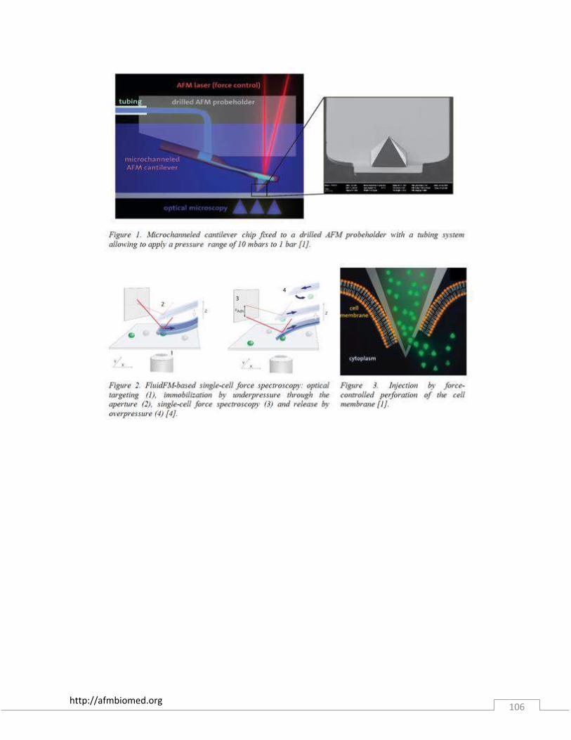
http://afmbiomed.org 106
http://afmbiomed.org 107
P-5 AFM Surface Analysis of Nanosilver Toxicity on Marine Heterotrophic
Bacteria
Nirav Patel, Farooq Azam, and Ratnesh Lal
Department of Bioengineering, UC San Diego, La Jolla, CA USA Marine colloid particles influence the interfacial structure and functions of marine bacteria, shaping how they sense and shape their external environment. Understanding how bacteria interact and associate with nanoparticles in the ocean has implications for nutrient cycling and toxicological effects in the marine food web. In this study, we aim to elucidate the morphological changes in the microbial surface from bacterial interactions with silver nanoparticles, their effects on bacterial cell mortality, and what the resulting ecological consequences may be using atomic force microscopy. Preliminary experiments with a marine isolate (Alteromonas sp. TW7) show the distribution of nanoparticles associated with exopolymer networks and microbial surfaces, demonstrating the application of AFM in visualizing single bacteria-nanoparticle interactions.
http://afmbiomed.org 108
P-6
A sticky tale: Sample preparation technique determines cell surface receptor mobility and adhesion
Thomas Mueller
University of Birmingham, Birmingham, United Kingdom
Cell surface proteins have become common targets for interrogation via atomic force microscopy (AFM). While the majority of studies use live cells in order to replicate in vivo conditions, this is not always feasible. For example, cells which exhibit poor natural adhesion to a countersurface need to be immobilised, or have their mobility restricted, in order to successfully perform AFM adhesion measurements. In the case of tissue sections, fixation – traditionally via acetone or paraformaldehyde (PFA) – is required to preserve the structure of the tissue as well as any applied immunohistochemical staining necessary in order to identify regions of interest. Prior to interrogating tissue sections using AFM, it is therefore necessary to understand the effects of the required sample preparation for comprehensive analysis and interpretation of the obtained results. In order to understand the effects that fixation has on AFM measurements, cells were immobilised onto poly-L-lysine coated glass coverslips, and were subsequently fixed using acetone or PFA. To be able to probe the specificity and affinity of the targeted surface proteins, measurements on the AFM were conducted using a custom chemically modified AFM tip. The experiments show that the stiffness of the cell membrane changes depending on the fixative, which can be implicated in the observed difference of successful binding events between AFM tip and cell surface proteins. An up to three-fold decrease in successful binding events was recorded from measurements on rigid cells, when compared to cells that exhibit some degree of membrane mobility. The experiments show that the stiffness of the cell membrane changes depending on the fixative, which can be implicated in the observed difference of successful binding events between AFM tip and cell surface proteins. An up to three-fold decrease in successful binding events was recorded from measurements on rigid cells, when compared to cells that exhibit some degree of membrane mobility. Furthermore, commonly used AFM adhesion descriptors such as (i) the peak retraction force and (ii) the work of adhesion are affected significantly when cell membrane flexibility is reduced. The implications of this research are of high importance given it will allow us to progress from interrogating isolated cells in vitro to measuring affinities in fixed tissue sections of organs containing complex arrangements of cells with multiple receptor affinities.
http://afmbiomed.org 109
P-7
Atomic Force Microscopy Study on Photosystem II-rich Region of Thylakoid Membrane in Chlamydomonas reinhardtii during State
Transition Witchukorn Phuthong [1], Zubin Huang [2], Mark L. Heinnickel [3], Friedrich Prinz [1], and Arthur
R. Grossman [3]
[1] Stanford University, [2] Applied Materials Inc., and [3] Carnegie Institution for Science State transition, a process that plants and algae use to balance the absorption cross-section of photosystem I (PSI) and photosystem II (PSII), appears to be important for maintaining optimal photosynthetic rates under conditions of natural irradiance. In green algae such as Chlamydomonas reinhardtii, state transitions can represent a significant portion of non-photochemical quenching (70-80%). In state 1, the mobile light harvesting complexes (LHCIIs) are associated with PSII. The cells transition to state 2 as the plastoquinone pool becomes reduced (e.g. under high light) and the mobile LHCIIs and some PSII core subunits become phosphorylated and move to PSI. These changes in organization of photosynthetic complexes within the thylakoid membranes have been deduced from biochemical and biophysical analyses; we now have the means to observe them using Atomic Force Microscopy (AFM). In a temperature-controlled, vibration-minimized chamber in which thylakoid membrane samples are maintained in an aqueous environment, we can use AFM to visualize dimeric PSII complexes protruding from the membranes; based on electron microscopic analyses, these protruding structures represent the PsbP, PsbO, and PsbQ subunits of the PSII oxygen evolving complex. From preliminary studies, we note that PSII organization/features from cells maintained in state 1 and state 2 differ in two respects: (i) nearest neighbor distance of PSII particles is greater in state 2 than in state 1; (ii) resolution of the features of the PSII particles is reduced in state 2. Our work suggests new opportunities for in situ analysis of the dynamic regulatory processes that modulate photosynthetic electron transport.
http://afmbiomed.org 110
P-8
Estimating water content of individual fibrin fibers
Alina Popescu[1] and Emilios K. Dimitriadis[2]
[1] National Cancer Institute and [2] National Institute for Biomedical Imaging and Bioengineering, Bethesda, MD USA
We used high-resolution atomic force microscopy (AFM) imaging of fibrin fibers under varied physiological conditions to gain insights into individual fibrin fiber structure and water content. In physiological buffer or in water, fibrin fibers appear flattened, having heights one order of magnitude smaller (20-100 nm) than diameters (0.1-1 µm); thicker fibers being proportionately more flattened. High resolution imaging in liquid reveals the periodic structure of the fibers, with a repeat of 23 nm, corresponding to the half-staggering of 45 nm fibrinogen monomers making up the fibers, thick or thin alike. These images also clearly reveal fibers’ longitudinal composition of protofibrils which appear to be uniformly distributed across the fiber. Exchange of the physiological buffer with water increases the cross-sectional area of individual fibers by up to a factor of two irrespective of the diameter of the fibers. Upon drying, the cross-sectional area decreases by around 80% and the decrease is bigger for thicker fibers. This is the first direct measurement of the density of individual fibers, which shows that thick fibers contain proportionately more water than thin fibers. According to a fractal structural model for the cross section of fibrin fibers, we obtained a fractal dimension of 1.1 for the distribution of protofibrils within the cross section of the fiber, which implies an almost hollow fiber with minimal internal structural content. During rehydration, the cross-sectional area increases, again independently of the fiber diameter, but the fiber cross-section never goes back to its original size, suggesting irreversible structural changes caused by dehydration.

http://afmbiomed.org 111
P-9 Exploring the structure of exosomes with nanofilaments using atomic
force microscopy Jung-Reem Woo [1] , Shivani Sharma [1, 2] , Kingshuk Das [3], and James K. Gimzewski [1, 2]
[1] Department of Chemistry and Biochemistry, [2] California NanoSystems Institute, and [3] Department of Pathology and Laboratory Medicine, David Geffen School of Medicine, UC Los
Angeles, Los Angeles, CA USA Exosomes, 40-100 nm sized vesicles secreted by mammalian cells, are of importance in cell-cell communications. Due to their small size, the structure of exosomes was not well-characterized and exosomes were believed to have a “cup-shaped” structure based on the electron microscope (EM) images. However, our atomic force microscopy (AFM) images have revealed that exosomes have a globular structure. Recently, we have found the nanofilament structures on glioblastoma-derived exosomes using AFM peak force mode and the exosomes with nanofilaments showed increased uptake into the cells compared with normal control exosomes. In conclusion, we have identified the nanofilament structure of exosomes and the nanofilaments may have implications on exosome-mediated cell-cell interactions.
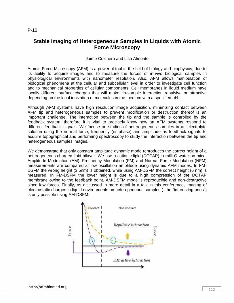
http://afmbiomed.org 112
P-10
Stable Imaging of Heterogeneous Samples in Liquids with Atomic Force Microscopy
Jaime Colchero and Lisa Almonte
Atomic Force Microscopy (AFM) is a powerful tool in the field of biology and biophysics, due to its ability to acquire images and to measure the forces of in-vivo biological samples in physiological environments with nanometer resolution. Also, AFM allows manipulation of biological phenomena at the cellular and subcellular level in order to investigate cell function and to mechanical properties of cellular components. Cell membranes in liquid medium have locally different surface charges that will make tip-sample interaction repulsive or attractive depending on the local ionization of molecules in the medium with a specified pH. Although AFM systems have high resolution image acquisition, minimizing contact between AFM tip and heterogeneous samples to prevent modification or destruction thereof is an important challenge. The interaction between the tip and the sample is controlled by the feedback system, therefore it is vital to precisely know how an AFM systems respond to different feedback signals. We focuse on studies of heterogeneous samples in an electrolyte solution using the normal force, frequency (or phase) and amplitude as feedback signals to acquire topographical and performing spectroscopy to study the interaction between the tip and heterogeneous samples images. We demonstrate that only constant amplitude dynamic mode reproduces the correct height of a heterogeneous charged lipid bilayer. We use a cationic lipid (DOTAP) in milli Q water on mica. Amplitude Modulation (AM), Frecuency Modulation (FM) and Normal Force Modulation (NFM) measurements are compared at low oscillation amplitude using dynamic AFM modes. In FM-DSFM the wrong height (3.5nm) is obtained, while using AM-DSFM the correct height (6 nm) is measured. In FM-DSFM the lower height is due to a high compression of the DOTAP membrane owing to the feedback point. AM-DSFM mode is reproducible and non-destructive since low forces. Finally, as discussed in more detail in a talk in this conference, imaging of electrostatic charges in liquid environments on heterogeneous samples (=the “interesting ones”) is only possible using AM-DSFM.

http://afmbiomed.org 113
P-11
New emerging technique of Electrostatic Force MIcroscopy AFM gives insight into the DNA-protein Interaction Path
Parminder Kaur, Dong Wu, Jiangguo Lin, Preston Countryman, Pan Hai,Hong Wang
North Carolina State University
We present a new exquisitely sensitive AFM technique, sensitive Dual-Resonance-frequency-Enhanced Electrostatic force Microscopy (DREEM) to resolve the DNA paths within protein –DNA complexes. To establish a baseline and illustrate the power of the technique, nucleosomal arrays extracted from HELA cells as well reconstituted in our lab were used. The DREEM images of these arrays reveal the DNA-histone model similar to the model in the literature already established. Further, we study the interaction of telomeric DNA with the TRF2 protein to understand the assembly of t-loop formation, the internal genomic structure of influenza virus and SA2 protein (part of the cohesion complex) with the telomeric DNA.
http://afmbiomed.org 114
P-12
Hydrophobic interaction governs unspecific adhesion of staphylococci
Nicolas Thewes and Karin Jacobs
Saarland University, Germany
Unspecific adhesion of bacteria is usually the first step of biofilm formation on abiotic surfaces. Alongside long-ranged van der Waals and electrostatic forces, short-ranged hydrophobic interaction plays an important role. To characterize the forces involved during approach and retraction of an individual bacterium to and from a surface, single cell force spectroscopy is applied using apathogenic species S. carnosus isolate TM300.

http://afmbiomed.org 115
P-13
Polymeric Ultrasound Contrast Agents Mechanical Properties
Baptiste Sarrazin [1], Patrick Guenoun [1], Nicolas Tsapis [2], Ludivine Mousnier [2], Nicolas Taulier [3], and Wladimir Urbach [3]
[1] CEA Saclay France, [2] Galien Institute, South Paris University, and [3] Pierre ans Marie
Curie University, Paris
The Ultrasound Contrast Agents (UCAs) are micro or nanocapsules which, when injected into the blood, can improve the contrast of ultrasound or MRI images [1]. UCAs can also be used as drug carriers by delivering the active ingredient to diseased targeted tissue by cavitation or vaporization, thereby enhancing efficacy while reducing deleterious effect on healthy organs. The studied UCAs consist of a polymer shell (poly(lactic-co-glycolic acid), PLGA) surrounding a liquid fluorinated core (perfluorooctyl bromid, PFOB). This new kind of particles differs from traditional UCAs which are gas filled thin shells. The main challenge to describe the behavior of UCAs subjected to ultrasonic waves is to get experimental data about their mechanical properties. Guédra et al. [2] used several models (Church, Rayleigh - Plesset, Kelvin-Voigt, Zener) - which take into account elasticity, stiffness and viscosity - to describe the influence of shell compressibility on the ultrasonic properties of UCAs. In this way, the aim of the present study is to experimentally measure mechanical properties of UCAs at the nanometer scale to feed the models. Atomic Force Microscopy (AFM) is able to probe such mechanical properties [3]. This technique allows characterizing single particles using contact model taking into account AFM tip and object geometries. In the present work, we evaluated the local indentation of individual PLGA/PFOB microcapsules (MCs) with different T/R ratios (T = shell thickness, R = capsule radius) on hard surface by measuring the force-displacement curves with an AFM cantilever tip. Hertz (fully elastic) and Johnson-Kendall-Roberts (JKR, elasto-adhesive model) models4 are employed to analyze the force vs. deformation curves and compute compressive elastic moduli (Young's modulus) of each MC. As a preliminary study, PLGA films and full microparticles are probed. Particular attention is paid to the calibration of cantilever stiffness and tip geometry which are critical parameters to interpret force curves. Moreover, frequency and temperature have been investigated in order to be closer to real experimental conditions of use for UCAs. Young's modulus of bulk PLGA and PLGA full microparticles show a similar behavior at low and high frequencies (≈ 3 GPa at 1 and 2000 Hz). Concerning liquid filled MCs, apparent elasticity is decreasing from bulk polymer with the capsule thickness. Thus, UCAs mechanical behavior is found to depend on capsule thickness and thermal history. Widely, this study raises some questions about the interpretation of AFM force curves on thick and hard capsules. References: [1] Pisani, E. et al., Langmuir 22, 4397–4402 (2006). [2] Guédra, M. et al., J. Acoust. Soc. Am. 135, 1044–1055 (2014). [3] Butt, H.-J., Cappella, B. & Kappl, M., Surf. Sci. Rep. 59, 1–152 (2005). [4] Barthel, E. et al, J. Phys. Appl. Phys. 41, 163001 (2008).
http://afmbiomed.org 116
P-14 Elasticity of pulmonary arteries within human lung tissue: Application
of AFM to study pulmonary arterial hypertension Delphine Sicard [1], Fei Liu [2], Kyoung Moo Choi [1], Qi Tan [1], Laura E. Fredenburgh [3] and
Daniel J. Tschumperlin [1]
[1] Department of Physiology and Biomedical Engineering, Mayo Clinic, Rochester, MN USA [2] Molecular and Integrative Physiological Sciences, Department of Environmental Health, Harvard School of Public Health, Boston, MA USA [3] Division of Pulmonary and Critical Care Medecine,
Department of Medecine, Brigham and Women’s Hospital, Boston, MA USA
Pulmonary arterial hypertension (PAH) is a severe disease characterized by a sustained elevation of pulmonary vascular pressure 1. Often idiopathic, PAH can also be genetic, induced by drug and toxin or associated with HIV infection, connective tissue disease or portal hypertension 2. At lung tissue level, PAH is characterized by a progressive vascular remodeling that includes intimal fibrosis, medial hypertrophy, vessel obstruction and plexiform lesions in the pulmonary arteries and arterioles 3. This remodeling leads to an increase of pulmonary vascular resistance and a progressive pulmonary arterial stiffening which ultimately results in right ventricular failure with high mortality 4. In our study, we are particularly interested in the stiffness of pulmonary arteries, as changes in tissue mechanics may alter cell function within the remodeled vessels of PAH patients. Therefore, we evaluated the mechanical properties of pulmonary arteries from healthy and PAH patients using Atomic Force Microscopy (AFM) in micro indentation. Based on the work of Liu et al.5 describing micro-mechanical characterization of lung tissue, you compared the Young’s modulus of pulmonary arteries for different classifications of PAH patients as well as healthy controls. Through this project, we will also report on the influence of key experimental parameters including sample thickness and tips radius on the measured values of vessel wall elasticity.
References: [1] Farber H. W. et al., N. Engl. J Med., 2004, 351, 1655-1665. [2] Simonneau G. et al., JACC, 2013, 62, 25, D34-41. [3] Archer S. et al., Circulation, 2000, 102, 2781-2791. [4] Fredenburgh L. E. et al., TCM, 2009, 19, 2, 31-37. [5] Liu F. et al., J. Vis. Exp., 2011, 54, e2911.

http://afmbiomed.org 117
P-15
Characterisation of the mechanical properties of blood and artificial clots
Celine Heu1,2, Luke Poggi1, Renee Whan2, Melissa Knothe Tate1
[1] MechBio Team, Graduate School of Biomedical Engineering, UNSW Australia [2]Biomedical
Imaging Facility (BMIF) - Mark Wainwright Analytical Centre, Lowy Cancer Research Centre, UNSW Australia
In a series of ovine studies (Knothe et al., 2010, Knothe and Springfield, 2005, Knothe Tate et al., 2007) and limited human studies, (Knothe and Springfield, 2005) remarkable tissue genesis by periosteum-derived stem cells is observed to fill critical sized bone defects via endochondral and intramembranous ossification. Surprisingly, defects bounded by periosteum in situ and filled by naturally occurring haematoma are filled with proliferative woven bone within two weeks of surgery. In contrast, packing defects with morcellised bone graft from the iliac crest retards healing and slows ingression of stem cells from the periosteal niche(Knothe Tate et al., 2007, Knothe et al., 2010). Our working hypothesis is that stem cell ingression into the defect decreases with increasing stiffness of the material filling the defect zone as well as biochemical cues intrinsic to graft and/or haematoma filling the defect. To begin to characterise and harness the biophysical properties of the haematoma and fibrin clot as endogenous tissue scaffolds, this study aimed specifically to measure by Atomic Force microscopy (Bioscope Catalyst, Bruker) the Young’s modulus of natural ovine haematoma, as well as ovine plasma and fibrin clots of three different compositions. In addition, the study aimed to characterise the emergent architecture of plasma and fibrin clots by multiphoton microscopy over a thirty minute period after initiation of clotting. Our overarching aim is to provide the basis for additional research on the impact of clot stiffness on mesenchymal stem cell ingression from periosteum in the context of bone fracture repair. This study provides a new experimental platform to investigate the spatiotemporal dynamics of clot development and the potential role of clot stiffness and composition on periosteum stem cell migration and de novo tissue genesis.

http://afmbiomed.org 118
P-16 Studying in situ protein adsorption and bacterial adhesion via fast scanning AFM
and force spectroscopy
Christian Spengler, Nicolas Thewes, Thomas Faidt, Christian Kreis, and Karin Jacobs
Saarland University, Department of Experimental Physics, 66123 Saarbrücken, Germany
Protein adsorption and bacterial adhesion are the first steps in biofilm formation. Hereby, proteins serve as a conditioning layer for the further attachment of bacteria and other organisms. Hence, the understanding and control of protein layers and their interaction with bacteria is an important task relevant to life sciences and engineering. We study the protein adsorption in situ on a single protein level. For this purpose, we use a fast scanning AFM operating in liquid under flow conditions which reveals single protein adsorption events. The surfaces for these experiments are hydrophilic and hydrophobized silicon substrates with different oxide layer thickness and very smooth artificial tooth samples. Additionally, we characterize the bactericidal activity of proteins in their adsorbed state using purified peptidoglycan. Furthermore, we perform single cell force spectroscopy to investigate the adhesion of bacteria to these protein layers in comparison to the bare surface.
http://afmbiomed.org 119
P-17
Atomic force microscopy reveals distinct regional nanomechanical properties of the extracellular matrix in healthy, aged, and
osteoarthritic native human menisci
Jeanie Kwok [1,3], Shawn Grogan, PhD [3], Brian Meckes [2], Fernando Arce, PhD [1,2], Ratnesh Lal, PhD [1,2], and Darryl D'Lima, MD, PhD [3]
[1] UC San Diego, Department of Mechanical and Aerospace Engineering, [2] UC San Diego, Department of Bioengineering, [3] Shiley Center for Orthopaedic Research and Education at
Scripps Clinic
The meniscus plays an important role in knee joint loading. Degeneration of the meniscus due to aging leads to unfavorable changes in joint loading dynamics that often contribute to the development of osteoarthritis (OA). Aging-related biochemical and biomechanical changes in the extracellular matrix (ECM) influence the degenerative process as well as OA development, however not much information is available about the mechanical environment of the ECM in the meniscus. Here, we used atomic force microscopy (AFM) to characterize nanomechanical properties of the ECM in native human meniscus tissue from young, aged, and OA patients. AFM elasticity measurements show aging-dependent differences: for young meniscus, the elastic moduli were unimodal in distribution for each region; for older patients, the elastic moduli distribution was bimodal and included stiffer values. OA tissue showed the broadest distribution in elastic moduli with no prominent modes, which is indicative of increased mechanical homogeneity in the ECM. In aged and OA patients, increases in the elastic moduli correlated with reduced proteoglycan content as identified by histological analysis. Significantly, AFM revealed distinct regional nanomechanical profiles that underlie the degenerative changes in the ECM with aging and OA development. Such understanding would help develop novel therapeutics and tissue engineering for the prevention and treatment of OA.

http://afmbiomed.org 120
P-18
Investigating the Impact of Antigen Density on Antigen-antibody Binding Efficiency with AFM
Zhijie Zhao [1], Youjie Fan[1], and Bin Li [2]
[1] Ningbo University, China and [2] SINAP, CAS, China
The antigen-antibody interaction is vital important due to its remarkable functions within biological immune systems and widely used in practical applications. However the antigen/antibody density on the impact of antigen-antibody interaction is poorly studied at the single-molecule level because of the lack of an appropriate platform which can enable to create a programmed distribution of antigen/antibody with the nanoscale. Here,we study the antigen-antibody binding efficiency as a function of antigen density. Using DNA origami as a platform, the density of antigen, digoxin here, can be controlled with a certain area at the nanometer scale, then the antigen-antibody complexes are determined by atomic force microscopy. Experimental results show that the binding ratio of digoxin-antibody is obviously depending on the density of the digoxin fixed on DNA origami, i.e. the binding efficiency of antigen-antibody changed greatly with a varied density of antigen. For a pattern composed of multiple antigen molecules, the geometry of the antigen distribution on DNA origami also contribution to the changeable binding efficiency. The method, combining the high-resolution of AFM with the nano-patterning capability of DNA origami, should be helpful to study antigen-antibody interactions at the single molecular level.
http://afmbiomed.org 121
P-19
CFTR is involved in polyphenol-induced swelling of the endothelial glycocalyx
Wladimir Peters, Kristina Kusche-Vihrog, Hans Oberleithner, Hermann Schillers
Institute of Physiology II, Robert-Koch-Straße 27b, D-48149 Münster, Germany
The luminal side of the endothelial cells is lined by a viscous liquid surface layer, rich in carbohydrates, the endothelial glycocalyx (eGC). The eGC together with plasma proteins, lipids, and ions form a complex interface between blood and tissue with specific functions, mainly the protection of the endothelium and the regulation of vascular permeability. The eGC is a dynamic structure whose functional properties vary by ongoing changes in composition and thickness of this extremely soft layer. Nanoindentations with colloidal probes were performed to simultaneously measure height and stiffness of eGC under various physiological conditions. It was shown that polyphenol-rich compounds induce swelling of the eGC and polyphenols are also known as modulators of specific ion channels. Our goal was to investigate whether the chloride channel CFTR is involved in the polyphenol-induced eGC-swelling and thus in the regulation of vascular function. The impact of the polyphenols resveratrol, (-)-epicatechin, and quercetin on mechanical properties of living endothelial cells was monitored to understand the physiological mechanism behind eGC dynamics. Results: The tested polyphenols lead to eGC-swelling with a parallel decrease in cortical stiffness. EGC-swelling, but not the change in cortical stiffness, was prevented by inhibition of CFTR. Polyphenol-induced eGC-swelling could be mimicked by cytochalasin D, an actin depolymerizing agent. Conclusion: In the vascular endothelium polyphenols soften cortical actin and activate CFTR which in turn induces eGC-swelling. Our findings imply that CFTR plays an important role in the maintenance of vascular homeostasis and may explain the vasoprotective properties of polyphenols.

http://afmbiomed.org 122
P-20
AFM-based sarcolemmal surface analysis of living cardiomyocytes unveils unexpected mitochondrial shift in heart failure
Véronique Lachaize[1,2,4], Gaël Genet [4], Céline Guilbeau-Frugier [4,5], Childerick Severac
[2], Jean-Michel Sénard [3,4], Céline Galés [2,4], and Etienne Dague [1,2,3]
[1] CNRS ; LAAS , F-31077 Toulouse Cedex 4, France [2] CNRS ; ITAV-UMS3039 ; F31106 Toulouse, France [3] Université de Toulouse ; UPS, INSA, INP, ISAE ; UT1, UTM, LAAS, ITAV ; F-31077 Toulouse Cedex 4, France [4] Institut des Maladies Métaboliques et Cardiovasculaires, INSERM UMR 1048, Toulouse, France. [5] Centre de Microscopie Électronique Appliquée à la
Biologie, Faculté de Médecine Rangueil, 31062 Toulouse, France.
In most cardiovascular diseases, like heart failure (HF), important modifications of the extracellular matrix associated with an increase in tissue stiffness are well known to contribute to the alteration of heart function. Recently, the loss of T-tubules, sarcolemmal invaginations of Cardiomyocytes (CMs), was identified as a general hallmark of HF. However, it is still unkown if T-tubules per se or the overall sarcolemma is altered during the heart failure. In this study, we used Atomic Force Microscopy (AFM) to directly characterize both surface topography and nanomechanical properties of sarcolemma of living adult CMs from healthy or failing mice hearts. In parallel, intracellular components of the CMs were visualized using Transmission Electronic Microscopy (TEM). CMs were isolated from mice and kept alive in non-beating conditions all along the experiment in MEM modified medium at 37°C under 5% CO2 flow using the perfusing cell from Bruker. We analyzed thestructure and the stiffness of healthy CMs or failing CMs (post-myocardial infarction conditions) by AFM in Force Volume mode. Indeed, we analyzed a post myocardial infarction kinetics (3 days and 15 days post-MI) to follow potential evolution of structural CMs defects over time. The elasticity of cell surface CMs was quantified by the Young Modulus (E) calculated according to the Hertz model). CMs isolated from healthy mice demonstrated highly structured cell surface membranes with a periodic organization in crests and holes along myofilaments (Fig 1A), with Young’s moduli values ranging from 18-20 kPa. The elasticity map indicates that crests are stiffer than holes. This is correlated with the presence of 2-3 packed subsarcolemmal mitochondria in each crest as seen by TEM. After 3 days post-MI,n we noticed a complete loss of crests/holes periodicity and a significant decrease of cell surface elasticity, with a membrane becoming smooth, with Young’s moduli values ranging from 5-15 kPa. This can be explained by the subsarcolemmal mitochondria (SSM) death (Fig 1B) as observed in TEM. However, 15days post MI, CMs exhibited an overall sarcolemma disorganization with still a general loss of crests/holes periodicity but associated with a significant increase of cell surface elasticity (Fig 1C) with increased Young’s moduli values ranging from 20-40 kPa. While smooth area of the CM cell surface can be still explained by the SSM death, TEM analysis revealed that the elasticity increase could be explained by the capability of interfibrillar mitochondria (IFM) to detach from the cell interior and to move toward the cell surface forming irregular cell surface heaps. These IFM heaps were confirmed by AFM-tomography analysis showing Young’s moduli increase in depth. Overall these results suggest for the first time that initial sarcolemmal stress leading to subsarcolemmal mitochondria death could underlie subsequent T-tubules disarray and heart failure setting.

http://afmbiomed.org 123

http://afmbiomed.org 124
P-21
Using the FluidFM to connect neurons, a step towards building engineered networks
Mathias J. Aebersold, Tomaso Zambelli, and Janos Vörös
Laboratory of Biosensors and Bioelectronics, Institute for Biomedical Engineering, ETH Zurich
Creating networks of neurons with controlled topology is a key technology to study the fundamental principles of how information is processed and stored in the nervous system. Investigating the mechanisms between individual cells is made possible by simplifying the complexity of the vast number of cells and connections within the brain down to a well-defined network in vitro. Current techniques rely on directed growth on structured surfaces and only more recently allow for in situ changes in connections [1]. Here we propose a method to create membrane tubes without relying on surface patterning. The recently developed FluidFM technology [2] which combines an atomic force microscope (AFM) with a nanofluidic channel in the cantilever allows for precise control of the pressure at the 300 nm sized aperture at the tip of the cantilever. Using suction, the cell membrane is temporarily fixed to the cantilever and a membrane tube can be pulled out by moving the cantilever away from the surface while monitoring the force exerted on the cell which is a critical advantage over using optical tweezers [3]. The membrane tube can then be released again by applying a positive pressure. In this work we show that it is possible to repeatedly pull out membrane tubes longer than 50 µm and release them at target. In addition we demonstrate the possibility to pick up existing neurites and reposition them using the same technique. References: [1] Dermutz, H. et al. Local polymer replacement for neuron patterning and in situ neurite guidance. Langmuir 30, 7037–46 (2014). [2] Meister, A. et al. FluidFM: combining atomic force microscopy and nanofluidics in a universal liquid delivery system for single cell applications and beyond. Nano Lett. 9, 2501–7 (2009). [3] Pascoal, P., Kosanic, D., Gjoni, M. & Vogel, H. Membrane nanotubes drawn by optical tweezers transmit electrical signals between mammalian cells over long distances. Lab Chip 10, 2235–41 (2010).

http://afmbiomed.org 125
P-22 The role of glycocalyx in cellular interactions between lung carcinoma
cells and the endothelium
Katarzyna Malek-Zietek, Marta Targosz-Korecka, and Marek Szymonski Center for Nanometer-scale Science and Advanced Materials, NANOSAM, Faculty of Physics,
Astronomy and Applied Computer Science, Jagiellonian University, Reymonta 4, 30-059 Krakow, Poland
A detailed understanding of the tumor cell extravasation mechanism is crucial for designing any therapeutic treatment against circulating tumor cells (CTCs) in the bloodstream. The endothelial glycocalyx (eGC) is an intravascular sugar-rich compartment that facilitates gliding of cells in the vascular network, particularly the CTCs. For several pathophysiological states of endothelial cells (ECs), such as inflammatory processes, eGC collapses, gradually shrinks and drops its negative charge. Under such conditions permeability of CTCs through endothelium is higher and creation of metastases is more likely. Adhesive interactions between cancer cells and the endothelium strongly depend on the structure of glycocalyx. Therefore, in the presented study we characterized the eGC of Primary Pulmonary Artery Endothelial Cells (PHAEC) and lung carcinoma cells (A549) cultured on fibronectin-rich surface. Nanoindentation spectroscopy with a spherical AFM probe was used to determine the eGC thickness and eGC stiffness before (reference) and after treatment cells with heparinase I, heparin and TNF-α. The results revealed that inflammation state of cells caused a notable reduction in the eGC thickness and the eGC stiffness in comparison to control cells. Later, the adhesive interactions between lung cancer cells and ECs have been studied. A novel approach was employed to attach the living cancer cell to the tipless AFM probe. The interactions were validated for living cells containing either native eGC, eGC damaged by inflammation (TNF-α), or enzymatically digested eGC. Multiparameter analysis of the measured force-distance curves demonstrated that reduction of glycocalyx caused stronger adhesive interactions between A549 and PHAEC. What is more, the strongest adhesion occured in inflammatory state mediated by expression of cell adhesion molecules. Therefore, we conclude that the structure of the eGC strongly affects the adhesion process between CTCs and the endothelium.
http://afmbiomed.org 126
P-23
Diagnostic of vascular dysfunction in endothelium nano-mechanics
Magdalena Jaglarz [1], Katarzyna Malek-Zietek [1], Elzbieta Buczek [2], Marta Targosz-Korecka [1], Stefan Chlopicki [2], Marek Szymonski [1]
[1] Center for Nanometer-scale Science and Advanced Materials, Department of Physics of
Nanostructures and Nanotechnology, Institute of Physics, Jagiellonian University, Reymonta 4, 30-059 Krakow, Poland, [2] Jagiellonian Centre for Experimental Therapeutics
(JCET),Bobrzynskiego 14, 30-348 Krakow,Poland The vascular endothelium plays a key role in regulating blood pressure and ensuring the homeostasis of blood. The relationship between the increase/decrease in the elasticity parameter of endothelial cells, and their condition broadened the description of the phenotype of endothelial cells with the characteristics of nano - mechanical properties[1]. Application of the AFM nanoindentation method to the vascular tissue will allow to characterize the mechanical properties of the vascular endothelium in the ex vivo model of various types of diseases. In particular, changes taking place in the endothelial and the subendothelial basal lamina layer are interested. The main purpose of this research was to conduct analysis of changes in the structure and elasticity properties of the inner layer of the mouse aorta, in particular changes involving endothelium in healthy and diseased tissue. First of all, the main attention was paid to the sample preparation (technique of section of the aorta slices) and immobilize them on a glass slide as well as chosen suitable measurement environment [2,3]. Elastic properties (reduced Young's modulus) of the inner tissue surface have been characterized using nanoindentation spectroscopy with an colloidal AFM tip. In terms of methodology, we conducted measurements of endothelial nano-mechanics of the ex vivo non-fixed aorta in a murine model of hypertension as well as in diabetes, induced by N-nitro-L-arginine (L-NAME) and in db/db mice, respectively. Both models are associated with the development of endothelium dysfunction, which affects the ability to contraction / diastole of the entire vessel. The morphology of the tissue (with a sharp AFM tip) and the qualitative analysis showed that the above-mentioned diseases revealed nano-mechanical changes in the inner layer of the aorta. [1] Fels J, Callies C, Kusche-Vihrog K, Oberleithner H (2010) Nitric oxide release follows endothelial nanomechanics and not vice versa, Pflugers Arch - Eur J Physiol (2010) 460:915–923 [2] Kristina Kusche-Vihrog, Katarina Urbanova, Anja Blanque´, Marianne Wilhelmi, Hermann Schillers, Katrin Kliche, Hermann Pavensta¨dt, Eva Brand, Hans Oberleithner C-Reactive Protein Makes Human Endothelium Stiff and Tight Hypertension(2011) [3] Anne Wiesinger, Wladimir Peters, Daniel Chappell, Dominik Kentrup, Stefan Reuter, Hermann Pavenstädt, Hans Oberleithner, Philipp Kümpers Nanomechanics of the Endothelial Glycocalyx in Experimental Sepsis PLoS ONE(2013)
http://afmbiomed.org 127