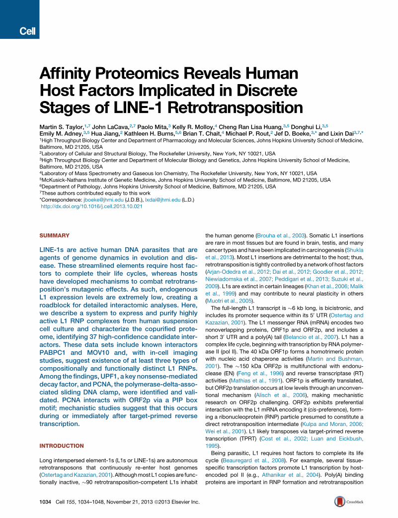
Affinity Proteomics Reveals Human Host Factors...
15
Affinity Proteomics Reveals Human Host Factors Implicated in Discrete Stages of LINE-1 Retrotransposition Martin S. Taylor, 1,7 John LaCava, 2,7 Paolo Mita, 3 Kelly R. Molloy, 4 Cheng Ran Lisa Huang, 3,5 Donghui Li, 3,5 Emily M. Adney, 3,5 Hua Jiang, 2 Kathleen H. Burns, 5,6 Brian T. Chait, 4 Michael P. Rout, 2 Jef D. Boeke, 3, * and Lixin Dai 3,7, * 1 High Throughput Biology Center and Department of Pharmacology and Molecular Sciences, Johns Hopkins University School of Medicine, Baltimore, MD 21205, USA 2 Laboratory of Cellular and Structural Biology, The Rockefeller University, New York, NY 10021, USA 3 High Throughput Biology Center and Department of Molecular Biology and Genetics, Johns Hopkins University School of Medicine, Baltimore, MD 21205, USA 4 Laboratory of Mass Spectrometry and Gaseous Ion Chemistry, The Rockefeller University, New York, NY 10021, USA 5 McKusick-Nathans Institute of Genetic Medicine, Johns Hopkins University School of Medicine, Baltimore, MD 21205, USA 6 Department of Pathology, Johns Hopkins University School of Medicine, Baltimore, MD 21205, USA 7 These authors contributed equally to this work *Correspondence: [email protected] (J.D.B.), [email protected] (L.D.) http://dx.doi.org/10.1016/j.cell.2013.10.021 SUMMARY LINE-1s are active human DNA parasites that are agents of genome dynamics in evolution and dis- ease. These streamlined elements require host fac- tors to complete their life cycles, whereas hosts have developed mechanisms to combat retrotrans- position’s mutagenic effects. As such, endogenous L1 expression levels are extremely low, creating a roadblock for detailed interactomic analyses. Here, we describe a system to express and purify highly active L1 RNP complexes from human suspension cell culture and characterize the copurified prote- ome, identifying 37 high-confidence candidate inter- actors. These data sets include known interactors PABPC1 and MOV10 and, with in-cell imaging studies, suggest existence of at least three types of compositionally and functionally distinct L1 RNPs. Among the findings, UPF1, a key nonsense-mediated decay factor, and PCNA, the polymerase-delta-asso- ciated sliding DNA clamp, were identified and vali- dated. PCNA interacts with ORF2p via a PIP box motif; mechanistic studies suggest that this occurs during or immediately after target-primed reverse transcription. INTRODUCTION Long interspersed element-1s (L1s or LINE-1s) are autonomous retrotransposons that continuously re-enter host genomes (Ostertag and Kazazian, 2001). Although most L1 copies are func- tionally inactive, 90 retrotransposition-competent L1s inhabit the human genome (Brouha et al., 2003). Somatic L1 insertions are rare in most tissues but are found in brain, testis, and many cancer types and have been implicated in carcinogenesis (Shukla et al., 2013). Most L1 insertions are detrimental to the host; thus, retrotransposition is tightly controlled by a network of host factors (Arjan-Odedra et al., 2012; Dai et al., 2012; Goodier et al., 2012; Niewiadomska et al., 2007; Peddigari et al., 2013; Suzuki et al., 2009). L1s are extinct in certain lineages (Khan et al., 2006; Malik et al., 1999) and may contribute to neural plasticity in others (Muotri et al., 2005). The full-length L1 transcript is 6 kb long, is bicistronic, and includes its promoter sequence within its 5 0 UTR (Ostertag and Kazazian, 2001). The L1 messenger RNA (mRNA) encodes two nonoverlapping proteins, ORF1p and ORF2p, and includes a short 3 0 UTR and a poly(A) tail (Belancio et al., 2007). L1 has a complex life cycle, beginning with transcription by RNA polymer- ase II (pol II). The 40 kDa ORF1p forms a homotrimeric protein with nucleic acid chaperone activities (Martin and Bushman, 2001). The 150 kDa ORF2p is multifunctional with endonu- clease (EN) (Feng et al., 1996) and reverse transcriptase (RT) activities (Mathias et al., 1991). ORF1p is efficiently translated, but ORF2p translation occurs at low levels through an unconven- tional mechanism (Alisch et al., 2006), making mechanistic research on ORF2p challenging. ORF2p exhibits preferential interaction with the L1 mRNA encoding it (cis-preference), form- ing a ribonucleoprotein (RNP) particle presumed to constitute a direct retrotransposition intermediate (Kulpa and Moran, 2006; Wei et al., 2001). L1 likely transposes via target-primed reverse transcription (TPRT) (Cost et al., 2002; Luan and Eickbush, 1995). Being parasitic, L1 requires host factors to complete its life cycle (Beauregard et al., 2008). For example, several tissue- specific transcription factors promote L1 transcription by host- encoded pol II (e.g., Athanikar et al., 2004). Poly(A) binding proteins are important in RNP formation and retrotransposition 1034 Cell 155, 1034–1048, November 21, 2013 ª2013 Elsevier Inc.
Transcript of Affinity Proteomics Reveals Human Host Factors...
Affinity Proteomics Reveals HumanHost Factors Implicated in DiscreteStages of LINE-1 RetrotranspositionMartin S. Taylor,1,7 John LaCava,2,7 Paolo Mita,3 Kelly R. Molloy,4 Cheng Ran Lisa Huang,3,5 Donghui Li,3,5
Emily M. Adney,3,5 Hua Jiang,2 Kathleen H. Burns,5,6 Brian T. Chait,4 Michael P. Rout,2 Jef D. Boeke,3,* and Lixin Dai3,7,*1High Throughput Biology Center and Department of Pharmacology and Molecular Sciences, Johns Hopkins University School of Medicine,
Baltimore, MD 21205, USA2Laboratory of Cellular and Structural Biology, The Rockefeller University, New York, NY 10021, USA3High Throughput Biology Center and Department of Molecular Biology and Genetics, Johns Hopkins University School of Medicine,
Baltimore, MD 21205, USA4Laboratory of Mass Spectrometry and Gaseous Ion Chemistry, The Rockefeller University, New York, NY 10021, USA5McKusick-Nathans Institute of Genetic Medicine, Johns Hopkins University School of Medicine, Baltimore, MD 21205, USA6Department of Pathology, Johns Hopkins University School of Medicine, Baltimore, MD 21205, USA7These authors contributed equally to this work
*Correspondence: [email protected] (J.D.B.), [email protected] (L.D.)http://dx.doi.org/10.1016/j.cell.2013.10.021
SUMMARY
LINE-1s are active human DNA parasites that areagents of genome dynamics in evolution and dis-ease. These streamlined elements require host fac-tors to complete their life cycles, whereas hostshave developed mechanisms to combat retrotrans-position’s mutagenic effects. As such, endogenousL1 expression levels are extremely low, creating aroadblock for detailed interactomic analyses. Here,we describe a system to express and purify highlyactive L1 RNP complexes from human suspensioncell culture and characterize the copurified prote-ome, identifying 37 high-confidence candidate inter-actors. These data sets include known interactorsPABPC1 and MOV10 and, with in-cell imagingstudies, suggest existence of at least three types ofcompositionally and functionally distinct L1 RNPs.Among the findings, UPF1, a key nonsense-mediateddecay factor, and PCNA, the polymerase-delta-asso-ciated sliding DNA clamp, were identified and vali-dated. PCNA interacts with ORF2p via a PIP boxmotif; mechanistic studies suggest that this occursduring or immediately after target-primed reversetranscription.
INTRODUCTION
Long interspersed element-1s (L1s or LINE-1s) are autonomous
retrotransposons that continuously re-enter host genomes
(OstertagandKazazian, 2001). Althoughmost L1copiesare func-
tionally inactive, �90 retrotransposition-competent L1s inhabit
1034 Cell 155, 1034–1048, November 21, 2013 ª2013 Elsevier Inc.
the human genome (Brouha et al., 2003). Somatic L1 insertions
are rare in most tissues but are found in brain, testis, and many
cancer types andhavebeen implicated in carcinogenesis (Shukla
et al., 2013). Most L1 insertions are detrimental to the host; thus,
retrotransposition is tightly controlledbyanetworkof host factors
(Arjan-Odedra et al., 2012; Dai et al., 2012; Goodier et al., 2012;
Niewiadomska et al., 2007; Peddigari et al., 2013; Suzuki et al.,
2009). L1s are extinct in certain lineages (Khan et al., 2006; Malik
et al., 1999) and may contribute to neural plasticity in others
(Muotri et al., 2005).
The full-length L1 transcript is �6 kb long, is bicistronic, and
includes its promoter sequence within its 50 UTR (Ostertag and
Kazazian, 2001). The L1 messenger RNA (mRNA) encodes two
nonoverlapping proteins, ORF1p and ORF2p, and includes a
short 30 UTR and a poly(A) tail (Belancio et al., 2007). L1 has a
complex life cycle, beginning with transcription by RNA polymer-
ase II (pol II). The 40 kDa ORF1p forms a homotrimeric protein
with nucleic acid chaperone activities (Martin and Bushman,
2001). The �150 kDa ORF2p is multifunctional with endonu-
clease (EN) (Feng et al., 1996) and reverse transcriptase (RT)
activities (Mathias et al., 1991). ORF1p is efficiently translated,
but ORF2p translation occurs at low levels through an unconven-
tional mechanism (Alisch et al., 2006), making mechanistic
research on ORF2p challenging. ORF2p exhibits preferential
interaction with the L1 mRNA encoding it (cis-preference), form-
ing a ribonucleoprotein (RNP) particle presumed to constitute a
direct retrotransposition intermediate (Kulpa and Moran, 2006;
Wei et al., 2001). L1 likely transposes via target-primed reverse
transcription (TPRT) (Cost et al., 2002; Luan and Eickbush,
1995).
Being parasitic, L1 requires host factors to complete its life
cycle (Beauregard et al., 2008). For example, several tissue-
specific transcription factors promote L1 transcription by host-
encoded pol II (e.g., Athanikar et al., 2004). Poly(A) binding
proteins are important in RNP formation and retrotransposition

(Dai et al., 2012), and DNA repair factors affect L1 retrotranspo-
sition, most likely in TPRT (Suzuki et al., 2009). Host genomes
have evolvedmechanisms to restrict potentially harmful L1 retro-
transposition, employing DNA methylation (Hata and Sakaki,
1997), RNAi pathways (Soifer et al., 2005; Yang and Kazazian,
2006), APOBEC3 cytidine deaminases (Niewiadomska et al.,
2007), and RNA helicase MOV10 (Arjan-Odedra et al., 2012;
Goodier et al., 2012). Two recent studies identified protein and
RNA interactors in L1 RNP complexes via an ORF1 tag (Goodier
et al., 2013; Mandal et al., 2013). However, although host factors
have been found, technical limitations have hindered isolation of
pure, active RNP complexes in analytically tractable quantities.
Although L1 is highly active in germline and brain, it is effec-
tively repressed in most somatic cells (Muotri et al., 2005). To
evaluate L1 activity in cell culture, it is often paired with a consti-
tutive, high-level promoter (Moran et al., 1996). Fractions con-
taining expressed L1 RNP complexes have been prepared by
sucrose cushion velocity sedimentation of cell lysates (Kulpa
and Moran, 2005, 2006). These provided valuable insights into
mechanism but suffer from low purity, limiting their usefulness
for studying L1-host interactomes. Traditional epitope tagging
of ORF polypeptides has been used to study proteins copurifying
with the L1 RNP (Doucet et al., 2010; Goodier et al., 2012, 2013),
but these conferred insufficient yield and purity for comprehen-
sive proteomics with high specific activity.
In contrast, new methods implementing cryomilling/rapid
affinity capture provide outstanding purification that significantly
improves interactomic surveys (Cristea et al., 2005; Domanski
et al., 2012; Oeffinger et al., 2007). Intact cells are flash-frozen
in liquid nitrogen and are milled in solid phase (cryomilling).
Native intermolecular interactions are preserved during milling,
producing a fine powder well-suited to subsequent affinity isola-
tion of protein complexes (pullouts). Micron-scale antibody-con-
jugated magnetic beads enable rapid purifications with binding
times as short as 5 min and facilitate binding of large complexes
excluded from pores of traditional matrices (Oeffinger et al.,
2007). When coupled with Isotopic Differentiation of Interactions
as Random or Targeted (I-DIRT), stable interactions formed
in vivo are distinguished from postextraction artifacts (Tackett
et al., 2005). These techniques have been applied to the study
of protein complexes from many systems (Di Virgilio et al.,
2013; Domanski et al., 2012; Lee et al., 2008; Oeffinger et al.,
2007).
In this study, we constructed a series of inducible L1 expres-
sion vectors containing either synthetic (ORFeus-Hs) (An et al.,
2011) or native (L1RP) L1 (Kimberland et al., 1999) elements
and screened epitope tags on both L1 ORFs for effects on
retrotransposition. Active constructs were expressed in suspen-
sion culture at a large scale, and RNPs were purified using cryo-
milling and rapid capture, facilitating enzymatic, transcriptomic,
and proteomic assays. Purified RNPs had �70-fold higher spe-
cific activity than those made by previous techniques. RNA
sequencing (RNA-seq) revealed L1 RNA as an abundant compo-
nent along with U6 small nuclear RNA (snRNA), commonly
observed as part of hybrid elements with L1 (Buzdin et al.,
2002). The ORF1p to ORF2p ratio was unexpectedly low in
tandem purified L1 particles. Cell imaging revealed that most
cells fail to express ORF2p. Mass spectrometric (MS) character-
C
ization identified 37 high-confidence interactors, including
known interactors PABPC1 and MOV10, and revealed that
ORF1 and ORF2 RNPs exhibit both overlapping and distinct
interactomes. UPF1, a helicase critical for nonsense-mediated
decay (NMD), and PCNA, the polymerase-associated sliding
clamp, were further validated for roles in the L1 life cycle.
RESULTS
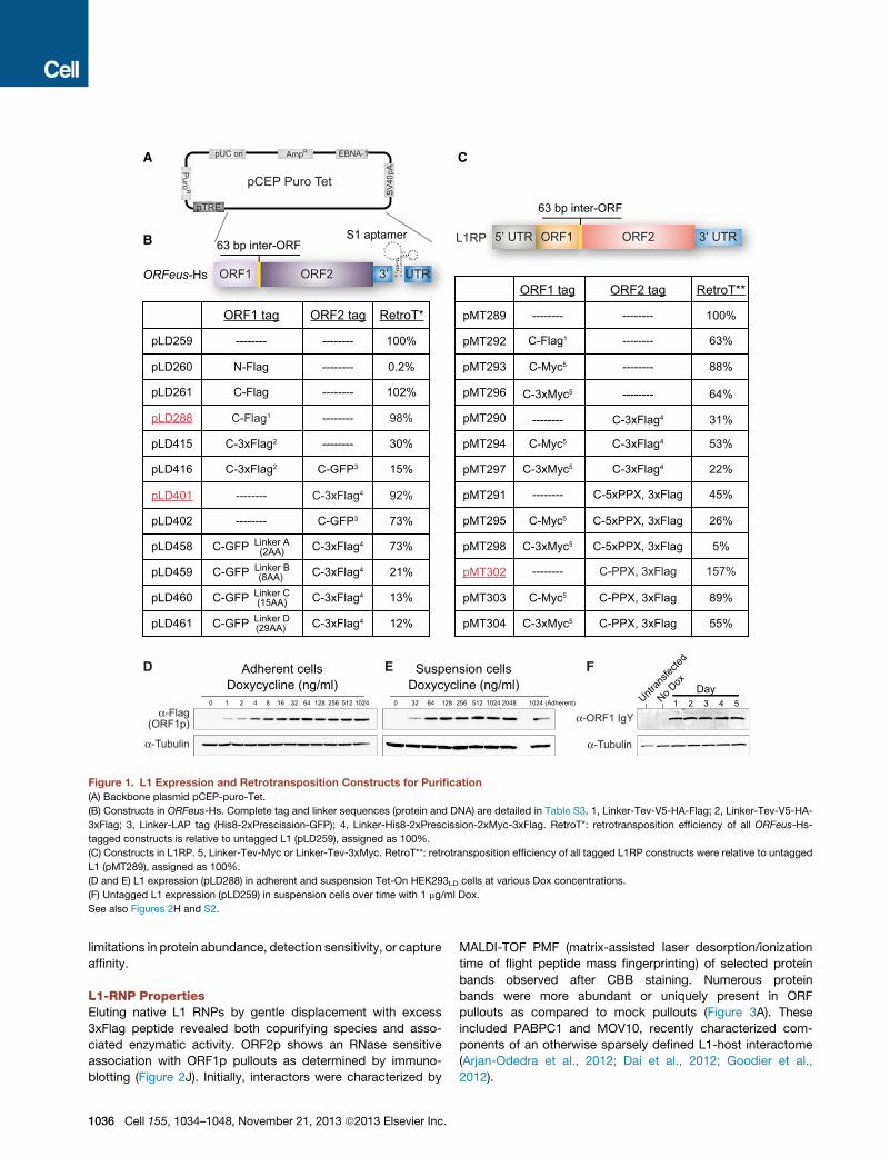
Producing Highly Purified L1-RNPsTo isolate L1 RNP complexes at high yield and purity, we used
synthetic human L1ORFeus-Hs, which has high activity and pro-
duces > 40-fold more RNA and 5- to 10-fold more ORF1p than a
native counterpart, while encoding identical proteins (An et al.,
2011). Uncontrolled L1 expression slows or eliminates growth;
therefore, we leveraged expression from a tetracycline-regu-
lated mini-CMV promoter (pTRE, Figure 1A) (O’Donnell et al.,
2013). Due to a lack of appropriate antibodies, especially for
ORF2p, we designed many epitope tag combinations and
measured retrotransposition efficiency relative to untagged
ORFeus-Hs (Figure 1B, Supplemental Information, and Tables
S1, S2, and S3). Constructs pLD288 (ORF1p-Flag) and pLD401
(ORF2p-3xFlag) were best, as they combined high L1 activity
with short tag sequences.
We introduced these constructs into adherent Tet-On
HEK293TLD (Dai et al., 2012) and selected transfected cells for
1 week or more with puromycin. Doxycycline (Dox) concentra-
tion and times had similar effects in monolayer and suspension
cultures (Figures 1D–1F); expression of both ORFs peaked
24 hr postinduction. Although ORF1p levels were relatively sta-
ble over the next 3 days, ORF2p levels decreased within 48 hr
(Figures 1F, 2H, and S2).
Multiliter suspension cultures were induced, harvested, and
cryomilled, and pullouts were optimized (Figures 2A–2D and
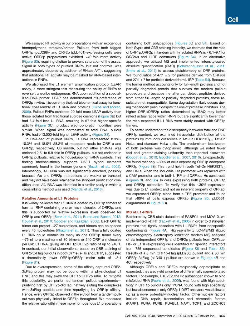
S1 available online). Both L1 proteins were readily observed by
Coomassie brilliant blue (CBB) staining (Figures 2E and 2F).
We also tagged native L1RP, which has lower protein expression
levels (Figure 1C). Surprisingly, the C-terminal Flag and 3xFlag
tag combinations on ORF1p and ORF2p that worked well in
ORFeus-Hs were poorly tolerated in L1RP. We designed and
screened new tags and linkers; a single Myc-tag at the C termi-
nus of ORF1p was tolerated (pMT293 and pMT303) but did not
provide high-quality affinity isolations. By shortening the linker
on ORF2p-3xFLAG, we preserved L1RP retrotransposition effi-
ciency (pMT302 and pMT303). An improved aORF1p antibody
obviated need for an epitope-tagged L1RP ORF1p, and thus,
pMT302 (L1RP with ORF2p-3xFlag) was selected (Figures 2E
and 2F).
Both synthetic (pLD401) and native (pMT302) constructs gave
robust ORF2p expression (Figures 2G and 2H). Taking into
account the different detection sensitivities exhibited by the
two tags (Hernan et al., 2000), ORF1p is apparently expressed
1,000- to 10,000-fold higher than ORF2p (Figures 2G and S2).
Comparing ORFeus-Hs and L1RP constructs, ORF1p levels
differ by �2.5-fold, but ORF2p levels differ by �20- to 40-fold
compared to a purified standard (Figures 2H, 2I, and S2).
The expression constructs, cell lines, and approach collec-
tively constitute an analytical suite unhindered by typical
ell 155, 1034–1048, November 21, 2013 ª2013 Elsevier Inc. 1035
C
SV
40pAP
uroR
EBNA-1AmpRpUC ori
pCEP Puro Tet
pTRE
A
B
Doxycycline (ng/ml)
α-Flag(ORF1p)
512256128643216421 8 1024 51225612864320 1024 2048 1024 (Adherent)0
Adherent cells Suspension cellsDoxycycline (ng/ml)
D E F
ORFeus-Hs
S1 aptamer
ORF1 ORF2
63 bp inter-ORF
3’ UTR
ORF1 ORF2
63 bp inter-ORF
3’ UTR5’ UTRL1RP
α-Tubulin
Day1 5432Untr
ansfe
cted
No Dox
α-ORF1 IgY
α-Tubulin
-------- -------- 100%pMT289
C-Myc5 -------- 88%pMT293
-------- C-5xPPX, 3xFlag 45%pMT291
C-Myc5 C-5xPPX, 3xFlag 26%pMT295
C-Myc5 C-PPX, 3xFlag 89%pMT303
C-Flag1 -------- 63%pMT292
-------- 31%pMT290 C-3xFlag4
C-Myc5 53%pMT294 C-3xFlag4
-------- 64%pMT296 C-3xMyc5
22%pMT297 C-3xFlag4C-3xMyc5
5%C-5xPPX, 3xFlagpMT298 C-3xMyc5
C-PPX, 3xFlag 55%pMT304 C-3xMyc5
-------- C-PPX, 3xFlag 157%pMT302
ORF1 tag ORF2 tag RetroT**
pLD415 C-3xFlag2 -------- 30%
C-3xFlag2pLD416 C-GFP3 15%
C-GFP3pLD402 -------- 73%
pLD259 -------- -------- 100%
pLD260 N-Flag -------- 0.2%
pLD261 C-Flag -------- 102%
C-Flag1 -------- 98%pLD288
-------- C-3xFlag4 92%pLD401
ORF1 tag ORF2 tag RetroT*
pLD458 73%C-3xFlag4C-GFP Linker A(2AA)
C-3xFlag4 21%pLD459 C-GFP Linker B(8AA)
C-3xFlag4 13%pLD460 C-GFP Linker C(15AA)
C-3xFlag4 12%pLD461 C-GFP Linker D(29AA)
Figure 1. L1 Expression and Retrotransposition Constructs for Purification
(A) Backbone plasmid pCEP-puro-Tet.
(B) Constructs in ORFeus-Hs. Complete tag and linker sequences (protein and DNA) are detailed in Table S3. 1, Linker-Tev-V5-HA-Flag; 2, Linker-Tev-V5-HA-
3xFlag; 3, Linker-LAP tag (His8-2xPrescission-GFP); 4, Linker-His8-2xPrescission-2xMyc-3xFlag. RetroT*: retrotransposition efficiency of all ORFeus-Hs-
tagged constructs is relative to untagged L1 (pLD259), assigned as 100%.
(C) Constructs in L1RP. 5, Linker-Tev-Myc or Linker-Tev-3xMyc. RetroT**: retrotransposition efficiency of all tagged L1RP constructs were relative to untagged
L1 (pMT289), assigned as 100%.
(D and E) L1 expression (pLD288) in adherent and suspension Tet-On HEK293LD cells at various Dox concentrations.
(F) Untagged L1 expression (pLD259) in suspension cells over time with 1 mg/ml Dox.
See also Figures 2H and S2.
limitations in protein abundance, detection sensitivity, or capture
affinity.
L1-RNP PropertiesEluting native L1 RNPs by gentle displacement with excess
3xFlag peptide revealed both copurifying species and asso-
ciated enzymatic activity. ORF2p shows an RNase sensitive
association with ORF1p pullouts as determined by immuno-
blotting (Figure 2J). Initially, interactors were characterized by
1036 Cell 155, 1034–1048, November 21, 2013 ª2013 Elsevier Inc.
MALDI-TOF PMF (matrix-assisted laser desorption/ionization
time of flight peptide mass fingerprinting) of selected protein
bands observed after CBB staining. Numerous protein
bands were more abundant or uniquely present in ORF
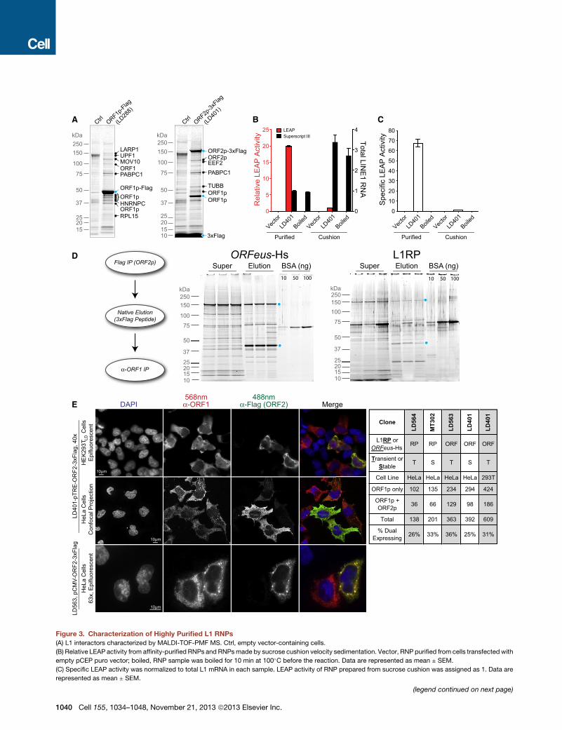
pullouts as compared to mock pullouts (Figure 3A). These
included PABPC1 and MOV10, recently characterized com-
ponents of an otherwise sparsely defined L1-host interactome
(Arjan-Odedra et al., 2012; Dai et al., 2012; Goodier et al.,
2012).
We assayed RT activity in our preparations with an exogenous
homopolymeric template/primer. Pullouts from both tagged
ORF1p (pLD288)- and ORF2p (pLD401)-expressing cells were
active; ORF2p preparations exhibited �40-fold more activity
(Figure S3), requiring dilution to prevent saturation of the assay.
Signal in both types of purified RNPs, but not controls, was
approximately doubled by addition of RNase A/T1, suggesting
that additional RT activity may be masked by RNA-based inter-
actions in RNPs.
We also used the L1 element amplification protocol (LEAP)
assay, a more stringent test measuring the ability of RNPs to
reverse transcribe endogenous RNA upon addition of a special-
ized DNA primer. LEAP has demonstrated cis-preference of
ORF2p in vitro; it is currently the best biochemical assay for func-
tional coassembly of L1 RNA and proteins (Kulpa and Moran,
2006). Pullout RNPs exhibited 20-fold more LEAP activity than
those isolated from traditional sucrose cushions (Figure 3B) but
had 3.4-fold less L1 RNA, resulting in 67-fold higher specific
activity (Figure 3C); product electrophoretic mobilities were
similar. When signal was normalized to total RNA, pullout
RNPs had >13,000-fold higher LEAP activity (Figure S3).
In RNA-seq of pullout RNPs, L1 RNA represented 8.3%–
10.3% and 18.0%–28.2% of mappable reads for ORF1p and
ORF2p, respectively. U6 snRNA, but not other snRNAs, was
enriched 2.5- to 5.9-fold in ORF2p pullouts, but not enriched in
ORF1p pullouts, relative to housekeeping mRNA controls. This
finding mechanistically supports U6/L1 hybrid elements
commonly found in the human genome (Buzdin et al., 2002).
Interestingly, Alu RNA was not significantly enriched, possibly
because Alu and ORF2p interactions are weaker or transient
and may not have been retained in the stringent purification con-
dition used. Alu RNA was identified in a similar study in which a
crosslinking method was used (Mandal et al., 2013).
Relative Amounts of L1 ProteinsIt is widely believed that L1 RNA is coated by ORF1p trimers to
form an RNP containing one or two molecules of ORF2p, and
this is supported by relative expression levels observed for
ORF1p and ORF2p (Beck et al., 2011; Burns and Boeke, 2012;
Doucet et al., 2010; Goodier and Kazazian, 2008). Each ORF1p
trimer can protect �27 nucleotides, and trimers can be spaced
every 45 nucleotides (Khazina et al., 2011). Thus a fully coated
L1 RNA could contain as many as one ORF1p trimer every
�75 nt to a maximum of 80 trimers or 240 ORF1p molecules
per 6kb L1 RNA, giving an ORF1p:ORF2p ratio of up to 240:1.
In contrast, our initial observations, based on CBB staining of
ORF2-3xFlag pullouts in both ORFeus-Hs and L1RP, suggested
a dramatically lower ORF1p:ORF2p molar ratio of �3:1
(Figure S1).
Due to overexpression, some of the affinity captured ORF2-
3xFlag protein may not be bound within a physiological L1
RNP, and this may skew the ORF1p:ORF2p ratio. To mitigate
this possibility, we performed tandem pullout experiments—
purifying first by ORF2p-3xFlag, natively eluting the complexes
with 3xFlag peptide and then repurifying by ORF1p affinity.
Hence, every ORF2p observed in the elution from a tandem pull-
out was physically linked to ORF1p throughout. We measured
the relative ratio within these more homogenous L1 preparations
C
containing both polypeptides (Figures 3D and S4). Based on
both Sypro and CBB staining intensity, we estimate that the ratio
of ORF1p:ORF2p in tandem affinity isolated RNPs is�6:1–9:1 for
ORFeus and L1RP constructs (Figure S4). In an alternative
approach, we utilized MS and implemented intensity-based
absolute quantification (iBAQ) (Schwanhausser et al., 2011;
Smits et al., 2013) to assess stoichiometry of ORF proteins.
We found ratios of 47:1 ± 2 for particles derived from ORFeus
and 27:1 ± 7 for particles derived from L1RP (Table S4). Because
the former method accounts only for full-length proteins and not
partially degraded protein that survives the tandem pullout
procedure and because the latter can detect peptides derived
from either full-length or partially degraded proteins, these re-
sults are not incompatible. Some degradation likely occurs dur-
ing the tandem pullout despite the use of protease inhibitors. The
higher ORF1:ORF2p ratios determined by iBAQ may better
reflect actual ratios within RNPs but are significantly lower than
the ratio expected if L1 RNA were stably coated with ORF1p
trimers.
To better understand the discrepancy between total and RNP
ORF1p content, we examined intracellular distribution of the
proteins by immunofluorescence in Tet-On HEK293TLD, Tet-On
HeLa, and standard HeLa cells. The predominant localization
of both proteins was cytoplasmic, although we noted fewer
foci and greater staining uniformity than reported elsewhere
(Doucet et al., 2010; Goodier et al., 2007, 2010). Unexpectedly,
we found that only �30% of cells expressing ORF1p coexpress
ORF2p (Figure 3E). This trend held in both Tet-On HEK293TLDand HeLa, when the inducible Tet promoter was replaced with
a CMV promoter, and in both L1RP and ORFeus-Hs constructs
(Figures 3E and S5). In cells expressing both proteins, ORF1p
and ORF2p colocalize. To verify that this �30% expression
was due to L1 context and not an inherent property of ORF2p,
we expressed ORF2p alone from a TRE promoter and found
that >90% of cells express ORF2p (Figure S5, pLD561,
diagrammed in Figure 5B).
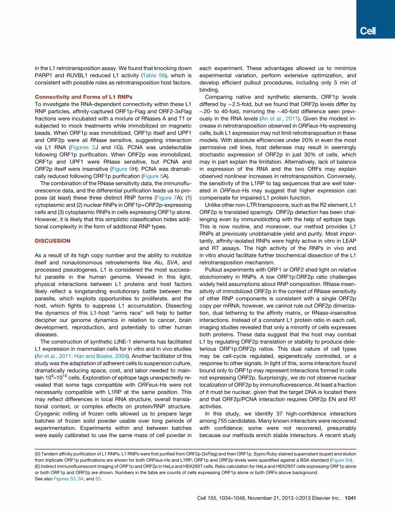
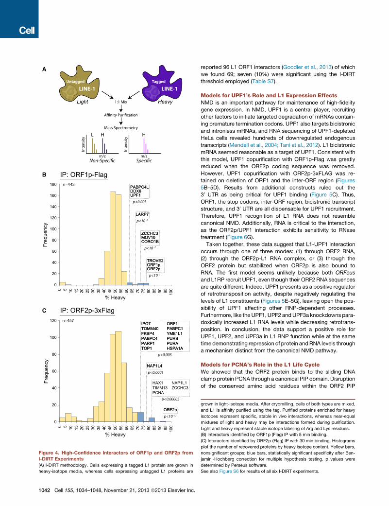
MS of L1-RNPsBolstered by CBB stain detection of PABPC1 and MOV10, we
implemented I-DIRT (Tackett et al., 2005) in order to distinguish
proteins that tightly associate with L1 RNPs from nonspecific
contaminants (Figure 4A). High-sensitivity LC-MS/MS (liquid
chromatography electrospray ionization tandem MS) analyses
of six independent ORF1p and ORF2p pullouts from ORFeus-
Hs- or L1RP-expressing cells identified 37 specific interactors
from 755 sequenced candidates (Figure S6 and Table S5).
Results of a 5 min ORF1p-Flag (pLD288) pullout and a 30 min
ORF2p-3xFlag (pLD401) pullout are shown in Figures 4B and
4C, respectively.
Although ORF1p and ORF2p reciprocally coprecipitate as
expected, they also yield a number of differentially coprecipitated
factors. For example, TROVE2, theRo autoantigen known to bind
misfolded RNA (Fuchs et al., 2006), was found with high speci-
ficity in ORF1p pullouts only. PCNA, found with high specificity
but low abundance in only ORF2p I-DIRT analyses, was followed
up as a novel potentially nuclear factor. Other nuclear factors
include DNA repair, transcription and chromatin factors
(PARP1, PURA, PURB, RUVBL1, NAP1, TOP1, and ZCCHC3
ell 155, 1034–1048, November 21, 2013 ª2013 Elsevier Inc. 1037
1
6
54
3
2
a
f
e
d
c
b
A B C D
kDa
E F G
H I
J
ORFeus-H
s
L1RP
ORF1p-F
lag
ORF2p-3x
Flag
ORFeus-H
s
L1RP
250kDa
150
100
75
50
37
25201510
ORF1ORF1p-Flag
kDa250150
100
75
50
37
252015
ORF2p-3xFlag260
160
11080
60
5040
30
20
kDa
kDa
160
110
50
Dox (hrs) 48 0 24 48 0 24 48
L1RPMT302
NoTag
ORFeus-HsLD401
0.05 0.1 0.2 0.4 0.8 1.6 3.2ng
α-Flag α-Flag α-Flag
α-Flag
α-ORF1p
ORF2p-3xFlagα-Flag
CK2α-3xFLAGα-Flag
kDa5040
L1RP ORFeus-Hs
α-ORF1
LD40
1
LD28
8
LD25
9
MT302
MT293
MT289
RNase
BSA
RNase
BSA
Super Beads
160α-Flag(ORF2p)
40α-ORF1
kDa
Figure 2. Interactomics Workflow Applied to L1 Produces Tractable Quantities of Both ORF Proteins from Both L1RP and ORFeus-Hs(A) Transposon-expressing human cell cultures are grown in suspension and are induced for 24 hr.
(B) Frozen cell ‘‘BBs’’ (�3mm diameter spherical globules of cells previously frozen in LN2) are cryomilled using stainless steel ball bearings under liquid nitrogen,
resulting in a fine powder stored at �80�C.(C) Antibody-conjugated mm-scale magnetic medium is used for rapid pullout.
(D) Gel-based proteomics: purified proteins are subjected to MALDI-TOF-PMF for identification of major species (1–6) or LC-MS/MS (a–f).
(E) ORF1p and copurified factors for ORFeus-Hs (pLD288) and L1RP (pMT289) by ORF1p purification.
(legend continued on next page)
1038 Cell 155, 1034–1048, November 21, 2013 ª2013 Elsevier Inc.
proteins), an apoptosis regulator (HAX1), and a nuclear import
factor (IPO7). These results are consistent with the possibility of
ORF1p and ORF2p having both shared and distinct host
interactomes. Notably, PABPC1, PABPC4, MOV10, UPF1, and
ZCCHC3 were specific interactors in both ORF1p and ORF2p
RNP preparations. although all of these hits are candidates for
mechanistic studies, we focused initial follow-up on UPF1 and
PCNA. UPF1 was chosen because of its high abundance and
obvious potential connections between bicistronic L1 mRNA
and NMD.
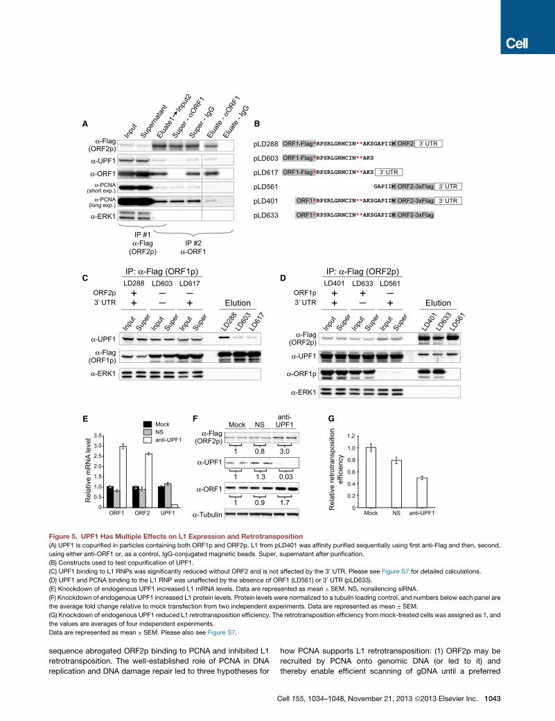
UPF1 Interaction Requires ORF2 and Downregulates L1ExpressionWe validated the L1/ UPF1 interaction by immunoblotting for
UPF1 in ORF1p-Flag and ORF2p-3xFlag pullouts from cell lines
expressing pLD288 and pLD401, respectively. UPF1 was de-
tected in both ORF1p and ORF2p elutions and was copurified
through tandem pullouts from pLD401-expressing cells (Fig-
ure 5A). The L1/ UPF1 interaction in purified fractions was sensi-
tive to RNase treatment (Figures 6G and 6H, see below).
In addition to recognizing mRNAs bearing premature non-
sense codons, UPF1 affects expression of intronless and bicis-
tronic mRNAs (e.g., MAP3K14 and ARHGEF18) (Mendell et al.,
2004). Within the L1 mRNA, a 63 nt inter-ORF region contains
two additional stop codons in frame with ORF1 (Figure 5B). We
hypothesized that the three inter-ORF stop codons in conjunc-
tion with a long 30 UTR-mimic (�4 kb ORF2 plus the native
30 UTR) might be recognized by UPF1 (Hogg and Goff, 2010). It
is known that UPF1 also binds to and scans 30 UTRs of many
cellular mRNAs (Shigeoka et al., 2012). We constructed several
plasmids with different combinations of ORF, interORF, and
30 UTR sequences to map interacting sequences (Figure 5B).
ORF1p affinity isolations demonstrate that removal of ORF2p
results in higher yields of both ORF1p protein and L1 RNA but
results in a dramatically reduced yield of UPF1; removal of the
30 UTR had no effect (Figure S7 and 5C). In ORF2p affinity isola-
tions, removal of the 30 UTR similarly had no effect on the yield of
UPF1, but, in contrast to our hypothesis, removal of ORF1p also
had no effect on UPF1 yield (Supplemental Information and Fig-
ures 5D and S7). Although the yield of ORF2p in the three
purifications differed slightly, the UPF1 and ORF2p signal inten-
sities precisely correlate (r2 = 0.995) (Figure S7G). In summary,
the ORF2 coding sequence or ORF2p expression, rather than
the interORF sequence, ORF1p sequence, bicistronic context,
or 30 UTR, enables the L1-UPF1 interaction.
Next, we investigated the impact of UPF1 on L1 expression by
knocking down endogenous UPF1 by RNA interference. L1
mRNA and polypeptides were all upregulated in the knockdown
cells. Surprisingly, L1 retrotransposition efficiency decreased
(F) ORF2p and copurified factors for ORFeus-Hs (pLD401) and L1RP (pMT302) b
(G) Comparison of ORF1p-Flag (pLD288) andORF2p-3xFlag (pLD401) expression
see Figure S2 for detailed calculations.
(H) ORF2p expression levels in L1RP versus ORFeus-Hs. CK2a-3xFlag was load
(I) ORF1p expression levels in L1RP versus ORFeus-Hs. 2 mg protein loaded in e
(J)ORF1p-ORF2p interactions areRNase sensitive. L1wasaffinity purifiedwith ant
mock treatedwithBSA. ‘‘Super,’’ proteins released into the supernatant; ‘‘beads,’’
See also Figures S1 and S2.
C
by �50% (Figures 5E–5G and S7C–S7F). To minimize the possi-
bility of off-target RNAi effects, we coexpressed L1 with five
different anti-UPF1 shRNAs. Four of these reduced retrotrans-
position significantly by as much as 90%, suggesting that
UPF1 depletion decreases retrotransposition. Knockdown of
two other proteins that act in concert with UPF1 in NMD,
UPF2, andUPF3a also reduced retrotransposition and increased
L1 mRNA levels (Table S6).
PCNA Binding to an ORF2 PIP Box Is Critical forRetrotranspositionPCNA was found as a low-abundance but high-specificity inter-
actor of ORF2p, but notORF1p.We identified a canonical PCNA-
interacting protein (PIP) box (Qxx[V/L/M/I]xx[F/Y][F/Y]) in resi-
dues 407–415 of ORF2p, located between the EN and RT
domains (Figure 6A). Within the PIP sequence, four amino acid
residues (I407, I411, Y414, and Y415) are highly conserved
across L1 ORF2s from diverse species, suggesting a crucial
role for a PIP box in ORF2 binding to PCNA. Mutation of these
residues to alanine (A) disrupted ORF2p/PCNA copurification,
whereas mutation of Q408, less conserved in ORF2p, does not
affect it (Figure 6B). Mutations that disrupt the ORF2p /PCNA
interaction correspondingly decrease retrotransposition effi-
ciency of L1, whereas the Q408A mutation shows no effect on
retrotransposition (Figure 6C). Overall, these data support an
ORF2p/PCNA interaction through a PIP box, and this interaction
is critical for retrotransposition. We also knocked down endoge-
nous PCNA using four small hairpin RNAs (shRNAs) and
observed downregulation of L1 retrotranspositionwell correlated
with decreased PCNA levels (r2 = 0.93; Figure 6D). To investigate
the L1 life cycle stage atwhich L1/PCNA interaction functions,we
assayed formaintenance of this interaction in cells expressing an
ORF2p EN domain catalytic mutant (H230A) (Figure 6E), which
was previously shown to abolish DNA nicking and L1 retrotrans-
position (Feng et al., 1996). Copurification of PCNA was greatly
reduced in RNPs isolated from the endonuclease domainmutant
(Figure 6E). To rule out the possibility that mutations in the EN
domain disrupt overall ORF2p folding, we repeated the experi-
ment with six additional well-studied EN mutants (E43A,
D145A, R155A, T192Y, I204Y, and D205G; Figures 6F and S7)
(Repanas et al., 2007, 2009; Weichenrieder et al., 2004). We
measured RT activity of these EN mutants, and all were highly
active, suggesting proper folding (Figure S7). Similarly, a catalytic
RTmutant (D702Y) also abolishedORF2p-PCNA interaction (Fig-
ure 6F). These observations suggest that PCNAmay be recruited
to ORF2 after completion of the target DNA nicking and during or
after L1 complementary DNA (cDNA) synthesis steps.
Because PCNA is involved in DNA repair pathways, we also
studied other L1 RNP factors with possible roles in DNA repair
y ORF2p (anti-Flag) purification.
levels in puro-selected adherent cells; 20mg protein loaded in each lane. Please
ed as control. 20 mg protein loaded in each lane.
ach lane.
i-ORF1Dynabeads frompLD401and then treated on-beadwithRNasesA/T1or
sample retainedonbeadsafterwashing and thenelutedwith LDSsamplebuffer.
ell 155, 1034–1048, November 21, 2013 ª2013 Elsevier Inc. 1039
A
D
E
250kDa
150
100
75
50
37
25201510
(LD40
1)
ORF2p-3x
Flag
Ctrl
ORF1p
ORF2p-3xFlagORF2pEEF2PABPC1
TUBBORF1p
3xFlag
250kDa
150
100
75
50
37
252015
LARP1UPF1MOV10
PABPC1
ORF1p
ORF1p}
ORF1
HNRNPC
RPL15
ORF1p-Flag
ORF1p-F
lag
(LD28
8)
Ctrl B C
Rel
ativ
e LE
AP
Act
ivity Total LIN
E1 R
NA
0
5
10
15
20
25
0
1
2
3
4LEAPSuperscript III
Purified CushionVec
tor
LD40
1
Boiled
Vector
LD40
1
Boiled
Spe
cific
LE
AP
Act
ivity
0
10
20
30
40
50
60
70
80
Purified CushionVec
tor
LD40
1
Boiled
Vector
LD40
1
Boiled
Flag IP (ORF2p)
Native Elution(3xFlag Peptide)
α-ORF1 IP
250kDa
150
100
75
50
37
25201510
Elution BSA (ng)ORFeus-Hs
Super10 50 100
250kDa
150100
75
50
37
25201510
Elution BSA (ng)L1RP
Super10 50 100
HeL
a C
ells
Con
foca
l Pro
ject
ion
HE
K29
3TC
ells
Epi
fluor
esce
ntLD
HeL
a C
ells
63x,
Epi
fluor
esce
nt
DAPI Merge568nm
α-ORF1488nm
α-Flag (ORF2)
10µm
10µm
10µm
LD40
1-pT
RE
-OR
F2-3
xFla
g, 4
0xLD
563,
pC
MV-
OR
F2-3
xFla
g
CloneLD564
MT302
LD563
LD401
LD401
L1RP orORFeus-Hs
RP RP ORF ORF ORF
Transient orStable
T S T S T
Cell Line HeLa HeLa HeLa HeLa 293T
ORF1p only 102 135 234 294 424
ORF1p +ORF2p 36 66 129 98 186
Total 138 201 363 392 609
% DualExpressing 26% 33% 36% 25% 31%
Figure 3. Characterization of Highly Purified L1 RNPs
(A) L1 interactors characterized by MALDI-TOF-PMF MS. Ctrl, empty vector-containing cells.
(B) Relative LEAP activity from affinity-purified RNPs and RNPsmade by sucrose cushion velocity sedimentation. Vector, RNP purified from cells transfected with
empty pCEP puro vector; boiled, RNP sample was boiled for 10 min at 100�C before the reaction. Data are represented as mean ± SEM.
(C) Specific LEAP activity was normalized to total L1 mRNA in each sample. LEAP activity of RNP prepared from sucrose cushion was assigned as 1. Data are
represented as mean ± SEM.
(legend continued on next page)
1040 Cell 155, 1034–1048, November 21, 2013 ª2013 Elsevier Inc.
in the L1 retrotransposition assay. We found that knocking down
PARP1 and RUVBL1 reduced L1 activity (Table S6), which is
consistent with possible roles as retrotransposition host factors.
Connectivity and Forms of L1 RNPsTo investigate the RNA-dependent connectivity within these L1
RNP particles, affinity-captured ORF1p-Flag and ORF2-3xFlag
fractions were incubated with a mixture of RNases A and T1 or
subjected to mock treatments while immobilized on magnetic
beads. When ORF1p was immobilized, ORF1p itself and UPF1
and ORF2p were all RNase sensitive, suggesting interaction
via L1 RNA (Figures 2J and 6G). PCNA was undetectable
following ORF1p purification. When ORF2p was immobilized,
ORF1p and UPF1 were RNase sensitive, but PCNA and
ORF2p itself were insensitive (Figure 6H). PCNA was dramati-
cally reduced following ORF1p purification (Figure 5A).
The combination of the RNase sensitivity data, the immunoflu-
orescence data, and the differential purification leads us to pro-
pose (at least) these three distinct RNP forms (Figure 7A): (1)
cytoplasmic and (2) nuclear RNPs in ORF1p+ORF2p-expressing
cells and (3) cytoplasmic RNPs in cells expressing ORF1p alone.
However, it is likely that this simplistic classification hides addi-
tional complexity in the form of additional RNP types.
DISCUSSION
As a result of its high copy number and the ability to mobilize
itself and nonautonomous retroelements like Alu, SVA, and
processed pseudogenes, L1 is considered the most success-
ful parasite in the human genome. Viewed in this light,
physical interactions between L1 proteins and host factors
likely reflect a longstanding evolutionary battle between the
parasite, which exploits opportunities to proliferate, and the
host, which fights to suppress L1 accumulation. Dissecting
the dynamics of this L1-host ‘‘arms race’’ will help to better
decipher our genome dynamics in relation to cancer, brain
development, reproduction, and potentially to other human
diseases.
The construction of synthetic LINE-1 elements has facilitated
L1 expression in mammalian cells for in vitro and in vivo studies
(An et al., 2011; Han and Boeke, 2004). Another facilitator of this
studywas the adaptation of adherent cells to suspension culture,
dramatically reducing space, cost, and labor needed to main-
tain 109–1010 cells. Exploration of epitope tags unexpectedly re-
vealed that some tags compatible with ORFeus-Hs were not
necessarily compatible with L1RP at the same position. This
may reflect differences in local RNA structure, overall transla-
tional context, or complex effects on protein/RNP structure.
Cryogenic milling of frozen cells allowed us to prepare large
batches of frozen solid powder usable over long periods of
experimentation. Experiments within and between batches
were easily calibrated to use the same mass of cell powder in
(D) Tandem affinity purification of L1 RNPs. L1 RNPs were first purified fromORF2
from triplicate ORF1p purifications are shown for both ORFeus-Hs and L1RP; O
(E) Indirect immunofluorescent imaging of ORF1p andORF2p in HeLa andHEK29
or both ORF1p and ORF2p are shown. Numbers in the table are counts of cells
See also Figures S3, S4, and S5.
C
each experiment. These advantages allowed us to minimize
experimental variation, perform extensive optimization, and
develop efficient pullout procedures, including only 5 min of
binding.
Comparing native and synthetic elements, ORF1p levels
differed by �2.5-fold, but we found that ORF2p levels differ by
�20- to 40-fold, mirroring the �40-fold difference seen previ-
ously in the RNA levels (An et al., 2011). Given the modest in-
crease in retrotransposition observed in ORFeus-Hs-expressing
cells, bulk L1 expressionmay not limit retrotransposition in these
models. With absolute efficiencies under 20% in even the most
permissive cell lines, host defenses may result in seemingly
stochastic expression of ORF2p in just 30% of cells, which
may in part explain the limitation. Alternatively, lack of balance
in expression of the RNA and the two ORFs may explain
observed nonlinear increases in retrotransposition. Conversely,
the sensitivity of the L1RP to tag sequences that are well toler-
ated in ORFeus-Hs may suggest that higher expression can
compensate for impaired L1 protein function.
Unlike other non-LTR transposons, such as the R2 element, L1
ORF2p is translated sparingly. ORF2p detection has been chal-
lenging even by immunoblotting with the help of epitope tags.
This is now routine, and moreover, our method provides L1
RNPs at previously unobtainable yield and purity. Most impor-
tantly, affinity-isolated RNPs were highly active in vitro in LEAP
and RT assays. The high activity of the RNPs in vivo and
in vitro should facilitate further biochemical dissection of the L1
retrotransposition mechanism.
Pullout experiments with ORF1 or ORF2 shed light on relative
stoichiometry in RNPs. A low ORF1p:ORF2p ratio challenges
widely held assumptions about RNP composition. RNase insen-
sitivity of immobilized ORF2p in the context of RNase sensitivity
of other RNP components is consistent with a single ORF2p
copy per mRNA; however, we cannot rule out ORF2p dimeriza-
tion, dual tethering to the affinity matrix, or RNase-insensitive
interactions. Instead of a constant L1 protein ratio in each cell,
imaging studies revealed that only a minority of cells expresses
both proteins. These data suggest that the host may combat
L1 by regulating ORF2p translation or stability to produce dele-
terious ORF1p:ORF2p ratios. This dual nature of cell types
may be cell-cycle regulated, epigenetically controlled, or a
response to other signals. In light of this, some interactors found
bound only to ORF1p may represent interactions formed in cells
not expressing ORF2p. Surprisingly, we do not observe nuclear
localization of ORF2p by immunofluorescence. At least a fraction
of it must be nuclear, given that the target DNA is located there
and that ORF2p/PCNA interaction requires ORF2p EN and RT
activities.
In this study, we identify 37 high-confidence interactors
among 755 candidates. Many known interactors were recovered
with confidence; some were not recovered, presumably
because our methods enrich stable interactors. A recent study
p (3xFlag) and then ORF1p. Sypro Ruby stained supernatant (super) and elution
RF1p and ORF2p levels were quantified against a BSA standard (Figure S4).
3T cells. Ratio calculation for HeLa and HEK293T cells expressing ORF1p alone
expressing ORF1p alone or both ORFs above background.
ell 155, 1034–1048, November 21, 2013 ª2013 Elsevier Inc. 1041
A
B
C
Untagged
LINE-1
Tagged
LINE-1
Light Heavy
L HH
Inte
nsity
m/z
Inte
nsity
m/z
1:1 Mix
Mass Spectrometry
0
20
40
60
80
100
120
50 10 15 20 25 30 35 40 45 50 55 60 65 70 75 80 85 90 95 100
n=457
0
20
40
60
80
100
120
140
160
180
50 10 15 20 25 30 35 40 45 50 55 60 65 70 75 80 85 90 95 100
n=443
NAP1L4NAP1L4p<0.0001
HAX1TIMM13PCNA
NAP1L1ZCCHC3
p<0.00005
ORF2pORF2pp<10 -13
TROVE2ORF1pORF2p
TROVE2ORF1pORF2p
p<10 -11
ZCCHC3MOV10CORO1B
ZCCHC3MOV10CORO1B
p<10 -7
IPO7TOMM40FKBP4PABPC4PARP1TOP1
ORF1PABPC1YME1L1PURBPURAHSPA1A
IPO7TOMM40FKBP4PABPC4PARP1TOP1
ORF1PABPC1YME1L1PURBPURAHSPA1A
p<0.005
LARP7LARP7p<10 -6
PABPC4LDDX6UPF1
PABPC4LDDX6UPF1
p<0.003
Freq
uenc
y
% Heavy
Freq
uenc
y
% Heavy
IP: ORF1p-Flag
IP: ORF2p-3xFlag
Figure 4. High-Confidence Interactors of ORF1p and ORF2p from
I-DIRT Experiments
(A) I-DIRT methodology. Cells expressing a tagged L1 protein are grown in
heavy-isotope media, whereas cells expressing untagged L1 proteins are
1042 Cell 155, 1034–1048, November 21, 2013 ª2013 Elsevier Inc.
reported 96 L1 ORF1 interactors (Goodier et al., 2013) of which
we found 69; seven (10%) were significant using the I-DIRT
threshold employed (Table S7).
Models for UPF1’s Role and L1 Expression EffectsNMD is an important pathway for maintenance of high-fidelity
gene expression. In NMD, UPF1 is a central player, recruiting
other factors to initiate targeted degradation of mRNAs contain-
ing premature termination codons. UPF1 also targets bicistronic
and intronless mRNAs, and RNA sequencing of UPF1-depleted
HeLa cells revealed hundreds of downregulated endogenous
transcripts (Mendell et al., 2004; Tani et al., 2012). L1 bicistronic
mRNA seemed reasonable as a target of UPF1. Consistent with
this model, UPF1 copurification with ORF1p-Flag was greatly
reduced when the ORF2p coding sequence was removed.
However, UPF1 copurification with ORF2p-3xFLAG was re-
tained on deletion of ORF1 and the inter-ORF region (Figures
5B–5D). Results from additional constructs ruled out the
30 UTR as being critical for UPF1 binding (Figure 5C). Thus,
ORF1, the stop codons, inter-ORF region, bicistronic transcript
structure, and 30 UTR are all dispensable for UPF1 recruitment.
Therefore, UPF1 recognition of L1 RNA does not resemble
canonical NMD. Additionally, RNA is critical to the interaction,
as the ORF2p/UPF1 interaction exhibits sensitivity to RNase
treatment (Figure 6G).
Taken together, these data suggest that L1-UPF1 interaction
occurs through one of three modes: (1) through ORF2 RNA,
(2) through the ORF2p-L1 RNA complex, or (3) through the
ORF2 protein but stabilized when ORF2p is also bound to
RNA. The first model seems unlikely because both ORFeus
and L1RP recruit UPF1, even though their ORF2 RNA sequences
are quite different. Indeed, UPF1 presents as a positive regulator
of retrotransposition activity, despite negatively regulating the
levels of L1 constituents (Figures 5E–5G), leaving open the pos-
sibility of UPF1 affecting other RNP-dependent processes.
Furthermore, like the UPF1, UPF2 andUPF3a knockdowns para-
doxically increased L1 RNA levels while decreasing retrotrans-
position. In conclusion, the data support a positive role for
UPF1, UPF2, and UPF3a in L1 RNP function while at the same
time demonstrating repression of protein andRNA levels through
a mechanism distinct from the canonical NMD pathway.
Models for PCNA’s Role in the L1 Life CycleWe showed that the ORF2 protein binds to the sliding DNA
clamp protein PCNA through a canonical PIP domain. Disruption
of the conserved amino acid residues within the ORF2 PIP
grown in light-isotope media. After cryomilling, cells of both types are mixed,
and L1 is affinity purified using the tag. Purified proteins enriched for heavy
isotopes represent specific, stable in vivo interactions, whereas near-equal
mixtures of light and heavy may be interactions formed during purification.
Light and heavy represent stable isotope labeling of Arg and Lys residues.
(B) Interactors identified by ORF1p (Flag) IP with 5 min binding.
(C) Interactors identified by ORF2p (Flag) IP with 30 min binding. Histograms
plot the number of recovered proteins by heavy isotope content. Yellow bars,
nonsignificant groups; blue bars, statistically significant specificity after Ben-
jamini-Hochberg correction for multiple hypothesis testing. p values were
determined by Perseus software.
See also Figure S6 for results of all six I-DIRT experiments.
A
D
B
C
pLD288 ORF1-Flag ORF2*RPSRLGRNCIN**AKSGAPIIM 3’ UTR
pLD401 ORF1 ORF2-3xFlag*RPSRLGRNCIN**AKSGAPIIM 3’ UTR
pLD633 ORF1 ORF2-3xFlag*RPSRLGRNCIN**AKSGAPIIM
pLD561 ORF2-3xFlagGAPIIM 3’ UTR
pLD603 ORF1-Flag*RPSRLGRNCIN**AKS
pLD617 ORF1-Flag*RPSRLGRNCIN**AKS 3’ UTR
F
1 0.8 3.0
α-Flag(ORF2p)
1 1.3 0.03
α-UPF1
1 0.9 1.7
α-ORF1
α-Tubulin
anti-UPF1NSMock
α-PCNA(short exp.)
α-PCNA(long exp.)
α-ORF1
α-Flag(ORF2p)
α-UPF1
α-ERK1
IP #1α-Flag
(ORF2p)IP #2
α-ORF1
Inpu
tSu
pern
atan
tEl
uate
1
Inpu
t2
Supe
r - α
ORF1Su
per -
IgG
Elua
te -
αORF1
Elua
te -
IgG
α-Flag(ORF1p)
α-UPF1
Elution
α-ERK1
α-Flag(ORF2p)
α-ORF1p
α-ERK1
α-UPF1
Inpu
tSu
per
Inpu
tSu
per
Inpu
tSu
per
LD28
8LD
603
LD61
7
LD288 LD603 LD617IP: α-Flag (ORF1p)
3’ UTRORF2p
Elution
Inpu
tSu
per
Inpu
tSu
per
Inpu
tSu
per
LD40
1LD
633
LD56
1
LD401 LD633 LD561IP: α-Flag (ORF2p)
3’ UTRORF1p
E
Rel
ativ
e m
RN
A le
vel
ORF1 ORF2 UPF10
0.5
1.0
1.5
2.0
2.5
3.0
3.5
MockNSanti-UPF1
G
1.0
1.2
0
0.2
0.4
0.6
0.8
Mock NS anti-UPF1
Rel
ativ
e re
trotra
nspo
sitio
nef
ficie
ncy
Figure 5. UPF1 Has Multiple Effects on L1 Expression and Retrotransposition
(A) UPF1 is copurified in particles containing both ORF1p and ORF2p. L1 from pLD401 was affinity purified sequentially using first anti-Flag and then, second,
using either anti-ORF1 or, as a control, IgG-conjugated magnetic beads. Super, supernatant after purification.
(B) Constructs used to test copurification of UPF1.
(C) UPF1 binding to L1 RNPs was significantly reduced without ORF2 and is not affected by the 30 UTR. Please see Figure S7 for detailed calculations.
(D) UPF1 and PCNA binding to the L1 RNP was unaffected by the absence of ORF1 (LD561) or 30 UTR (pLD633).
(E) Knockdown of endogenous UPF1 increased L1 mRNA levels. Data are represented as mean ± SEM. NS, nonsilencing siRNA.
(F) Knockdown of endogenous UPF1 increased L1 protein levels. Protein levels were normalized to a tubulin loading control, and numbers below each panel are
the average fold change relative to mock transfection from two independent experiments. Data are represented as mean ± SEM.
(G) Knockdown of endogenous UPF1 reduced L1 retrotransposition efficiency. The retrotransposition efficiency frommock-treated cells was assigned as 1, and
the values are averages of four independent experiments.
Data are represented as mean ± SEM. Please also see Figure S7.
sequence abrogated ORF2p binding to PCNA and inhibited L1
retrotransposition. The well-established role of PCNA in DNA
replication and DNA damage repair led to three hypotheses for
C
how PCNA supports L1 retrotransposition: (1) ORF2p may be
recruited by PCNA onto genomic DNA (or led to it) and
thereby enable efficient scanning of gDNA until a preferred
ell 155, 1034–1048, November 21, 2013 ª2013 Elsevier Inc. 1043
C D
0
20
40
60
80
100
120
Rel
ativ
eR
etro
trans
posi
tion
(%)
Rel
ativ
e A
mou
nt (%
)
WT
Q408A
I407A
I411A
Y414A
Y415A
E F
0
20
40
60
80
100
120
shLuc
shPCNA A
shPCNA C
shPCNA B
shPCNA D
Retrotransposition
PCNA mRNA
HeLa
HEK-293TLD
A 401 411 421
Conservation
gi|307098 Human gi|3599320 Mouse gi|1791243 Ratgi|2981631 Doggi|82071112 Rice fish gi|34392575 Zebrafish gi|34392560 Zebrafish gi|34392563 Zebrafish gi|34392555 Pufferfish gi|34392557 Pufferfish gi|34392550 Pufferfish gi|34392553 Pufferfish gi|34392566 Zebrafish gi|34392569 Zebrafish gi|34392572 Zebrafish gi|141475 Frog gi|10140689 Rice gi|2129709 Arabidopsis gi|22245 Zea mays
Rel
a
TT-DPTEIQTTIREYYKHLYANK-TT-DPEEIQNTIRSFYKRLYSTK-TT-DSEEIQKIIRSYYKNLYSTK-TT-NTKEIQTILKTYYEQVYANK-IETDQEKIQQCFHEYYKNLYSETNVK-DKESILRTVKDFYETLFKAK-LS-ESSEIRKRAVNFYQDLYKSE-TS-DPVLMRRLAVRYYSNLFAAE-IT-DTARIRRHGTCFYKELFKSY-IS-DSSEIRKYAAGFYKDLYRSE-LV-EPGQIKKRAVEFFSSLYESE-LT-EPGQLRKRATEFYSALYSSE-VE-QHEEILEEIRSYYEKLFCTE-AK-GNIEILEEIKHFYEDLFKAK-IE-GNEEILKEIKKYYEELFKTQ-LE-DPEAIRDRARSFYQNLFSPD-CI-SQEGIKRMAEVFYENLFSSE-HK-DEMNKGAIAEAYFSDLFKST-TS--QEDKLQEAHRHFLEILGTR-
ORF2 EN RT C
B
G H
IgG
Flag
IgG
Flag
IgG
Flag
WT
Q40
8A
Y41
4AY
415A
WT Q408AY414AY415A
WT
I407
A
IgG
Flag
IgG
Flag
IgG
Flag
WT I411A
InputI407A
Elution
α-Flag(ORF2p)
α-PCNAα-Tubulin
α-Flag(ORF2p)α-PCNA
α-Tubulin
I411
A
IP: α-Flag(ORF2p)
α-PCNA
α-ORF1
α-UPF1
α-Flag (ORF2p)
α-ERK1
RNase BSA
Inpu
tSu
per 1
Supe
r 2
Supe
rBe
ads
Supe
rBe
ads
IP: α-Flag (ORF2p)
Elution
WT
D145A
R155A
D702Y
WT
D145A
R155A
D702Y
InputIP: α-Flag (ORF2p)
α-PCNA
α-ERK1
α-ORF1
α-Flag (ORF2p)
α-Flag(ORF1p)
α-PCNA
α-UPF1
α-ERK1
RNase BSA
Inpu
tSu
per 1
Supe
r 2
Supe
rBe
ads
Supe
rBe
ads
IP: α-Flag (ORF1p)
α-PCNA
α-ORF1
α-UPF1
α-Flag (ORF2p)
LD401(Full L1)
LD561(No-ORF1)
LD567(H230A) Elution
Inpu
tSu
per
Inpu
tSu
per
Inpu
tSu
per
LD40
1LD
561
LD56
7
IP: α-Flag (ORF2p)
Figure 6. PCNA Specifically Interacts with a Conserved Region within ORF2p in an EN- and RT-Dependent Way
(A) Partial sequence alignment of LINE ORF2p from various species indicating the PIP box, a known PCNA interaction motif, outlined in red. PIP: QXX(V/L/
M/I)XX(F/Y)(F/Y).
(B) PCNA specifically coimmunoprecipitates with ORF2p, and this interaction is dependent on the PIP box.
(C) The PIP box is important for L1 retrotransposition activity in both HeLa and HEK293T cells. The retrotransposition efficiency of the wild-type L1 was assigned
as 1; values are the average of three independent experiments; SE is shown.
(D) Knockdown of endogenous PCNA decreased L1 retrotransposition, and data are represented as mean ± SEM.
(E) PCNA-ORF2p coimmunoprecipitation is abolished in theORF2p ENmutant. UPF1, but not PCNA, binding is retained in amutant ORF2p lacking endonuclease
activity (H230A).
(legend continued on next page)
1044 Cell 155, 1034–1048, November 21, 2013 ª2013 Elsevier Inc.
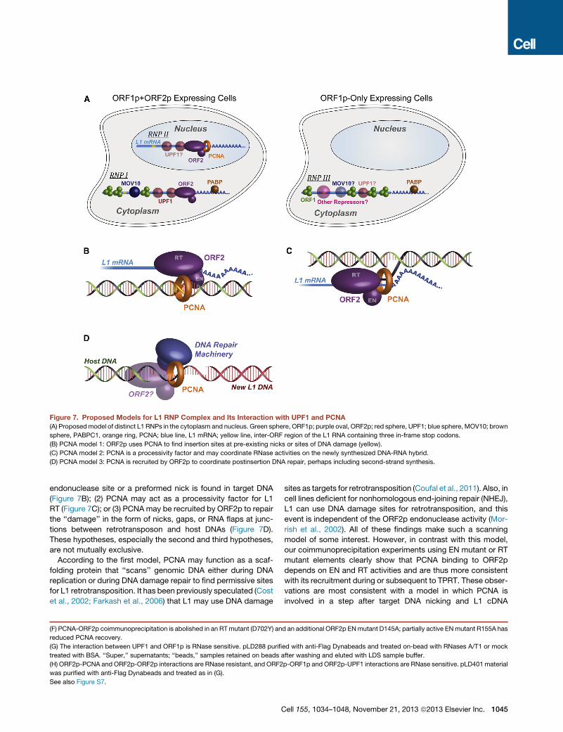
Figure 7. Proposed Models for L1 RNP Complex and Its Interaction with UPF1 and PCNA(A) Proposedmodel of distinct L1 RNPs in the cytoplasm and nucleus. Green sphere, ORF1p; purple oval, ORF2p; red sphere, UPF1; blue sphere, MOV10; brown
sphere, PABPC1, orange ring, PCNA; blue line, L1 mRNA; yellow line, inter-ORF region of the L1 RNA containing three in-frame stop codons.
(B) PCNA model 1: ORF2p uses PCNA to find insertion sites at pre-existing nicks or sites of DNA damage (yellow).
(C) PCNA model 2: PCNA is a processivity factor and may coordinate RNase activities on the newly synthesized DNA-RNA hybrid.
(D) PCNA model 3: PCNA is recruited by ORF2p to coordinate postinsertion DNA repair, perhaps including second-strand synthesis.
endonuclease site or a preformed nick is found in target DNA
(Figure 7B); (2) PCNA may act as a processivity factor for L1
RT (Figure 7C); or (3) PCNAmay be recruited by ORF2p to repair
the ‘‘damage’’ in the form of nicks, gaps, or RNA flaps at junc-
tions between retrotransposon and host DNAs (Figure 7D).
These hypotheses, especially the second and third hypotheses,
are not mutually exclusive.
According to the first model, PCNA may function as a scaf-
folding protein that ‘‘scans’’ genomic DNA either during DNA
replication or during DNA damage repair to find permissive sites
for L1 retrotransposition. It has been previously speculated (Cost
et al., 2002; Farkash et al., 2006) that L1 may use DNA damage
(F) PCNA-ORF2p coimmunoprecipitation is abolished in an RTmutant (D702Y) an
reduced PCNA recovery.
(G) The interaction between UPF1 and ORF1p is RNase sensitive. pLD288 purifi
treated with BSA. ‘‘Super,’’ supernatants; ‘‘beads,’’ samples retained on beads a
(H) ORF2p-PCNA and ORF2p-ORF2p interactions are RNase resistant, and ORF2
was purified with anti-Flag Dynabeads and treated as in (G).
See also Figure S7.
C
sites as targets for retrotransposition (Coufal et al., 2011). Also, in
cell lines deficient for nonhomologous end-joining repair (NHEJ),
L1 can use DNA damage sites for retrotransposition, and this
event is independent of the ORF2p endonuclease activity (Mor-
rish et al., 2002). All of these findings make such a scanning
model of some interest. However, in contrast with this model,
our coimmunoprecipitation experiments using EN mutant or RT
mutant elements clearly show that PCNA binding to ORF2p
depends on EN and RT activities and are thus more consistent
with its recruitment during or subsequent to TPRT. These obser-
vations are most consistent with a model in which PCNA is
involved in a step after target DNA nicking and L1 cDNA
d an additional ORF2p ENmutant D145A; partially active ENmutant R155A has
ed with anti-Flag Dynabeads and treated on-bead with RNases A/T1 or mock
fter washing and eluted with LDS sample buffer.
p-ORF1p and ORF2p-UPF1 interactions are RNase sensitive. pLD401 material
ell 155, 1034–1048, November 21, 2013 ª2013 Elsevier Inc. 1045
synthesis. All EN mutants maintained high RT activity, and
several of them have crystal structures closely resembling the
wild-type enzyme, minimizing the possibility that EN mutations
disrupt overall folding of ORF2p and thereby the physical inter-
action between ORF2p and PCNA.
Further, PCNA is known to be capable of recruiting repair
enzymes like lesion bypass polymerases and ligases (Beattie
and Bell, 2011; Ulrich, 2011) and, most interestingly, has been
specifically implicated in recruiting RNase H2 to RNA/DNA
hybrids in genomic DNA (Bubeck et al., 2011). Mammalian L1
elements do not encode a recognizable RNase H activity, unlike
most other retrotransposons (including non-LTR elements like
L1 in other organisms such as the Drosophila I factor), even
though the activity is presumably required to degrade the original
RNA strand, allowing second DNA strand synthesis. Thus,
RNase H binding activity of PCNA would be highly consistent
with a role in DNA elongation during reverse transcription and
provide a convenient link to the RNase H domain of RT that is
‘‘missing’’ from mammalian L1 elements. Although this model
may be appealing, the lack of interaction with the RT mutant
presents a challenge to it; however, it is formally possible that
a few base pairs of synthesis are required to enable PCNA
engagement.
Other copurified factors may be associated with nuclear L1
RNPs through PCNA or in relation to PCNA. PURA, PURB,
MEPCE, and PCNA have been shown to be associated with
PCNA clamp-loaders RFC1–5 (Havugimana et al., 2012; Kubota
et al., 2013). Similarly, RUVBL1 are copurified ring-shaped pro-
teins implicated in DNA damage response (Jha and Dutta,
2009; Rosenbaum et al., 2013). PARP1 may provide a link be-
tween nuclear and mitochondrial-derived interactors of ORF2p
via DNA damage repair and apoptosis (Hong et al., 2004), and
interestingly, PARP1 modifies both PCNA and TOP1, which are
both high-confidence interactors of ORF2p (Simbulan-Rosen-
thal et al., 1999). We did not recover any APOBEC3 family cyti-
dine deaminases, which are known inhibitors of LINE1 activity
with mixed nuclear and cytoplasmic localizations (Bogerd
et al., 2006); however, these proteins may not be expressed
highly (or at all) in HEK293T cells, and this represents a general
limitation of this study. Knockdown of endogenous PARP1 and
RUVBL1 resulted in reduced retrotransposition, suggesting
additional roles for other enzymes involved in generic DNA repair
in the retrotransposition pathway.
Distinct RNPs at Different Life Cycle Stages?Our data are consistent with isolation of L1 RNPs of at least three
distinct types, (Figure 7A). First, some RNPs purified using
ORF1p are likely isolated from cells not expressing ORF2p,
perhaps a state in which the host has defeated L1 by preventing
ORF2p expression (RNP III). Focusing on the tandem pullout
(Figure 5A), we identify two additional distinct populations:
(1) the elution, containing all detectable ORF1p, the majority of
UPF1, and only a trace amount of PCNA (RNP I) and (2) the
supernatant depleted for ORF1p but containing the vast majority
of the PCNA and a minority—but nevertheless substantial
amount—of UPF1 (RNP II).
Taken together with the idea that PCNA may be tightly and
specifically bound to ORF2p only after initiation of TPRT, this
1046 Cell 155, 1034–1048, November 21, 2013 ª2013 Elsevier Inc.
suggests that these two populations of L1 RNPs, while both re-
taining ORF2p, interact with ORF1p, PCNA, and UPF1 in distinct
ways—RNP I is the tandem-purified RNP, present in the cyto-
plasm and primarily containing ORF1p/ORF2p/UPF1 in which
UPF1 is active in a negative regulatory capacity, and RNP II is
a distinct complex in nuclei, primarily containing ORF2p/PCNA
with lesser amounts of UPF1 and seemingly lacking ORF1p.
This suggests that ORF1p may be absent in steps subsequent
to EN activity. Interestingly, UPF1 has been linked to Pol d in
DNA replication/repair (Azzalin and Lingner, 2006) and may
play a second role here as a permissive host factor; this could
explain the unexpectedly reduced retrotransposition in the
context of UPF1 knockdown.
EXPERIMENTAL PROCEDURES
Materials and experimental procedures are detailed in the Extended Experi-
mental Procedures.
SUPPLEMENTAL INFORMATION
Supplemental Information includes Extended Experimental Procedures, seven
figures, and seven tables and can be found with this article online at http://dx.
doi.org/10.1016/j.cell.2013.10.021.
ACKNOWLEDGMENTS
We thank Jeffrey Han for gifts of antibodies and helpful discussions; Phil Cole
and CarolynMachamer for helpful discussion and critical reading of the manu-
script; Jennifer Wang, Chih-Yung Lee, and Geraldine Seydoux for assistance
with confocal microscopy and helpful discussions; and Yana Li, Dan Leahy,
Jennifer Kavran, and Jacqueline McCabe for help with suspension cell culture.
The DNA template for artwork in Figure 7 was adapted from http://www.
dragonartz.net with permission. This work was supported in part by NIH grant
U54 GM103511 to M.P.R. and B.T.C., grants R01 GM36481 and U54
GM103520 to J.D.B., and grant P41 GM103314 to B.T.C.
Received: May 13, 2013
Revised: August 25, 2013
Accepted: September 30, 2013
Published: November 21, 2013
REFERENCES
Alisch, R.S., Garcia-Perez, J.L., Muotri, A.R., Gage, F.H., and Moran, J.V.
(2006). Unconventional translation of mammalian LINE-1 retrotransposons.
Genes Dev. 20, 210–224.
An, W., Dai, L., Niewiadomska, A.M., Yetil, A., O’Donnell, K.A., Han, J.S., and
Boeke, J.D. (2011). Characterization of a synthetic human LINE-1 retrotrans-
poson ORFeus-Hs. Mob. DNA 2, 2.
Arjan-Odedra, S., Swanson, C.M., Sherer, N.M., Wolinsky, S.M., and Malim,
M.H. (2012). Endogenous MOV10 inhibits the retrotransposition of endoge-
nous retroelements but not the replication of exogenous retroviruses. Retrovir-
ology 9, 53.
Athanikar, J.N., Badge, R.M., and Moran, J.V. (2004). A YY1-binding site is
required for accurate human LINE-1 transcription initiation. Nucleic Acids
Res. 32, 3846–3855.
Azzalin, C.M., and Lingner, J. (2006). The human RNA surveillance factor UPF1
is required for S phase progression and genome stability. Curr. Biol. 16,
433–439.
Beattie, T.R., and Bell, S.D. (2011). The role of the DNA sliding clamp in
Okazaki fragment maturation in archaea and eukaryotes. Biochem. Soc.
Trans. 39, 70–76.
Beauregard, A., Curcio, M.J., and Belfort, M. (2008). The take and give
between retrotransposable elements and their hosts. Annu. Rev. Genet. 42,
587–617.
Beck, C.R., Garcia-Perez, J.L., Badge, R.M., and Moran, J.V. (2011). LINE-1
elements in structural variation and disease. Annu. Rev. Genomics Hum.
Genet. 12, 187–215.
Belancio, V.P., Whelton, M., and Deininger, P. (2007). Requirements for poly-
adenylation at the 30 end of LINE-1 elements. Gene 390, 98–107.
Bogerd, H.P., Wiegand, H.L., Hulme, A.E., Garcia-Perez, J.L., O’Shea, K.S.,
Moran, J.V., and Cullen, B.R. (2006). Cellular inhibitors of long interspersed
element 1 and Alu retrotransposition. Proc. Natl. Acad. Sci. USA 103, 8780–
8785.
Brouha, B., Schustak, J., Badge, R.M., Lutz-Prigge, S., Farley, A.H., Moran,
J.V., and Kazazian, H.H., Jr. (2003). Hot L1s account for the bulk of retrotrans-
position in the human population. Proc. Natl. Acad. Sci. USA 100, 5280–5285.
Bubeck, D., Reijns,M.A., Graham, S.C., Astell, K.R., Jones, E.Y., and Jackson,
A.P. (2011). PCNA directs type 2 RNase H activity on DNA replication and
repair substrates. Nucleic Acids Res. 39, 3652–3666.
Burns, K.H., and Boeke, J.D. (2012). Human transposon tectonics. Cell 149,
740–752.
Buzdin, A., Ustyugova, S., Gogvadze, E., Vinogradova, T., Lebedev, Y., and
Sverdlov, E. (2002). A new family of chimeric retrotranscripts formed by a full
copy of U6 small nuclear RNA fused to the 30 terminus of l1. Genomics 80,
402–406.
Cost, G.J., Feng, Q., Jacquier, A., and Boeke, J.D. (2002). Human L1 element
target-primed reverse transcription in vitro. EMBO J. 21, 5899–5910.
Coufal, N.G., Garcia-Perez, J.L., Peng, G.E., Marchetto, M.C., Muotri, A.R.,
Mu, Y., Carson, C.T., Macia, A., Moran, J.V., and Gage, F.H. (2011). Ataxia
telangiectasia mutated (ATM) modulates long interspersed element-1 (L1) ret-
rotransposition in human neural stem cells. Proc. Natl. Acad. Sci. USA 108,
20382–20387.
Cristea, I.M., Williams, R., Chait, B.T., and Rout, M.P. (2005). Fluorescent pro-
teins as proteomic probes. Mol. Cell. Proteomics 4, 1933–1941.
Dai, L., Taylor, M.S., O’Donnell, K.A., and Boeke, J.D. (2012). Poly(A) binding
protein C1 is essential for efficient L1 retrotransposition and affects L1 RNP
formation. Mol. Cell. Biol. 32, 4323–4336.
Di Virgilio, M., Callen, E., Yamane, A., Zhang, W., Jankovic, M., Gitlin, A.D.,
Feldhahn, N., Resch, W., Oliveira, T.Y., Chait, B.T., et al. (2013). Rif1 prevents
resection of DNA breaks and promotes immunoglobulin class switching.
Science 339, 711–715.
Domanski, M., Molloy, K., Jiang, H., Chait, B.T., Rout, M.P., Jensen, T.H., and
LaCava, J. (2012). Improved methodology for the affinity isolation of
human protein complexes expressed at near endogenous levels. Bio-
techniques 0, 1–6.
Doucet, A.J., Hulme, A.E., Sahinovic, E., Kulpa, D.A., Moldovan, J.B., Kopera,
H.C., Athanikar, J.N., Hasnaoui, M., Bucheton, A., Moran, J.V., and Gilbert, N.
(2010). Characterization of LINE-1 ribonucleoprotein particles. PLoS Genet. 6,
e1001150.
Farkash, E.A., Kao, G.D., Horman, S.R., and Prak, E.T. (2006). Gamma radia-
tion increases endonuclease-dependent L1 retrotransposition in a cultured
cell assay. Nucleic Acids Res. 34, 1196–1204.
Feng, Q., Moran, J.V., Kazazian, H.H., Jr., and Boeke, J.D. (1996). Human L1
retrotransposon encodes a conserved endonuclease required for retrotrans-
position. Cell 87, 905–916.
Fuchs, G., Stein, A.J., Fu, C., Reinisch, K.M., and Wolin, S.L. (2006). Structural
and biochemical basis for misfolded RNA recognition by the Ro autoantigen.
Nat. Struct. Mol. Biol. 13, 1002–1009.
Goodier, J.L., and Kazazian, H.H., Jr. (2008). Retrotransposons revisited: the
restraint and rehabilitation of parasites. Cell 135, 23–35.
Goodier, J.L., Zhang, L., Vetter, M.R., and Kazazian, H.H., Jr. (2007). LINE-1
ORF1 protein localizes in stress granules with other RNA-binding proteins,
including components of RNA interference RNA-induced silencing complex.
Mol. Cell. Biol. 27, 6469–6483.
C
Goodier, J.L., Mandal, P.K., Zhang, L., and Kazazian, H.H., Jr. (2010). Discrete
subcellular partitioning of human retrotransposon RNAs despite a common
mechanism of genome insertion. Hum. Mol. Genet. 19, 1712–1725.
Goodier, J.L., Cheung, L.E., and Kazazian, H.H., Jr. (2012). MOV10 RNA
helicase is a potent inhibitor of retrotransposition in cells. PLoS Genet. 8,
e1002941.
Goodier, J.L., Cheung, L.E., and Kazazian, H.H., Jr. (2013). Mapping the LINE1
ORF1 protein interactome reveals associated inhibitors of human retrotrans-
position. Nucleic Acids Res. 41, 7401–7419.
Han, J.S., and Boeke, J.D. (2004). A highly active synthetic mammalian retro-
transposon. Nature 429, 314–318.
Hata, K., and Sakaki, Y. (1997). Identification of critical CpG sites for repres-
sion of L1 transcription by DNA methylation. Gene 189, 227–234.
Havugimana, P.C., Hart, G.T., Nepusz, T., Yang, H., Turinsky, A.L., Li, Z.,
Wang, P.I., Boutz, D.R., Fong, V., Phanse, S., et al. (2012). A census of human
soluble protein complexes. Cell 150, 1068–1081.
Hernan, R., Heuermann, K., and Brizzard, B. (2000). Multiple epitope tagging of
expressed proteins for enhanced detection. Biotechniques 28, 789–793.
Hogg, J.R., and Goff, S.P. (2010). Upf1 senses 3’UTR length to potentiate
mRNA decay. Cell 143, 379–389.
Hong, S.J., Dawson, T.M., and Dawson, V.L. (2004). Nuclear and mitochon-
drial conversations in cell death: PARP-1 and AIF signaling. Trends Pharmacol.
Sci. 25, 259–264.
Jha, S., and Dutta, A. (2009). RVB1/RVB2: running rings around molecular
biology. Mol. Cell 34, 521–533.
Khan, H., Smit, A., and Boissinot, S. (2006). Molecular evolution and tempo of
amplification of human LINE-1 retrotransposons since the origin of primates.
Genome Res. 16, 78–87.
Khazina, E., Truffault, V., Buttner, R., Schmidt, S., Coles, M., and Weichen-
rieder, O. (2011). Trimeric structure and flexibility of the L1ORF1 protein in
human L1 retrotransposition. Nat. Struct. Mol. Biol. 18, 1006–1014.
Kimberland, M.L., Divoky, V., Prchal, J., Schwahn, U., Berger, W., and Kaza-
zian, H.H., Jr. (1999). Full-length human L1 insertions retain the capacity for
high frequency retrotransposition in cultured cells. Hum. Mol. Genet. 8,
1557–1560.
Kubota, T., Nishimura, K., Kanemaki, M.T., and Donaldson, A.D. (2013). The
Elg1 replication factor C-like complex functions in PCNA unloading during
DNA replication. Mol. Cell 50, 273–280.
Kulpa, D.A., and Moran, J.V. (2005). Ribonucleoprotein particle formation is
necessary but not sufficient for LINE-1 retrotransposition. Hum. Mol. Genet.
14, 3237–3248.
Kulpa, D.A., and Moran, J.V. (2006). Cis-preferential LINE-1 reverse transcrip-
tase activity in ribonucleoprotein particles. Nat. Struct. Mol. Biol. 13, 655–660.
Lee, D.J., Busby, S.J.W., Westblade, L.F., and Chait, B.T. (2008). Affinity isola-
tion and I-DIRT mass spectrometric analysis of the Escherichia coli O157:H7
Sakai RNA polymerase complex. J. Bacteriol. 190, 1284–1289.
Luan, D.D., and Eickbush, T.H. (1995). RNA template requirements for target
DNA-primed reverse transcription by the R2 retrotransposable element. Mol.
Cell. Biol. 15, 3882–3891.
Malik, H.S., Burke, W.D., and Eickbush, T.H. (1999). The age and evolution of
non-LTR retrotransposable elements. Mol. Biol. Evol. 16, 793–805.
Mandal, P.K., Ewing, A.D., Hancks, D.C., and Kazazian, H.H., Jr. (2013).
Enrichment of processed pseudogene transcripts in L1-ribonucleoprotein
particles. Hum. Mol. Genet. 22, 3730–3748.
Martin, S.L., and Bushman, F.D. (2001). Nucleic acid chaperone activity of the
ORF1 protein from the mouse LINE-1 retrotransposon. Mol. Cell. Biol. 21,
467–475.
Mathias, S.L., Scott, A.F., Kazazian, H.H., Jr., Boeke, J.D., and Gabriel, A.
(1991). Reverse transcriptase encoded by a human transposable element.
Science 254, 1808–1810.
ell 155, 1034–1048, November 21, 2013 ª2013 Elsevier Inc. 1047
Mendell, J.T., Sharifi, N.A., Meyers, J.L., Martinez-Murillo, F., and Dietz, H.C.
(2004). Nonsense surveillance regulates expression of diverse classes of
mammalian transcripts and mutes genomic noise. Nat. Genet. 36, 1073–1078.
Moran, J.V., Holmes, S.E., Naas, T.P., DeBerardinis, R.J., Boeke, J.D., and
Kazazian, H.H., Jr. (1996). High frequency retrotransposition in cultured
mammalian cells. Cell 87, 917–927.
Morrish, T.A., Gilbert, N., Myers, J.S., Vincent, B.J., Stamato, T.D., Taccioli,
G.E., Batzer, M.A., and Moran, J.V. (2002). DNA repair mediated by endonu-
clease-independent LINE-1 retrotransposition. Nat. Genet. 31, 159–165.
Muotri, A.R., Chu, V.T., Marchetto, M.C., Deng, W., Moran, J.V., and Gage,
F.H. (2005). Somatic mosaicism in neuronal precursor cells mediated by L1
retrotransposition. Nature 435, 903–910.
Niewiadomska, A.M., Tian, C., Tan, L., Wang, T., Sarkis, P.T., and Yu, X.F.
(2007). Differential inhibition of long interspersed element 1 by APOBEC3
does not correlate with high-molecular-mass-complex formation or P-body
association. J. Virol. 81, 9577–9583.
O’Donnell, K.A., An, W., Schrum, C.T., Wheelan, S.J., and Boeke, J.D. (2013).
Controlled insertional mutagenesis using a LINE-1 (ORFeus) gene-trap mouse
model. Proc. Natl. Acad. Sci. USA 110, E2706–E2713.
Oeffinger, M., Wei, K.E., Rogers, R., DeGrasse, J.A., Chait, B.T., Aitchison,
J.D., and Rout, M.P. (2007). Comprehensive analysis of diverse ribonucleopro-
tein complexes. Nat. Methods 4, 951–956.
Ostertag, E.M., and Kazazian, H.H., Jr. (2001). Biology of mammalian L1 retro-
transposons. Annu. Rev. Genet. 35, 501–538.
Peddigari, S., Li, P.W., Rabe, J.L., and Martin, S.L. (2013). hnRNPL and nucle-
olin bind LINE-1 RNA and function as host factors to modulate retrotransposi-
tion. Nucleic Acids Res. 41, 575–585.
Repanas, K., Zingler, N., Layer, L.E., Schumann, G.G., Perrakis, A., and Wei-
chenrieder, O. (2007). Determinants for DNA target structure selectivity of the
human LINE-1 retrotransposon endonuclease. Nucleic Acids Res. 35, 4914–
4926.
Repanas, K., Fuentes, G., Cohen, S.X., Bonvin, A.M., and Perrakis, A. (2009).
Insights into the DNA cleavage mechanism of human LINE-1 retrotransposon
endonuclease. Proteins 74, 917–928.
Rosenbaum, J., Baek, S.H., Dutta, A., Houry, W.A., Huber, O., Hupp, T.R., and
Matias, P.M. (2013). The emergence of the conserved AAA+ ATPases Pontin
and Reptin on the signaling landscape. Sci. Signal. 6, mr1.
Schwanhausser, B., Busse, D., Li, N., Dittmar, G., Schuchhardt, J., Wolf, J.,
Chen, W., and Selbach, M. (2011). Global quantification of mammalian gene
expression control. Nature 473, 337–342.
1048 Cell 155, 1034–1048, November 21, 2013 ª2013 Elsevier Inc.
Shigeoka, T., Kato, S., Kawaichi, M., and Ishida, Y. (2012). Evidence that the
Upf1-related molecular motor scans the 30-UTR to ensure mRNA integrity.
Nucleic Acids Res. 40, 6887–6897.
Shukla, R., Upton, K.R., Munoz-Lopez, M., Gerhardt, D.J., Fisher, M.E.,
Nguyen, T., Brennan, P.M., Baillie, J.K., Collino, A., Ghisletti, S., et al. (2013).
Endogenous retrotransposition activates oncogenic pathways in hepatocellu-
lar carcinoma. Cell 153, 101–111.
Simbulan-Rosenthal, C.M., Rosenthal, D.S., Iyer, S., Boulares, H., and Smul-
son, M.E. (1999). Involvement of PARP and poly(ADP-ribosyl)ation in the early
stages of apoptosis and DNA replication. Mol. Cell. Biochem. 193, 137–148.
Smits, A.H., Jansen, P.W.T.C., Poser, I., Hyman, A.A., and Vermeulen, M.
(2013). Stoichiometry of chromatin-associated protein complexes revealed
by label-free quantitative mass spectrometry-based proteomics. Nucleic
Acids Res. 41, e28.
Soifer, H.S., Zaragoza, A., Peyvan, M., Behlke, M.A., and Rossi, J.J. (2005).
A potential role for RNA interference in controlling the activity of the human
LINE-1 retrotransposon. Nucleic Acids Res. 33, 846–856.
Suzuki, J., Yamaguchi, K., Kajikawa, M., Ichiyanagi, K., Adachi, N., Koyama,
H., Takeda, S., and Okada, N. (2009). Genetic evidence that the non-homolo-
gous end-joining repair pathway is involved in LINE retrotransposition. PLoS
Genet. 5, e1000461.
Tackett, A.J., DeGrasse, J.A., Sekedat, M.D., Oeffinger, M., Rout, M.P., and
Chait, B.T. (2005). I-DIRT, a general method for distinguishing between spe-
cific and nonspecific protein interactions. J. Proteome Res. 4, 1752–1756.
Tani, H., Imamachi, N., Salam, K.A., Mizutani, R., Ijiri, K., Irie, T., Yada, T.,
Suzuki, Y., and Akimitsu, N. (2012). Identification of hundreds of novel UPF1
target transcripts by direct determination of whole transcriptome stability.
RNA Biol. 9, 1370–1379.
Ulrich, H.D. (2011). Timing and spacing of ubiquitin-dependent DNA damage
bypass. FEBS Lett. 585, 2861–2867.
Wei, W., Gilbert, N., Ooi, S.L., Lawler, J.F., Ostertag, E.M., Kazazian, H.H.,
Boeke, J.D., and Moran, J.V. (2001). Human L1 retrotransposition: cis prefer-
ence versus trans complementation. Mol. Cell. Biol. 21, 1429–1439.
Weichenrieder, O., Repanas, K., and Perrakis, A. (2004). Crystal structure of
the targeting endonuclease of the human LINE-1 retrotransposon. Structure
12, 975–986.
Yang, N., and Kazazian, H.H., Jr. (2006). L1 retrotransposition is suppressed
by endogenously encoded small interfering RNAs in human cultured cells.
Nat. Struct. Mol. Biol. 13, 763–771.