Affective Computing and...
26
Affective Computing and Interaction: Psychological, Cognitive and Neuroscientific Perspectives Didem Gökçay Middle East Technical University, Turkey Gülsen Yildirim Middle East Technical University, Turkey Hershey • New York InformatIon scIence reference
Transcript of Affective Computing and...

Affective Computing and Interaction:Psychological, Cognitive and Neuroscientific Perspectives
Didem GökçayMiddle East Technical University, Turkey
Gülsen YildirimMiddle East Technical University, Turkey
Hershey • New YorkInformatIon scIence reference

Director of Editorial Content: Kristin KlingerDirector of Book Publications: Julia MosemannAcquisitions Editor: Lindsay JohnstonDevelopment Editor: Dave DeRiccoPublishing Assistant: Milan Vracarich Jr.Typesetter: Milan Vracarich Jr., Casey ConapitskiProduction Editor: Jamie SnavelyCover Design: Lisa Tosheff
Published in the United States of America by Information Science Reference (an imprint of IGI Global)701 E. Chocolate AvenueHershey PA 17033Tel: 717-533-8845Fax: 717-533-8661E-mail: [email protected] site: http://www.igi-global.com
Copyright © 2011 by IGI Global. All rights reserved. No part of this publication may be reproduced, stored or distributed in any form or by any means, electronic or mechanical, including photocopying, without written permission from the publisher.Product or company names used in this set are for identification purposes only. Inclusion of the names of the products or com-panies does not indicate a claim of ownership by IGI Global of the trademark or registered trademark.
Library of Congress Cataloging-in-Publication Data
Affective computing and interaction : psychological, cognitive, and neuroscientific perspectives / Didem Gokcay and Gulsen Yildirim, editors. p. cm. Includes bibliographical references and index. ISBN 978-1-61692-892-6 (hardcover) -- ISBN 978-1-61692-894-0 (ebook) 1. Human-computer interaction. 2. Human-machine systems. 3. Affect (Psychology)--Computer simulation. I. Gokcay, Didem, 1966- II. Yildirim, Gulsen, 1978- QA76.9.H85A485 2011 004.01'9--dc22 2010041639
British Cataloguing in Publication DataA Cataloguing in Publication record for this book is available from the British Library.
All work contributed to this book is new, previously-unpublished material. The views expressed in this book are those of the authors, but not necessarily of the publisher.

1
Copyright © 2011, IGI Global. Copying or distributing in print or electronic forms without written permission of IGI Global is prohibited.
DOI: 10.4018/978-1-61692-892-6.ch001
Chapter 1
Neurophysiology of EmotionsAyşen Erdem
Hacettepe University, Turkey
Serkan KaraismailoğluHacettepe University, Turkey
INTRODUCTION
Complex organisms must not only perceive and evaluate changes in their internal and external en-vironment, but also generate appropriate responses to survive. In general, emotions help an organism to survive by embodying a multitude of nervous system functions for perception of the valuable components of the external environment. At the
same time, emotions are a way for directly com-municating our internal world to others through non-verbal means (Öztürk et al., 2005). Emotions aid an individual to interact with his/her external –or more specifically social- environment in a flexible way, while also helping in the regulation of an individual’s internal world.
Emotional information is used for personal, as well as social decision making of an individual with
AbsTRACT
Emotions embody goal-directed behavior for survival and adaptation through the perception of varia-tions in the environment. At a physiological level, emotions consist of three complementary components: Physical sensation, emotional expression and subjective experience. At the level of anatomical structures though, trying to segregate distinct components is impossible. Our emotions are resulting products of compatible and coordinated cortical and sub-cortical neural mechanisms originating from several ana-tomical structures. In this chapter, an overview of the three physiological components and underlying anatomical constructs will be presented.

2
Neurophysiology of Emotions
respect to the surrounding dynamical processes. Briefly, we can say that an emotion is an evaluative response. These responses are physiological and behavioral impulses that prepare the organisms for escaping or approaching dangerous versus safe objects. Actually, emotions consist of not a single but a multitude of responses in presence of a stimulus (Öztürk et al., 2005).
In summary, we can say that ‘emotions may be characterized as a response to an environmen-tal event that allow for goal-directed behavior in the adaptation of the organism to changing environmental demands. This response involves cognitive, affective, behavioral, and autonomic sub-systems’ (Oatley & Jenkins, 1996; Hagemann et al., 2003). At a physiological level, most of the recent research in affective neuroscience strive to elucidate the prominent components of neural networks of emotion. When considered from this perspective, there are three distinct but comple-mentary components of emotions (Bownds, 1999):
1. Physical sensation: Consists of the compo-nents within the autonomic nervous system which we call physiological arousal.
2. Emotional expression: Consists of our be-haviours such as facial expressions, gestures and posture for reflecting our feelings such as sadness, anger, happiness.
3. Subjective experience: Consists of the personal feeling of our current emotion such as fear, anger, and happiness.
It is extremely difficult to identify all emotional states that we go through. Because of this, emotions can be simply categorized as basic (primary) and social (secondary). The basic emotions are innate and related to the anatomical structures in the limbic system (for ex. Amygdala). They consist of organized automatic and stereotypical behaviours (Izard, 2009), that are consistent across cultures. These basic emotions are essential for survival, evolution and development. There are six basic emotions that are widely accepted: happiness,
sadness, anger, fear, disgust, and surprise. These emotions can further be grouped as pleasant (hap-piness, surprise) and unpleasant (sadness, anger, fear, disgust). The pleasant emotions consist of positive affect causing approach to stimuli whereas the unpleasant emotions consist of negative af-fect that is repellant (Izard, 2009). On the other hand, social emotions differ from the basic emo-tions primarily because they are learned through social interaction as the individual grows. Social emotions embody personal experiences such as guilt, embarrassment/shame, and pride (Damasio, 1994). Other than these, it is also necessary to consider background emotional processes such as mood, which are elicited in a totally different way.
PHYsICAL sENsATION AND AUTONOMIC NERVOUs sYsTEM
When we investigate areas that are responsible of the complementary components of emotions, we would have to say that the hypothalamus, brain stem, and medulla spinalis are specifically responsible from the physical sensations which include physiologic components of the autonomic nervous system (see Figure 1).
Hypothalamus and the Autonomic Nervous system
Although small in physical size, hypothalamus is an extremely important structure. It acts as a relay station conjoining incoming signals from a lot of different areas. It consists of different cell groups and its basic responsibility is in the ho-meostatic ogranization of the internal environment (maintenance of internal stability). Hypothalamus controls most of the endocrinal, vegetative and emotional behaviour of the body. Several nuclei contained within the hypothalamus play differ-ent roles: For example the excitation of lateral and posterior areas increases blood pressure and heart rate while the excitation of the preoptic area

3
Neurophysiology of Emotions
reduces these. Similarly, while the frontal areas activate mechanisms responsible from decrease of the body temperature, the posterior areas activate heat preserving mechanisms. The side areas of hypothalamus are known as the centers for thirst and hunger. The area close to the medial part of the hypothalamus serves as the satiety center. In addition, hypothalamus controls excretion of water from kidneys, and birth through the release of oxytocine hormone (Guyton & Hall, 2006).
With the inclusion of a multitude of centers in the brain stem and medulla spinalis, the hy-pothalamus is incharge of the activation of the autonomic nervous system and the regulation of reflex responses. Under any emotional stimulus, the signals incoming to the hypothalamus induce and effect on the autonomic nervous system. What is the importance of the autonomic nervous system then? What is its duty?
As mentioned earlier, the autonomic nervous system maintains the homeostasis of our internal envionment. It works without our conscious will. There are two main parts of the autonomic nervous system: sympathetic and parasympathetic. Other than a few exceptions, these two systems coun-teract. While one system increases activity in and organ, the other supresses it. The parasympathetic
system is active under resting or peaceful condi-tions, maintains energy storage and preservation. Its effects are local. Sympathetic system on the other hand, is also named as the system for ‘fight or flight’. It prepares the individual for a fleeing or fighting response.
What are the main effects imposed by the sym-pathetic system? In case of danger, our heart rate and blood pressure increases, because blood and oxygen consumption by our vital organs has in-creased. Respiratory rate and depth also increases to satisfy the increased oxygen demand. Bronchi in the respiratory tract widen. On the contrary, the digestive tract’s blood demands decrease. The digestive functions slow down, or even stop the blood demand in this area. The muscles which provide motor movements in this area are relaxed, prohibiting defecation and micturation. Production of urine slows down. All other secretions such as saliva are decreased. Glucose in the bloodstream increases, because our vital organs and muscles are in need of energy. Our pupils are dilated to be able to immerse more light. Muscle tone increases and reflexes speed up. We became more alert. The effects of the neurotransmitters (adrenalin ve noradrenalin) that induce these changes, especially those that effect the heart, start late and end late.
Figure 1. Main structures those are responsible from the physical sensation of emotion

4
Neurophysiology of Emotions
This is why our heart still keeps pounding for a while even when the danger is over. On the other hand, by the induction of the neurotransmitter acetylcholine, the parasympathetic system induces chages that counteract with these. Heart rate and blood pressure decreases. Respiratory rate de-creases, causing the bronchi to constrict. Digestion and absorption speed up, causing increased blood delivery to this area. Secretions are increased, the pupils constrict. In short, it becomes important to restore the consumed energy (Guyton & Hall, 2006; Hagemann et al., 2003; for an overview, see Lovallo & Sollers, 2000).
EMOTIONAL EXPREssION AND THE LIMbIC sYsTEM
When we say that there are three complementary components in the physiology of emotions, we also have to specify that it is extremely difficult to isolate the underlying areas exclusively ac-cording to the functionality of these components. This is because many areas in the brain have as-sumed responsibility for a multitude of differing tasks. Hypothalamus is an outstanding example for this. Hypothalamus not only effects physical
sensation, but also has many effects on emotional expressions and behaviours. For example, if we stimulate lateral hypothalamus, we cause the ani-mal to exhibit agressive, fighting behaviour. When we remember that this same area is implicated in thirst and hunger related behaviour, we can find an answer to why animals become nervous when they are hungry. On the contrary, when a medial nucleus of the hytpothalamus is activated, the animal becomes relaxed, eats less and feels nutritional satisfaction causing satiety. In a normal animal, the rage feeling is held under control by the inhibiting signals that come from the medial nuclei of hypothalamus (Guyton & Hall, 2006).
Historically, it was Paul Broca who first in-dicated that there is a specific area in the medial aspect of the brain, around corpus callosum and brain stem, which is separated from the surround-ing cortex with clear borders. Broca named this area as the limbic lobe (limbus: border), because of its distinct borders around the brain stem. Limbic lobe embodies the cingulate gyrus, para-hippocampal gyrus and the hippocampal forma-tion consisting of dentate gyrus and subiculum (see Figure 2). Broca thought that this area was prominent for the olfactory sense having its major inputs from the olfactory bulb, without foresee-
Figure 2. Limbic lobe

5
Neurophysiology of Emotions
ing the importance of this region for emotional processes. It was James Papez (Papez, 1937) who coined the first idea about the relationship of the limbic lobe and emotion.
Based on the existing knowledge regarding organization of emotional expressions by hypo-thalamus, identification of emotions by cortex and manipulations of emotions by cognitive pro-cesses, Papez defined a new pathway called the ‘Papez circuit’. According to this, hippocampal formation processes the incoming information from the cingulate gyrus and forwards it to the mammillary body of the hypothalamus through fornix. The projections going through the mam-millary body run through the posterior part of the thalamus and loop back to the cingulate gyrus. In Papez circuit, the interaction between the hypo-thalamus and cingulate gyrus is bidirectional, providing cortical control for the emotional ex-pression (through cingulate to hypothalamus), as well as emotional experience (through hypo-thalamus to cingulate).
The initial definitions of the limbic system started out with the Papez circuit. Due to the consequential similarity between the structures involved in Broca’s limbic lobe and Papez circuit, this region of the brain which is involved in emo-tions is named as the ‘limbic system’. Interestingly, the person who made the limbic system popular was neither Broca nor Papez. By extending the limbic system to include the septal area, nucleus accumbens, amygdala and orbitofrontal cortex, it was the physiologist Paul McLean who accentu-ated the limbic system.
Advancing research showed later that some of the main structures within the Papez circuit such as hippokampus were not actually related to emotions. Papez had included hippocampus in the circuit because rabies cases were characterized by the Rabies virus’ attacks to the hippocampus and the symptoms associated with Rabies were related to emotional changes such as terror and rage. As a matter of fact, Papez was being misled by thinking that hippocampus was coordinating
hypothalamus’ actions; although we now know that it is the amygdala which coordinates the activities of hypothalamus.
Heinrich Kluver and Paul Bucy (1939) con-ducted some experiments on monkeys after the proposal of the Papez circuit for emotions. They observed abnormal behaviour which they later named as ‘Kluver-Bucy syndrome’ in animals that underwent bilateral temporal lobectomy (removal of the temporal lobe in both hemispheres). One of the most important behavioral changes was visual agnosia: although the visual perception was good, visual recognition was mediocre. These animals would put everything within their visual area into their mouth, unlike other animals (Sometimes, similar behaviour is also observed in human patients with temporal lobe lesions). The lobecto-mized monkeys were trying to contract everything around them physically, showed hyperactivity and hypersexuality. In addition, they also exhibited dif-ferences in their emotional behaviour. While they were agressive, wild and fearful towards humans beforehands, they became extremely tame after the surgery. No agressive displays were observed when they ware handled. Furthermore, while normal monkeys became fearful when they saw a snake, their natural predator, the lobectomized ones did not display any signs of fear. In short, the lobectomized monkeys were void of fear and agression. Later research proved that similar sypotoms are observed even when amygdala is removed exclusively. Therefore a rather specific region, amygdala, started to receive all the atten-tion regarding the control of emotional behaviour.
The Amygdala and Its Outputs for Emotional Expression
Amygdala was first identified at the beginning of the 19th century as a structure resembling almond in the medial temporal lobe. Amygdala is a complex structure consisting of approximately 10 nuclei, and it participates in most behavioral functions (LeDoux, 2007). Although the term ‘amygdaloid
6
Neurophysiology of Emotions
complex’ better reflects the complexity of this structure, we will keep using the term amygdala for the remaining parts of this text.
Around the end of 1930’s researchers have noticed that after damage to the amygdala, fear response, as well as feeding and sexual responses are altered (Klüver & Bucy, 1939). Downer (1961) proved the important contribution of amygdala to agressive behaviour through an interesting experiment. He removed the amygdala of Rhesus monkeys unilaterally. At the same time, he dis-connected the two hemispheres by severing the connections that carried information through the corresponding areas of the brain on both sides. During the experiments alternatingly, one eye was closed while the animal was exposed to the sight of natural predators. This way, he was able to form an animal model such that the intact amygdala only receives visual input from the seeing eye which is co-located inside the same hemisphere. According to Downer, the emotional behaviour of the animal should be determined by the seeing eye. When the animal saw the predator with the eye on the same side with the lesioned amygdala, he presented symptoms similar to Kluver-Bucy syndrome. But when the animal saw the preda-tor with the eye on the same side with the intact amygdala, it showed natural emotional behaviour consisting of aggression and fear.
More interestingly, amygdala also takes part in the regulation of acquired (or learned) emo-tional responses. The best example regarding classical fear conditioning response is probably the experiment done by Joseph LeDoux and his colleagues. In this experiment, rats are trained using a specific sound such that after the sound is presented, the animal receives an electrical shock to its foot. The fear response of the animals is recorded in terms of their blood pressure and duration of freezing behavior. While the animals showed no specific response to this sound before training, they showed increased blood pressure and freezing behavior after training. By the help of this paradigm LeDoux (1995; 2000) was able
to isolate the neural circuitry between the sound and fear, which implicated that the the medial geniculate nucleus (MGN) of the Thalamus is the key player. The incoming auditory information is carried from Thalamus to the amygdala through direct connections. Furthermore, when these direct connections are severed, this fear response is no longer observed. Continuing studies of LeDoux showed that amygdala’s nuclei confined to the central area project directly to the reticular nucleus of the mid-brain, which plays an important role in the formation of the freezing response. In a similar way, the projections from the reticular nucleus enter the hypothalamus causing increased blood pressure (LeDoux, 2000).
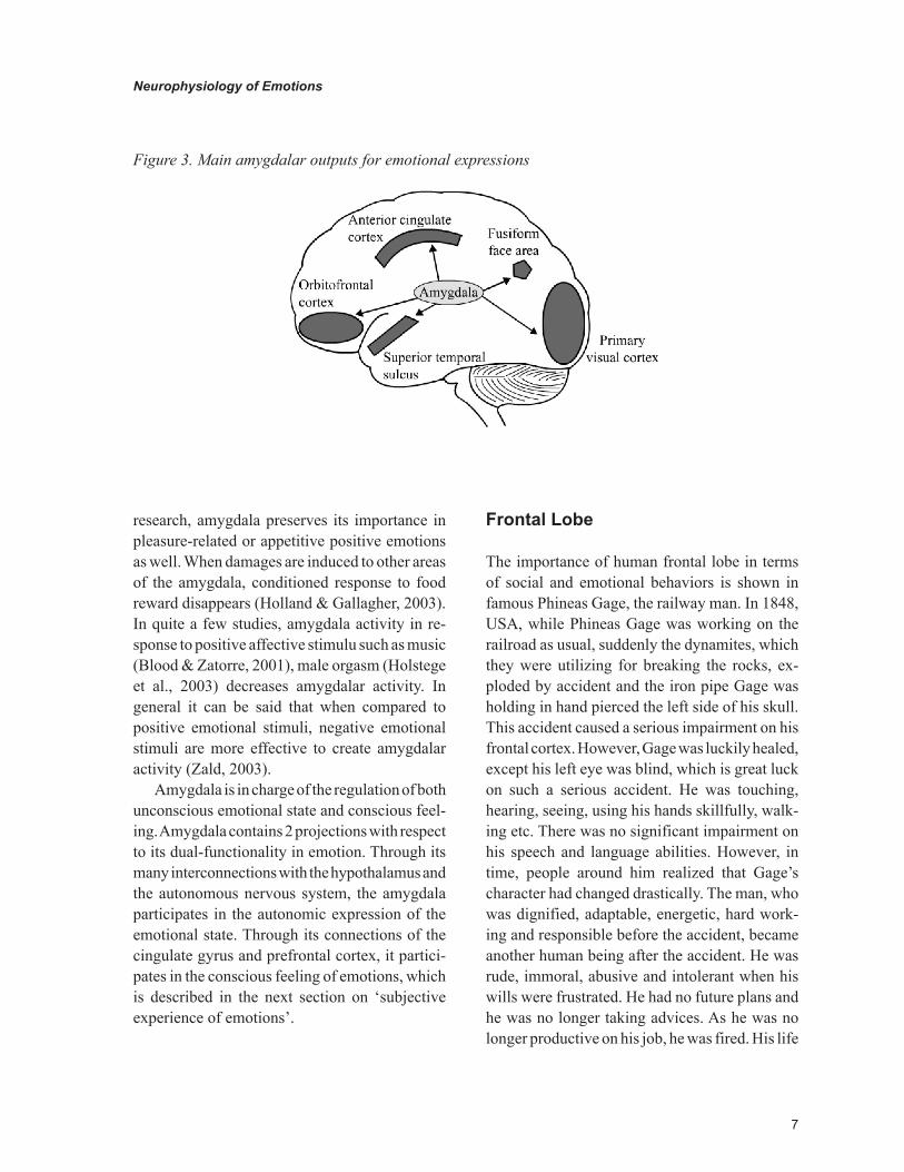
Amygdala exhibits its manipulatory presence in several cognitive and behavioral functions in-cluding attention, perception, and explicit memory. It is a general belief that the amygdala manages the processes in the evaluation of the emotional meaning of an external stimulus, which in turn helps in the organization of cognitive functions. Amygdala exhibits its modulatory role on a wide area in the brain, including STS (superior tempo-ral sulcus) (Amaral et al., 2003), primary visual cortex, fusiform face area, orbitofrontal kortex (e.g., Davidson et al., 1990; Schmidt & Fox, 1999) and anterior cingulate cortex (see Figure 3). Outputs from the amygdala alter the cognitive processes within cortical areas by causing release of hormones and neurotransmitters (please see the appendix for a brief overview of the neu-rotransmitters related to emotions). For example, by modulating the activity of superior temporal sulcus, amygdala may contribute to the perception of motion (Amaral et al., 2003; Chouchourelou et al., 2006): such as perceiving slow movements performed by sad people and fast pacing by angry people (Pollick et al., 2001). Furthermore, explicit memories regarding the emotional situations are enforced through amygdalar outputs leading to the hippocampus (LeDoux, 2007).
Although fear and mechanisms related to fear constitute the most prominent areas in current
7
Neurophysiology of Emotions
research, amygdala preserves its importance in pleasure-related or appetitive positive emotions as well. When damages are induced to other areas of the amygdala, conditioned response to food reward disappears (Holland & Gallagher, 2003). In quite a few studies, amygdala activity in re-sponse to positive affective stimulu such as music (Blood & Zatorre, 2001), male orgasm (Holstege et al., 2003) decreases amygdalar activity. In general it can be said that when compared to positive emotional stimuli, negative emotional stimuli are more effective to create amygdalar activity (Zald, 2003).
Amygdala is in charge of the regulation of both unconscious emotional state and conscious feel-ing. Amygdala contains 2 projections with respect to its dual-functionality in emotion. Through its many interconnections with the hypothalamus and the autonomous nervous system, the amygdala participates in the autonomic expression of the emotional state. Through its connections of the cingulate gyrus and prefrontal cortex, it partici-pates in the conscious feeling of emotions, which is described in the next section on ‘subjective experience of emotions’.
Frontal Lobe
The importance of human frontal lobe in terms of social and emotional behaviors is shown in famous Phineas Gage, the railway man. In 1848, USA, while Phineas Gage was working on the railroad as usual, suddenly the dynamites, which they were utilizing for breaking the rocks, ex-ploded by accident and the iron pipe Gage was holding in hand pierced the left side of his skull. This accident caused a serious impairment on his frontal cortex. However, Gage was luckily healed, except his left eye was blind, which is great luck on such a serious accident. He was touching, hearing, seeing, using his hands skillfully, walk-ing etc. There was no significant impairment on his speech and language abilities. However, in time, people around him realized that Gage’s character had changed drastically. The man, who was dignified, adaptable, energetic, hard work-ing and responsible before the accident, became another human being after the accident. He was rude, immoral, abusive and intolerant when his wills were frustrated. He had no future plans and he was no longer taking advices. As he was no longer productive on his job, he was fired. His life
Figure 3. Main amygdalar outputs for emotional expressions

8
Neurophysiology of Emotions
was ruined more and more and he probably died because of status epilepticus1 eventually.
At first this incident was not assessed as it deserved, since Gage’s abilities such as attention, sensation, memory, language and intelligence were not impaired. The degeneration which arouse in his character and social life did not attract attention at that time, because the most widely accepted view was that if speech, sensation and memory were not impaired then social life and executive func-tions could not be impaired. This conflict could very lately strike the attention of the scientific community. In 1935, John, Fulton and Carlyle reported that chimpanzees were calmed when their frontal cortex is removed (lobotomy) and called this effect as the “calming effect of frontal lobectomy” (Horwitz, 1998). After a few months of this report, a Portuguese neuropsychiatrist, Egas Moinz conducted the first prefrontal lobotomy in humans. In order to cure an emotional illness which accompanies an acute mental illness, he removed the orbital frontal cortex, cutting the limbic association link.
After many years, the researchers, who inquired Gage incident again, displayed that the iron pipe did not harm the regions responsible for motor and language functions, but it did affect both hemisphere’s ventro-medial prefrontal cortex, but especially the left one. Some other incidents got into the literature where frontal lobe is damaged. The fact that these patients showed similar impair-ments to Gage’s, made it clear that especially the prefrontal cortex has a crucial role in planning the future, behaving according to social codes, deciding appropriately in order to survive and acting appropriately.
In recent years, countless studies demonstrated that the prefrontal region is very closely related to positive and negative emotions.
Frontal lobe has an important role on the de-velopment of any kind of spontaneous behaviors, such as facial expression and speech. It is also crucial in controlling the processes necessary to perceiving the emotion from the other lobes (un-
derstanding the facial expressions, in particular) (Lane & Nadel, 2006). Similarly, frontal lobe rectifies the behaviors depending on the internal and external information. If the information is not processed properly, then the behaviors are corrupted. For example, the mammalians’ primary visual cortex sends information both to the visual association area and to the prefrontal cortex and amygdala (Lane & Nadel, 2006). We know that the visual pathways reaching to amygdala have an important role in regulating the fear response. For instance in a study conducted on cats, when encountered with a cat shaped as a “Halloween profile”, the cats respond with similar postures and approach the perineal or head region of the stimulus cat. A piloerection on the back and tail region, slowed respiration, perspiration on the paws and dilatation on the pupil are observed on these cats. This affective response to the Halloween configuration is typical; however no such response is seen when the Picasso cat (a cat profile which does not resemble to a regular cat) is used as stimulus. The cats pay little attention to the Picasso cat. The “Halloween profile” cat threatens the cats, while Picasso cat does not. If there is a lesion in cats’ visual cortex naturally they do not respond to the stimulus (“Halloween profile” cat), because their perception is seriously impaired. The cats with impaired amygdala are oriented to the stimulus and approaches to it; however they do not respond affectively (no piloerection, no autonomic response). The cats with frontal lesions, however, are oriented to the stimulus, but rather than approaching to it, they avoid it (nor an autonomic response).
All of these examples show that the visual pathways reaching amygdala are very crucial for the formation of fear response to the species specific visual stimulus. The visual pathways to the prefrontal cortex, on the other hand, have a role on the formation of the behavior pattern ap-propriate to the species specific typical stimulus. Moreover, the primates, whose amygdala or frontal lobe is impaired, are incompetent in perceiving

9
Neurophysiology of Emotions
the species specific stimuli (e.g. facial expressions or vocalization) (Lane & Nadel, 2006).
All emotions are expressed with stereotypical somatic motor responses (especially the motor movements of facial muscles) and visceral motor system. This necessitates the activity of the central nervous structures which govern the preganglionic autonomic neurons located in the brain stem and the spinal cord. It is interesting that the more the voluntary facial expressions reflect the real emo-tions (e.g. fear, anger, happiness), the more the autonomic response gets stronger. Furthermore, physiological responses may start with the stimuli coming from the forebrain: for instance with odor, music, watching a movie. This neuronal activity that started on the forebrain reaches the visceral and somatic motor nuclei which coordinate the emotional behaviors through hypothalamus and brainstem reticular formation. Among these structures hypothalamus is the central structure which regulates both the visceral and the somatic motor components. Hypothalamus functions like this, through the synapses it forms with reticular formation.
Reticular Formation
Rete means “web” in Latin. In fact, when this formation is examined histologically, it seems as a structure which is located on the inner regions of the brain stem with cells extending to every-where and as a web-like structure formed from those cells’ bundles. The main jobs of reticular formation are visceral and motor control, sensory control and control of consciousness.
The short neurons located in RF have important role on the regulation of stereotypical or coordi-nated movements (e.g. cardiovascular functions, respiration, eye movements, chewing, swallow-ing). RF provides the coordinated activation of autonomic and somatic effector systems which are in charge during these activities. For example, RF is crucial in regulating the movements during the chewing action (the chin gets open and closed
rapidly) in order to prevent biting of the tongue or the cheeks; or in providing the emotional face expression in accordance with the stimuli coming from the superior centers (limbic system, cerebral cortex and cerebellum). Hence, facial expressions are not only started with the voluntary motor activity beginning from the motor cortex on the posterior frontal lobe, but it also starts with the stimuli coming from forebrain regions in order to provide emotional expression.
RF does not only get information from hypo-thalamus, but also from the fibers descending from the limbic system on the forebrain region. Affect-ing the somatic and motor behaviors, these fibers elicit the emotion expression, independent from the motor cortex which sends stimuli in order to carry out voluntary motor movements. Therefore, there are two parallel ways which provide us to accomplish the emotional expressions voluntarily or involuntarily.
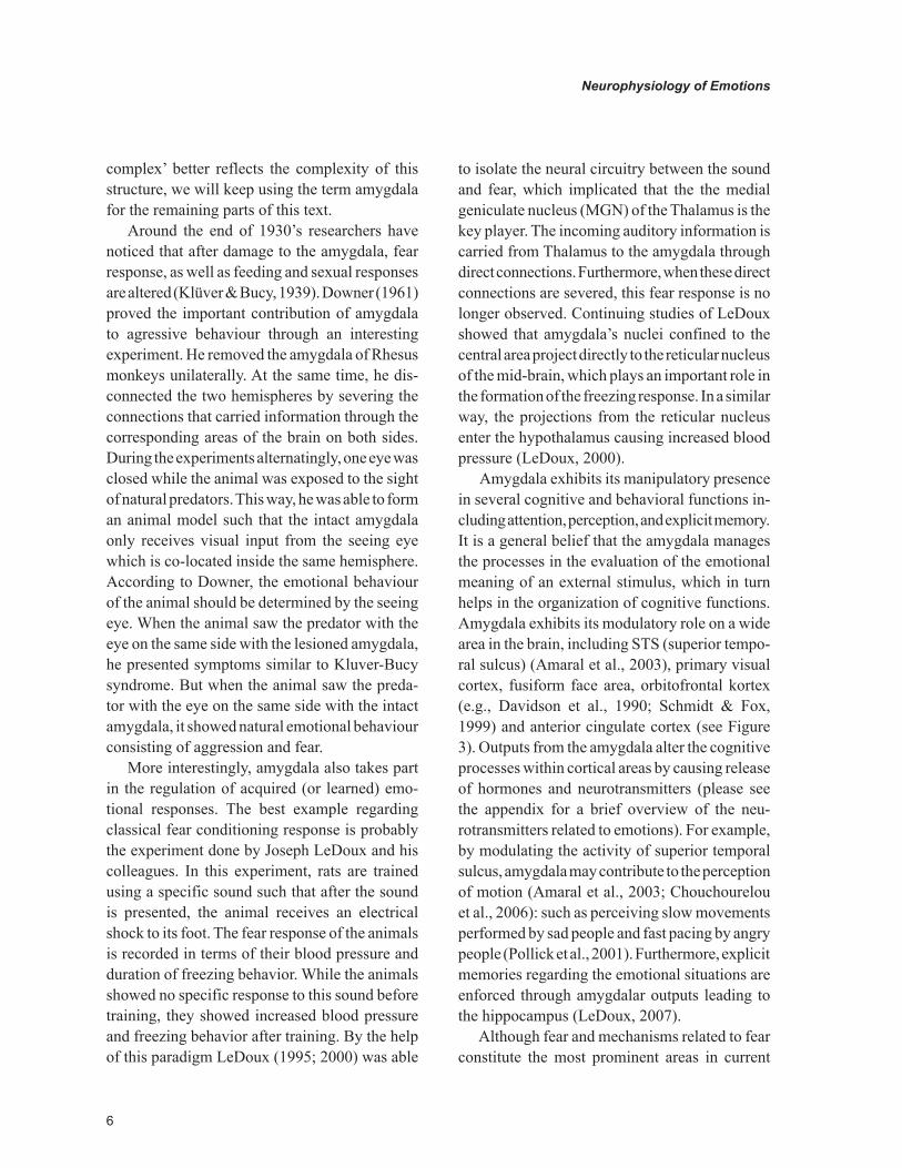
The first one is the pathway descending from the motor cortex on the posterior frontal lobe which provides the voluntary motor contractions. The other one is the involuntary pathway which reaches to the brain stem and reticular system from medial and ventral forebrain and hypothalamus, and which stimulates the visceral and somatic motor neurons around here (see Figure 4).
Best example for this is the facial paralysis that is built up because of damage on the pathway between primary motor cortex and facial nerve (corticobulbar tractus)2. The motor nucleus of the facial nerve is located in Pons and the fibers com-ing from this nucleus innervate all the mimic muscles on the superior and inferior parts of the face. If there is damage on this motor nerve’s nucleus or on it until it reaches the mimic muscles, a motor function loss occurs on the same half of the face and no muscle can be used. However, if damage occurs before the facial nerve leaves the brain stem, i.e. somewhere between the motor cortex and the motor nucleus located on the brain stem, the manifestation will be different. The up-per parts of the face are stimulated by motor

10
Neurophysiology of Emotions
Figure 4. Voluntary and involuntary motor innervations of facial nerve
Figure 5. Different aspect of upper and lower motor neuron lesion of facial nevre. Smooth shaded area represents the lower motor neuron lesion of facial nevre which causes whole plegia of right side of face and dotted shaded are area represents the upper motor neuron lesion of facial nevre which causes semi-plegia of left side of face
11
Neurophysiology of Emotions
cortex bilaterally. The lower face, on the other hand, is stimulated only by contralateral motor cortex. Thus, if damage occurs somewhere be-tween the motor cortex and the motor nucleus located on the brain stem, only the opposite lower part of the face will lose motor functions. However, it is very interesting that although these patients cannot perform voluntary motor activity on the paralyzed part of the face, they can still perform facial expressions responding to the emotional stimuli. This situation, which is called volintary-emotional dissociation, is actually the proof of the fact that the stimuli which elicit the emotional expression are not coming from motor cortex, but they are coming directly from the brain’s emotional systems to the facial nucleus. This pathway is mediated by the reticular forma-tion. On the contrary, in the emotional facial paresis, patients can move their mimic muscles voluntarily; but they cannot perform spontaneous emotional expressions on the muscles of the op-posite side of the face (see Figure 5).
sUbJECTIVE EXPERIENCE OF EMOTIONs
The Amygdala and Its Inputs for Emotional subjective Evaluations
As mentioned earlier, many regions of the brain participate in a multitude of tasks. Because of this, it is not surprising that the structures involved in the expression of emotion also participate in emotional subjective evaluation. For example, while amygdala participates in emotional expres-sion through its outputs, it is also responsible in the subjective evaluation of emotions through its inputs. For the processing of inputs, it wouldn’t be wrong to say that the lateral amygdala acts as a door-keeper. This region is the main area receiving major inputs from all sensory systems –visual, auditory, somatosensory (including pain), olfac-tory and taste. The information coming from the
olfactory and taste systems is forwarded to other amydaloid nuclei. The amygdala also receives inputs from the other areas of the brain, facilitat-ing processing of an array of different types of information. For example, auditory input comes from two resources: auditory part of the thalamus and auditory cortex. The auditory information coming from the Thalamus is weakly encoded with respect to frequency characteristics, generating a signal which has less precision but allows faster processing. On the other hand, the imcoming cortical inputs arrive from not primary but higher association cortices, providing much more detailed (already processed) information in comparison to thalamic inputs. However the processing associ-ated with these signals is slow due to the large number of underlying synaptic connections. For several emotions, especially fear, accurate and fast processing of the arriving information to the amygdala is of ultimate importance. Forming fast, primitive emotional responses are indispensable for emergencies. In other words, amygdala facili-tates responding to sudden, unexpected stimuli impulsively. For example, we can generate an emotional response to a snake that we see in a meadow. This kind of fast response is preparatory for the amygdala before the arrival of slow inputs from upstream cortices (LeDoux, 1995; LeDoux, 2000). Later, when they project into the lateral amygdala, these sensory inputs establish connec-tions with the other nuclei inside the amygdala. In order for these sensory inputs to have an effect on the behavioral response, they must go through inter-maygdalar connections.
Amygdala is the most critical structure to perceive and analyse facial expressions such as fear and anger (Adolphs et al., 1994; Adolphs et al., 1999; Young et al., 1996). For example, just catching a glimpse of the white part in the eyes of a fearful person is adequate to stimulate the amygdala (Whalen et al., 2004). Therefore we can say that even hints of danger are sufficient to trigger this system (Amaral et al., 2003; Sato et al., 2004). Amygdala, also receives inputs from

12
Neurophysiology of Emotions
the visual cortex, especially from the fusiform face area, which allows its participation in facial expression recognition in a very complex way. When subjects are shown angry faces sublimi-nally (30 msec) versus neutral faces for 170 msec, they consciously recollect only seeing neutral faces. However, PET studies indicate amygdalar activity for this type of subliminal presentation of emotional faces, while amygdalar activation is absent for similar subliminal presentation of neutral faces (Pitkanen et al., 1997). In addition, when amygdala is severed bi-laterally, it is known that fear, anger and disgust expressions can not be recognized correctly (Morris et al., 1996).
Prefrontal Cortex
The lower part of prefrontal cortex which is close to the eyes is named as orbitofrontal cortex (OFC), and the activity therein allows evaluation of posi-tive or negative emotions (for reviews, see Bechara et al., 2000). In several fMRI experiments, it has been shown that the OFC activation is associated with affective content of positive stimuli in many domains such as sampling food (Kringelbach et al., 2003), vision (Rolls, 2000), exposure to odors (Rolls et al., 2003a), hearing music (Blood & Zatorre, 2001), or even for somatosensory stimuli (Rolls et al., 2003b). Still, it is not clear how OFC effects the formation of positive affective reac-tions. Prefrontal cortex sends massive projections to the nucleus accumbens in subcortex (Zahm, 2000). And the stimulation of the nucleus accum-bens causes positive affective reactions (Peciña & Berridge, 2000). Due to the projections of OFC to nucleus accumbens (Davidson et al., 2000), it is possible that OFC is indirectly participating in the regulation of positive affective reactions, or OFC may also directly be in charge of the formation of positive affective states. It is interesting that dam-ages inflicted upon the prefrontal cortex result only in subtle affective deficits but not an entire loss in positive affective reactions (Berridge, 2003). Due to this probably it can be inferred that OFC
mainly participates in the regulation of voluntary emotions and associated emotional strategies.
Studies performed in non-human primates indicate that OFC responds to emotional stimuli, but does not participate in decisions regarding the response itself (Wallis & Miller, 2003; Padoa-Schioppa & Assad, 2006). This brings up the question ‘Which area then performs the deci-sions for emotional behaviour?’ For example, consider a woman hearing some sounds coming from the entrance in the middle of the night. She may panic at first, but then when she hears her husband’s voice at the door, she relaxes. In this scenario, due to input from the auditory stream to the amygdala, the woman initially believes there is danger. In the danger situation, OFC considers the information received and sends inputs to the amygdala to remove its inhibition. The uninhibited amygdala alerts the autonomic nervous system for alarm due to emotional arousal. But when the woman understands she is not in danger, by hear-ing her spouse’s voice, the same cycle beginning with the auditory input is repeated with this new emotional information. However, this time, using a different pathway directed from the OFC to the amygdala’s central nuclei, the hypothalamic au-tonomic center is inhibited, which in turn triggers autonomic homeostasis and the woman relaxes. Therefore OFC does not directly embark in the planning of action (Barbas, 2007). However, the lateral prefrontal cortex (LPFC) may assume an executive role for deciding how to act. Because the LPFC obtains many signals from the visual and auditory association cortices, for considering detailed environmental situations. In addition, there are bidirectional connections between the lateral and orbitofrontal cortices (Barbas, 2007). Shortly, many areas within the prefrontal cortex have critical importance for the perception of the current and varying emotional situation. While lateral prefrontal cortex is responsible from the basic goal-states which are directed by emotions, medial prefrontal cortex is responsible from the

13
Neurophysiology of Emotions
cognitive presentation of the primary emotional situation (Esslen et al., 2004).
Cingulate Cortex
The segregation of emotion and cognition in the prefrontal cortex is also found in the cingulate cortex of the limbic system. Cingulate cortex can be divided into two part based upon cytoarchi-tecture and function: Anterior cingulate cortex (ACC) and Posterior cingulate cortex (PCC) (Bush et al., 2000; Esslen et al., 2004). The dor-sal part of ACC is responsible from cognitive functions, complex motor control, motivation and participates in working memory whereas the ventral part is responsible from the evalua-tion of motivational information and meaning of emotions such as happiness, sadness, and disgust as well as arrangement of emotional responses. While ACC exhibits executive features, PCC embarks in evaluative roles. PCC activates in happiness and sadness (Esslen et al., 2004), but just like the prefrontal cortex, ACC activates for both positive and negative stimulants (Breiter et al., 1997; Firestone et al., 1996; Mathew et al., 1997). Although the cingulate cortex assumes multitude of functions, it is especially considered as the neuronal marker of positive affective reac-tions. More importantly, it is considered essential for the formation of positive affective reactions (Esslen et al., 2004). In humans, when cingulate gyrus is damaged operatively to cure pain, the patients develop depression). In rats, damage to the cingulate cortex induces ignorance with respect to reward versus punishment (Bussey et al., 1997).
Ventral striatum systems
Ventral striatum is an area where lots of affective states such as reward are reflected. Nucleus ac-cumbens, which is a sub-component of ventral striatum, is primarily activated by anticipation of monetary reward (Knutson et al., 2001). The reward system, overlaps with emotions but it is
a complex system itself, consisting of many sub-components with duties commissioned to a wide range of brain areas including medial temporal cortex, orbitofrontal cortex, dopaminergic neurons and amygdala, as well as ventral striatum. Major cognitive components of the reward system consist of ‘the detection of past rewards, the prediction and expectation of future rewards and the use of information about future rewards to control goal-directed behaviour’ (Schultz, 2000). Ventral striatum plays a central role in the reward system especially in reward detection and goal representa-tion. (For an overview, see Schultz, 2000).
Also stimulants such as cocaine or amphet-amines generate a positive affective state which correlates with increased dopamine levels in ventral striatum, especially in nucleus accumbens (Di Chiara & Imperato, 1988; Esslen et al., 2004). In addition, electrical stimulation of nucleus ac-cumbens causes smiling laughter and euphoria (Okun et al., 2004; Esslen et al., 2004).
Empathy and Mirror Neurons
Empathy can be defined as putting ourselves in someone else’s place and considering feeling and thinking like this other person. The term ‘empathy’ comes from Greek, the ‘em’ prefix means ‘in’ or ‘at’ and ‘pathos’ means feeling. Empathy is spe-cifically important in establishing relationships and socialization. The mirror neurons which are first discovered by accident in macaque monkeys are thought to be related to empathy as well. In the early studies of Rizzolatti and his colleagues, while collecting single cell neuron recordings from the premotor cortex of monkeys for study-ing the hand and mouth movements, neurons are observed to fire when the monkeys handled food. Interestingly, the same set of neurons fired, also when someone else in the room handled food just as if the monkey itself was handling it. The mirror neurons are named this way because they not only fire when the animal itself makes a movement, but they also fire when someone else makes the

14
Neurophysiology of Emotions
same movement (Rizzolatti et al., 1996; Gallese et al., 1996).
Neuroimaging and electrophysiology studies show the existence of the mirror neurons in the premotor and motor cortex of humans as well (Harrison & Critchley, 2007). Earlier, it was thought that mirror neurons were participating only in imitations and action recognition. But later studies indicate that these neurons also indulge in higher level roles such as intention, cognition and emotion. For example, while watching emotional facial expressions, the mirror neuron activity is observed to be weak in patients with social cog-nitive deficits such as autism, or in patients with developmental psychopathy. In humans, facial expressions, especially the interpretation of facial expression of others carry important roles in the establishment of empathy. For this, somatosensory, motor, and limbic systems are all at work. Carr et al. (2003) has shown that amygdala and insula participates in facial expression observation, as well as execution. In a study by Wicker et al. (2003), when a person observes someone with disgust expression, the same disgust expression develops on his face while at the same time, the anterior insula portion of his brain activates. In another study by Singer et al. (2004), when a person observes a beloved one to be subjected to a painful shock stimulus, a co-activation occurs in the anterior insula and rostral anterior cingulate cortex. As a result, we can say that while people generate their own emotional resposes, they are affected by the emotional responses they observe on others.
LATERALIZATION
The existence of cerebral asymmetry among the complex cognitive cortical functions makes us speculate that a similar asymmetry may be exhibited for the generation, expression and management of emotions as well. In addition, studies with right or left hemisphere lesion patients
indicate that there are differences in the right and left hemispheres with respect to emotional responses. For example, patients with lesions in the right hemisphere exhibit much less illness-related negative emotional responses such as depression, uncontrollable crying compared to the patients with left hemisphere lesions (Fedoroff et al., 1992; House et al., 1990; Jorge & Robinson, 2002). Patients with left frontal lobe lesions are more inclined to have depression while patients with right frontal lobe lesions display symptoms related to mania (Robinson et al., 1984; Sackeim et al., 1982). In short, there exists a difference between the right versus left hemispheres of the brain with respect to positive versus negative af-fect. When one considers that right frontal systems are associated with negative (withdrawal-related) emotional states such as sadness, anger, fear, and disgust, but left frontal regions are associated with positive (approach-related) emotional states such as happiness, the lesions in the correspond-ing areas are better understood (Davidson, 2001; Davidson and Irwin, 1999: Ahern and Schwartz, 1985; Davidson et al., 1990).
Apart from this, it has also been shown that the right hemisphere of the brain is more effective in the perception and expression of emotions. The predominant speech production and processing/understanding areas are located in the left hemi-sphere, Broca’s and Wernicke’s areas, respectively. However, the counterpart of the Wernicke’s area in the right temporal lobe is specially known to take part in the comprehension or discimination of emotional content in speech. By the help of the structures in this area, we are capable to sense the underlying emotions within the talks of people speaking in a language that is illegitimate to us. Similarly, the counterpart of Broca’s area in the right temporal lobe is implicated in the process-ing of emotional intonations of speech. Patients with lesions in this area present with monotonic speech, making it almost impossible to identify whether they are angry or happy while talking. Similarly, although the left hemisphere is more

15
Neurophysiology of Emotions
active in face recognition, the right hemisphere is more active in the recognition of facial expres-sions (Haxby et al., 2000).
CONCLUsION
Although streotypical in many animals, emotional behaviour in humans is very different. Previ-ous experiences, mood, expectations and social environment may alter the emotional response. For example, riding a roller coaster may induce fear in one person, but little or no reaction in others. In reality, exclusive mechanisms under-lying individual emotional responses have not been understood well. Probably the main reason underlying this is the engagement of a multitude of structures in very different functional roles. Presumably the amygdala and its neocortical input-output connections play a significant role in the upstream processes of emotion. In addition, while the subjective experience of emotion requires intact cerebral cortex, the emotional expression does not require the cortical processes, because the neuronal circuitry between the amygdala, hypothalamus and brain stem are implicated in the emotional expression.
REFERENCEs
Adolphs, R., Tranel, D., Damasio, H., & Damasio, A. (1994). Impaired recognition of emotion in fa-cial expressions following bilateral damage to the human amygdala. Nature, 372(6507), 669–672. doi:10.1038/372669a0
Adolphs, R., Tranel, D., Hamann, S., Young, A. W., Calder, A. J., & Phelps, E. A. (1999). Recog-nition of facial emotion in nine individuals with bilateral amygdala damage. Neuropsychologia, 37, 1111–1117. doi:10.1016/S0028-3932(99)00039-1
Ahern, G. L., & Schwartz, G. E. (1985). Differen-tial lateralization for positive and negative emotion in the human brain: EEG spectral analysis. Neu-ropsychologia, 23, 745–755. doi:10.1016/0028-3932(85)90081-8
Altemus, M., Deuster, P. A., Gallıven, E., Carter, C. S., & Gold, P. W. (1995). Suppression of hypothalmic-pituitary-adrenal axis responses to stress in lactating women. The Journal of Clinical Endocrinology and Metabolism, 80, 2954–2959. doi:10.1210/jc.80.10.2954
Amaral, D. G., Behniea, H., & Kelly, J. L. (2003). Topographic organization of projections from the amygdala to the visual cortex in the macaque mon-key. Neuroscience, 118, 1099–1120. doi:10.1016/S0306-4522(02)01001-1
Argiolas, A. (1992). Oxytocin stimulation of penile erection. Pharmacology, site, and mechanism of action. Annals of the New York Academy of Scienc-es, 652, 194–203. doi:10.1111/j.1749-6632.1992.tb34355.x
Barbas, H. (2007). Flow of information for emotions through temporal and orbitofrontal pathways. Journal of Anatomy, 211, 237–249. doi:10.1111/j.1469-7580.2007.00777.x
Bechara, A., Damasio, H., & Damasio, A. R. (2000). Emotion, decision making and the orbito-frontal cortex. Cerebral Cortex, 10(3), 295–307. doi:10.1093/cercor/10.3.295
Berman, M. E., & Coccaro, E. F. (1998). Neu-robiologic correlates of violence: relevance to criminal responsibility. Behavioral Sci-ences & the Law, 16(3), 303–318. doi:10.1002/(SICI)1099-0798(199822)16:3<303::AID-BSL309>3.0.CO;2-C
Berridge, K. C. (2003). Pleasures of the brain. Brain and Cognition, 52, 106–128. doi:10.1016/S0278-2626(03)00014-9
16
Neurophysiology of Emotions
Blood, A. J., & Zatorre, R. J. (2001). Intensely pleasurable responses to music correlate with activity in brain regions implicated in reward and emotion. Proceedings of the National Academy of Sciences of the United States of America, 98, 11818–11823. doi:10.1073/pnas.191355898
Bownds, D. M. (1999). Biology of Mind - origins and structures of mind, brain, and consciousness. Maryland: Fitzgerald Science Press.
Breiter, H. C., Gollub, R. L., Weisskoff, R. M., Kennedy, D. N., Makris, N., & Berke, J. D. (1997). Acute effects of cocaine on human brain activity and emotion. Neuron, 19(3), 591–611. doi:10.1016/S0896-6273(00)80374-8
Bush, G., Luu, P., & Posner, M. I. (2000). Cogni-tive and emotional influences in anterior cingulate cortex. Trends in Cognitive Sciences, 4, 215–222. doi:10.1016/S1364-6613(00)01483-2
Bussey, T. J., Everitt, B. J., & Robbins, T. W. (1997). Dissociable effects of cingulate and medial frontal cortex lesions on stimulus reward learning using a novel Pavlovian autoshaping procedure for the rat: Implications for the neurobiology of emo-tion. Behavioral Neuroscience, 111(5), 908–919. doi:10.1037/0735-7044.111.5.908
Caldwell, J. D. (1992). Central oxytocin and female sexual behavior. Annals of the New York Academy of Sciences, 652, 166–179. doi:10.1111/j.1749-6632.1992.tb34353.x
Carmichael, M. S., Humbert, R., Dixen, J., Palmi-sano, G., Greenleaf, W., & Davidson, J. M. (1987). Plasma oxytocin increases in the human sexual response. The Journal of Clinical Endocrinol-ogy and Metabolism, 64, 27–31. doi:10.1210/jcem-64-1-27
Carr, L., Iacoboni, M., Dubeau, M. C., Mazziotta, J. C., & Lenzi, G. L. (2003). Neural mechanisms of empathy in humans: a relay from neural sys-tems for imitation to limbic areas. Proceedings of the National Academy of Sciences of the United States of America, 100, 5497–5502. doi:10.1073/pnas.0935845100
Carter, C. S. (1992). Oxytocin and sexual behavior. Neuroscience and Biobehavioral Reviews, 16, 131–144. doi:10.1016/S0149-7634(05)80176-9
Chouchourelou, A., Matsuka, T., Harber, K., & Shiffrar, M., (2006). The visual analysis of emo-tional actions. Social Neuroscience, 1(1), 63-/74.
Damasio, A. R. (1994). Descartes’ error: emo-tion, reason, and the human brain. New York: Grosset / Putnam.
Davidson, R. J. (2001). Towards a biology of personality and emotion. Annals of the New York Academy of Sciences, 935, 191–207. doi:10.1111/j.1749-6632.2001.tb03481.x
Davidson, R. J., Ekman, P., Saron, C. D., Senu-lis, J. A., & Friesen, W. V. (1990). Approach-withdrawal and cerebral asymmetry: emotional expression and brain physiology I. Journal of Personality and Social Psychology, 58, 330–341. doi:10.1037/0022-3514.58.2.330
Davidson, R. J., & Irwin, W. (1999). The func-tional neuronatomy of emotion and affective style. Trends in Cognitive Sciences, 3, 11–21. doi:10.1016/S1364-6613(98)01265-0
Davidson, R. J., Jackson, D. C., & Kalin, N. H. (2000). Emotion, plasticity, context, and regulation: perspectives from affective neurosci-ence. Psychological Bulletin, 126(6), 890–909. doi:10.1037/0033-2909.126.6.890
Di Chiara, G., & Imperato, A. (1988). Drugs abused by humans preferentially increase syn-aptic dopamine concentrations in the mesolimbic system of freely moving rats. Proceedings of the National Academy of Sciences of the United States of America, 85, 5274–5278. doi:10.1073/pnas.85.14.5274
Domes, G., Heinrichs, M., Michel, A., Berger, C., & Herpertz, S. C. (2007). Oxytocin improves “mind-reading” in humans. Biological Psychiatry, 61, 731–733. doi:10.1016/j.biopsych.2006.07.015

17
Neurophysiology of Emotions
Downer, J. L., & De, C. (1961). Changes in visual gnostic functions and emotional behavior following unilateral temporal pole damage in the “split-brain” monkey. Nature, 191, 50–51. doi:10.1038/191050a0
Esslen, M., Pascual-Marqui, R. D., Hell, D., Kochi, K., & Lehmann, D. (2004). Brain areas and time course of emotional processing. Neu-roImage, 21, 1189–1203. doi:10.1016/j.neuroim-age.2003.10.001
Fedoroff, J. P., Starkstein, S. E., & Forrester, A. W. (1992). Depression in patients with acute trau-matic injury. The American Journal of Psychiatry, 149, 918–923.
Ferguson, J. N., Aldag, J. M., Insel, T. R., & Young, L. J. (2001). Oxytocin in the medial amygdala is essential for social recognition in the mouse. The Journal of Neuroscience, 21(20), 8278–8285.
Ferguson, J. N., Young, L. J., & Insel, T. R. (2002). The neuroendocrine basis of social recognition. Frontiers in Neuroendocrinology, 23, 200–224. doi:10.1006/frne.2002.0229
Firestone, L. L., Gyulai, F., Mintun, M., Adler, L. J., Urso, K., & Winter, P. M. (1996). Human brain activity response to fentanyl imaged by positron emission tomography. Anesthesia and Analge-sia, 82(6), 1247–1251. doi:10.1097/00000539-199606000-00025
Gallese, V., Fadiga, L., Fogassi, L., & Rizzolatti, G. (1996). Action recognition in the premotor cortex. Brain, 119, 593–609. doi:10.1093/brain/119.2.593
Guyton, A. C., & Hall, J. E. (2006). The Auto-nomic Nervous System and the Adrenal Medulla. In Textbook of Medical Physiology (11th ed., pp. 748–760). Philadelphia, Pennsylvania: Elsevier Inc.
Hagemann, D., Waldstein, S. R., & Thayera, J. F. (2003). Thayer Central and autonomic nervous system integration in emotion. Brain and Cognition, 52, 79–87. doi:10.1016/S0278-2626(03)00011-3
Harrison, N. A., & Critchley, H. D. (2007). Af-fective neuroscience and psychiatry. The British Journal of Psychiatry, 191, 192–194. doi:10.1192/bjp.bp.107.037077
Haxby, J. V., Hoffman, E. A., & Gobbini, M. I. (2000). The distributed human neural system for face perception. Trends in Cognitive Sciences, 4, 223–233. doi:10.1016/S1364-6613(00)01482-0
Heinrichs, M., Baumgartner, T., Kirschbaum, C., & Ehlert, U. (2003). Social support and oxyto-cin interact to suppress cortisol and subjective responses to psychosocial stress. Biological Psychiatry, 54, 1389–1398. doi:10.1016/S0006-3223(03)00465-7
Holland, P. C., & Gallagher, M. (2003). Double dissociation of the effects of lesions of basolateral and central amygdala on conditioned stimulus-potentiated feeding and Pavlovian-instrumental transfer. The European Journal of Neurosci-ence, 17, 1680–1694. doi:10.1046/j.1460-9568.2003.02585.x
Holstege, G., Georgiadis, J. R., Paans, A. M., Meiners, L. C., van der Graaf, F. H., & Reinders, A. A. (2003). Brain activation during human male ejaculation. The Journal of Neuroscience, 23, 9185–9193.
Horwitz, N. H. (1998). John F. Fulton (1899-1960). Neurosurgery , 43(1), 178–184. doi:10.1097/00006123-199807000-00129
House, A., Dennis, M., Warlow, C., Hawton, K., & Moltneux, A. (1990). Mood disorders after stroke and their relation to lesion location. Brain, 113, 1113–1129. doi:10.1093/brain/113.4.1113
Huber, D., Veinante, P., & Stop, R. (2005). Va-sopressin and oxytocin excite distinct neuronal populations in the central amygdala. Science, 308, 245–248. doi:10.1126/science.1105636
Izard, C. E. (2009). Emotion theory and re-search. Annual Review of Psychology, 60, 1–25. doi:10.1146/annurev.psych.60.110707.163539

18
Neurophysiology of Emotions
Jorge, R., & Robınson, R. G. (2002). Mood disorders following traumatic brain injury. Neu-roRehabilitation, 17, 311–324.
Klein, D. F., Skrobola, A. M., & Garfınkel, R. S. (1995). Preliminary look at the effects of preg-nancy on the course of panic disorder. Anxiety, 1, 227–232.
Klüver, H., & Bucy, P.C. (1939). Preliminary Analysis of Functions of the Temporal Lobes in Monkeys. Archives of Neurology and Psychiatry, 42 (6), 979-1 000.
Knutson, B., Fong, G. W., Adams, C. M., Varner, J. L., & Hommer, D. (2001). Dissociation of reward anticipation and outcome with event-related fMRI. Neuroreport, 12, 3683–3687. doi:10.1097/00001756-200112040-00016
Knutson, B., Wolkowitz, O. M., Cole, S. W., Chan, T., Moore, E. A., & Johnson, R. C. (1998). Selec-tive alteration of personality and social behavior by serotonergic intervention. The American Journal of Psychiatry, 155(3), 373–379.
Kosfeld, M., Heinrichs, M., Zak, P. J., Fisch-bacher, U., & Fehr, E. (2005). Oxytocin increases trust in humans. Nature, 435(7042), 673–676. doi:10.1038/nature03701
Kringelbach, M. L., O’Doherty, J., Rolls, E. T., & Andrews, C. (2003). Activation of the human orbitofrontal cortex to a liquid food stimulus is correlated with its subjective pleasantness. Cerebral Cortex, 13, 1064–1071. doi:10.1093/cercor/13.10.1064
Lane, R. D., & Nadel, L. (2006). Facial Expression, Emotion, and Hemispheric Organization. In Kolb, B., & Taylor, L. (Eds.), Cognitive Neuroscience of Emotion (pp. 62–83). Oxford University Press.
LeDoux, J. (1995). Emotion: Clues from the brain. Annual Review of Psychology, 46, 209–235. doi:10.1146/annurev.ps.46.020195.001233
LeDoux, J. (2000). Emotion circuits in the brain. Annual Review of Neuroscience, 23, 155–184. doi:10.1146/annurev.neuro.23.1.155
LeDoux, J. (2007). The amygdala. Current Biology, 17(20), R868–R874. doi:10.1016/j.cub.2007.08.005
Linnoila, V. M. I., & Virkkunen, M. (1992). Ag-gression, suicidality, and serotonin. The Journal of Clinical Psychiatry, 53(supp. 10), 46–51.
Lovallo, W. R., & Sollers, J. J. III. (2000). Autonomic nervous system. In Fink, G. (Ed.), Encyclopedia of stress (Vol. 1, pp. 275–284). San Diego: Academic Press.
Mathew, R. J., Wilson, W. H., Coleman, R. E., Turkington, T. G., & DeGrado, T. R. (1997). Mari-juana intoxication and brain activation in mari-juana smokers. Life Sciences, 60(23), 2075–2089. doi:10.1016/S0024-3205(97)00195-1
McCarthy, M. M., McDonald, C. H., Brooks, P. J., & Goldman, D. (1996). An anxiolytic ac-tion of oxytocin is enhanced by estrogen in the mouse. Physiology & Behavior, 60(5), 1209–1215. doi:10.1016/S0031-9384(96)00212-0
Morris, J.S., Frith, C.D., Perrett, D.I., Rowland, D., Young, A.W., Calder, A.J., & Dolan, R.J. (1996). A differential neural response in the human amygdala to fearful and happy facial expressions. Nature, 31, 383(6603), 812-815.
Murphy, M. R., Checkley, S. A., Seckl, J. R., & Lightman, S. L. (1990). Naloxone inhibits oxytocin release at orgasm in man. The Journal of Clinical Endocrinology and Metabolism, 71, 1056–1058. doi:10.1210/jcem-71-4-1056
Neumann, I. D., Kromer, S. A., Toschi, N., & Ebner, K. (2000). Brain oxytocin inhibits the (re)activity of the hypothalamo–pituitary–adrenal axis in male rats: Involvement of hypothalamic and limbic brain regions. Regulatory Peptides, 96, 31–38. doi:10.1016/S0167-0115(00)00197-X

19
Neurophysiology of Emotions
Oatley, K., & Jenkins, J. M. (1996). Understanding emotion. Cambridge, MA: Blackwell.
Okun, M. S., Bowers, D., Springer, U., Sha-pira, N. A., Malone, D., & Rezai, A. R. (2004). What’s in a ‘smile?’ Intra-operative observa-tions of contralateral smiles induced by deep brain stimulation. Neurocase, 10, 271–279. doi:10.1080/13554790490507632
Öztürk, Ö., Eraslan, D., & Kayahan, B. (2005). Emosyon ve temel insan davranışlarının evrimsel gelişimi. Yeni Symposium, 43 (1), 14-19.
Padoa-Schioppa, C., & Assad, J. A. (2006). Neu-rons in the orbitofrontal cortex encode economic value. Nature, 441(7090), 223–226. doi:10.1038/nature04676
Papez, J. W. (1937). A proposed mechanism of emotion. Archives of Neurology and Psychiatry, 38, 725–743.
Peciña, S., & Berridge, K. C. (2000). Opioid eating site in accumbens shell mediates food intake and hedonic ‘liking’: Map based on microinjection Fos plumes. Brain Research, 863, 71–86. doi:10.1016/S0006-8993(00)02102-8
Pedersen, C. A. (1997). Oxytocin control of maternal behavior. Regulation by sex ste-roids and offspring stimuli. Annals of the New York Academy of Sciences, 807, 126–145. doi:10.1111/j.1749-6632.1997.tb51916.x
Pedersen, C. A., Vadlamudi, S. V., Boccia, M. L., & Amico, J. A. (2006). Maternal behavior deficits in nulliparous oxytocin knockout mice. Genes Brain & Behavior, 5(3), 274–281. doi:10.1111/j.1601-183X.2005.00162.x
Pitkanen, A., Savander, V., & LeDoux, J. E. (1997). Organization of intra-amygdaloid cir-cuitries in the rat: an emerging framework for understanding functions of amygdala. Trends in Neurosciences, 20, 517–523. doi:10.1016/S0166-2236(97)01125-9
Pollick, F. E., Paterson, H. M., Bruderlin, A., & Sanford, A. J. (2001). Perceiving affect from arm movement. Cognition, 82, B51–B61. doi:10.1016/S0010-0277(01)00147-0
Rizzolatti, G., Fadiga, L., Fogassi, L., & Gallese, V. (1996). Premotor cortex and the recognition of motor actions. Brain Research. Cognitive Brain Research, 3, 131–141. doi:10.1016/0926-6410(95)00038-0
Robinson, R. G., Kubos, K. L., Starr, L. B., Rao, K., & Price, T. R. (1984). Mood disorders in stroke patients. Importance of location of lesion. Brain, 107, 81–93. doi:10.1093/brain/107.1.81
Rolls, E. T. (2000). The orbitofrontal cortex and reward. Cerebral Cortex, 10(3), 284–294. doi:10.1093/cercor/10.3.284
Rolls, E. T., Kringelbach, M. L., & de Araujo, I. E. (2003a). Different representations of pleasant and unpleasant odours in the human brain. The European Journal of Neuroscience, 18, 695–703. doi:10.1046/j.1460-9568.2003.02779.x
Rolls, E. T., O’Doherty, J., Kringelbach, M. L., Francis, S., Bowtell, R., & McGlone, F. (2003b). Representations of pleasant and painful touch in the human orbitofrontal and cingulate cortices. Cerebral Cortex, 13, 308–317. doi:10.1093/cercor/13.3.308
Sackeim, H. A., Greenberg, M. S., Weiman, A. L., Gur, R. C., Hungerbuhler, J. P., & Geschwind, N. (1982). Hemispheric asymmetry in the expression of positive and negative emotions. Neurologic evidence. Archives of Neurology, 39, 210–218.
Sato, W., Yoshikawa, S., Kochiyama, T., & Matsumura, M. (2004). The amygdala processes the emotional significance of facial expressions: an fMRI investigation using the interaction between expression and face direction. Neuro-Image, 22, 1006–1013. doi:10.1016/j.neuroim-age.2004.02.030

20
Neurophysiology of Emotions
Schmidt, L. A., & Fox, N. A. (1999). Conceptual, biological, and behavioral distinctions among dif-ferent categories of shy children. In Schmidt, L. A., & Schulkin, J. (Eds.), Extreme fear, shyness and social phobia (pp. 47–66). Oxford: Oxford University Press.
Schultz, W. (2000). Multiple reward signals in the brain. Nature Reviews. Neuroscience, 1(3), 199–207. doi:10.1038/35044563
Singer, T., Seymour, B., O’Doherty, J., Kaube, H., Dolan, R. J., & Frith, C. D. (2004). Empathy for pain involves the affective but not sensory com-ponents of pain. Science, 303(5661), 1157–1162. doi:10.1126/science.1093535
Stern, K., & McClintock, M. K. (1998). Regula-tion of ovulation by human pheromones. Nature, 392(6672), 177–179. doi:10.1038/32408
Turner, R. A., Altemus, M., Enos, T., Cooper, B., & McGuinness, T. (1999). Preliminary research on plasma oxytocin in normal cycling women: investigating emotion and interpersonal distress. Psychiatry, 62(2), 97–113.
Wallis, J. D., & Miller, E. K. (2003). Neuronal activity in primate dorsolateral and orbital pre-frontal cortex during performance of a reward preference task. The European Journal of Neu-roscience, 18, 2069–2081. doi:10.1046/j.1460-9568.2003.02922.x
Wedekind, C., Seebeck, T., Bettens, F., & Paepke, A. J. (1995). MHC-dependent mate preferences in humans. Proceedings. Biological Sciences, 260(1359), 245–249. doi:10.1098/rspb.1995.0087
Whalen, P.J., Kagan, J., Cook, R.G., Davis, F.C., Kim, H., Polis, S., McLaren, D.G., Somerville, L.H., McLean, A.A., Maxwell, J.S., & John-stone, T. (2004). Human amygdala responsivity to masked fearful eye whites. Science, 17, 306 (5704), 2061.
Wicker, B., Keysers, C., Plailly, J., Royet, J. P., Gallese, V., & Rizzolatti, G. (2003). Both of us disgusted in My insula: the common neural basis of seeing and feeling disgust. Neuron, 40, 655–664. doi:10.1016/S0896-6273(03)00679-2
Williams, J. R., Carter, C. S., & Insel, T. (1992). Partner preference development in fe-male prairie voles is facilitated by mating or the central infusion of oxytocin. Annals of the New York Academy of Sciences, 652, 487–489. doi:10.1111/j.1749-6632.1992.tb34393.x
Williams, J. R., Insel, T. R., Harbaugh, C. R., & Carter, C. S. (1994). Oxytocin administered cen-trally facilitates formation of a partner preference in female prairie voles (Microtus ochrogaster). Journal of Neuroendocrinology, 6(3), 247–250. doi:10.1111/j.1365-2826.1994.tb00579.x
Young, A. W., Hellawell, D. J., Van De Wal, C., & Johnson, M. (1996). Facial expression processing after amygdalotomy. Neuropsychologia, 34(1), 31–39. doi:10.1016/0028-3932(95)00062-3
Zahm, D. S. (2000). An integrative neuroanatomi-cal perspective on some subcortical substrates of adaptive responding with emphasis on the nucleus accumbens. Neuroscience and Biobehavioral Reviews, 24(1), 85–105. doi:10.1016/S0149-7634(99)00065-2
Zald, D. H. (2003). The human amygdala and the emotional evaluation of sensory stimuli. Brain Research. Brain Research Reviews, 41, 88–123. doi:10.1016/S0165-0173(02)00248-5
ADDITIONAL READING
Adolphs, R., & Heberlein, A. S. (2002). Emotion. In Ramachandran, V. S. (Ed.), Encyclopedia of the Human Brain (pp. 181–191). USA: Elsevier Science Publishing.

21
Neurophysiology of Emotions
Bear, M. F., Connors, B. W., & Paradiso, M. A. (2007). Brain Mechanisms of Emotion. In Explor-ing the brain (pp. 563–584). Lippincott Williams & Wilkins.
Hari, R., & Kujala, M. V. (2009). Brain basis of human social interaction: from concepts to brain imaging. Physiological Reviews, 89(2), 453–479. doi:10.1152/physrev.00041.2007
Kandel, E. R., Schwartz, J. H., & Jessell, T. M. (2004). Emotional States and Feelings. In Iversen, S., Kupfermann, I., & Kandel, E. R. (Eds.), Prin-ciples of Neural Science (pp. 982–997). New York: McGraw-Hill Companies, Inc.
Lewis, M., Haviland-Jones, J. M., & Barrett, L. F. (2008). Emotional Networks in the Brain. In LeDoux, J. E., & Phelps, E. A. (Eds.), Handbook of Emotions (pp. 159–179). The Guilford Press.
Oatley, K. Keltne,r D., & Jenkins, J.M. (2006). Understanding Emotions. Blackwell publishing Ltd. Madlen MA USA.
Picard, R. W. (2000). Affective Computing. Cam-bridge, MA: MIT press.
Power, M., & Dalgleish, T. (2008). Cognition and Emotion: From order to disorder. Psychol-ogy Press.
Purves, D. (2008). Emotions. In Purves, D., Augustine, G. J., Fitzpatrick, D., Hall, W. C., LaMantia, A. S., McNamara, J. O., & White, L. E. (Eds.), Neuroscience (pp. 733–759). Sinauer Associates, Inc.
Rubinson, K., & Lang, E. J. (2008). The Auto-nomic Nervous System and Its Central Control. In Koeppen, B. M., & Stanton, B. A. (Eds.), Berne & Levy Physiology (pp. 218–230). Mosby Elsevier.
KEY TERMs AND DEFINITIONs
Lateral: Away from the midlinePosterior: Backside of the bodyDorsal: The backside of the animal’s body.
In two-legged animals, the term posterior may be used interchangeable with the term dorsal, but four-legged animals dorsal is synonymous with superior (above)
Ventral: The belly side of animals.Homeostasis: Maintenance of internal stability
ENDNOTEs
1 Long lasting brain seizure.2 This type of facial paralysis is also named as
facial nerve’s upper motor neuron syndrome
22
Neurophysiology of Emotions
APPENDIX
NEUROCHEMICALs, PHEROMONEs AND EMOTION
In nuclei of amygdala, the allocation of some neuromodulator and hormone receptors (including glu-cocorticoid and estrogen hormones) differs. Many peptide receptors (opioid peptides, oxytocin, vaso-pressin, corticotropin releasing factor and neuropeptide Y) are also located in amygdala. It is crucial to understand how these chemical systems interact in order to settle amygdala’s response. Here, we will give examples of some eminent chemicals’ effects.
In a dangerous/stressful situation the body can use all its potential with the intention of overcom-ing this situation. In case of danger, amygdala and other brain regions excite hypothalamic neurons and cause the release of corticotrophin releasing factor (CRF). Release of CRF excites the release of adrenocorticotropin (ACTH) from hypophysis, thus the cortisol release from adrenal glands increase. Cortisol spreads all over the body through blood stream and gets attached to the receptors located in the hippocampus in particular. When attached with adequate amount of cortisol, these receptors produce an effect of negative feedback on hypothalamus and so inhibit the CRF release. Keeping the cortisol level stable like this, hippocampus regulates the stress response which amygdala has triggered. If stress takes long, this prolonged stress may impair hippocampus’ functions. In various researches it is confirmed that prolonged stress constricts the dendrites, which eventually causes cell deaths in hippocampus. Therefore, the functions depending on hippocampus, such as explicit memory, get seriously impaired. The con-striction of dendrites is usually seen in CA3 region of hippocampus. The neurogenesis in dentate gyrus stops. These two conditions may illuminate the volume decrease in hippocampus in the other situations where stress and cortisol are increased. Stress does not only impair hippocampus functions, but it also impairs prefrontal cortex functions. The transmission in prefrontal cortex is mediated by glutamate which is an excitatory neurotransmitter. There are also inhibitory circuits in prefrontal region. Most of these inhibitory links are located in the output neurons situated in deeper layers. However, the most important neurotransmitter which provides the neuromodulation between excitatory and inhibitory circuits is dopa-mine. Animal experiments where dopamine effect is blocked with some dopamine antagonist chemicals resulted as if the prefrontal cortex was removed. In case of danger, the cells which release serotonin and norepinephrine are also activated and therefore serotonin and norepinephrine release is increased. These chemicals provide mostly the neuromodulation. In the instant of danger, since septum and hippocampus are active; enhancing the synaptic process, serotonin and norepinephrine cause vigilance, increased attention and anxiety. On the contrary, the decrease of serotonin level is correlated with enhanced ag-gression. For instance, in people who committed violent crime, serotonin level is found to be reduced (for reviews, see Linnoila & Virkkunen, 1992; Berman & Coccaro, 1998). In another study, Knutson et al. (1998) gave serotonin reuptake inhibitor to some of the volunteers, whereas they gave placebo to the other group. The volunteers were asked to fill some personal tests and then to play cooperative games in pairs. The group which took the serotonin reuptake inhibitor showed reduced hostility index and reduced negative effect on their personal tests. Moreover they showed fair amount of cooperation and affiliation.
One of the hormones which influence emotions and behaviors is oxytocin, which shows a neurotrans-mitter effect in the brain. Oxytocin actually means “quick birth” (oxys—quick and tokos—childbirth in Greek) because of its uterotonic activity. Oxytocin is released from supraoptic and paraventricular nuclei of hypothalamus into the posterior hypophisis (neurohypophysis) and interfuses into the blood stream from this point. This hormone has receptors on numerous brain regions such as limbic system,

23
Neurophysiology of Emotions
brain stem and olfactory system; and it is very influential on the autonomic nervous system, especially on parasymphatetic system. Oxytocin is mainly in charge of lactation, uterus contraction, maternal bonding and sexual interaction. Its effects on brain structures are also shown in various animal stud-ies. For example, oxytocin enhances social memory in rodents (Ferguson et al., 2001; Ferguson et al., 2002), while it reduces anxiety in social interactions (Neumann et al., 2000). Moreover, it has a role on pair-bonding. In a study conducted on prairie voles, Williams et al. (1992; 1994) demonstrated that oxytocin injection into some specific brain regions enhanced single partner selection, whereas oxytocin antagonist injection caused a reduction. Similarly, oxytocin is very crucial for affiliation in maternal care. For instance, Pederson and colleagues showed that oxytocin activity blockage disrupted maternal care (Pedersen, 1997; Pedersen et al., 2006). Various studies emphasized oxytocin’s role on sexual interaction. For example, for female rats, intracerebral infusion of oxytocin acts on hypothalamus and facilitates sexual behaviors (Caldwell, 1992). In another study conducted on rabbits and monkeys, oxytocin is showed to be the most important agent for the stimulation of penile erection (Argiolas, 1992). It can be said that oxytocin’s central role is to mediate social behaviors depending on amygdala. For instance, oxytocin inhibits the excitatory current which goes to brainstem from amygdala, and hence reduces the fear (McCarthy et al., 1996; Huber et al., 2005).
Similarly in humans, oxytocin suppresses anxiety which causes stress (Heinrichs et al., 2003). In a study about stress, Altemus et al. (1995) demonstrated that lactating mothers showed reduced stress response to the given exercises compared to mothers who nourish their babies with bottle. Likewise, mothers, who have a panic disorder, showed relief of symptoms during lactation (Klein et al., 1995). Furthermore, in terms of interpersonal relations, oxytocin provides a raise of the feeling of trust in taking risk, in humans (Kosfeld et al., 2005). Domes et al. (2007) showed that oxytocin enhance the ability of understanding others’ affective mental conditions from their facial expressions.
Apart from maternal oxytocin increase, oxytocin secretion can also be increased with touching (Turner et al., 1999). Interestingly, during both male and female sexual stimulation and orgasm, oxytocin secre-tion increases (Carmichael et al., 1987; Carter, 1992; Murphy et al., 1990). Moreover, oxytocin leads people to fidelity, monogamous sentiments, and that is why oxytocin is thought to be the biological substrate of love.
Pheromones
Odor has both visceral and behavioral effects on humans. To retch or to vomit because of a harmful odor or salivation and increased gastric activity because of a charming odor can be given as examples of visceral motor responses. Chemical signals that are transported through air are perceived by olfactory sensory neurons on the nasal cavity and are transferred to the olfactory bulb. The information coming from the olfactory bulb is transmitted directly and through thalamus to the neocortex. The afferent pathway which goes to orbitofrontal cortex through thalamus is responsible for the perception and discrimination of the odor; whereas the pathway which goes to amygdala and hippocampus regulates the emotional and motivational aspects of the odor.
It is assumed that the effects of odor on emotions can be via the mediation of the pheromones. Phero-mones’ etymologic origin stems from Greek words phrein (to carry) and horman (to excite). Although for numerous animals the functions of pheromones are understood very well, its importance for human beings is not yet clarified. There are various studies which illustrates the existence of pheromones in humans. In a study of Stern & McClintock (1998), a group of women who did not use any odor (donors) put a

24
Neurophysiology of Emotions
cotton pad on their armpit during 8 hours. When these odorless compounds are then smelled by another group of women (recipients), the menstrual cycle of the women from the recipient group extended or diminished in accordance with the menstrual cycle of the women from the donor group. Furthermore, with a similar experimental design, when the recipient women smelled men odors, the synchronization of the menstrual cycle is disrupted. Moreover relatives can recognize each other by their odors. Mother and infant can also differentiate their odors from others. While a similar structure can also be seen in siblings, this effect is not perceived among spouses. Therefore these studies demonstrate that body odors can determine kinship information on a deep genetic level. On this issue, Wedekind et al. (1995) has conducted a very interesting experiment. MHC (major histocompatibility complex) is a gene group which is coded for the human immune system and its differences are expressed via HLA types. Wedekind and colleagues made a group of women to smell t-shirts of men from different HLA groups. At the end, these women preferred the t-shirts of men who have the different HLA type than theirs.
![Emotion Modeling and Machine Learning in Affective Computing · great chance for affective computing. Picard, in her paper [50], stated that affective computing will be used in various](https://static.fdocuments.net/doc/165x107/5e76352e7b8092691e0a2355/emotion-modeling-and-machine-learning-in-affective-computing-great-chance-for-affective.jpg)