Affection of guttral pouch
25
Affection and treatment of Guttral Pouch Dr. Bikash Puri Assist. Professor Nepal Polytechnic Institute, Chitwan Dr. Bikash Puri 1
-
Upload
bikas-puri -
Category
Education
-
view
86 -
download
0
Transcript of Affection of guttral pouch
Dr. Bikash Puri 1
Affection and treatment of Guttral Pouch
Dr. Bikash PuriAssist. Professor
Nepal Polytechnic Institute, Chitwan

Dr. Bikash Puri 2
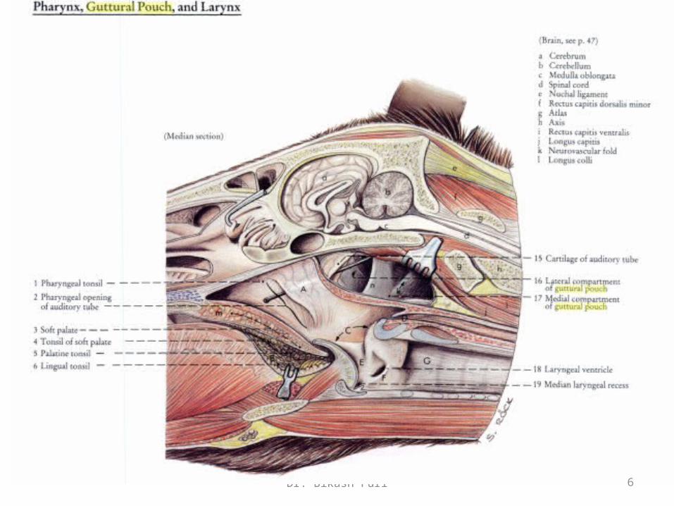
Anatomy•Present only in equines
•GP – large mucous sac which is ventral deverticulum of the eustachian tube
located in craninal cavity
•It is covered laterally by the Pterygoid muscles, parotid and mandibular
glands.
•The floor lies mainly on the pharynx and beginning of the Oesophagus.
•It connects the pharynx through the pharyngeal orifice of the eustachian
tube.
•The medial retropharyngeal lymph node lies between the pharynx and
ventral wall of the pouches.
Dr. Bikash Puri 3
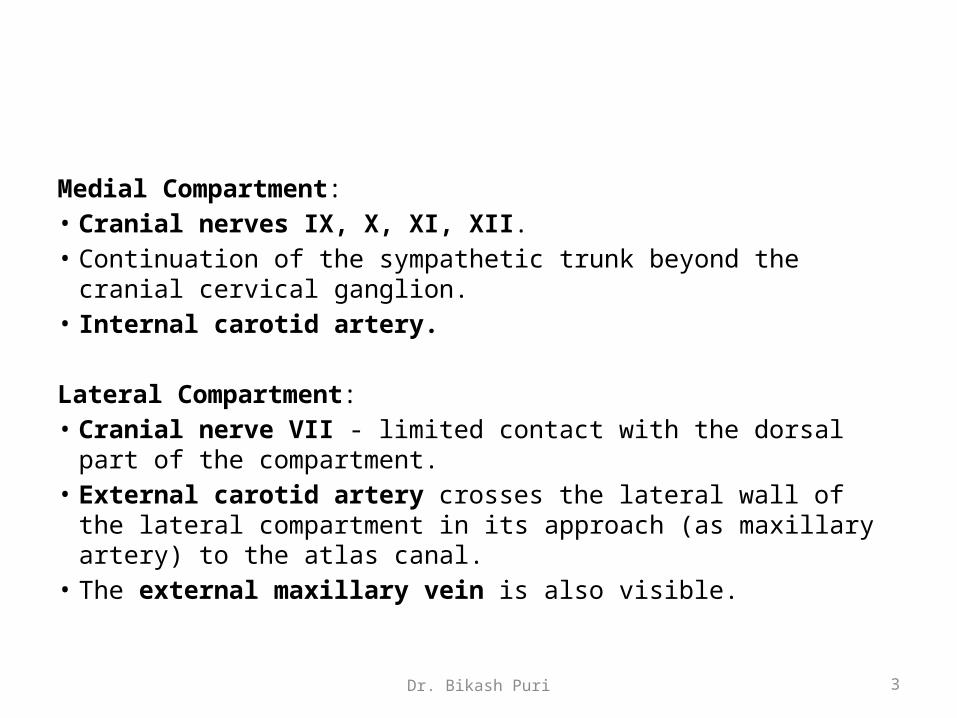
Medial Compartment:• Cranial nerves IX, X, XI, XII.• Continuation of the sympathetic trunk beyond the cranial cervical
ganglion.• Internal carotid artery.
Lateral Compartment:• Cranial nerve VII - limited contact with the dorsal part of the
compartment.• External carotid artery crosses the lateral wall of the lateral
compartment in its approach (as maxillary artery) to the atlas canal. • The external maxillary vein is also visible.

4Dr. Bikash Puri

Dr. Bikash Puri 5
Dr. Bikash Puri 6

Dr. Bikash Puri 7
Empyema
• Empyema of the guttural pouch refers to the accumulation of exudates
within a guttural pouch empyema should be considered whenever the
patient is affected with chronic mucopurulent nasal discharges.
Etiology:
– Most often secondary to other disease process.
– Respiratory tract infection caused by viral agents (Influenza), bacterial agents
(strangle) or combination of both.
– URT infections, especially caused by streptococcus equi
– Retropharyngeal abscess
– Trauma for example: stylohyoid fracture
Dr. Bikash Puri 8
Clinical signs
• Distention of guttral pouch forming a palpable, fluctuating visible swelling
behind the jaw
• Head is kept lowered during feeding or drinking
• Massive distension will interfere with swallowing and breathing (Dysphagia
and dyspnoea). Respiratory noise
• Pressure on the distended pouch may cause a mucopurulent nasal discharge
• Chronic cases may develop chondroids (inspissated pus with the appearance of
cottage cheese)
• Pharyngeal paralysis and dysphagia may be complication of an advanced disease
process.
Dr. Bikash Puri 9
Diagnosis• Based on history and clinical example: chronic nasal (mucopurulent and serosanginous)
discharge, pharyngeal distortion and dyspnoea and cranial nerve dysfunction.
• Pharyngeal endoscopy (stream of mucopurulent exudates from pharyngeal orifice of
guttural pouch.)
• Direct endoscopic examination of guttural pouch
• Centesis and lavage of the pouch: Catheterization of guttural pouch may be
accomplished either through the pharyngeal orifice or by percutaneous technique.
Radiographic examination
• A standing lateral view of skull is suggested to obtain shape and size
• Radiographic signs that suggests guttural pouch disease includes distension of the pouch and
observation of fluid line within it.
• Contrast radiograph may facilitate identification of the involved pouch and may contribute to the
appreciation of space occupying lesions.
Dr. Bikash Puri 10
Treatment • In patient with acute GPE
• Systemic antimicrobial therapy may enhance the resolution of condition within
10-14 days.
• If systemic therapy is ineffective or the case of chronic GPE
1. The pouch may be treated locally by utilizing an indwelling catheter.
2. Daily lavage of the pouch with 500ml of antiseptic or antimicrobial solution should be accomplished until
nasal discharge oblates.
3. A variety of solution can be used including 5-10% povidone iodine or a dilute solution of antimicrobials
based on sensitivity test.
4. Concurrent systemic antimicrobials therapy is useful
• When infection are refractory to systemic and local therapy, ventral drainage of
the pouch should be established surgically.
Dr. Bikash Puri 11
Prognosis1. Favorable with early and vigorous treatment
2. In case of chondroids- respond favorably
3. In advanced case – gurded.
• In patients with inspissations or chondroids of guttural pouch- medical
management be ineffective until the incriminated materials is removed
from the pouch
• Once the pouch has been entered surgically, a spoon can be used to remove
the foreign materials followed by copious lavage.
• Failure to remove all the semisolid materials from the pouch will predispose
to reoccurrence of the condition.
Dr. Bikash Puri 12
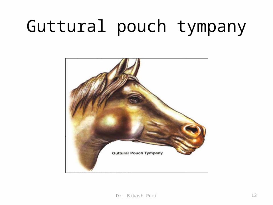
Tympanities or emphysema• Characterized by abnormal filling and distension of the GP with air.
• Usually observed in young cells, supporting its congenital occurance and
appears to affect female greater than male
• Most often occurs as unilateral but sometimes may be bilateral.
Etiology:
• The air apparently enters the pouches during expiration or when the
animal is swallowing due to the formation of gas.
Dr. Bikash Puri 13
Guttural pouch tympany
Dr. Bikash Puri 14
Clinical signs
• Diffuse painless, elastic, tympanic swelling in the parotid
region
• Unilateral distension of the pouch enough pressure on the
tissue to produce buldging in the area of the guttural pouch.
• In this case, if needle is inserted in previously distended
pouch and air is removed – the swelling subsides on both
sides.
Dr. Bikash Puri 15
Management • Effective management of this condition requires surgical innervation and depends upon
the cause of distension of the pouch.
Guidelines• If the pouch extends posterior from its normal position
• The excess portion of the pouch should be removed
• For this , the incision through the skin should be made over the most ventral and prominent part of the
pouch.
• Make 3 inch long incision and continuously divide the underlying tissue until the pouch is exposed.
• The pouch can be separated from the surrounding tissue by blunt dissection
• Continue the separation of the pouch until all of the excess portion is free from the surrounding tissue.
• The pouch is grasped with forceps and by some traction excess portion of the guttural pouch is
removed
• The wound is treated as an open wound.
Dr. Bikash Puri 16
Surgical entry and drainage of guttural pouch
Indication:
– Empyema
– Emphysema
– Food material in the pouch
Anesthesia and control
– Horse is controlled in lateral recumbency with the affected side up.
– Anesthesia is achieved either by-
• Local infiltration of local analgesics with tranquilizers or sedatives
• or general anesthetic agent.

Dr. Bikash Puri 17
Surgical approachesThree surgical approaches for guttural pouch-
1. Viborg’s triangle approach: for drainage of the guttural pouch in cases of empyema
and for treatment of tympanities.
2. Hyovertebrotomy approach: provides access through the dorsolateral aspect of the
guttural pouch and is used for removal of chondroids and inspissated pus; treatment of
guttural pouch mycosis.
3. Ventral or white house approach: provides the best surgical exposure to the dorsal
aspect of the guttural pouch for procedures such as
1. Ligating the internal carotid artery within the pouch in the treatment of guttural pouch
mycosis associated with epistaxis
2. Tympanities
3. Empyema
Dr. Bikash Puri 18
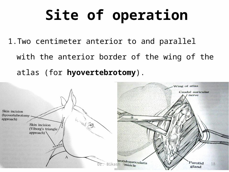
Site of operation
1. Two centimeter anterior to and parallel with the anterior border
of the wing of the atlas (for hyovertebrotomy).
Dr. Bikash Puri 19
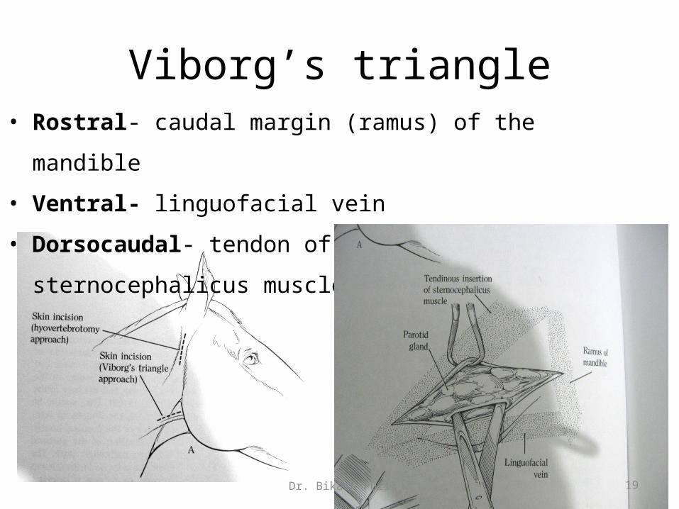
Viborg’s triangle• Rostral- caudal margin (ramus) of the mandible
• Ventral- linguofacial vein
• Dorsocaudal- tendon of insertion of sternocephalicus muscles

Dr. Bikash Puri 20
Surgical technique (hyovertebrotomy)
1. For removal of inspissated mass or chondroids or food materials.
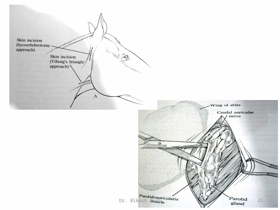
Dr. Bikash Puri 21

Dr. Bikash Puri 22

Dr. Bikash Puri 23
2. For drainage of pus and air removal from the guttural pouch (viborg’s triangle)
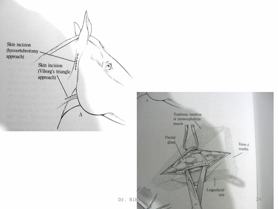
Dr. Bikash Puri 24
Dr. Bikash Puri 25
PO care
• Full course of antibiotics should be given
• Anti tetanus serum should be given immediately after
surgery
• Analgesics
• Animal should be given soft diet
• The pouch and the wound should be irrigated with non
irritant antiseptics solution daily until healing.