AESKUBLOT ANA-17 Pro Ref 4001peramed.com/peramed/docs/4001_EN.pdfProduct Ref. 4001 Product Desc....
13
AESKUBLOT ANA-17 Pro Ref 4001
Transcript of AESKUBLOT ANA-17 Pro Ref 4001peramed.com/peramed/docs/4001_EN.pdfProduct Ref. 4001 Product Desc....

AESKUBLOT ANA-17 Pro
Ref 4001


Product Ref. 4001
Product Desc. ANA-17 Pro
Manual Rev. No. 005 : 2013-02-28
Instruction Manual
Table of Contents
1 Intended Use .................................................................................................................. 1
2 Clinical Application and Principle of the Test ................................................................... 1
3 Kit Contents .................................................................................................................... 3
4 Storage and Shelf Life .................................................................................................... 3
5 Precautions of Use and General Introductions ................................................................ 4
6 Sample Collection, Handling and Storage ....................................................................... 5
7 Assay Procedure ............................................................................................................ 5
8 Qualitative Interpretation ................................................................................................. 8
9 Technical Data ................................................................................................................ 8
10 Performance Data ........................................................................................................... 9
11 Literature ........................................................................................................................ 9
AESKU.DIAGNOSTICS GmbH & Co. KG Mikroforum Ring 2 55234 Wendelsheim, Germany Tel: +49-6734-9627-0 Fax: +49-6734-9627-27 [email protected] www.aesku.com

Product Ref. 4001
Product Desc. ANA-17 Pro
Manual Rev. No. 005 : 2013-02-28
Page 1 of 9
1 Intended Use
AESKUBLOT ANA-17 Pro is a membrane based enzyme immunoassay for qualitative detection of IgG antibodies against dsDNA, nucleosomes, histones, SmD1, PCNA, Rib-P0, SS-A/Ro60kD, SS-A/Ro52kD, SS-B/La, CENP-B, SCL-70, U1-snRNP, AMA M2, Jo-1, Pm-Scl, Mi-2 and Ku in human serum or plasma. Antigens are located as parallel lines at exactly defined positions on a nitrocellulose membrane.
The assay is a tool in differential diagnosis of systemic rheumatic diseases.
2 Clinical Application and Principle of the Test
Anti-nuclear antibodies (ANAs) are an important tool for the differential diagnosis of systemic rheumatic diseases. The detection of autoantibodies in the Line Immuno Assay (LIA) with corresponding specific antigens allows a simple and reliable differentiation of ANAs by their specificity. ANAs are especially found in active and inactive systemic lupus erythematosus (SLE), mixed connective tissue diseases (MCTDs), scleroderma, Sjögren’s syndrome, primary biliary cirrhosis (PBC) and polymyositis. According to their relevance for the single autoimmune diseases, 17 antigens are arranged on an AESKUBLOT ANA-17 Pro -test strip (SLE, Sjögren’s syndrome, CREST-syndrome, scleroderma, MCTD, PBC and myositis). Antibodies against:
- Nucleosomes are directed against epitopes of the histone complex (nucleosome). In addition, anti-dsDNA and anti-histone antibodies can recognize epitopes of the nucleosome. In comparison to anti-dsDNA antibodies, anti-nucleosome antibodies are more sensitive and can provide a useful addition to the diagnosis of SLE (Chabre et al. 1995; Bruns et.al. 2000). Furthermore, they have pathogenetic significance in lupus nephritis (Van Bruggen et al. 1996; Amoura et al. 1999).
- dsDNA are regarded as being specific for SLE and have been observed in approximately 50-80 % of the patients.
- Histones are common in SLE patients. However they also occur in other connective tissue diseases. Antibodies to histones in the absence of other autoantibodies (especially anti-dsDNA) are a characteristic marker for drug-induced lupus erythematosus (Rubin 1999).
- SmD1 (Smith antigen) are directed against the core protein D1 of small nuclear ribonucleoproteins (snRNPs). Anti-Sm-antibodies as well as antibodies against double stranded DNA (dsDNA) are highly specific for SLE and thus are included in diagnostic and classification criteria for SLE.
- U1-snRNP are pathognomonic for MCTD but do also occur in SLE. A high titer of antibodies against this antigen is typical for the sharp’s syndrome.
- SS-A (Ro; soluble cytoplasmic and/or nuclear ribonucleoproteins of 52 kDa and 60 kDa) and antibodies against SS-B (La; 48 kDa protein associated with RNA-polymerase III) are mainly found in high titers for primary and secondary Sjögren’s syndrome but also in SLE, congenital heart block and neonatal lupus.
- Scl-70 are directed against DNA-topoisomerase I. They are highly specific for systemic scleroderma and are indicative of a severe course of the disease.
- CENP-B (80 kDa centromere protein B) are typical for the CREST-syndrome (69 % of CREST patients), which is a more protracted type of systemic sclerosis.
- Jo-1 are directed against histidyl-tRNA-synthetase (a cytoplasmic protein involved in protein biosynthesis) and are found in 20-40 % of patients with polymyositis and dermatomyositis.
- ribosomal P-proteins are directed against several phosphoproteins of the large ribosomal subunit. They occur in patients with systemic lupus erythematosus (Elkon et al. 1985) and in lupus patients with cerebral involvement (Bonfa et al. 1987).
Product Ref. 4001
Product Desc. ANA-17 Pro
Manual Rev. No. 005 : 2013-02-28
Page 2 of 9
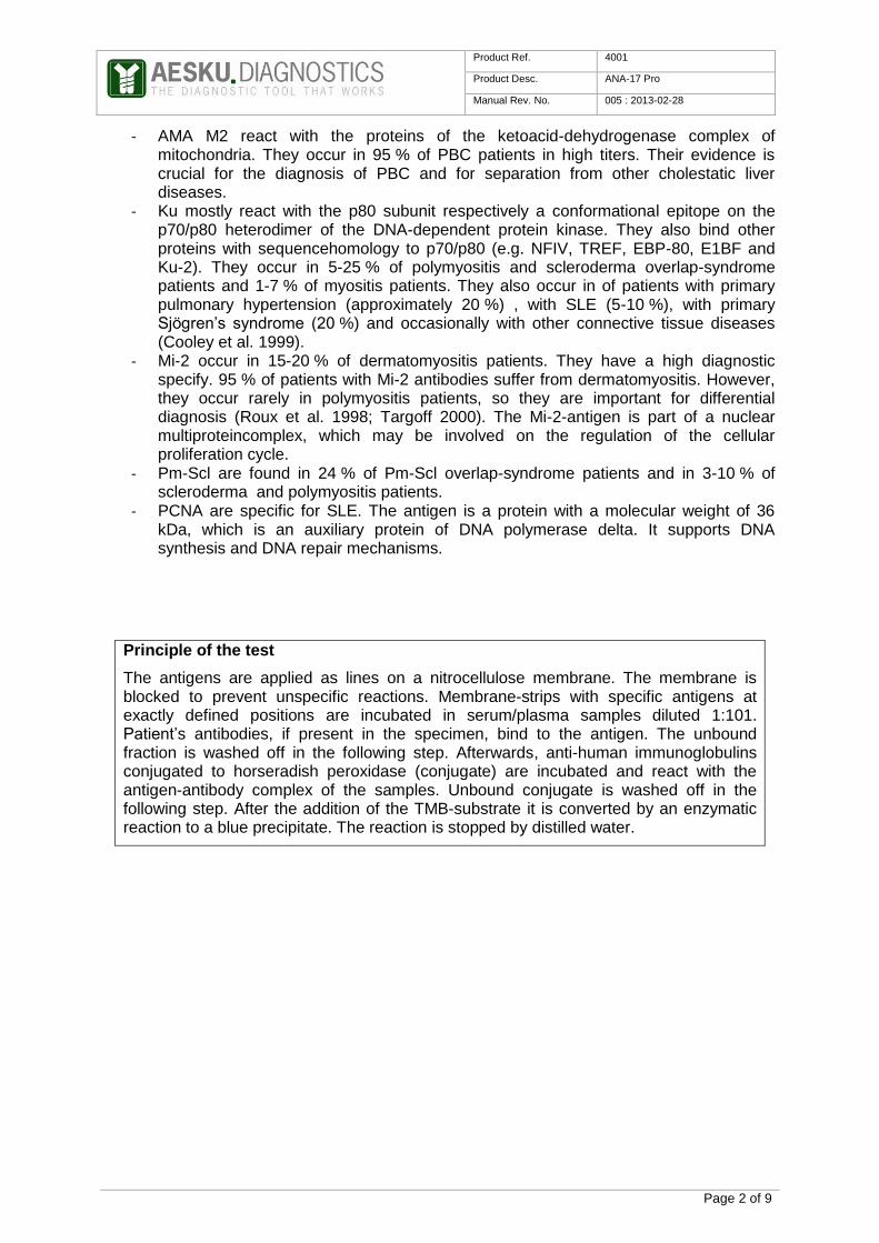
- AMA M2 react with the proteins of the ketoacid-dehydrogenase complex of mitochondria. They occur in 95 % of PBC patients in high titers. Their evidence is crucial for the diagnosis of PBC and for separation from other cholestatic liver diseases.
- Ku mostly react with the p80 subunit respectively a conformational epitope on the p70/p80 heterodimer of the DNA-dependent protein kinase. They also bind other proteins with sequencehomology to p70/p80 (e.g. NFIV, TREF, EBP-80, E1BF and Ku-2). They occur in 5-25 % of polymyositis and scleroderma overlap-syndrome patients and 1-7 % of myositis patients. They also occur in of patients with primary pulmonary hypertension (approximately 20 %) , with SLE (5-10 %), with primary Sjögren’s syndrome (20 %) and occasionally with other connective tissue diseases (Cooley et al. 1999).
- Mi-2 occur in 15-20 % of dermatomyositis patients. They have a high diagnostic specify. 95 % of patients with Mi-2 antibodies suffer from dermatomyositis. However, they occur rarely in polymyositis patients, so they are important for differential diagnosis (Roux et al. 1998; Targoff 2000). The Mi-2-antigen is part of a nuclear multiproteincomplex, which may be involved on the regulation of the cellular proliferation cycle.
- Pm-Scl are found in 24 % of Pm-Scl overlap-syndrome patients and in 3-10 % of scleroderma and polymyositis patients.
- PCNA are specific for SLE. The antigen is a protein with a molecular weight of 36 kDa, which is an auxiliary protein of DNA polymerase delta. It supports DNA synthesis and DNA repair mechanisms.
Principle of the test
The antigens are applied as lines on a nitrocellulose membrane. The membrane is blocked to prevent unspecific reactions. Membrane-strips with specific antigens at exactly defined positions are incubated in serum/plasma samples diluted 1:101. Patient’s antibodies, if present in the specimen, bind to the antigen. The unbound fraction is washed off in the following step. Afterwards, anti-human immunoglobulins conjugated to horseradish peroxidase (conjugate) are incubated and react with the antigen-antibody complex of the samples. Unbound conjugate is washed off in the following step. After the addition of the TMB-substrate it is converted by an enzymatic reaction to a blue precipitate. The reaction is stopped by distilled water.
Product Ref. 4001
Product Desc. ANA-17 Pro
Manual Rev. No. 005 : 2013-02-28
Page 3 of 9
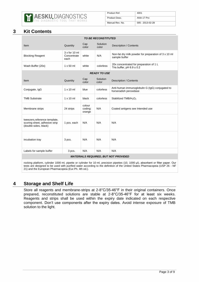
3 Kit Contents
TO BE RECONSTITUTED
Item Quantity Cap color
Solution color
Description / Contents
Blocking Reagent 3 x for 10 ml Concentrate each
white N/A Non-fat dry milk powder for preparation of 3 x 10 ml sample buffer
Wash Buffer (20x) 1 x 50 ml white colorless 20x concentrated for preparation of 1 L Tris buffer, pH 6.9 ± 0.2
READY TO USE
Item Quantity Cap color
Solution color
Description / Contents
Conjugate, IgG 1 x 10 ml blue colorless Anti-human immunoglobulin G (IgG) conjugated to horseradish peroxidase
TMB Substrate 1 x 10 ml black colorless Stabilized TMB/H2O2
Membrane strips 24 strips colour coding: orange
N/A Coated antigens see Intended use
tweezers,reference template, scoring sheet, adhesive strip (double-sides, black)
1 pcs. each N/A N/A N/A
incubation tray 3 pcs. N/A N/A N/A
Labels for sample buffer 3 pcs. N/A N/A N/A
MATERIALS REQUIRED, BUT NOT PROVIDED
rocking platform, cylinder 1000 ml, pipette or cylinder for 10 ml, precision pipettes (10, 1000 µl), absorbent or filter paper. Our tests are designed to be used with purified water according to the definition of the United States Pharmacopeia (USP 26 - NF 21) and the European Pharmacopeia (Eur.Ph. 4th ed.).
4 Storage and Shelf Life
Store all reagents and membrane-strips at 2-8°C/35-46°F in their original containers. Once prepared, reconstituted solutions are stable at 2-8°C/35-46°F for at least six weeks. Reagents and strips shall be used within the expiry date indicated on each respective component. Don’t use components after the expiry dates. Avoid intense exposure of TMB solution to the light.
Product Ref. 4001
Product Desc. ANA-17 Pro
Manual Rev. No. 005 : 2013-02-28
Page 4 of 9
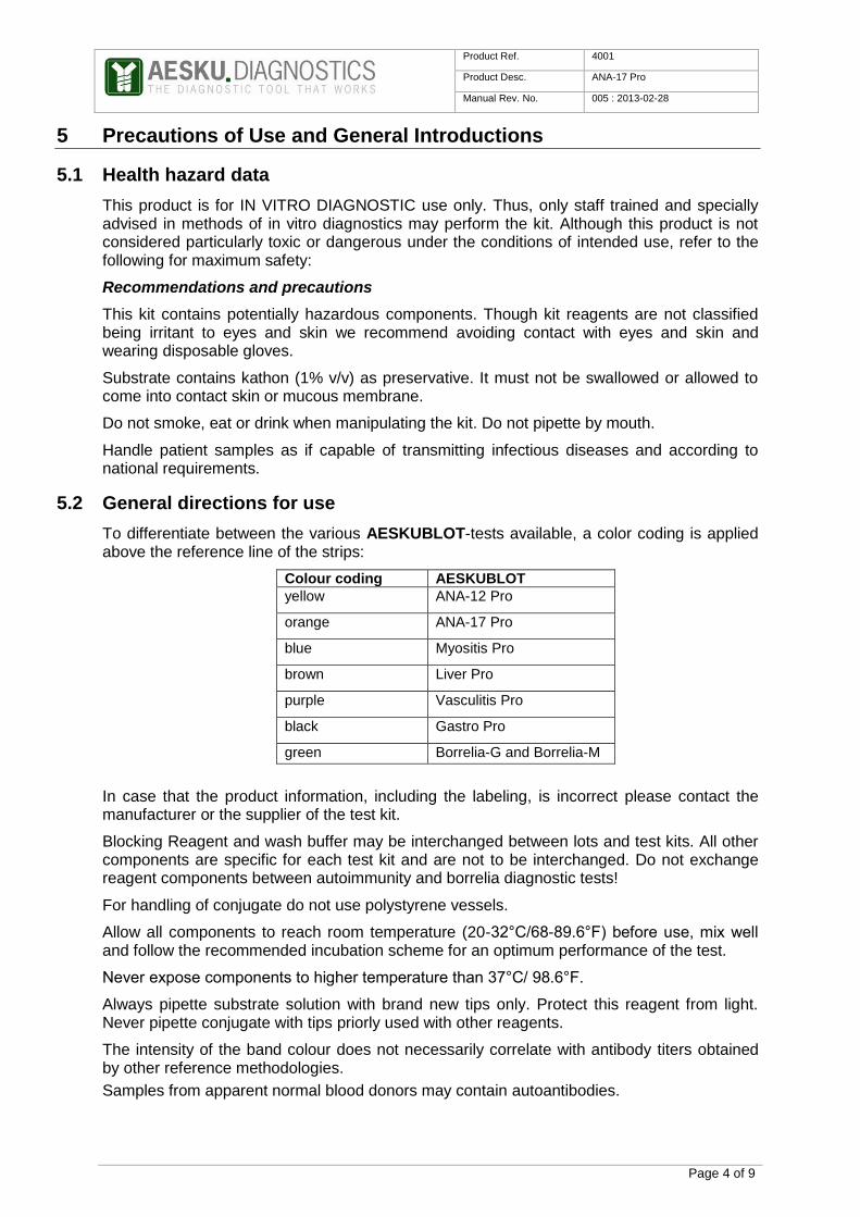
5 Precautions of Use and General Introductions
5.1 Health hazard data
This product is for IN VITRO DIAGNOSTIC use only. Thus, only staff trained and specially advised in methods of in vitro diagnostics may perform the kit. Although this product is not considered particularly toxic or dangerous under the conditions of intended use, refer to the following for maximum safety:
Recommendations and precautions
This kit contains potentially hazardous components. Though kit reagents are not classified being irritant to eyes and skin we recommend avoiding contact with eyes and skin and wearing disposable gloves.
Substrate contains kathon (1% v/v) as preservative. It must not be swallowed or allowed to come into contact skin or mucous membrane.
Do not smoke, eat or drink when manipulating the kit. Do not pipette by mouth.
Handle patient samples as if capable of transmitting infectious diseases and according to national requirements.
5.2 General directions for use
To differentiate between the various AESKUBLOT-tests available, a color coding is applied above the reference line of the strips:
Colour coding AESKUBLOT
yellow ANA-12 Pro
orange ANA-17 Pro
blue Myositis Pro
brown Liver Pro
purple Vasculitis Pro
black Gastro Pro
green Borrelia-G and Borrelia-M
In case that the product information, including the labeling, is incorrect please contact the manufacturer or the supplier of the test kit.
Blocking Reagent and wash buffer may be interchanged between lots and test kits. All other components are specific for each test kit and are not to be interchanged. Do not exchange reagent components between autoimmunity and borrelia diagnostic tests!
For handling of conjugate do not use polystyrene vessels.
Allow all components to reach room temperature (20-32°C/68-89.6°F) before use, mix well and follow the recommended incubation scheme for an optimum performance of the test.
Never expose components to higher temperature than 37°C/ 98.6°F.
Always pipette substrate solution with brand new tips only. Protect this reagent from light. Never pipette conjugate with tips priorly used with other reagents.
The intensity of the band colour does not necessarily correlate with antibody titers obtained by other reference methodologies.
Samples from apparent normal blood donors may contain autoantibodies.

Product Ref. 4001
Product Desc. ANA-17 Pro
Manual Rev. No. 005 : 2013-02-28
Page 5 of 9
If the patient sample contains elevated levels of immune complexes or other immunoglobulin aggregates, false positive results by non-specific binding cannot be ruled out.
A definite clinical diagnosis should not be based on the results of the performed test only, but should be made by the physician after all clinical and laboratory findings have been evaluated. The diagnosis is to be verified using different diagnostic methods.
6 Sample Collection, Handling and Storage
Use preferentially freshly collected serum/plasma samples. Blood withdrawal must follow national requirements. Do not use icteric, lipemic, hemolysed or bacterially contaminated samples. Sera with particles should be cleared by low speed centrifugation (<1000 x g). Blood samples should be collected in clean, dry and empty tubes.
After separation, the serum/plasma samples should be used during the first 8 h. Alternatively, the samples should be stored in tightly closed vials at 2-8°C/35-46°F for up to 48 h, or frozen at -20°C/-4°F for longer periods. Avoid repeated thawing and freezing. Do not use heat inactivated samples.
7 Assay Procedure
7.1 Preparations prior to starting
Confirm that no salt crystals have been formed in the concentrate. If this happened, dissolve the crystals by slightly warming, room temperature should be enough, the concentrate.
Dilute concentrated wash buffer 1:20 with distilled water (e.g. 950 ml plus 50 ml).
For preparation of sample buffer: add 10 ml wash buffer to one bottle Blocking Reagent and mix well.
7.2 Test Steps
Important notes:
Follow exactly this protocol. Make sure that the two components mentioned in the protocol are added to the tray in steps 2, 6, 9.
Do not let strip dry out during incubation steps.
Do not touch strip with fingers, use tweezers.
Remove diluted samples completely after incubation of strip to avoid carry over.
Continuously shake strip during incubation steps.
Give sample buffer, conjugate and substrate together with the wash buffer to one side of the incubation tray. Do not allow to flow over the strip.

Product Ref. 4001
Product Desc. ANA-17 Pro
Manual Rev. No. 005 : 2013-02-28
Page 6 of 9
Step Description
1. Ensure the preparations, from step 7.1 above, have been carried out prior to test begin.
2.
Put strip in correct orientation into incubation tray (reference line and colour coding upwards). Put 700 µl wash buffer and 300 µl sample buffer in the incubation tray. Moisten strip with the solution and incubate for 5 minutes with agitation.
CONTROLS & SAMPLES
3.
Pipette 10 µl serum/plasma sample into the designated incubation trays with sample buffer.
4.
Incubate for 30 minutes at 20-32°C/68-89.6°F with agitation. After that remove sample completely.
5.
Wash 3 times for 5 minutes with 1.5 ml wash buffer by agitation. Remove wash buffer after every washing step.
CONJUGATE
6.
Pipette 700 µl wash buffer and 300 µl conjugate into each incubation tray with strip.
7.
Incubate for 30 minutes at 20-32°C/68-89.6°F with agitation. Remove conjugate.

Product Ref. 4001
Product Desc. ANA-17 Pro
Manual Rev. No. 005 : 2013-02-28
Page 7 of 9
8.
Wash 3 times for 5 minutes with 1.5 ml wash buffer by agitation. Remove wash buffer after every washing step.
SUBSTRATE
9.
Pipette 700 µl dH2O and 300 µl substrate into each incubation tray with strip.
10.
Incubate for 15 minutes at 20-32°C/68-89.6°F with agitation, protected from intense light. Remove substrate.
STOP
11.
Pipette 2 ml dH2O into each incubation tray with strip. Incubate 1 minute with agitation. Remove dH2O. Repeat this step one time.
12. Remove strip of the incubation tray. Dry strip between filter paper
13. Analyze results within 24 h.

Product Ref. 4001
Product Desc. ANA-17 Pro
Manual Rev. No. 005 : 2013-02-28
Page 8 of 9
8 Qualitative Interpretation
8.1 Manual Analysis Test results can be considered valid, if:
- Functional control is visible - Cut-off control is visible - Colour intensity of cut-off control is weaker than colour intensity of functional control
Fix dried strip onto scoring sheet aligned with reference line. Align reference template with the strip reference line. Interpret results only in reference to cut-off control of each strip.
Each test kit contains a colour copy with all bands provable in the test.
The analysis is carried out by means of comparing the colour intensities of the bands with colour intensity of the cut-off control. The test is equivocal if the intensities do not significant differ. Is the colour more intensive the test result is positive, if the colour intensity is weaker, the test is negative.
The results can be recorded on the scoring sheet.
In case that the values of the controls do not meet the criteria, the test is invalid and has to be repeated. We recommend retesting samples that are borderline.
The following technical issues should as well be checked: expiry date of (prepared) reagents, storage conditions, pipettes, equipment, incubation conditions and washing methods.
If the samples tested show aberrant values or any kind of deviation or if the validation criteria are not met because of reasons outside the operator’s responsibility, please contact the manufacturer or the supplier of the test kit.
Medical laboratories might perform an in-house quality control by using their own controls and/or internal pooled sera, as stated in national regulations.
9 Technical Data
Sample material: serum or plasma
Sample volume: 10 µl of sample
Total incubation time: 112 minutes at 20-32°C/68-89.6°F
Storage: at 2-8°C/35-46°F; use original vials only.
Number of determinations: 24 tests

Product Ref. 4001
Product Desc. ANA-17 Pro
Manual Rev. No. 005 : 2013-02-28
Page 9 of 9
10 Performance Data
10.1 Relative Sensitivity and Specificity
In order to determine the positive agreement (relative sensitivity), 115 sera from IIF antibody-positive patients were tested in AESKUBLOT ANA-17 Pro. For determination of the
negative agreement (relative specificity), 50 sera from blood donors were analyzed.
Positive agreement: 99,1 % (114/115)
Negative agreement: 98 % (49/50)
Total agreement: 98,8 % (163/165)
11 Literature
Amoura Z, Piette J-C, Bach J-F, Koutouzov S (1995). The key role of nucleosomes in
lupus. Arthritis & Rheumatism. 42 (5):833–843.
Bonfa E, Golombek SJ, Kaufman LD, Skelly S (1987). Association between Lupus
Psychosis and Antiribosomal P Protein Antibodies. The New England Journal of Medicine.
317(5):265-271.
Bruns A, Bläss S, Hausdorf G, Burmester GR, Hiepe F (2000). Nucleosomes are major T
and B cell autoantigens in systemic lupus erythematosus. Arthritis & Rheumatism. 43
(10):2307–2315.
Chabre H, Amoura Z, Piette J-C, Godeau P, Bach J-F, Koutouzov S (1995). Presence of
nucleosome-restricted antibodies in patients with systemic lupus erythematosus. Arthritis &
Rheumatism. 38 (10):1485–1491.
Cooley HM, Melny BJ, GleesonR, Greco T, Kay TW (1999). Clinical and serological
associations of anti-Ku antibody. J Rheumatol. 26:563–567.
Elkon KB, AP, Foster CL (1985). Lupus autoantibodies target ribosomal P proteins. J Exp
Med. 162(2):459–71
Roux S, Seelig HP, Meyer O (1998). Significance of MI-2autoantibodies in polymyositis and
dermatomyositis. J Rheumatol. 25: 395–396.
Rubin RL (1999). Etiology and mechanismof drug-induced lupus. Curr opin Rheumatol.
11:357–365.
Targoff IN (2000). Update on myositis-specific and myositis-associated autoantibodies.
Current Opinion in Rheumatology. 12(6):475–481.
Van Bruggen MC, Kramers C, Berden JH (1996). Autoimmunity against nucleosomes and
lupus nephritis. Ann Med Interne. 147(7):485–9.
For further reading:
Peter JB, Shoenfeld Y (1996). Autoantibodies. Elsevier Sciences B.V., Amsterdam.
Tan EM, (1989). Antinuclear antibodies: diagnostic markers for autoimmune diseases and
probes for cell biology. Adv. Immunol. 44: 93–151.
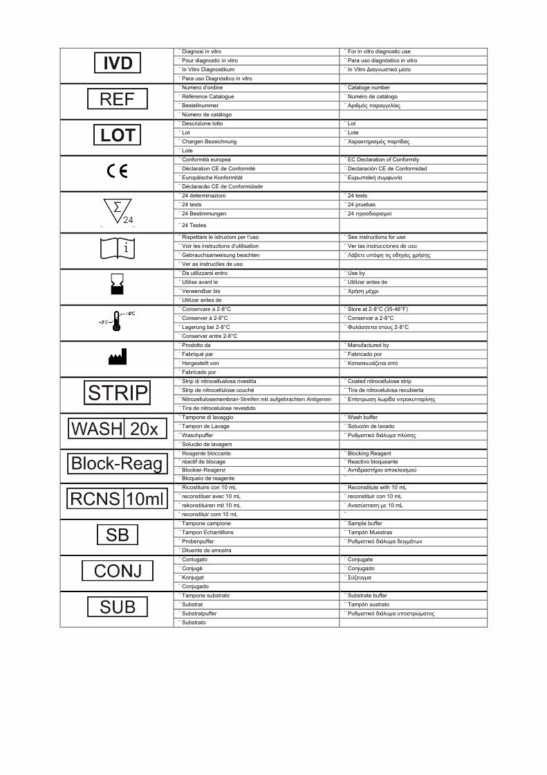
¨ Diagnosi in vitro ¨ For in vitro diagnostic use
¨ Pour diagnostic in vitro ¨ Para uso diagnóstico in vitro
¨ Ιn Vitro Diagnostikum ¨ In Vitro Διαγνωστικό μέσο
¨ Para uso Diagnóstico in vitro
¨ Numero d’ordine ¨ Cataloge number
¨ Référence Catalogue ¨ Numéro de catálogo
¨ Bestellnummer ¨ Αριθμός παραγγελίας
¨ Número de catálogo
¨ Descrizione lotto ¨ Lot
¨ Lot ¨ Lote
¨ Chargen Bezeichnung ¨ Χαρακτηρισμός παρτίδας
¨ Lote
¨ Conformità europea ¨ EC Declaration of Conformity
¨ Déclaration CE de Conformité ¨ Declaración CE de Conformidad
¨ Europäische Konformität ¨ Ευρωπαϊκή συμφωνία
¨ Déclaracão CE de Conformidade
¨ 24 determinazioni ¨ 24 tests
¨ 24 tests ¨ 24 pruebas
¨ 24 Bestimmungen ¨ 24 προσδιορισμοί
¨ 24 Testes
¨ Rispettare le istruzioni per l’uso ¨ See instructions for use
¨ Voir les instructions d‘utilisation ¨ Ver las instrucciones de uso
¨ Gebrauchsanweisung beachten ¨ Λάβετε υπόψη τις οδηγίες χρήσης
¨ Ver as instrucões de uso
¨ Da utilizzarsi entro ¨ Use by
¨ Utilise avant le ¨ Utilizar antes de
¨ Verwendbar bis ¨ Χρήση μέχρι
¨ Utilizar antes de
¨ Conservare a 2-8°C ¨ Store at 2-8°C (35-46°F)
¨ Conserver à 2-8°C ¨ Conservar a 2-8°C
¨ Lagerung bei 2-8°C ¨ Φυλάσσεται στους 2-8°C
¨ Conservar entre 2-8°C
¨ Prodotto da ¨ Manufactured by
¨ Fabriqué par ¨ Fabricado por
¨ Hergestellt von ¨ Κατασκευάζεται από
¨ Fabricado por
¨ Strip di nitrocelluslosa rivestita ¨ Coated nitrocellulose strip
¨ Strip de nitrocellulose couché ¨ Tira de nitrocelulosa recubierta
¨ Nitrozellulosemembran-Streifen mit aufgebrachten Antigenen ¨ Επίστρωση λωρίδα νιτροκυτταρίνης
¨ Tira de nitrocelulose revestido
¨ Tampone di lavaggio ¨ Wash buffer
¨ Tampon de Lavage ¨ Solución de lavado
¨ Waschpuffer ¨ Ρυθμιστικό διάλυμα πλύσης
¨ Solucão de lavagem
¨ Reagente bloccante ¨ Blocking Reagent
¨ réactif de blocage ¨ Reactivo bloqueante
¨ Blockier-Reagenz ¨ Αντιδραστήριο αποκλεισμού
¨ Bloqueio de reagente ¨
¨ Ricostituire con 10 mL ¨ Reconstitute with 10 mL
¨ reconstituer avec 10 mL ¨ reconstituir con 10 mL
¨ rekonstituiren mit 10 mL ¨ Ανασύσταση με 10 mL
¨ reconstituir com 10 mL ¨
¨ Tampone campione ¨ Sample buffer
¨ Tampon Echantillons ¨ Tampón Muestras
¨ Probenpuffer ¨ Ρυθμιστικό διάλυμα δειγμάτων
¨ Diluente de amostra
¨ Coniugato ¨ Conjugate
¨ Conjugé ¨ Conjugado
¨ Konjugat ¨ Σύζευγμα
¨ Conjugado
¨ Tampone substrato ¨ Substrate buffer
¨ Substrat ¨ Tampón sustrato
¨ Substratpuffer ¨ Ρυθμιστικό διάλυμα υποστρώματος
¨ Substrato