Aed guide revision_feb_14_2012_draft
22
DEFIBRILLATION AND THE AUTOMATIC EXTERNAL DEFIBRILLATOR A GUIDE Defibrillation and the AED, A guide Revised February 2012 Page 1 of 22
-
Upload
ichsan-izatul -
Category
Engineering
-
view
288 -
download
1
Transcript of Aed guide revision_feb_14_2012_draft

DEFIBRILLATION AND THE AUTOMATIC EXTERNAL DEFIBRILLATOR
A GUIDE
Defibrillation and the AED, A guide Revised February 2012 Page 1 of 22
Original document complied by Pat Standen, Grampians Trauma, Emergency & Critical Care Coordinator ACKNOWLEDGEMENTS Sue Garner, Clinical Educator, Intensive Care/Coronary Care Unit, Ballarat Health Services, Amber van Dreven, Clinical Educator, Emergency Department, Ballarat Health Services and Geoff McCurdy, Director of Pharmacy, Ballarat Health Services for reviewing the original document and providing their expert advice. Thank you to Wendy Porteous, Clinical Educator – Emergency Department, Ballarat Health Services for reviewing and revising this version 2012 Ballarat Health Services and Ambulance Victoria for so generously allowing their clinical practice guidelines to be used as a guide. For information regarding this Guide contact: Pat Standen Department of Health PO Box 712 Ballarat 3353 Email: [email protected] Phone: 03 5333 6026 Version Date Major Changes Page No 1.0 September 2005 1.1 January 2008 Basic Life Support Flowchart
AED Flowchart Competency Assessment form Basic Life Support Table
16 17 18 20
2.0 February 2012 Section 3 Basic life support flowchart
11-19 20
DISCLAIMER: Care has been taken to confirm the accuracy of the information presented in this guide, however, the authors, editors and publisher are not responsible for errors or omissions or for any consequences from application of the information in the guide and make no warranty, express or implied, with respect to the contents of the publication. Every effort has been made to ensure the clinical information provided is in accordance with current recommendations and practice. However, in view of ongoing research, changes in government regulations and the flow of other information, the information is provided on the basis that all persons undertake responsibility for assessing the relevance and accuracy of its content.
Defibrillation and the AED, A guide Revised February 2012 Page 2 of 22
TABLE OF CONTENTS
Page INTRODUCTION 4 SECTION 1 BRIEF REVIEW OF THE HEART 5
1.1 Anatomy 5 1.2 Coronary Arteries 6 1.3 The Conduction System 8
SECTION 2 THE ELECTROCARDIAGRAM 9 2.1 ECG Complex 9
2.2 ECG Grid Paper 10 SECTION 3 CARDIAC RHYTHMS AND DEFIBRILLATION 11 3.1 What is Defibrillation 11
3.2 Types of AEDs 11 3.3 Sequence of actions when using the AED 12 3.4 Position of Pads 12 3.5 Pads 15
3.6 Rhythms: Ventricular Tachycardia 16 Ventricular Fibrillation 17
3.7 Defibrillation Safety 18 3.8 Procedure 18
Basic Life Support Flowchart 20 REFERENCES 21 SUGGESTED FURTHER READING 21
Defibrillation and the AED, A guide Revised February 2012 Page 3 of 22
INTRODUCTION The purpose of this guide is to assist educators in the Grampians Region to design their own Health Service specific package for Registered Nurses Division 1, and others, required to use an Automatic External Defibrillator (AED). There are a number of these devices available for purchase; the aim of this guide is to provide generic information based on principles of care. It is the responsibility of each individual practitioner and Health Service to ensure appropriate education for all equipment and that competency in the use of the equipment is maintained. The use and education around the AED should be undertaken in conjunction with Basic Life Support (BLS) and Laryngeal Mask Airway (LMA) management. Please note: Any person wishing to use this material must review the materials prior to use and ensure they are suitable for the purpose, including the needs of educators and intended training recipients. Some aspects of the materials may not be relevant. The accuracy and appropriateness of the materials may not suit all circumstances, and the materials may not include all the information required. Any person using the materials does so at their own risk and shall accept responsibility for any actions arising from their use. No responsibility is taken for failure to update the materials and it is the responsibility of the person to ensure that the information contained in the materials is up to date and reflects current practice, law and guidelines.
Defibrillation and the AED, A guide Revised February 2012 Page 4 of 22
SECTION 1
1.0 A BRIEF REVIEW OF THE HEART
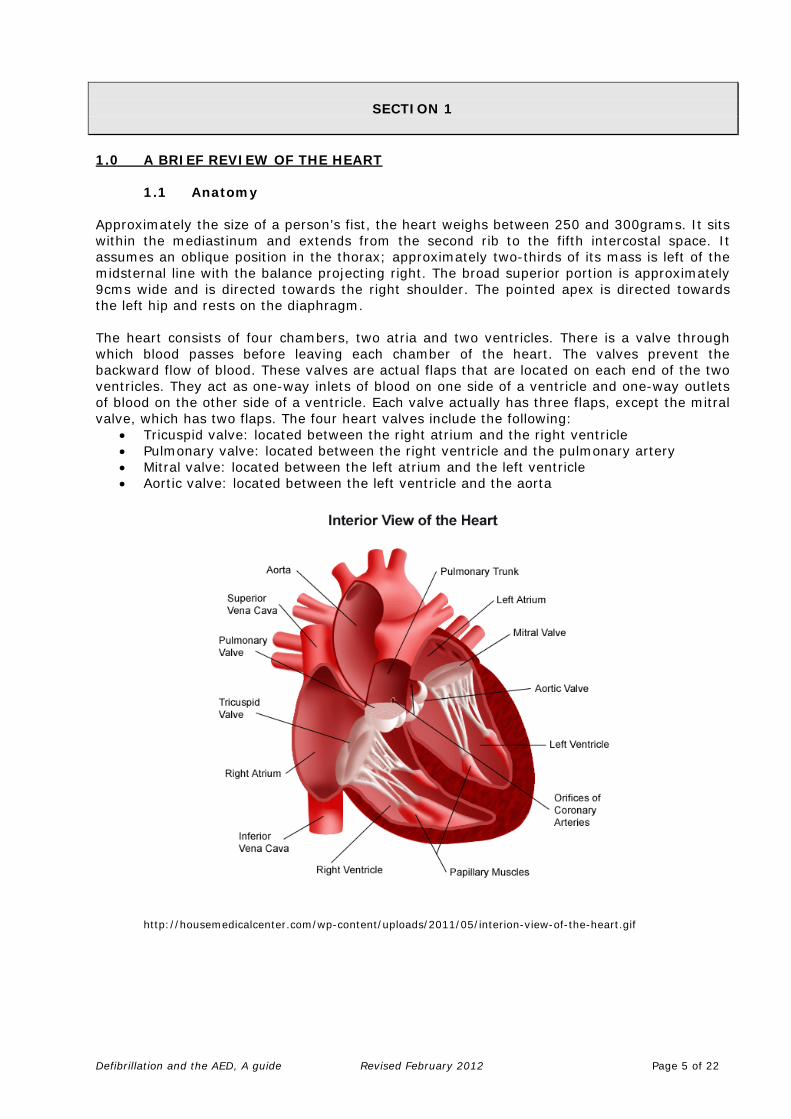
1.1 Anatomy Approximately the size of a person’s fist, the heart weighs between 250 and 300grams. It sits within the mediastinum and extends from the second rib to the fifth intercostal space. It assumes an oblique position in the thorax; approximately two-thirds of its mass is left of the midsternal line with the balance projecting right. The broad superior portion is approximately 9cms wide and is directed towards the right shoulder. The pointed apex is directed towards the left hip and rests on the diaphragm. The heart consists of four chambers, two atria and two ventricles. There is a valve through which blood passes before leaving each chamber of the heart. The valves prevent the backward flow of blood. These valves are actual flaps that are located on each end of the two ventricles. They act as one-way inlets of blood on one side of a ventricle and one-way outlets of blood on the other side of a ventricle. Each valve actually has three flaps, except the mitral valve, which has two flaps. The four heart valves include the following:
• Tricuspid valve: located between the right atrium and the right ventricle • Pulmonary valve: located between the right ventricle and the pulmonary artery • Mitral valve: located between the left atrium and the left ventricle • Aortic valve: located between the left ventricle and the aorta
http://housemedicalcenter.com/wp-content/uploads/2011/05/interion-view-of-the-heart.gif
Defibrillation and the AED, A guide Revised February 2012 Page 5 of 22

1.2 Coronary Arteries
Coronary arteries supply blood to the heart muscle. Like all other tissues in the body, the heart muscle needs oxygen-rich blood to function. The coronary arteries consist of two main arteries: the right and left coronary arteries, and their two branches, the circumflex artery and the left anterior descending artery.
http://www.cumc.columbia.edu/dept/rehab/images/Pages%20from%20cardiac-4_img_0.jpg
• The left coronary artery (LCA), divides into the left anterior descending artery and the circumflex branch, and supplies blood to the ventricles and left atrium.
• The right coronary artery (RCA), divides into the right posterior descending artery and a large marginal branch, supplies the ventricles, right atrium, and sinoatrial node.
• The circumflex artery (Cx) branches off the left coronary artery and encircles the heart muscle. This artery supplies blood to the back of the heart.
• The left anterior descending artery (LAD) branches off the left coronary artery and supplies blood to the front of the heart
Defibrillation and the AED, A guide Revised February 2012 Page 6 of 22
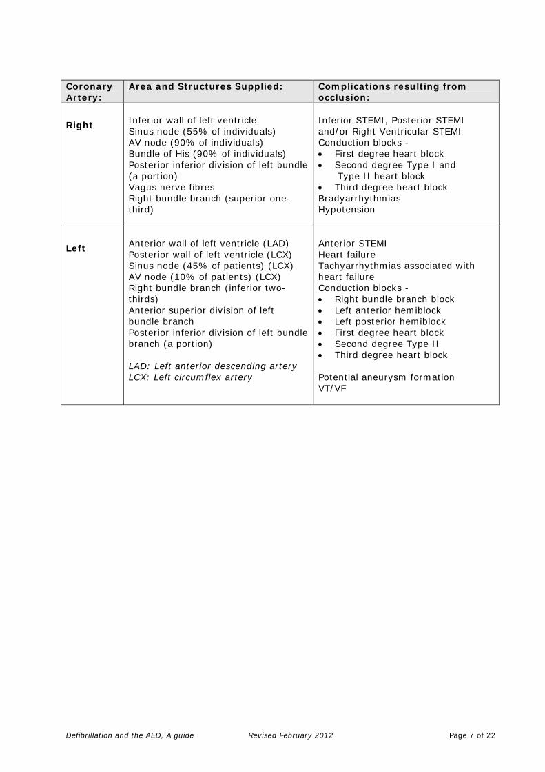
Coronary Artery:
Area and Structures Supplied: Complications resulting from occlusion:
Right
Inferior wall of left ventricle Sinus node (55% of individuals) AV node (90% of individuals) Bundle of His (90% of individuals) Posterior inferior division of left bundle (a portion) Vagus nerve fibres Right bundle branch (superior one-third)
Inferior STEMI, Posterior STEMI and/or Right Ventricular STEMI Conduction blocks - • First degree heart block • Second degree Type I and Type II heart block • Third degree heart block Bradyarrhythmias Hypotension
Left
Anterior wall of left ventricle (LAD) Posterior wall of left ventricle (LCX) Sinus node (45% of patients) (LCX) AV node (10% of patients) (LCX) Right bundle branch (inferior two-thirds) Anterior superior division of left bundle branch Posterior inferior division of left bundle branch (a portion) LAD: Left anterior descending artery LCX: Left circumflex artery
Anterior STEMI Heart failure Tachyarrhythmias associated with heart failure Conduction blocks - • Right bundle branch block • Left anterior hemiblock • Left posterior hemiblock • First degree heart block • Second degree Type II • Third degree heart block Potential aneurysm formation VT/VF
Defibrillation and the AED, A guide Revised February 2012 Page 7 of 22
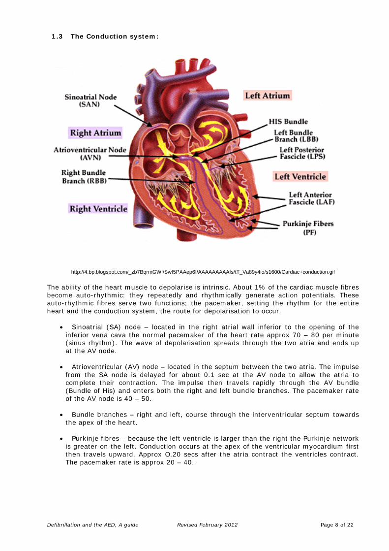
1.3 The Conduction system:
http://4.bp.blogspot.com/_zb7BqrrxGWI/Swf5PAAep6I/AAAAAAAAAIs/tT_Va89y4io/s1600/Cardiac+conduction.gif The ability of the heart muscle to depolarise is intrinsic. About 1% of the cardiac muscle fibres become auto-rhythmic: they repeatedly and rhythmically generate action potentials. These auto-rhythmic fibres serve two functions; the pacemaker, setting the rhythm for the entire heart and the conduction system, the route for depolarisation to occur.
• Sinoatrial (SA) node – located in the right atrial wall inferior to the opening of the inferior vena cava the normal pacemaker of the heart rate approx 70 – 80 per minute (sinus rhythm). The wave of depolarisation spreads through the two atria and ends up at the AV node.
• Atrioventricular (AV) node – located in the septum between the two atria. The impulse
from the SA node is delayed for about 0.1 sec at the AV node to allow the atria to complete their contraction. The impulse then travels rapidly through the AV bundle (Bundle of His) and enters both the right and left bundle branches. The pacemaker rate of the AV node is 40 – 50.
• Bundle branches – right and left, course through the interventricular septum towards
the apex of the heart. • Purkinje fibres – because the left ventricle is larger than the right the Purkinje network
is greater on the left. Conduction occurs at the apex of the ventricular myocardium first then travels upward. Approx O.20 secs after the atria contract the ventricles contract. The pacemaker rate is approx 20 – 40.
Defibrillation and the AED, A guide Revised February 2012 Page 8 of 22
SECTION 2
2.0 THE ELECTROCARDIOGRAPH 2.1 E.C.G. Complex
• P WAVE - The first wave represents depolarisation of the atria. The impulse usually comes from the SA node, which is the chief pacemaker of the heart and is situated in the right atrium. The general wave of depolarisation is downwards and towards the left hand side of the body. The P wave is generally not higher (amplitude) than 2-3mm and no longer than 0.10 – 0.11 of a second in duration. It is normally upright in Lead I, Lead II, aVF, V3 – V6. It may be inverted in aVR, may be biphasic in V1 and V2, and may be occasionally inverted in Leads I and II and aVL.
• PR INTERVAL – This interval represents the time taken for the impulse to go through the atria, across the AV node and junction and down the Bundle of His. It is measured from the beginning of the P wave to the beginning of the QRS. Normal duration 0.12 – 0.20 seconds.
• QRS – This complex follows the P wave, PR interval and represents the depolarisation of the ventricles. Depolarisation comes through the AV node, then goes through the septum and then through the left and right ventricles. The general wave is downwards and to the left of the body. The QRS is usually 0.04 – 0.10 of a second in duration and between 5mm and 125mm in amplitude, depending on the lead.
The Q wave is the first downward deflection preceding the R or S wave. If there is no R wave it is termed a QS wave. The Q wave is normally less than 0.04 seconds in duration, and less than a ¼ of the R wave. Q waves in Lead III and aVF may vary with inspiration. The R wave – first upward deflection of the QRS complex usually less than 0.01
second in duration. The R wave in V6 represents left ventricle activity. The R wave in V1 represents right ventricle activity. The S wave – first downward deflection following R wave is rarely deeper than
6mm and may be absent. An S wave in V1 represents left ventricle activity. An S wave in V6 represents right ventricle activity.
• ST SEGMENT –
This segment represents the refractory period of the ventricles. The point where the QRS joins the ST segment is called the “J” point. The ST segment duration varies with the cardiac rate and ranges from zero to 0.15 seconds. It is normally iso-electric (on the baseline) because positive and negative forces are equal during this period.
• T WAVE – This usually starts at the iso-electric line and varies in shape. It represents rapid repolarisation of the ventricles. Its amplitude is usually from 5mm – 10mm. It can be upright in Lead I, Lead II, V3 to V6 inverted in aVR and variable in other leads. Following the T wave a U wave of low voltage is sometimes seen, it may represent the slow repolarisation of the ventricles. It is usually difficult to see.
Defibrillation and the AED, A guide Revised February 2012 Page 9 of 22

• QT INTERVAL –
This interval measures the total time taken for depolarisation and repolarisation of the ventricle. It is measured from start of the QRS to end of the T wave. Its duration varies with age, sex and cardiac rate, but usually is about 0.35 – 0.42 seconds. As a general rule – QT should be less then ½ of the R – R interval. 2.2 ECG Grid Paper
http://yenoh93.medceu.com/images/sample6.jpg
Measurements of paper and the complex: • P wave – 0.20 sec • PR interval – 0.12 – 0.20 sec • QRS interval – 0.07 – 0.10 sec • ST segment – measurement not significant, elevation or depression is more
important • QT segment – measurement depending on heart rate
Calculation of Heart Rate Paper speed: 25mm/sec – therefore 5 large boxes = 25mm or 1 sec therefore 300boxes = 1 minute If heart rate regular: Measure the interval between the complexes (R R interval) and divide into 300 For example: 1 complex every large box = Rate 300bpm
1 complex every 2 large boxes = rate 150bpm 1 complex every 3 large boxes = rate 100bpm 1 complex every 4 large boxes = rate 75bpm 1 complex every 5 large boxes = rate 60bpm
OR – If the heart rate irregular: 15 large boxes = 3 secs therefore 30 large boxes = 6 secs Count the complexes in a 6 second interval and multiply by 10 to get the rate per minute.
Defibrillation and the AED, A guide Revised February 2012 Page 10 of 22

SECTION 3
3.0 CARDIAC RHYTHMS & DEFIBRILLATION 3.1 What Is Defibrillation?
“The passage of an electrical current across the myocardium of sufficient magnitude to depolarise a critical mass of myocardium, and enable restoration of coordinated electrical activity” Source: Deakin et al (2010) Mechanism of defibrillation It is thought that successful defibrillation occurs when a critical mass of myocardium is depolarised by the passage of an electric current (RCUK, 2011). This will then hopefully enable the sinoatrial node (the heart’s normal pacemaker) or another intrinsic pacemaker to regain control of the heartbeat (Jevon, 2009). REMEMBER: EARLY DEFIBRILLATION PROVIDES THE BEST CHANCE OF SURVIVAL IN
VF OR PULSELESS VT
Defibrillation refers to the current of electricity passing through the heart and it occurs at a random point in the cardiac cycle (unsynchronised). Defibrillation produces simultaneous depolarisation of the mass of myocardial cells and enables the resumption of organised electrical activity. 3.2 Types of automated external defibrillator AEDs are sophisticated, reliable, safe, computerised devices that deliver electric shocks to victims of cardiac arrest when the ECG rhythm is one that is likely to respond to a shock. Simplicity of operation is a key feature: controls are kept to a minimum, voice and visual prompts guide rescuers. Modern AEDs are suitable for use by both lay rescuers and healthcare professionals. All AEDs analyse the victim’s ECG rhythm and determine the need for a shock. The semi-automatic AED indicates the need for a shock, which is delivered by the operator, while the fully automatic AED administers the shock without the need for intervention by the operator. Some semi-automatic AEDs have the facility to enable the operator (normally a healthcare professional) to override the device and deliver a shock manually, independently of prompts. Examples of some of the automatic external defibrillators available:
Welch Allyn AED 10 Powerheart AED G3 http://www.welchallyn.com/products/en-us/x-11-ac-100-0000000001041.htm http://www.powerheart.com/products/phaed_g3auto.htm
Defibrillation and the AED, A guide Revised February 2012 Page 11 of 22

Phillips HeartStart FR2 Zoll AED http://www.medical.philips.com/main/products/resuscitation/index2.html http://www.zoll.com.au/products/aed_plus/literature/AED_Plus_Brochure.pdf
3.3 Sequence of actions when using an automated external defibrillator The following sequence applies to the use of both semi-automatic and automatic AEDs in a victim who is found to be unconscious and not breathing normally.
1. Follow the adult BLS sequence. Do not delay starting CPR unless the AED is available immediately. 2. As soon as the AED arrives: • If more than one rescuer is present, continue CPR while the AED is switched on. If you are alone, stop CPR and switch on the AED. • Follow the voice / visual prompts. • Attach the electrode pads to the patient’s bare chest. • Ensure that nobody touches the victim while the AED is analysing the rhythm. 3A. If a shock is indicated: • Ensure that nobody touches the victim. • Push the shock button as directed (fully-automatic AEDs will deliver the shock automatically). • Continue as directed by the voice / visual prompts. • Minimise, as far as possible, interruptions in chest compression. 3B. If no shock is indicated: • Resume CPR immediately using a ratio of 30 compressions to 2 rescue breaths. • Continue as directed by the voice / visual prompts. 4. Continue to follow the AED prompts until: • qualified help arrives and takes over OR • the victim starts to show signs of regaining consciousness, such as coughing, opening his eyes, speaking, or moving purposefully AND starts to breathe normally OR • you become exhausted.
3.4 Position of Pads The electrode positions are generally standard (you can refer to the instructions on your AED). The apex pad is placed over the 6th intercostal space, anterior chest wall, mid axillary line. The sternal pad is placed with its top in the 2nd intercostal space, just right of the sternum.
Defibrillation and the AED, A guide Revised February 2012 Page 12 of 22
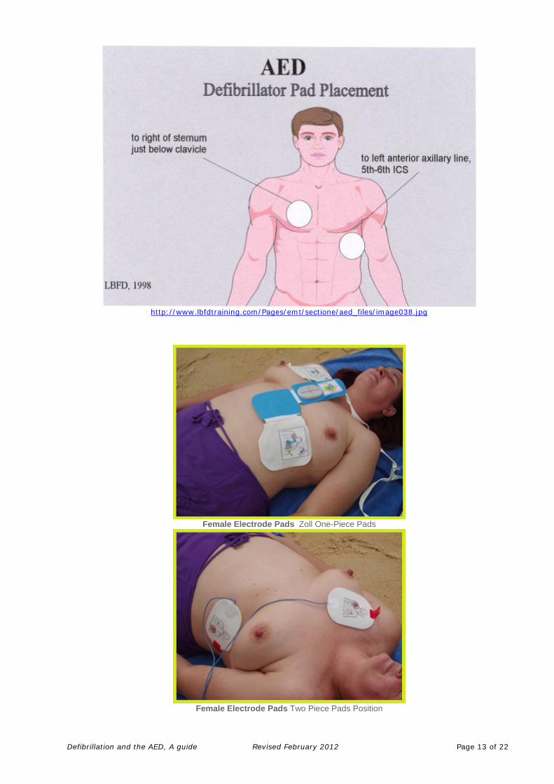
http://www.lbfdtraining.com/Pages/emt/sectione/aed_files/image038.jpg
Female Electrode Pads Zoll One-Piece Pads
Female Electrode Pads Two Piece Pads Position
Defibrillation and the AED, A guide Revised February 2012 Page 13 of 22

Male Electrode Pads Zoll One-Piece Pads
Male Electrode Pads Two Piece Pads Position
http://www.australiandefibrillators.com.au/use-an-aed.html
3.5 The Pads The standard defibrillation pads for AEDs are about the size of your hand and are made of soft thin foam coated on one side with a gel. The gel side is covered with a peel off backing which is removed prior to placement on the chest. The backing is removed from one pad at a time and the gel side firmly placed onto the patient’s bare chest (ensure moisture is wiped away and the entire pad smoothed on firmly). Good pad contact will reduce the risk of skin burns and reduce resistance to the current.
Defibrillation and the AED, A guide Revised February 2012 Page 14 of 22

http://images-mediawiki-sites.thefullwiki.org/03/2/5/3/92649233828269596.jpg
Defibrillation if the victim is wet As long as there is no direct contact between the user and the victim when the shock is delivered, there is no direct pathway that the electricity can take that would cause the user to experience a shock. Dry the victim’s chest so that the adhesive AED pads will stick and take particular care to ensure that no one is touching the victim when a shock is delivered. Defibrillation in the presence of supplemental oxygen There are no reports of fires caused by sparking where defibrillation was delivered using adhesive pads. If supplemental oxygen is being delivered by a face mask, remove the face mask and place it at least one metre away before delivering a shock. Do not allow this to delay shock delivery. Minimise interruptions in CPR The importance of early, uninterrupted chest compressions is emphasised throughout these guidelines. Interrupt CPR only when it is necessary to analyse the rhythm and deliver a shock. When two rescuers are present, the rescuer operating the AED applies the electrodes while the other continues CPR. The AED operator delivers a shock as soon as the shock is advised, ensuring that no one is in contact with the victim. CPR before defibrillation Provide good quality CPR while the AED is brought to the scene. Continue CPR whilst the AED is turned on, then follow the voice and visual prompts. Giving a specified period of CPR, as a routine before rhythm analysis and shock delivery, is not recommended.
3.6 Rhythms
There are four lethal rhythms Ventricular Tachycardia (VT) Ventricular Fibrillation (VF) Pulseless Electrical Activity Asystole
The two shockable rhythms, which the AED will recognise, are VT and VF. The AED reads the ECG from the pads applied to the chest. Some AEDs have screens that show the rhythms, some do not. The AED identifies the heart rhythms. The operator does not need to be able to identify the rhythm or decide if there is a need for defibrillation. It is however useful to have a basic knowledge of heart rhythms.
Defibrillation and the AED, A guide Revised February 2012 Page 15 of 22
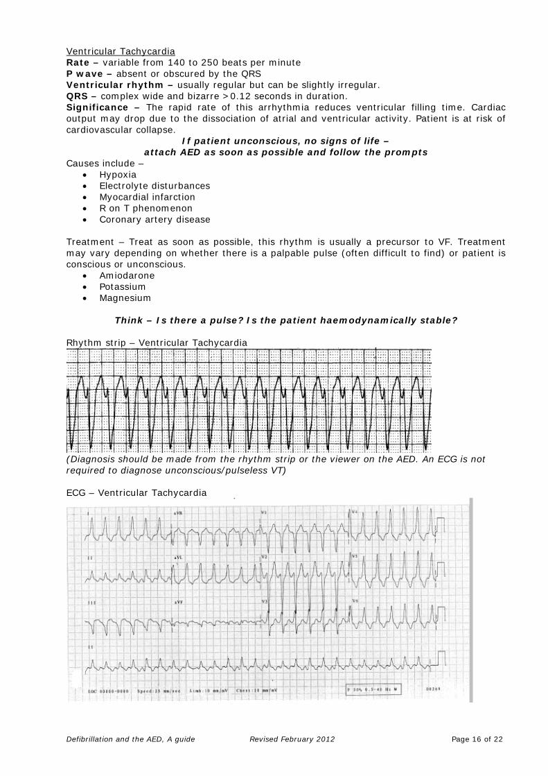
Ventricular TachycardiaRate – variable from 140 to 250 beats per minute P wave – absent or obscured by the QRS Ventricular rhythm – usually regular but can be slightly irregular. QRS – complex wide and bizarre >0.12 seconds in duration. Significance – The rapid rate of this arrhythmia reduces ventricular filling time. Cardiac output may drop due to the dissociation of atrial and ventricular activity. Patient is at risk of cardiovascular collapse.
If patient unconscious, no signs of life – attach AED as soon as possible and follow the prompts
Causes include – • Hypoxia • Electrolyte disturbances • Myocardial infarction • R on T phenomenon • Coronary artery disease
Treatment – Treat as soon as possible, this rhythm is usually a precursor to VF. Treatment may vary depending on whether there is a palpable pulse (often difficult to find) or patient is conscious or unconscious.
• Amiodarone • Potassium • Magnesium
Think – Is there a pulse? Is the patient haemodynamically stable?
Rhythm strip – Ventricular Tachycardia
(Diagnosis should be made from the rhythm strip or the viewer on the AED. An ECG is not required to diagnose unconscious/pulseless VT) ECG – Ventricular Tachycardia
Defibrillation and the AED, A guide Revised February 2012 Page 16 of 22
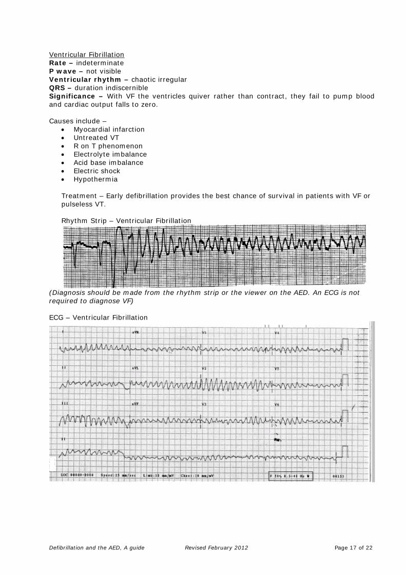
Ventricular Fibrillation Rate – indeterminate P wave – not visible Ventricular rhythm – chaotic irregular QRS – duration indiscernible Significance – With VF the ventricles quiver rather than contract, they fail to pump blood and cardiac output falls to zero. Causes include –
• Myocardial infarction • Untreated VT • R on T phenomenon • Electrolyte imbalance • Acid base imbalance • Electric shock • Hypothermia
Treatment – Early defibrillation provides the best chance of survival in patients with VF or pulseless VT.
Rhythm Strip – Ventricular Fibrillation
(Diagnosis should be made from the rhythm strip or the viewer on the AED. An ECG is not required to diagnose VF) ECG – Ventricular Fibrillation
Defibrillation and the AED, A guide Revised February 2012 Page 17 of 22

3.7 Defibrillation Safety Operators must be aware of the need for safety when using the AED. Considerations include:
• Adult pads cannot be cut down to a smaller size • There should be no contact with patient (by anyone) during defibrillation • Check pad area for jewellery, ECG electrodes, pacemaker (if pacemaker present adjust pads) and medication patches • Think about conductive surfaces (water, fluids, metal) • Explosive environment (oxygen, gases, fumes) • Do not operate in an unstable environment which may prevent the AED from
performing a valid assessment of the ECG signal (eg. rapidly moving vehicle) • Respond to all prompts within safety constraints. • Make sure all personnel are clear of the patient during analysis and prior to
initiating a shock. • Cellular phones, radios or other devices that emit electrical signals may interfere
with the analysis of the AED and their use should be discouraged within 6 feet. • The use of AED in children (below 8yrs/40kgs) requires special pads. If the special
pads are not available, an unmodified adult AED may be used in children older than 1 year
3.8 Procedure
• Collapsed patient • Remember chain of survival
1. Call for help. If you see someone collapse, immediately call for help and activate your emergency procedures. If there are other people around, choose someone specific and instruct them to call for help and explain the situation. This decreases confusion about who should do what and ensures that the call is being placed. 2. Check the victim's breathing and airway. If someone has collapsed, you should immediately determine whether they are breathing. If the victim is breathing, you know that they have a pulse. If the victim is not breathing, check the airway is clear then begin CPR at 30 chest compressions at a depth of 4-5cm then 2 breaths. 3. Locate an AED. If there is an AED nearby, ask a bystander to take over CPR while you apply the AED chest electrode pads to the victim. Uninterrupted CPR is an important factor in increasing the recovery rate of cardiac arrest patients. Always ensure that someone is providing CPR for the victim unless the AED is actively analysing or shocking the victim. 4. Turn on the AED. Follow the visual & voice prompts of the AED
Defibrillation and the AED, A guide Revised February 2012 Page 18 of 22
5. Attach the electrode pads to patients bare chest. (Expose the patients bare chest, male or female) First ensure that the adhesive AED pads are attached to a cable, which is plugged into the AED machine. Then bare the victim's chest including females and attach the adhesive AED pads in the appropriate locations. The AED should include a diagram (typically on the adhesive pads themselves) indicating where each pad goes. The ZOLL AED uses a one-piece chest pad that makes placement easy. 6. Always follow the instructions of the AED. Note: CPR should not be interrupted while the adhesive electrode pads are being applied.
Analysing the victims’ heart rhythm.
The AED may instruct you to “Stop CPR, do not touch patient, analysing”. The rescuer will then say "CLEAR!" to ensure that nobody is touching the victim while the AED analyses the victims heart rhythm. A shock is only indicated if the victim's heart is in ventricular fibrillation (VF) or ventricular tachycardia (VT). The AED will automatically analyse the heart rhythm of the victim and inform you, the rescuer, whether a shock is advised.
If you get a "no shock advised" instruction from the AED it can mean:
• the victim that you thought was pulse less does indeed have a pulse, or • the victim has now regained a pulse, or • the victim is pulseless but is not in a 'shockable' rhythm (i.e. not ventricular fibrillation (VT) or ventricular tachycardia (VT).
Follow the visual & voice prompts of the AED
Delivering a shock to the victim.
If the AED indicates that a shock is required, make sure that everyone is “CLEAR” of the victim. Tell everyone assisting you to stay clear of the victim and ensure that you are clear of the victim as well. Then press the shock button on the AED to deliver the first shock. Immediately following the shock, begin 2 minutes of CPR as instructed by the AED.
• Perform CPR in cycles of 30 chest compressions to 2 breaths for 2 mins or until the AED informs you to "Stop CPR"
Note: Do not remove the AED chest pads while performing CPR.
Follow the visual & voice prompts of the AED
Check the victim's rhythm.
After 2 minutes the AED will say “Stop CPR, analysing” The AED is now analysing the victim to see whether a shockable rhythm is present and instruct you again either “ Shock advised” or “No Shock advised”, continue CPR in intervals of 2 mins or until help arrives. If the AED gives a "no shock advised" message after any analysis, check the victim's pulse and breathing. If a pulse is present, monitor the victim's airway and provide assisted breathing as needed.
Follow the visual & voice prompts of the AED
Defibrillation and the AED, A guide Revised February 2012 Page 19 of 22

Defibrillation and the AED, A guide Revised February 2012 Page 20 of 22
REFERENCES Australian Resuscitation Council Guidelines, http://www.resus.org.au/ Deakin C et al (2010) European Resuscitation Council Guidelines for Resuscitation 2010. Section 3. Electrical therapies: Automated external defibrillators, defibrillation, cardioversion and pacing. Resuscitation; 81: 1293-1304. Defibrillation and AED Use, Agilent Technologies Defibrillation Training Course (Workbook), Surf Life Saving Australia (The life of the beach) Version 2, December 2003 Tortora, G.T and Grabowski, S.R. 1996 Principles of Anatomy and Physiology 8th Ed. Sydney: Harper Collins. Jevon P (2009) Advanced Cardiac Life Support. Oxford: Wiley Blackwell. Resuscitation Council (UK) (2011) Immediate Life Support. London: RCUK. http://medlib.med.utah.edu/kw/ecg/image_index/index.html#Vtachy http://medlib.med.utah.edu/kw/ecg/index.html http://www.cc.utah.edu/~mda9899/CPRTable.html http://www.resus.org.uk/pages/aed.pdf SUGGESTED FURTHER READING: Kenward, G., Castle, N. and Hodgetts, T.J. 2002 Should ward nurses be using automatic external defibrillators as first responders to improve the outcome from cardiac arrest? A systematic review of the primary research. Resuscitation vol 52, issue 1, pp31-37 Resuscitation Council UK, The use of Automated External Defibrillators http://www.resus.org.uk/pages/aed.pdf Royal College of Nursing Australia 2006 Position Statement: The Role of Nurses in the Management of Cardiorespiratory Arrest http://www.rcna.org.au/UserFiles/role_of_nurses_in_the_management_of_cardio_respiratory_arrest_revised_2006.pdf Chan P et al (2008) Delayed time to defibrillation after in-hospital cardiac arrest. New England Journal of Medicine; 358: 1, 9-17. Deakin C et al (2010a) European Resuscitation Council Guidelines for Resuscitation 2010. Section 4. Adult advanced life support. Resuscitation; 81: 1305-1352. Deakin C et al (2010b) European Resuscitation Council Guidelines for Resuscitation 2010 Section 3. Electrical therapies: Automated external defibrillators, defibrillation, cardioversion and pacing. Resuscitation; 81: 1293-1304.
Defibrillation and the AED, A guide Revised February 2012 Page 21 of 22

Gwinnutt C et al (2000) Outcome after cardiac arrest in adults in UK hospitals: effect of the 1997 guidelines. Resuscitation; 47: 125-135. Jevon P (2009) Advanced Cardiac Life Support. Oxford: Wiley Blackwell. Resuscitation Council (UK) (2011) Immediate Life Support. London: RCUK. Resuscitation Council (UK) (2010) Resuscitation Guidelines 2010. London: RCUK. tinyurl.com/ resus-2010-guidelines Spearpoint KG et al (2000) Early defibrillation and the chain of survival in in-hospital adult cardiac arrest: minutes count. Resuscitation; 44: 165-169.
Defibrillation and the AED, A guide Revised February 2012 Page 22 of 22