Adverse Reactions to Blood Products Medical Oncology Training Program Academic Half-Day Friday...
49
Adverse Reactions to Blood Products Medical Oncology Training Program Academic Half-Day Friday February 20 th 2009, 11:00 am – 12:00 pm Christine Cserti-Gazdewich, MD FRCPC FASCP Assistant Professor, University of Toronto, Faculty of Medicine Transfusion Medicine/Hematology, University Health Network [email protected] UHN TGH BTL 3EC-306 14-6303
-
Upload
stephen-robertson -
Category
Documents
-
view
215 -
download
0
Transcript of Adverse Reactions to Blood Products Medical Oncology Training Program Academic Half-Day Friday...

Adverse Reactions to Blood Products
Medical Oncology Training Program Academic Half-DayFriday February 20th 2009, 11:00 am – 12:00 pm
Christine Cserti-Gazdewich, MD FRCPC FASCP
Assistant Professor, University of Toronto, Faculty of MedicineTransfusion Medicine/Hematology, University Health Network
[email protected] TGH BTL 3EC-306
14-6303

Reactions to Master….• hemolytic transfusion reactions• FNHTRs• allergic & anaphylactic reactions• acute pain transfusion reactions• bacterial contamination• TRALI• TAGVHD• hypotensive transfusion reactions• posttransfusion purpura• stem cell reinfusion reactions• circulatory overload• IVIG adverse effects

Reportingwhathow

Our reporting obligations• WHAT:
– all transfusion reactions (no matter how mild) and transfusion-related errors
• TO WHOM:– hospital transfusion service
• relays information to:– the Transfusion Transmitted Injuries Surveillance
System (TTISS) at Public Health Agency of Canada
– CBS or Héma-Québec if related to the quality of the product
• DEPENDS UPON:– awareness & competence

Transfusion Hazards
Immune Non-Immune
Acute Allergic
TRALI
BaCon
AHTR / IBCT
FNHTR
TACO
Cold toxicity
Citrate toxicity
Hyperkalemia
Delayed DHTR
HLA – PTR
PTP
TA-GVHD
TTD
Fe overload
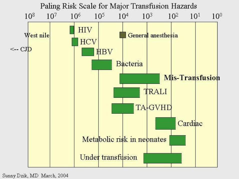

Deaths Due to Transfusion
• # 1 cause of transfusion-attributed death: TRALI
• overall risk of transfusion-related death: 1 in 200,000 chance
= risk of death from anesthesia in a fit person

Transfusion attributable symptoms, what they mean, &
how they happen
feverdyspneaallergy
cytopeniasdelayed infections

FeverHTR
BaConFNHTR

when is it a fever?
• T > 38ºC AND ↑ by Δ1ºC
OR
• the fever equivalent of chills or rigors

Acute Hemolytic Transfusion Reaction
• immune– active (recipient antibodies)– passive (donor antibodies)
• non-immune– devices physically destroying RBCs– thermal injury to RBCs (hot/cold storage
misadventure)– pressurized infusions – post-expiry infusions

Acute Hemolytic Transfusion Reaction
• active AHTR
–recipient has, and can keep making, specific antibodies against a transfused cellular product that bears vulnerable antigens
antibody-mediated hemolysis of transfused red cells
R
D
transfused allogeneic red blood cells
D host allo-antibody attacks alloantigen

Acute Hemolytic Transfusion Reaction
• passive AHTR
–product carries donor antibodies which are specific for a recipient’s cells
antibody-mediated hemolysis of recipient red cells
high plasma-volume containing products (FFP, platelets)
R

Causes of active AHTR• ABO mistake
– failure to avoid anticipated ABO antibody– group O patient given non-O RBC *
• anti-A and anti-B are naturally occurring antibodies and hemolysis is immediate
• missed or hidden non-ABO antibody– failure to find unanticipated non-ABO antibody– use of emergency ‘uncrossmatched’ blood– antibody not detected on antibody screen
* : ½ of these mistakes are due to hanging the properly labelled blood on the wrong patient, and many of the remainder are due to the recipient being wrongly characterized because of sampling errors

the point of the SCREEN (IAT): preventing “minor” RBC antigen-based HTRs…but sometimes failing
• relevant to finding recipient antibodies to non-ABO antigens
• utilizes O red cells
plasma
1
2
ABO
Rh(D)
Kell
Duffy
Kidd
etc

AHTR: clinical facts• presentation: fever/chills, hemoglobinuria, IV or
flank pain, ↓BP, nausea/vomiting, dyspnea, renal failure, DIC
• case fatality rate: <10%
• non-morbidity rate: >50%
• + “dose-response” risk (↑ blood = ↑ risk)
• cases are usually ABO-related• non-ABO antigens implicated in 13% of AHTRs

AHTR: How to pick it up…
patient’s cells
directCoomb’stest
look for evidence of red cell incompatibility • clerical error search
• re-type
• direct antiglobulin test (DAT), aka Coomb’s test
•assess plasma for hemolysis10cc (or only 3% of a unit) of blood is visible…!
• re-screen or re-crossmatch
• review labs indicative of hemolysis

Suspected HTR: What to doStop transfusion & run NS; check labels
Notify blood bank, send post-transfusion specimen & remainder of product
Stat lab work:
acute renal failure work-up:
electrolytes (hyperkalemia), creatinine
DIC work-up:
PT/INR, aPTT, fibrinogen
Support BP & maintain good urine output
Manage coagulopathy and bleeding

Bacterial Contamination (BaCon)INCIDENCE
• Risk of septic shock:– 1 in 10,000 PPC– 1 in 100,000 RBC
• Risk of death from septic shock:– case fatality rate: 24-60%– 1 in 40,000 PLT pools– 1in 1,000,000 RBC
• Key Message – Run transfusion slowly for 1st 15 min

BaCon
ETIOLOGY• donor bacteremia, skin plug, processing• most commonly:
– Yersinia enterocolitica– Serratia marcescens/liquefaciens– Staph aureus/epidermidis
PRESENTATION• fever, rigors, hypotension, renal failure, DIC• platelets more commonly implicated• RBC more severe

FNHTR: what it is
• = Febrile Non-Hemolytic Transfusion Reaction
• a diagnosis of exclusion– after ruling out:
• hemolytic transfusion reaction• bacterial contamination• underlying illness in patient
• common– 1/10 platelet transfusions– 1/300 red cell transfusions

FHNTR: why it happens•recipient “leuko-agglutinins”
–patient previously sensitized to antigens on leukocytes
•old transfusions•pregnancies
–host antibodies react with passenger leukocytes in blood product
•product “pyrogens”–residual leukocytes secrete cytokines–occurs more often with:
•non-leukoreduced products•products under warm storage•products near expiry
(STORAGE
LESION)
donor leukocytewith an HLA antigenrelevant to hostHLA alloantibody
donor leukocytesecretes cytokinesin storage
D D

FNHTR: how to manage• acetaminophen• meperidine (Demerol) for rigors
• PREVENTION?– acetaminophen
• may not work• may obscure an important febrile cue to stop
next transfusion
– fresh components• not always available
– plasma-reduced or washed products• risks product degradation/loss

IVIG & Immune Reactions• immune-replacement / immune-modifying
therapy = unique immune-mediated adverse effects, attributed to:– cytokines, vasoactive peptides, complement
activation via Ig dimerization
• common minor effects:– headache, fever, fatigue, chills, vomiting,
nausea, dizziness, diarrhea, flushing, abdominal pain, chest tightness, cough, muscle cramps, pruritis, backpan
• rarer, more serious effects: – sucrose-induced nephropathy, aseptic
meningitis, hemolysis, anaphylaxis, vascular thrombosis

DyspneaTACOTRALI
pulmonary allergic reaction (bronchospasm)

what to do?
• assess patient (focus on cardiorespiratory vitals & volume status)
• stop transfusion, notify blood bank
• stat CXR + ABG if hypoxic oximetry
• treat patient:– oxygenate if hypoxic– diurese if +evidence of volume overload

TACO• Transfusion-Associated Circulatory
Overload
• incidence:– 1 in 700 patients overall– 1 in 100 elderly recipients of blood
• specific clues:– fast infusion rate– compensated chronic anemia (hyperdynamic
before transfusion)– known congestive heart failure risk– orthopnea, ↑JVP, cyanosis, ↑HR, ↑BP

TRALI: what it is• Transfusion Related Acute Lung Injury
= NON-cardiogenic pulmonary edema
• definition:– new acute lung injury (PaO2/FiO2 <300)
– onset during or within 6h of transfusion– excludes:
• TACO• underlying CHF• other pre-transfusion pulmonary morbidities
• under-recognized & under-reported– estimates: 1 in 1200 to 1 in 5000

TRALI: why it happens•donor “leuko-agglutinins”
–multiparous donor sensitized to WBC antigens –passive antibodies venously pumped through lungs & encounter host leukocytes & react–explains ≥ 75% of cases
•product toxins–cellular products release biologically active lipids–interact with illness-“primed” pulmonary endothelium to produce tissue damage
recipient leukocytewith an HLA antigenrelevant to donorHLA alloantibody
donor cells release biologically active lipids
D
H

TRALI: outcomes• symptoms & signs:
– dyspnea, cough, hypoxia, fever, BP ↑ or ↓, WBC ↓
• resolves in 24-72 hours• 72% require mechanical ventilation• death in 5-10%
– most common cause of transfusion-attributed death now
• supportive care is key– diuretic utility in non-cardiogenic edema?
• prevention:– defer implicated donors – new mandates may restrict use of female plasma

Allergymucocutaneous vs multisystemic
IgA deficiency & other causes

The Allergic spectrum
• hives < 2/3 of body surface area– mild urticaria, pruritis, erythema, flushing
• hives > 2/3 of body surface area
• angioedema (subcutaneous rather than cutaneous)
• respiratory:– bronchospasm
• wheezing, stridor, hoarseness, dyspnea, hypoxia, psychologic sense of asphyxia/doom
• gastrointestinal instability:– nausea/vomiting/abdominal cramping/diarrhea
• cardiovascular instability:– hypotension, chest pain, tachycardia
• anaphylactoid / anaphylactic reaction
90%

Why allergic reactions happen•Recipient-donor allergen-IgE interactions
–classic allergic IgE antibodies and antigens, with one or the other passively infused and interacting in host
•drugs, foods, chemicals
–likely the most common explanation–no routine tests or traceback methods to confirm–URTICARIA to ANAPHYLAXIS
•Recipient IgG sensitization to normal donor proteins
–recipient has a rare polymorphism or deficiency of a certain protein compared to most donors
•eg: IgA, haptoglobin, complement, albumin, α1anti-trypsin, transferrin
–exposure to proteins = immunizing event–anti-protein IgG
=ANAPHYLACTOID REACTION

a few words on IgA deficiency• 1 in 500 are IgA deficient
• 1 in 1200 have anti-IgA IgG
(ie- proof of sensitization)– mysteries:
• why/how IgG raised without known exposure • why ½ don’t react if exposed later
– explains why reactions may be observed in a 1st transfusion

How often & how severely they happen• within 1-45 min of transfusion, but up to 3 h
afterwards
• if severe: – as little as 10cc may be anaphylactic
• dose-responsive
• relapsing
• frequencies:– minor: 1/100– severe allergic: 1 in 10 000 to 40 000
– fatal anaphylaxis: 1 in 1 000 000

Management of Allergic Reactions
• stop transfusion if >2/3rd BSA involved or non-cutaneous (visceral) allergic symptoms
• allergic anti-histamines– diphenhydramine (Benadryl), cetirizine (Reactine)
± H2R blockers – ranitidine (Zantac)
• epinephrine, β2 receptor agonist bronchodilators• corticosteriods• ACLS: vasopressors, ventilatory support
• future transfusions:– modify the patient:
• premedication– modify the product:
• ± plasma depletion / washing • obtain from donors deficient in the inciting allergen/antigen

HypotensionHTR
BaConAllergy
Bradykinin

Bradykinin-mediated hypotension• bradykinin surge from contact activation:
• for donors or recipients on ACE inhibitors: – reduced bradykinin breakdown from cross-
reactive suppression of enzyme from drug
• recipients of bradykinin: – flushing, hypotension, dyspnea, hypoxia

CytopeniasRBC (HTR)
platelets (HLA PTR, PTP)WBC (TRAIN)
pancytopenia (TA-GVHD)

RBC decreases after transfusion• if immediate & not related to hemorrhage:
– investigate for acute hemolytic transfusion reaction
• usually related to ABO antibodies (preformed) & suspected from fever/hypotension
• if delayed (3-14 days after transfusion):– investigate for delayed hemolytic transfusion
reaction (DHTR)• usually non-ABO antibodies that took time to “re-
surge” after the offending red cell transfusion• may not have symptoms of hemolysis (fever,
hemoglobinuria)
– 1 in 6715 transfusions– in future: transfuse blood negative for antigen

Platelet decreases after transfusion
•HLA (human leukocyte antige) sensitization
–recipient makes HLA antibodies from past exposure to WBCs
•other transfusions
•pregnancies
•transplants
–platelets express 73% of circulating HLA class I (A & B) antigens–recipient can destroy platelets bearing vulnerable HLA antigens–the cause of 80-90% of platelet transfusion refractoriness
•HPA (human platelet alloantigen) sensitization
–recipient with a polymorphic or missing platelet glycoprotein makes antibody against the common glycoprotein type
•pregnancies
•other transfusions
–recipient can destroy platelets bearing vulnerable HPA antigens–leads to PTP (post-transfusion purpura)–fatal in 8%–fortunately RARE!

Platelet-Specific Antigens in PTP
Platelet Antigen System
Protein Antigen
AllelesAntigen Frequency
HPA-1 GPIIIa
HPA-1a = PlA1
HPA-1b = PlA2
97%3%
HPA-2 GPIbHPA-2AHPA-2b
99%14%
HPA-3 GPIIbHPA-3aHPA-3b
85%66%
HPA-4 GPIIaHPA-4aHPA-4b
>99%<1%
HPA-5 GPIaHPA-5aHPA-5b
99%20%
But fortunately only 28% make the antibody if exposed
Most frequently involved polymorphism (75% of PTP cases)

Post transfusion neutropenia
•donor anti-granulocyte antibodies
–although usually implicated in TRALI, may also (or instead) cause neutropenia of host’s neutrophils–passive alloimmune reaction

TA-GVHD• Transfusion-Associated Graft Versus Host
Disease
• passenger lymphocytes “engraft” in recipient– who:
• immune compromised• immune recognition failure
– what happens: • bone marrow aplasia (empyting from attack) =
pancytopenia (all counts down)• fever, rash• liver dysfunction, diarrhea• death in 90%

Vulnerable recipients•immune compromised
–congenital immunodeficiency–fetuses, premature neonates–hematologic malignancies (eg lymphoma)–stem cell transplants–high dose chemotherapies with immune-ablation potential–nucleoside analogue drugs (eg fludarabine for chronic lymphocytic leukemia)
•immune “oblivious”–host’s HLA type is foreign to transfused lymphocytes, which resemble the host enough to be tolerated
•eg. donor is HLA “homozygous haploidentical” with respect to recipient
•related donors
•1 in 18 000 – 39 000 of strangers
A1 A2B7 B9C4 C8
recipient tissue type
A2 A2B9 B9C8 C8
donor lymphocyte

Delayed onset infectionsViruses (H A/B/C V, HIV, CMV, HTLV, WNV, PVB19, HHV8)
Prions (vCJD)
Others (protozoal, helminthic, spirochetal, rickettsial)

Viral testing of each donation• all donors:
– HIV Ab, HIV NAT – HCV Ab, HCV NAT – HBsAg, HBcAb– Syphilis– HTLV-1/2 Ab– WNV NAT
• selected cases/products:– CMV Ab – bacterial culture (pre-pooled/high volume platelets only)– Parvovirus B19 (fractionated products only)
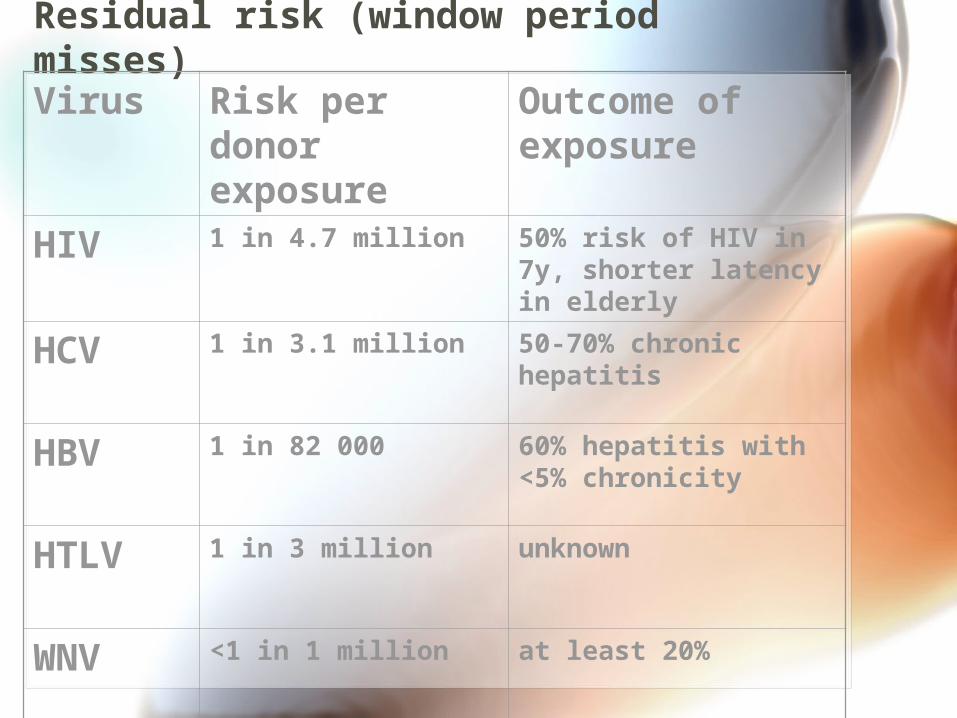
Residual risk (window period misses)
Virus Risk per donor exposure
Outcome of exposure
HIV 1 in 4.7 million 50% risk of HIV in 7y, shorter latency in elderly
HCV 1 in 3.1 million 50-70% chronic hepatitis
HBV 1 in 82 000 60% hepatitis with <5% chronicity
HTLV 1 in 3 million unknown
WNV <1 in 1 million at least 20%

Other transfusion transmissible infections
•Malaria•Toxoplasmosis•Leishmaniasis•Filariasis•Ehrlichiosis•Babesiosis•Trypanasoma cruzi (Chagas)•Borrelia burgdorferi•Rocky mountain spotted fever (rickettsial group)•Q fever (Coxiella burnetti)•variant CJD
at this time,screeningby donor history alone is performed in order to prevent the entry of these infections in the donor supply