Advances In Musculoskeletal Intervention - pedrad · Advances In Musculoskeletal Intervention Neil...
69
Advances In Musculoskeletal Intervention Neil Johnson, MB.BS, M.Med FRANZCR William Shiels, DO Cincinnati Children’s Hospital Nationwide Children’s Hospital
Transcript of Advances In Musculoskeletal Intervention - pedrad · Advances In Musculoskeletal Intervention Neil...

Advances In Musculoskeletal Intervention
Neil Johnson, MB.BS, M.Med FRANZCR
William Shiels, DO
Cincinnati Children’s Hospital Nationwide Children’s Hospital

Disclosures
• Dr. Johnson CCHMC is a Research Site for Philips Medical
– Research Agreement / I.R. Animal Lab
– No Personal Financial Benefits
• Dr. Shiels
• Basic MSK Intervention
• Beyond Basics – Core Biopsy
– Treating Lesions
– Screws, Bone Grafts and Hardware
• Two Important Lesions – Histiocytosis (LCH)
– Aneurysmal Bone Cyst
• Advanced Guidance and Fusion Imaging
• A Little Politics
OUTLINE

MSK Intervention: Basics
• Image Guidance
– CT / CT Fluoroscopy
– Ultrasound
– Standard Fluoroscopy
– Cone Beam CT +/- Guidance
– Combined / Fusion Imaging
• “Needle” Biopsy
– Cytology
– Small Diameter < 4mm
• Automated Gun
• True Cut (Slot) Type Devices: Fibrous Lesions
X

MSK Intervention: Basics
• Abscess Drainage
– Similar To Other Sites
• Joint Injections
– MRI Arthrography
– Steroid Injections
• Joint /Tendon Sheath
• Bursa
• Marking Deep Lesions for Surgery
• Foreign Body Removal

MSK Intervention: Beyond Basics
• Deep Large Core Bone Biopsy
– Equipment
– Guidance
• Malignant Tumor Biopsy
– Intelligent Approach Paths
– Viable Tissue – “The Edge Is The Target”
– Exceptions: When Even Good Biopsies Go Bad
• Screws, Routers and Bone Grafts
– Orthopedics Through Small Holes

Beyond The Basics NF1 Malignant Nerve Sheath Tumor: ? Mets to Sternum and T1
Ultrasound Guided Biopsy of Sternum for Diagnosis

2 Months: Metastasis Enlarged
*
Mediastinum

Palliative R.F. Ablation of Sternal Met Ultrasound guidance
*

Tendon Sheath Steroid Injections
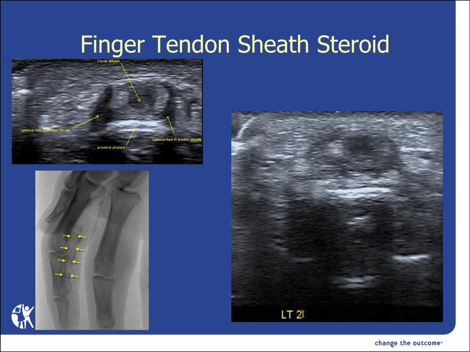
Finger Tendon Sheath Steroid

On An Awake (Smiling!) Patient

I.R. In The Operating Room

Cone Beam CT + Guidance: The Complex Angled Approach
• Planning 3D Low Dose CT Equivalent
• Complex Angled Approach (On Screen Guidance)
• High Quality Fluoroscopy

Post Traumatic Physeal Bar

Ultrasound: Avoiding Major Structures
Netter Radial Nerve?
Desmoplastic Fibroma
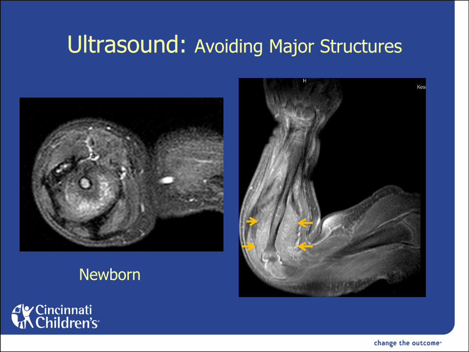
Ultrasound: Avoiding Major Structures
Newborn

Newborn: Forearm Biopsy
“Fibromatosis Coli”

Mediastinal Germ Cell Tumor: SVC Syndrome
*

Bone Tumor Biopsy: Co-Ordination with Surgery
Radiographics 2007; 27: 189-206

Always Co-Ordinate with Surgery
Radiographics 2007; 27: 189-206

Caution……

Posterior Component… Not Sampled (Sciatic Nerve + Surgery)

Original Diagnosis:
Benign Desmoplastic Fibroma Variant So…Open Curettage and Bone Graft……
BUT……

Two Special Lesions
• Langerhans Cell Histiocytosis
– Solitary Bone Lesion
• Aneurysmal Bone Cyst (ABC)

Langerhans Cell Histiocytosis
• Histiocytoses:
– Group of proliferative disorders arising from histiocytes, a common progenitor cell in bone marrow.
• 3 types of Histiocytes (dendritic cells)
– Langerhans cell: Epidermis
– Mononuclear Cell/Macrophage: Dermis
– Dermal dendritic cell: Dermis
• LCH and non – LCH Histiocytoses
Courtesy Dr. Joseph Palumbo, MD CCHMC

Types of Histiocytes: “Its Too Complicated for Radiologists”
CD 34+
CD 14+
DDC
MΦ
CD14- LC
Fitpatrick’s Dermatology in General Medicine, pp 106 CD1a

• Infectious? Disseminated, spontaneous remission of milder forms
– CMV, EBV, HHV-6, HHV-8 implicated; none proven
• Neoplastic ?
• Reactive Clonal Disorder ?
LCH Pathogenesis –Theories

• Congenital Self-Healing Reticulo-histiocytosis
– AKA Hashimoto-Pritzker disease
• Eosinophilic Granuloma
• Hand-Schuller-Christian disease
• Letterer-Siwe disease
Histiocytosis Clinical Types Old Classification

• Single system
– Isolated Bone Lesions (Best Prognosis***)
• Multisystem
• Disseminated
– Widespread, multi-organ disease (Poorest Prognosis)
Histiocytosis Clinical types Current Classification

LCH Isolated Bone Lesion Skull
Langerhans Cell Histiocytosis (LCH)

LCH: Ultrasound Guidance Biopsy and Steroid Infiltration
Depo Medrol 40 mg (Methylprednisolone Acetate)
Brain

LCH Skull

LCH Primary I.R. Treatment

Biopsy and Steroids

But….
*
*
*

2 Months Post Steroids
* *
*
At Diagnosis

LCH Rib

LCH Rib

LCH Rib: Ultrasound and Cone Beam CT

Post Procedure……..
But…

Histology: No Active Lesion

9 Year Old - LCH Pubis

LCH Pubis: Curettage, Steroids and Percutaneous Bone Graft

LCH Pubis: Curettage, Steroids and Percutaneous Bone Graft

20 Months Post Treatment

Incision for Pubic Ramus Surgical Approach:
(Giant Cell Tumor)
Right Leg Abdomen

LCH: Acetabulum Roof

LCH: Steroids and Bone Graft

LCH: Steroids and Bone Graft Primary and Only Treatment
5 Months

Aneurysmal Bone Cyst
• Expansile Lytic Vascular Lesion of Bone
• 1.4 / Million Individuals
• Usually < 20 Years Old
• Male = Female
• Occurs In All Bones
– Most Common:
• Pelvis
• Spine ( Posterior Elements)
• Long Bones
Cottalorda, Arch Orthop Trauma Surgery (2007) 127: 105-114

Aneurysmal Bone Cyst
• 70% Primary
• 30% Secondary
– Chondroblastoma
– Osteoblastoma
– Giant Cell Tumor
– Fibrous Dysplasia
– Malignant Bone Tumors
• *** Telangiectatic OsteoSarcoma ***
Aneurysmal Bone Cyst
• Differentiation from Unicameral Bone Cyst (UBC)
– Single Cyst Vs Multiple Cysts
– Fluid Level Less Likely in UBC
– UBC Less Expansile
• BUT
– Complicated UBC (Fracture) May Be Difficult
– Biopsy Required
• UBC: Simple Cyst Lining Vs ABC
• UBC Different Histology

Aneurysmal Bone Cyst

2nd Procedure: Curettage, Bone Graft and Steroids

12 Months

Percutaneous Bone Grafting: ABC
* *
Aneurysmal Bone Cyst
• Causation: Primary ABC
– Venous Obstructive Lesion
• Post Traumatic
• Post Infection
– Vascular Malformation
– Benign Neoplasm
• 16:17 q22:p13 Translocation [1]
• TRE17 / USP6 Oncogene Translocation [2]
[1] Panoutsakopoulos G, et.al. Recurrent t(16;17)(q22;p13) in aneurysmal bone cysts. Genes Chromosomes Cancer. 1999;26:265-266. [2] Ye Y, et.al. TRE17/USP6 oncogene translocated in aneurysmal bone cyst induces matrix metalloproteinase production via activation of NF-xB. Oncogene. 2010;29:3619-3629

Aneurysmal Bone Cyst
• Treatment Options
– Traditional Open Surgery
• 12-71 % “Recurrence” [1]
• Significant Complications
– Blood Loss, Loss of Function (Plates / Screws), Infection
– Radiotherapy
• Secondary Malignancy
– Percutaneous Sclerotherapy
• STS
• Ethibloc
• Doxycycline

Aneurysmal Bone Cyst
• Treatment Options
– Hybrid
• Minimally Invasive CT Guided (<1cm Incision)
• Curettage / Routing / Aspiration
• Steroid Soaked Percutaneous Bone Graft
– Image Guided Doxycycline (Dr. Shiels)
• Ultrasound or CT Guided
• Minimally Invasive
• Cysts Individually Targeted
• Doxycycline Suppresses Multiple Cellular Abnormalities
– Metalo Matrix Proteins (MMP)
– VEGF

Tumors:
• Biopsy Guidance Ultrasound Vs CT
• Avoidance of Major Structures
• Color Doppler: Identifying Viable Tumor
Ewing’s Sarcoma

17 Year old Male 5 months Left Hip Pain

Primary Ultrasound Guidance

Ultrasound Guidance

Same Patient……. Diagnosis Please

2.5 Year Interval: Diagnosis ….

2.5 Year Interval: Diagnosis Please….

Hybrid Ultrasound Guidance ? Best of Both ?
Magnetic Field Plate Under Patient

IR / OR of the future Image Guided Orthopedics: We Need To Be There
• Surgery
– General, Ortho, Neuro
• Pulmonology /GI
• Oncology
• Led by IR ….Or Not