Advances in analytical methodologies for - DiVA portal360221/FULLTEXT03.pdf · Advances in...
54
Advances in analytical methodologies for studies of the platinum metallome in malignant cells exposed to cisplatin Yvonne Nygren Akademisk avhandling Som med vederbörligt tillstånd av Rektorsämbetet vid Umeå Universitet för avläggande av filosofie doktorsexamen vid Teknisk naturvetenskapliga fakulteten, framläggs till offentligt försvar vid Kemiska institutionen, Umeå Universitet, sal KB3B1, Stora hörsalen, KBC, fredagen den 26/11, kl 13:00. Avhandlingen kommer att försvaras på engelska. Fakultetsopponent: Prof. Bente Gammelgaard, Department of Pharmaceutics and Analytical Chemistry Faculty of Pharmaceutical Sciences, University of Copenhagen [email protected]
Transcript of Advances in analytical methodologies for - DiVA portal360221/FULLTEXT03.pdf · Advances in...

Advances in analytical methodologies for studies of the platinum metallome in malignant cells exposed to cisplatin
Yvonne Nygren
Akademisk avhandling Som med vederbörligt tillstånd av Rektorsämbetet vid Umeå Universitet för avläggande av filosofie doktorsexamen vid Teknisk naturvetenskapliga fakulteten, framläggs till offentligt försvar vid Kemiska institutionen, Umeå Universitet, sal KB3B1, Stora hörsalen, KBC, fredagen den 26/11, kl 13:00. Avhandlingen kommer att försvaras på engelska. Fakultetsopponent: Prof. Bente Gammelgaard,
Department of Pharmaceutics and Analytical Chemistry Faculty of Pharmaceutical Sciences, University of Copenhagen [email protected]

Title Advances in analytical methodologies for studies of the platinum metallome in malignant cells exposed to cisplatin Svensk titel Förbättrade analytiska metodologier för studier av platina-metallomet i maligna celler exponerade för cisplatin. Keywords Method development, Metallome, Inductively coupled plasma mass spectrometry, Electrospray ionization mass spectrometry, Liquid chromatography, Organic modifier, Cisplatin, Platinum Language: English ISBN: 978-91-7459-085-2 Number of Pages: 44+ Five papers
Abstract The scientific progress about the important chemotherapeutic drug substance cisplatin (CDDP) and its function has often been rendered by data difficult to interpret, and still many questions about its mode of action remains to be clarified by the scientific community. However, studies of CDDP possess a high complexity due to; i) low intracellular concentration, ii) many potential biomolecule targets, iii) poor or unknown stability of the intact drug and its biomolecule adducts and iv) complex and varying sample matrices. Metallomic studies, using advanced analytical techniques may contribute to clarify the interactions between CDDP and intracellular biomolecules. For a successful outcome sample preparation conditions as well as separation and detection techniques must be carefully selected and optimized to achieve accurate results and correct interpretation of data. This thesis describes some new and improved analytical methodologies for characterizing the Pt metallome in CDDP-exposed malignant cells. The developed methods are based on powerful liquid chromatography (LC) methods hyphenated to sensitive detection by inductively coupled plasma- (ICP) and electrospray ionization mass spectrometry (ESIMS). Consideration has also been taken about sample preparation conditions. By selecting “chemically inert” sample preparation (cell lysis by osmosis) and separation (using only nonreactive or no additatives) conditions we could avoid the formation of platinum artifact compounds previously described in the literature (Paper I and II). Using oxygen containing organic solvents with high boiling points (dimethylformamide; DMF, 1,4-dioxane, n-propanol and ethanol) as alternatives to acetonitrile in the LC separations, significant improvements were achieved in ICPMS sensitivity and robustness. When evaluated in combination with chromatographic performance and ESIMS detection the overall best performance was achieved with n-propanol (Paper II, III and IV). From the studies in Paper II we could show that free intact CDDP can be found in malignant cells, as supporting evidence for passive or endocytotic uptake of the drug and further estimate a half-life for intracellular CDDP to about 15 minutes. Such data has not been shown before. In Paper V, the above improved LC methods were used to demonstrate differences in the platinum and cupper metallome from sensitive and resistant T289 melanoma cells exposed to CDDP at near clinical levels.
I

In a wider perspective we have shown the potential of using hydrophilic liquid interaction chromatography (HILIC) hyphenated to ICPMS detection as a general approach for analysis of hydrophilic metallo-compounds (Paper II). Taking advantage of the superior ICPMS performance using n-propanol gradients for reversed phase liquid chromatography (RPLC) possess a true alternative and /or complimentary technique to size exclusion chromatography (SEC) commonly applied within metallomic studies of biomolecules (Paper V). Using n-propanol in HILIC as well as in RPLC enables parallel detection by ICP- and ESIMS using only one set of chromatographic parameters (Paper III and IV), something commonly called for by scientists in the field.
II

This thesis includes the five following papers, which are referred to in the text by their Roman numerals (Ι) M.Q.T.Tran, Y Nygren, C Lundin, P Naredi and E Björn
Evaluation of cell lysis methods for platinum metallomic studies of human malignant cells Analytical Biochemistry 396 (2010) 76-82
(ΙΙ) Y Nygren, P Hemström, C Åstot, P Naredi and E Björn Hydrophilic interaction liquid chromatography (HILIC) coupled to inductively coupled plasma mass spectrometry (ICPMS) utilizing a mobile phase with a low-volatile organic modifier for the determination of cisplatin, and its monohydrolyzed metabolite Journal of Analytical Atomic Spectrometry 23 (2008) 948-954
(III) P Hemström, Y Nygren, E Björn and K Irgum Alternative organic solvents for HILIC separation of cisplatin species with on-line ICPMS detection. Journal of Separation Science 31 (2008) 599-603
(IV) Y Nygren and E Björn Liquid chromatography mobile phase selection for the combined use of LC-ICPMS and LC-ESIMS. Journal of Chromatography 1217 (2010) 4980-4986
(V) Y Nygren, C Åstot, P Naredi and E Björn
The platinum and cupper metallome from sensitive and resistant T289 melanoma cells exposed to Cisplatin studied by a multi-analytical approach. Manuscript in preparation
III

Also by the author but not included in this thesis (i) E Björn, Y Nygren, T Nguyen, C Ericson, M Nöjd, P Naredi
Determination of platinum in human subcellular microsamples by inductively coupled plasma mass spectrometry Analytical Biochemistry 363 (2007)135-142
IV

Authors contribution Paper (I) Partly planned and performed the study. Partly supervised M.Q.T. Tran, a diploma student that was doing most of the experimental work. Initially developed the analytical method used for the analysis. Paper (II) Major responsibility for planning of the study. Assembled and optimized the analytical experimental setup and performed the experimental analysis. Wrote the initial draft of the paper and participated in completing it with co-authors. Paper (III) Planned and performed some of the analytical experiments of the study together with P. Hemström. Paper (IV) Major responsibility for planning of the study and writing the paper. Assembled and optimized the analytical experimental setup and performed the experimental analysis Paper V) Major responsibility for planning of the study and writing the manuscript. Assembled and optimized the analytical experimental setup and performed the experimental analysis.
V
List of Abbreviations ICPMS Inductively Coupled Plasma Mass Spectrometry ESIMS Electrospray Ionization Mass Spectrometry HPLC High Performance Liquid Chromatography HILIC Hydrophilic Interaction Liquid Chromatography RPLC Reversed Phase Liquid Chromatography NPLC Normal Phase Liquid Chromatography SEC Size Exclusion Chromatography AC Affinity Chromatography LOD Limit of Detection S/N Signal to Noise CDDP cis-Diammindichloroplatinum, Cisplatin PBS Phosphate buffered saline
VI

Table of Contents 1. Introduction 1 2. Platinum anti-tumor drugs; their use and function 3
2.1 CDDP and its use as an anti-tumour agent 4 2.2 Mode of action of and resistance 4
towards CDDP in cancer treatment 2.3 Analytical challenges in studies
of cisplatin and its metallome 7
3. Sampling and Sample Treatment 12 3.1 Preparation of cell lysate 12 3.2 Summary of Papers 13 4. Liquid Separation 14 4.1 Reversed phase liquid chromatography 15
4.2 Hydrophilic Interaction Liquid Chromatography 16
4.3 Size Exclusion Chromatography 17 4.4 Summary of papers 17
5. Detection 24
5.1 Inductively Coupled Plasma Ionization Mass Spectrometry (ICPMS) 24
5.2 Electrospray Ionization Mass Spectrometry (ESIMS) 28
5.3 Combining ICPMS and ESIMS in metallomic work 31
VII

6. Quantitative and/or Qualitative Evaluation 33
6.1 Calibration methods for quantitative analysis 33
6.2 Considerations in qualitative analysis 35 6.3 Summary of Papers 36 7. Concluding Remarks and Future Trends 38 8. Acknowledgement 39 9. References 41
VIII

1. Introduction Analytical chemistry is a science concerning development and improvement of methods for detection and determination of chemical substances (species) in various matrices. Determining the presence and identity of a species in a given sample represents one of the two main parts, namely qualitative analysis, while finding the relative and/or absolute concentration represents quantitative analysis. The analytical chain often includes several events important for the outcome of the analysis. In planning, sampling, sample pretreatment, separation of sample components as well as in the detection, evaluation and data interpretation steps, quantitative and qualitative unreliabilities can be introduced which all together in the end contribute to a result that can be difficult to interpret and sometimes even totally incorrect. With proper knowledge, awareness, experience, and thorough planning these combined unreliablilities can however be decreased to a minimized level. In developing a method the main aim for the analytical chemist is to achieve a robust and reproducible system, often with a high power of detection to allow, analyzing low abundant species in complex matrices. Within life-science it is of crucial importance to produce reliable and correctly interpreted analytical data to avoid the risk of drawing wrong conclusions when using the data to understand biological functions and reactions. Analyzing samples of biological origin such as blood plasma, tissue, urine or cell lysates is a challenge even when using advanced and modern instrumentation and requires skill and experience of the researcher developing the method. Following the eras of genomics, proteomics and metabolomics, analytical chemistry is now for sure facing metallomics, reflected in the number of papers currently being published in the area. Determining the metallome, defined as the entirety of metal or metalloid species within a cell or tissue type, extensive analytical protocols combining different techniques are often required as the metallome often comprises a range of analytes differing in size and hydrofobicity. Metal-binding compounds often play essential roles regulating biological reactions and physiological functions in cells and organs. Metalloenzymes catalyses important biological functions, metal-binding biomolecules are involved in degeneration processes and furthermore, drugs containing metal atoms are frequently used in medicine. An extensively explored area is platinum containing anti-tumor drugs which are very efficient and widely used in treatment of solid tumors, although
- 1 -

their use is hampered by side effects and acquired resistance to the drugs. The mechanisms for drug function and resistance are still today poorly understood. Metallomic studies offer a way to improve the knowledge about the faith of these drugs in the organism, however, to achieve an accurate results and a correct interpretation of the analytical data, the development and use of refined methodologies is of crucial importance. The aim of this thesis was to develop analytical tools for fundamental studies on intracellular metabolism of the platinum-containing anti-tumor drug cis-Diammindichloroplatinum (CDDP, cisplatin) with the ultimate aim to better understand the mode of action and cell resistance towards such drugs. The central objectives were to; i) accurately quantify the uptake of total platinum and CDDP in malignant cells , ii) identify intracellular metal-binding ligands, verify the occurrence of the complexes in a “real environment” and quantify their concentration and iii) characterize how the Pt-metallome differ in CDDP sensitive and resistant cell lines.
- 2 -
2. Platinum anti-tumor drugs; their use and function
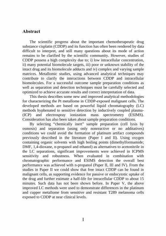
When CDDP was first synthesized in 1845 by Michele Peyrone [1], no one could have foreseen its future role in medicine. The new platinum compound, containing two ammines and two chlorines was similar with “Reyset’s salt” but showed to have different physiological properties. This finding was in 1893 proposed by Alfred Werner [2] to be explained by a square planar geometry of the compound, accommodating a cis- and trans-isomer with different properties (Figure 1).
Figure 1. Cisplatin (Peyrone´s) and Transplatin (Reyset´s) CDDP´s property as an anti-tumor agent was later discovered accidently by Dr Roosenberg [3]. In evaluating if electric or magnetic dipole fields affected bacterial cell division, platinum electrodes were included in the growth chamber set up. The experiment showed that differences in cell growth were not dependent on the electric field but rather to electrolytic products formed at the platinum electrodes. Later, as the structure of CDDP was identified as the active complex the astonishing result that the compound completely inhibited development of Sarcoma-180 tumors in mice [4, 5], was published. The first clinical trials were performed in 1971 and subsequently the approval by the US Food and Drug Administration (FDA) was granted in 1978. In an article describing Rosenberg´s discovery [6] the author discusses how unlikely the success of CDDP is. However, since the approval in 1978 it has become one of the most efficient and best selling anti-tumor drugs in the world.
- 3 -

2.1 CDDP and its use as an anti-tumour agent CDDP was the first platinum anti-tumour agent but are nowadays belonging to a family of platinum compounds which during a long time have been successfully and widely used for treatment of several types of solid tumors. Of the three compounds clinically approved in Europe and USA; cisplatin (CDDP), carboplatin and oxaliplatin (Figure 2), CDDP is considered as a complete cure for testicular cancer and to be one of the most efficient anti-tumor drugs for treatment of melanoma, ovarian, head and neck as well as non-small-cell lung cancers.
Figure 2. Clinically approved platinum-containing anti-tumor agents; cisplatin, carboplatin and oxaliplatin 2.2 Mode of action of and resistance towards CDDP in cancer
treatment 15 years ago Lippard and Berg [7] published a quite simple picture (Figure 3) illustrating the, at that time, gathered dominant hypothesis of the pathway from intravenous injection of CDDP to nuclear DNA as the therapeutic target.
- 4 -

Figure 3. Scematic illustration of cisplatin uptake
after intravenous injection from 1994. The publication makes some statements that are still supported in more recent scientific literature. Once entered into the blood stream CDDP is stabilized by the high (>100 mM) chloride concentration [8] whereas after entering into the cells by passive diffusion, hydrolysis is favored by the much lower (4-20 mM) intracellular chloride concentration [9, 10, 11]. The highly reactive mono- and di-aqua metabolites subsequently enter the nucleus and react with DNA by forming intrastrand crosslinks between adjacent purines [12]. Only 1 % of the intracellular CDDP is estimated to react with DNA; however it is still considered to be the main pathway causing anti tumoral activity [13]. Since then, extensive research has revealed a more complex picture of CDDP entering the cell, which recently was summarized by Gottesman et al [14] (Figure 4). Today, the majority of researchers agree upon a parallel passive and active uptake of CDDP into the cells. Some possible pathways
- 5 -

have been pointed out as important. Passive uptake is still supported by the fact that the cell does not seem to be saturated neither against exposure time or drug concentration [15, 16] but questioned by the theory that decreased uptake of CDDP by passive diffusion, commonly seen in resistant cell lines, must involve changes in the membrane composition, which not have been shown [17, 18, 19].
Figure 4. Proposed pathways for in- and efflux of CDDP (Reprinted with permission from [14]).
Several routes for active uptake of CDDP have recently been proposed. The first indication on active uptake of CDDP was made by Byfield et al observing that proliferating cells were more sensitive to the drug than resting ones [20]. Endocytosis is considered as an active uptake as it is energy dependent and has been suggested as a route for protection of intact CDDP until reaching the nucleus and DNA [21, 22]. Further, Na+, K+ ATPases, and organic cation transporters have been suggested as possibly
- 6 -

influx systems for CDDP [23, 24]. However one of the most supported theories and evaluated systems are the Cupper transporter 1 (Ctr1) membrane protein. Ctr1 is the first protein in a chain of proteins responsible for the in- and efflux of cupper. As cupper binds to this protein a conformational change induces a gated channel for cupper to entrance the cell. Via Atox1, a chaperone, Cu is further directed to the ATP7A and ATP7B systems for further transport to cell compartments and of excess cupper ions out from the cell. This system is by many scientists considered to be a possible transporter also for CDDP as indicated by an onset down-regulation of Ctr1 when exposing cells to CDDP [25, 26, 27]. Together with the general but non-proven apprehension that the glutathione (GSH) transferase system binds and transports cisplatin out from the cell [28], this is today the main theory behind the fact that accumulation of platinum in resistant cells has shown to be considerably lower than in sensitive. In addition to the above described theories, i.e. decreased accumulation of intracellular CDDP (Ctr1, ATP7B etc) and detoxification of cisplatin by thiols (GSH, proteins) improved repair of DNA [29, 30] is generally accepted as an important intracellular mechanisms by which cells acquire resistance to CDDP. 2.3 Analytical challenges in studies of cisplatin and its metallome A large number of studies have been performed about CDDP and its intracellular effects. Metallomic studies of Pt-based drugs possess a high complexity due to; i) low concentration in their real environment, ii) many potential biomolecule targets, iii) poor or unknown stability of the intact drugs and their biomolecule adducts and iv) complex and varying sample matrices. Most results for CDDP have been obtained from in-vitro experiments or incubation of standards offering a way to simplify the matrice and work with species at a clearly elevated concentration compared to what would be found in clinical samples. This has in fact in some cases lead to doubtful and contradictory results depending on the experimental conditions. The vast majority of CDDP studies are done in the scientific areas of medicine and molecular biology, however during the last years an extensive effort has been made to improve the detection and identification of metal-biomolecule compounds by using advanced analytical methodologies in order to evaluate CDDP’s function.
- 7 -

Powerful separation techniques in hyphenation with detection techniques mainly based on mass spectrometry has been widely employed in these studies. In parallel, sample preparation methods, using mild conditions, intended to preserve the original structures of target molecules has also been strived to be developed. Inductively Coupled Plasma Mass Spectrometry (ICPMS) is nowadays the most common technique used for metal element analysis, while Electrospray Ionization Mass Spectrometry (ESIMS) is the preferred technique for structure determinations, which is necessary for the final identification of metal-biomolecule species. Chromatographic separations have most frequently been performed with Size Exclusion Chromatography (SEC), Reversed Phase Liquid Chromatography (RPLC) and Ion Exchange Chromatography (IXC). The analytical studies of CDDP that have been performed so far mainly concern; i) stability in aqueous media, ii) interaction with blood molecules, iii) urine excretion, iv) accumulation and distribution in tissue, and v) various interactions within the cell. 2.3.1 CDDP stability in aqueous media In studies concerning the stability and reactivity of CDDP in aqueous media different chromatographic methods have been developed with the aim to detect CDDP and its hydrolysis products. Several of the publications use ion-pairing or ion exchange chromatography as an alternative to pure reversed-phase liquid chromatography in order to increase the retention of the charged hydrolysis products to enable separation of intact cisplatin and the hydrolysed forms [31-33]. However even if using ion exchange chromatography for the charged, hydrolysed species, retention for CDDP, as a neutral molecule, was mostly non-affected. In addition most of the early studies were done with either UV absorption spectrometry or off line element detection (GFAAS) giving only a preliminary identification of the species and rather poor detection limits [34, 35]. In an important contribution on the reactivity of CDDP, El-Khateeb et al [36] concluded from NMR studies that CDDP and its metabolites readily react with several additatives commonly used in HPLC and that precautions must be taken to avoid adding components that compete with natural ligands in the sample. This has also has been discussed by Heudi et al [37]. Later published methods have shown significant improvements in achieving excellent
- 8 -
detection limits by the use of chromatography coupled on-line to ICPMS detection [38, 39]. 2.3.2 CDDP interaction with blood molecules It is known that intravenously administrated CDDP, to a high extent, interacts with blood proteins and it has been shown that only 20% of the intact CDDP remains free in the blood stream 24h after administration [40]. Blood proteins known to have a role in transport of essential substances (human serum albumin; HSA, transferrin; Tf,) have been studied for a long time with different analytical approaches. HSA, has shown to have a high capacity for binding Pt-based drugs [41], while Tf does not interact to a high extent with such compounds [42a,b]. Even though the studies show the potential of using hyphenated techniques and proteomic approaches in studies of metal-protein interactions they also demonstrate the need of refined analytical methodologies in order to fully evaluate such interactions. 2.3.3 CCDP urine excretion Analysis of urine has been performed to evaluate the biological time course for CDDP and to determine its half-life [43, 44]. One main obstacle in interpreting these data is the variety of treatment schemes and dosages used. 2.3.4 CDDP accumulation and distribution in tissue In order to evaluate the treatment with CDDP many studies on the uptake and distribution in different target organs has been performed. The overall conclusion, whether using X-Ray Fluorescence [45] gamma camera spectroscopy [46] or ICPMS [47], is that liver, kidney and prostate are the organs with the highest levels of accumulated platinum after CDDP exposure.
- 9 -

2.3.5 Intracellular CCDP interactions As already mentioned, once entered into the cells, CDDP interacts with DNA as the primary therapeutic target. This interaction has been studied by X-ray crystallography, NMR and immunoassays [48-50] while lately HPLC-ICP-MS also has been used for characterization of the adducts [52, 52]. Other intracellular targets that have been subjected to investigation are mainly metallo-thionines [53, 54] and glutathione (GSH) [55-57a], both considered to play a role in detoxification of CDDP in the cell. The question how and if CDDP enters into the cell by protein-mediated active transport has lately promoted the analysis of other possible protein targets within cell systems. So far, striving for a better understanding of CDDP reactivity towards cellular biomolecules and set the basis for reliable analytical mehodologies, mainly in-vitro incubation with proteins has been evaluated. Efforts have been made to identify binding sites, metal-protein stochiometry and conformational changes of both CDDP and the protein in their interaction. Proteins commonly used in these types of experiments are superoxide dismutase, cytochrome C, insulin and ubiquitin. Recently, Franz et al [57b] published a study on CDDP’s interaction with the methionine-rich motifs thought to be the binding sites for CDDP in the Ctr1-protein as a potential influx transporter for the drug. Despite the large number of papers published in the area, so far only one publication [58] has shown a positive identification of a platinum-binding biomolecule (Pt-GSH) other than DNA in a real sample. This fact certainly reflects the analytical challenges future scientists have to encounter. In general it can be said that the scientific progress about CDDP and its function has often been rendered by data difficult to interpret. The large number of patients that has been totally recovered after cisplatin treatment supports the continued use, still many questions about its mode of action remains to be clarified by the scientific community. Mechanisms causing cell resistance to and side-effects of the drug still hamper its effectiveness in treatment of patients. From my point of view, analytical chemistry is playing an essential role in the continued work about CDDP´s mechanism of action. As pointed out in the introduction, by experience, thorough planning and proper knowledge, conclusions drawn from misinterpreted analytical data can be
- 10 -

avoided. By use of modern instrumentation and by well thought-out strategies, determining the intracellular platinum pathways should be of great advantage for cancer treatment in the future.
- 11 -

3. Sampling and Sample Treatment Evaluation of interactions between CDDP and biomolecules has so far mostly been done by in-vitro incubations. Extending their application to in-vivo studies implies a complexity that makes the sample preparation as well as the analysis more difficult. In addition, samples from animals or patients are not easily acquired as it requires ethical permissions and surgery. In between in-vitro and in-vivo preparations tumor specific in-vitro-grown cells, exposed to the drug, can offer an alternative more similar to real samples than pure in-vitro incubations. Throughout this thesis in-vitro grown T289 melanoma cancer cells have been used for the different studies. Cells for all studies were grown in RPMI-1640 medium supplemented with 10% fetal calf serum. The cells were grown to between 80 and 95 % confluence before exposed to varying concentrations of CDDP. After exposure, cells were either incubated in CDDP free medium for selected times and subsequently washed with phosphate-buffered saline (PBS) or directly washed. After the washing step cells were loosened and the cells were collected by centrifugation before treated with varying lysis protocols. 3.1 Preparation of cell lysate Three different lysis protocols have been evaluated in this thesis, freeze-thaw, osmosis and chemical detergent (Triton X-100) based. In the freeze-thaw method cells were cycled five times between 20 min freezing at -20 °C and 20 minutes thawing at +37 °C. For osmosis, cells were mixed with MQ-water, placed on ice for 4 hours while occasionally vigorously shaking was performed. In the chemical detergent based method a reagent mixture was added to the cells and kept on ice for 30 minutes. Before speciation analysis of CDDP the lysates were further treated on a 5 kDa cutoff centrifugation unit in order to discard macromolecules and remaining debris from the cells. Prior SEC and RP chromatography the cell samples were subjected to a 0.45 um filtering to discard particles and thereby preserve the column performance.
- 12 -

3.2 Summary of Papers In paper I the different lysis methods were evaluated as sample treatment procedures for platinum metallomic studies. The highest absolute lysis efficiency was achieved by the chemical detergent method. However, it could be concluded that reagents in the buffer reacted with CDDP making this method unsuitable for this application (analysis of reactive compounds like CDDP). Of the three different methods osmosis gave the highest CDDP recovery likely because the method is chemically inert and carried out under a constant low temperature. In Paper V osmosis was used for cell lysis, however Paper II published in 2008 used the chemical detergent based method. The HPLC-ICPMS chromatogram from cell lysates in Paper II resembles the appearance of the corresponding analysis of CDDP mixed with chemical reagent buffer in Paper I and some of the observed peaks in Paper II are likely reaction products from buffer reagents. The results from Paper I and II hence show the crucial importance of carefully selecting methods for sample preparations to avoid artifacts in the final analysis.
- 13 -
4. Liquid Separation
“To most people a solution is an answer to a problem while for a chemist a solution is things still mixed-up”
Chromatography is a partitioning process that separates solutes in a mixture by mass transfer between two different phases; i) the stationary phase which is held static and ii) the mobile phase, which contains the dissolved sample, moving through the column past or through the stationary phase usually driven by a mechanical pump. The interaction of sample components between the two different phases, causes what is called retention. Differences in hydrophobicity for the stationary and mobile phases constitute to the so called selectivity factor for the different solutes passing through the column. Most commonly, the stationary phase is either hydrophobic, retaining non-polar or weakly polar compounds (reversed phase liquid chromatography, RPLC) or hydrophilic with a strong affinity for polar compounds (normal phase liquid chromatography, NPLC). In other modes of chromatography, analytes can be separated according to their size (Size exclusion chromatography, SEC) or by their affinity to a certain ligand molecule (affinity chromatography, AC). Three main models for retention have been described for liquid chromatography in the literature; the partition model [59, 60], the adsorption model [61, 62] and the solvophobic theory [63]. For complex sample matrices the separation becomes a crucial step within the analytical chain for a successful outcome of the results. A liquid chromatography system in general consists of a pumping system, an injection valve, the separation column and a suitable detector to detect the solutes as schematically illustrated in Figure 5. Modes of chromatography used in this thesis are RPLC, Hydrophilic Interaction Liquid Chromatography (HILIC) and SEC which are briefly described below.
- 14 -
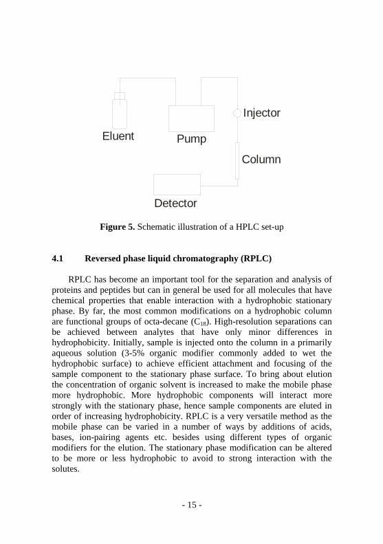
Eluent Pump
Injector
Column
Detector
Figure 5. Schematic illustration of a HPLC set-up 4.1 Reversed phase liquid chromatography (RPLC) RPLC has become an important tool for the separation and analysis of proteins and peptides but can in general be used for all molecules that have chemical properties that enable interaction with a hydrophobic stationary phase. By far, the most common modifications on a hydrophobic column are functional groups of octa-decane (C18). High-resolution separations can be achieved between analytes that have only minor differences in hydrophobicity. Initially, sample is injected onto the column in a primarily aqueous solution (3-5% organic modifier commonly added to wet the hydrophobic surface) to achieve efficient attachment and focusing of the sample component to the stationary phase surface. To bring about elution the concentration of organic solvent is increased to make the mobile phase more hydrophobic. More hydrophobic components will interact more strongly with the stationary phase, hence sample components are eluted in order of increasing hydrophobicity. RPLC is a very versatile method as the mobile phase can be varied in a number of ways by additions of acids, bases, ion-pairing agents etc. besides using different types of organic modifiers for the elution. The stationary phase modification can be altered to be more or less hydrophobic to avoid to strong interaction with the solutes.
- 15 -

4.2 Hydrophilic Interaction Liquid Chromatography (HILIC) HILIC is a special case of normal phase liquid chromatography in which highly hydrophobic mobile phases as hexane has been exchanged for semi-aqueous mobile phases. The stationary phase is hydrophilic and often charged at some region of the pH-scale. During the separation, the water content in the mobile phase will act as an integral part of the stationary phase as a water-enriched layer will form within the stationary phase due to its hydrophilic character.
Figure 6. The zwitterionic surface of a Zic-HILIC column
(Reprinted with permission from [64b])
With charged groups added to the stationary phase (as for example in Zic-HILIC columns), see Figure 6, the retention will be influenced by electrostatic forces in addition to hydrogen bonding, dipole-dipole interaction and polarizability of the solutes [64a/b, 65]. Separations on charged HILIC columns requires the addition of salt to brake the electrostatic forces for ionic analyte elution. This can in fact be a disadvantage as the achieved signal intensity, when hyphenated to MS detection, can be severely suppressed by these types of additatives which consequently should be kept at a minimum concentration. In general, to
- 16 -

bring about elution the concentration of organic solvents in the mobile phase is decreased, hence contrary to RPLC, components are eluted in order of decreasing hydrophobicity. 4.3 Size Exclusion Chromatography (SEC) In SEC the solutes within a sample mixture are separated by their size and form rather than their hydrophobicity as in reversed phase and hydrophilic interaction chromatography. The technique is widely used for sample clean-up and analysis of whole proteins and is considered to be a fairly non-denaturating chromatographic technique; hence the preservation of the native protein form is more likely to be higher than for other chromatographic techniques. SEC columns are packed with a stationary phase (can be polymeric or silica based) with varying pore-sizes. Smaller molecules are more retained within the pores than larger; hence the components are eluted in order of decreasing size. However the chromatographic resolution in general is rather low and complimentary separation techniques has to be used when analyzing complex mixtures in order to fully determine (ideally) each component. In addition the requirement of high concentrations of non-volatile salts in the mobile phase is a disadvantage in the coupling to both ICP- and ESIMS detection. 4.4 Summary of papers In paper (II) we developed a very successful method to separate and quantify CDDP and its mono-hydrolysed metabolite by Zic-HILIC chromatography coupled to ICPMS. In planning the study the aim was to achieve a good separation between cisplatin and its monohydrolysed metabolite, achieve low enough detection limits to analyze lysates from CDDP-exposed cells and to avoid the formation of artifacts during the analysis. The strategy to achieve the aims was to use HILIC chromatography with uncommon mobile phase organic modifiers and without using derivative agents or reactive additatives to the mobile phase, avoiding unwanted reactions between the eluent and the analytes. Due to that Zic-HILIC chromatography in common are accomplished with high concentrations of acetonitrile, known to deteriorate ICPMS signal-to-noise ratios and also to react with CDDP [36,66-69], we considered using another
- 17 -

mobile phase in the chromatography step in order to optimise ICPMS detection to achieve lower detection limits. Dimethylformamide was selected mainly based on its high boiling point which would be advantageous to minimize the plasma solvent load. The method showed to be 35 times more sensitive than if using acetonitrile in the chromatographic separation and we were able to detect a decreasing concentration of intracellular free CDDP in T289 melanoma cells up to 1 h after exposure while the total amount of Pt were shown to be rather constant (Table 1). In complimentary experiments (using the same chromatographic method) in our laboratory a study has been done where cell batches exposed to CDDP and subsequently incubated at 4 and 37 °C respectively, were differentiated by a 10-fold higher cisplatin concentration in the cells incubated at 4 °C than in 37 °C [70]. This result indicates that the measured CDDP really is intracellular as the intracellular metabolism is slower at lower temperature and that the difference in concentration between cells incubated at 4 and 37 °C is expected to be less if the determined cisplatin would be of extracellular origin, e.g. adsorbed to the outer surface of the cell membrane. The finding supports the theory that at least some of the CDDP uptake into the cells takes place by passive diffusion over the cell membrane or possible by endocytosis as the majority of papers describing active uptake indicates conformational changes for cisplatin by lost ligands upon uptake. We did at no time observe any hydrolyzed derivative of the drug, indicating that this form readily reacts with other intracellular molecules as soon as it forms and/or is lost in the sample treatment procedures. Whether intact CDDP enters the cell by passive or active uptake, interactions between CDDP and intracellular components e.g. proteins, GSH, DNA etc will occur. The biological half-life for CDDP due to the rate of such reactions has not been demonstrated experimentally until recently when Gibson et al [28] presented a half-life of 75 and 120 minutes for CDDP spiked in to cell lysates (non-viable) and water respectively. The result (however preliminary) from our study indicates that the intracellular half-life of CDDP in viable cells, as expected, is shorter but still as long as 15-20 min (Table 1). To our knowledge this is the first published result on the intracellular half-life for CDDP.
- 18 -

Table 1. Determined total platinum and cis-DDP concentrations in whole cell lysates of in-
vitro grown T289 human malignant melanoma cells exposed to 50 µM cis-DDP in growth
medium for 1 h followed by incubation in cis-DDP free medium for different times
Incubation
time (min)
Total Pt (ng Pt /g) cis-DDP (ng Pt /g) % cis-DDP of total Pt
0a 500 110 23
15a 620 64 10
30a 580 16 2.7
60a 650 6.8 1.0
Further, the results from Paper II showed that HILIC-ICPMS can be a potent hyphenated technique for the analysis of hydrophilic metal compounds and that the use of a low-volatile mobile phase may be a generic approach to improve analyte sensitivity and ICPMS robustness when using mobile phases with high amounts of organic solvents. It was however concluded that even if DMF was superior in ICPMS it is not a suitable solvent for structure determination by ESIMS, as described more in detail under the detection part. The usefulness of the developed separation method was later also demonstrated in Paper (I) in evaluating different cell lysis methods for platinum metallomic studies. In Paper III we more thoroughly evaluated a number of organic solvents for HILIC chromatography in hyphenation with ICPMS. Here an interesting finding was that the retention time of CDDP was fairly constant when varying the DMF concentration between 20 and 80% in the eluent and that a “true” HILIC mechanism seem to occur only at DMF levels above 80%. Varying the ammonium-acetate buffer strength from 250 to 0 µM did not affect the CDDP retention (however mono-hydroxy CDDP was affected), indicating that CDDP retention was not due to electrostatic interactions to the stationary phase. We hence ascribed the retention to some specific interaction between CDDP and the zwitterionic stationary phase as
- 19 -

in a control experiment on bear silica (non-charged hydrophilic phase) cisplatin had no retention and was eluted in the void volume using eluents with less than 80 % DMF. The fact that CDDP seem to have a weak retention on hydrophilic stationary phases is rather peculiar and has not yet been fully explained. Considering its strong polarity it should theoretically be well retained on a hydrophilic stationary phase. In fact, this complicates the strategy for developing a chromatographic method; however the use of Zic-HILIC chromatography gave a capacity factor for CDDP superior to methods previously published. Ion-pairing reagents have been frequently used to enhance the retention for the hydrolysed metabolites in RPLC but have little or no effect on CDDP itself. In addition these types of additatives are undesirable as they will compete with natural biological ligands binding the analytes and also extend the column equilibration time for reproducible retention times. From the study the overall conclusion was that among the tested solvents 1,4-dioxane and n-propanol yielded the best chromatographic behavior for HILIC separation of CDDP and its mono-hydrolysed metabolite as well as showed preliminary good performance in the hyphenation to ICPMS detection. With experience from Paper II and III we further evaluated this successful approach in Paper IV by examine several organic solvents for elution in reversed phase chromatography. As pointed out in many chromatography theory books [71], reversed phase chromatography can be carried out by using different organic modifiers but in practice a very high percentage of the separations are done with either methanol or acetonitrile. As both these solvents severely deteriorates ICPMS signal-to-noise ratios (discussed in detection part) the aim of the study was again to identify an alternative solvent giving preserved chromatographic and improved ICPMS performance as well as being compatible with ESIMS for structure determinations. The main reason for using reversed phase chromatography in metallomic studies is the analysis of proteins and peptides adducts with metal ions or molecules. In the case of CDDP it has previously been shown that a rather large fraction of the accumulated platinum reacts with proteins and it is also believed that reaction with endogenous thiol-containing peptides such as glutathione can be a detoxification pathway for CDDP. In this study we therefore selected a CDDP-cytochromeC complex and two different peptides for the evaluation. Considering chromatographic performance, eluent strength and compatibility to both ICPMS and ESIMS the overall best performance was acquired with 1-propanol. A moderate
- 20 -

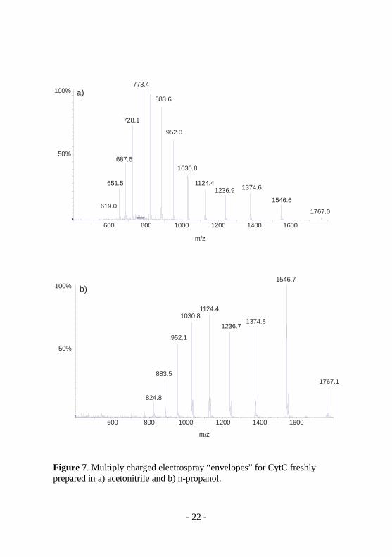
decrease in column efficiency could be observed when using n-propanol instead of acetonitrile when using a column packed with 3 µm particles while the decrease in efficiency was negligible when using a column packed with 5 um particles. The efficiency drop on the 3 um particle column could however be circumvented by increasing the column temperature from ambient to 40 °C. An additional aspect when analyzing protein-metal complexes is the importance of using chromatographic methods less prone to denaturate proteins than routine reversed phase chromatography. This is important for limiting losses of the metal ion or molecule attached to the protein during the chromatographic step which naturally is more critical when the metal species is non-covalently bounded. As a complement to the results from Paper III the ability to denaturate proteins was later evaluated (unpublished data)by a simple experiment infusing solutions of two different proteins in the different solvents (acetonitrile and n-propanol) into a standard electrospray ion source. Using the simple assumption that a more denaturated protein is more feasible to proton uptake, hence giving a electrospray protein “envelope” with higher charges [72], gave a significantly lower denaturation grade of the Cytochrome C with n-propanol than with acetonitrile (Figure 7, unpublished data).
- 21 -
600 800 1000 1200 1400 1600
m/z
773.4
883.6
728.1
952.0
687.61030.8
651.5 1124.4 1374.61236.91546.6
619.01767.0
100%
100%
50%
50%
600 800 1000 1200 1400 1600
m/z
1546.7
1124.41030.8
1374.81236.7
952.1
883.51767.1
824.8
a)
b)
Figure 7. Multiply charged electrospray “envelopes” for CytC freshly prepared in a) acetonitrile and b) n-propanol.
- 22 -
This indicates that using n-propanol in RPLC, in addition to the advantages considering hyphenation to ICPMS (described in the detection part), might preserve the native protein form better than for example acetonitrile. This method could indeed be a preferable alternative to size exclusion chromatography when analysing whole proteins as well as peptides bounded to metal species as the resolution with RPLC is superior to that of SEC. This was also actually shown in Paper V where both SEC and C4-RPLC were applied in a top-down approach for the analysis of whole proteins as an increase of resolved protein peaks could be observed in the RPLC method.
- 23 -

5. Detection 5.1 Inductively Coupled Plasma Ionization Mass Spectrometry
(ICPMS)
Today, ICPMS is a well-established, successfully and widely used technique for the analysis of trace elements and isotope measurements. The combination of a plasma ion source with mass spectrometric detection offers efficient atomization and ionization and low instrumental detection limits as well as selective detection of mass isotopes covering most elements of the periodic table. ICP-MS is robust, sensitive, and suitable for quantification as the response for a certain compound in general is independent of molecular structure; therefore there is in most cases no need for species-specific standards for the quantification. A major drawback in metallomic work is however that ICPMS does not give any information about molecular structure. 5.1.1 Principles of operation Figure 8 shows a schematic overview of a standard type ICP quadrupole MS. Typically, a liquid sample is introduced via a pneumatic nebulizer converting it into an aerosol that can be introduced to the plasma. The aerosol is passed through a spray chamber to remove large droplets that would otherwise be inefficiently vaporized in the plasma and cause excessive cooling. It is also important with a homogene droplet size distribution to avoid large signal fluctuations. From the spray chamber the aerosol is directed into the plasma, which has a temperature of 3000-10000 K generated by coupling radio frequence (rf) energy to argon gas electrons via an induction coil. In the plasma, particles from the spray chamber is ideally converted to free elemental, singly positively charged ions formed by collision with charged particles or by radiation. Positively charged ions, extracted and focused by an ion lens system are subsequently directed into the mass spectrometer while stepwise reducing the pressure by high performance turbo vacuum pumps. Three different types of commercial mass analysers has been used with ICP ionization; quadropole, magnetic sector and time-of-flight, of which by far the most common is the qudropole as also used in this thesis. The quadropole consists of four rods to which variable DC and AC potentials are applied. At a certain setting only one
- 24 -

selected m/z will have a stable path through the spectrometer, while other m/z will be lost. Adjusting the settings on a millisecond scale allows fast sequential multi-element determinations.
Figure 8. Schematic presentation of an ICP quadropole MS system. 5.1.2 Avoiding spectroscopic interferences by use of a
collision/reaction cell Spectroscopic interferences occur when the difference in m/z ratio between two different ions is smaller than what the resolving power of the instrument can separate. For a quadrupole the resolution is about 1 amu or at the best slightly better than that. Interferences can be caused by elemental isotopes, doubly charged ions and polyatomic ions. Polyatomic ions are formed by reaction between elements from the plasma (Ar), entrained air (N, O), the solvent, additatives and the sample matrix. Since the introduction in the late 1990’s of collision/reaction cell (CRC) technology [73] in combination with quadrupole analysers, selective removal of interfering species as well as selecting analyte-reaction gas products as alternatives for the determination has been possible and offers a remarkable improvement in detection limit and accuracy for certain elements. As example, sulphur (32S+) is interfered by oxygen (16O2
+) ions formed in the plasma. Reacting 32S+ with oxygen applied in the collision cell converts
- 25 -

sulphur to 32S16O+ which is determined at m/z 48 where no interferences occur as 48O3+ does not readily form in the collision cell. 5.1.3 Hyphenation of liquid chromatography to ICPMS Speciation analysis is a term that describes analytical activities of identifying and/or measuring the quantities of one or more individual chemical species in a sample. A chemical species can be described as a specific form of an element defined as an isotopic composition, electronic or oxidation state, and/or complex or molecular structure. With respect to environmental and biological issues it has been understood that factors like distribution, mobility and availability of elements is not only dependent on their total concentration but rather to the forms and/or associations they undergo in natural systems. In speciation studies, a chromatographic step to separate the different species prior to the ICPMS detection is crucial. Liquid chromatography has been used in hyphenated systems to elemental detection for a long time and due to the possibilities to vary the mobile and stationary phases, in addition to that aqueous sample introduction is the most preferred and simple introduction method for ICPMS, the technique has become very versatile for the ICPMS user. ICPMS interfaces are constructed in a manner so that they are easily coupled to liquid chromatography and since the first publication on ICPMS as an element-specific detector for HPLC by Houk et al in 1986 [74] the number of publications is steadily increasing. Still, the low tolerance of organic solvents introduced to ICPMS is a major drawback which has to be considered in developing separation methods intended for use in hyphenation with ICPMS. Introducing and decomposing organic solvents in the plasma will require extra energy which will result in lower atomization and ionization efficiencies as well as cause plasma instability [75, 76]. Black carbon will eventually form from the carbon content in the solvents. This will cause depositions on instrumental parts and lead to deteriorated performance of the detection. The sampler and skimmer cones in the MS interface can eventually become partly or completely clogged which in worst case can lead to complete loss of signal. In order to overcome the problems observed, chilled spray chambers to reduce evaporation of the organic solvent accumulated in the spray chamber [77-80], and thereby reduce the plasma solvent load, are often used in combination with addition of oxygen to the plasma to enhance the combustion of remaining carbon.
- 26 -

However, even if taking those measures, applications such as reversed phase liquid chromatography, where high amounts of organic solvents are used, must still be considered as problematic. 5.1.4 Summary of papers In Paper (III) an extensive study of the ICPMS as well as chromatographic performance for selected organic solvents were performed. From an ICPMS perspective the different solvents, selected as potential alternatives to acetonitrile, were chosen due to their relatively high boiling points and oxygen/carbon ratio content in order to decrease the solvent vapour plasma load and enhance combustion of remaining carbon in the plasma. Ethanol, n-propanol, 1,4-dioxane and dimethylformamide, all showed superior performance compared to acetonitrile when monitoring the platinum ICPMS signal variation during a solvent gradient from 0 to 60%. (Figure 9).
0
20
40
60
80
100
120
0 5 10 15 20Time (min)
195 Pt
sig
nal i
nten
sity
(cou
nts
x 10
3 )
0
10
20
30
40
50
60
70
% o
rgan
ic m
odifi
er
DMF
Dioxane
Propanol
Etanol
Acetonitril
gradient profile
a)
Figure 9. Signal variation during gradient elution with different solvents.
- 27 -
There was no need to add additional oxygen to the plasma and no carbon deposition could be observed on instrumental parts except for ethanol as with the lowest boiling points (78 °C) after an extended run time showed a tendency to form black carbon deposits. In average the signal intensity decreased to about 70 % of the starting value, while acetonitrile caused a signal suppression of 90 % leaving only 10 % of the starting signal intensity remaining. As peptides and proteins, commonly elute between 30 and 50 % of organic modifier, the gain in signal intensity can be predicted to be between 2 and 4 times when using the alternative solvents. This was also shown in the study as a 2.5-fold increase of the peak intensity was observed for the CDDP-Cytochrome C complex while for the peptide bacitracin a 10-fold enhancement of the peak intensity was achieved when using n-propanol instead of acetonitrile. The enhanced ICPMS performance for DMF was already observed from Paper II giving a 35-fold increase of the peak intensity (compared with acetonitrile) for CDDP when analyzed by Zic-HILIC chromatography. However in Paper IV DMF was found to be a too weak organic modifier for reversed phase chromatography and was subsequently not further evaluated for this purpose. In Paper V the applicability of using n-propanol as organic modifier was further demonstrated using both SEC and RP chromatography to explore differences of cupper and platinum speciation in sensitive and resistant melanoma T289 cell lines. 5.2 Electrospray Ionization Mass Spectrometry (ESIMS) In the beginning of the last century, long before its application to mass spectrometry, Zeleny [81] showed that as a result of an applied electrical field at appropriate conditions a liquid will disperse into electrically charged droplets. Later, a few years after first introducing electrospray for MS applications [82] Fenn reported on the multiple charged ion formation in 1988 [83]. About the same time Alexandrov et al [84], developed a method they called “extraction of ions from solution at atmospheric pressure, EIS AP. In this thesis the term electrospray ionization (ESI) will refer to the above mechanism and be used for the further discussion.
- 28 -

5.2.1 Principles of operation To separate and detect analytes in a mass spectrometer, formed ions in the ion source must be transferred into the gas-phase. In ESI three major steps can be defined in the production of gas-phase ions; production of charged droplets at the ESI capillary tip, solvent evaporation causing shrinkage of the charged droplets eventually leading to very small, highly charged droplets and finally forming gas-phase ions from these small droplets. This is achieved by applying an electric field between the capillary outlet and the inlet of the mass spectrometer as shown in Figure 10.
Figure 10. Schematic of major processes of ion formation in ESI. Two main mechanisms has been suggested for forming gas-phase ions from desolvated small charged droplets; i) the charged residue model and ii) the ion evaporation theory. None of them will however be described in detail here but interested readers are recommended to read the original works by Dole [85] and Iribarne and Thompson respectively [86].
- 29 -

In comparison with ICPMS, ESIMS is considered as a soft ionization techniques. Preserving the molecular structure in the ionization step makes it possible to determine the molecular mass and after collision induced dissociation or in-source fragmentation, the structure of the molecule can also be determined from its fragmentation pattern. All types of mass analysers can be used in connection with ESI, in the studies covered by this thesis single and triple quadropoles as well as time-of-flight (TOF) analysers have been used. The function of the different analyzers will not be describe in detail, however some of the important differences between quadrupoles and time-of-flight instruments is that the resolving power is much better and in addition the analyzable mass range is wider for TOFs [87], which means that higher mass accuracy can be obtained and that larger biomolecules can be analysed by TOF analyzers. 5.2.2 Hyphenation of liquid chromatography to ESIMS The key to achieve separation and maintain ESIMS performance in LC-MS is finding chromatographic conditions compatible with the electrospray processes. The preset requirements for a successful outcome is that analyte ions can be created in solution, that the solvents used in the mobile phase can be electrosprayed and that buffers compatible with ESI can be employed. In practice this severely limits chromatography modes like ion exchange and size exclusion chromatography due to the necessary addition of relatively high concentrations of non-volatile salts leading to severe suppression of the ESIMS signal intensity. As the process for desolvation and ionization is dependent of the volality and surface tension of the organic solvent it also excludes using low-volatile solvents with high surface tension values. It is possible to use post-column dilution techniques to enhance the performance but as ESIMS is a concentration sensitive technique this leads to poorer detection limits. Despite the limitations, ESI interfaces possess a construction that are easily coupled to liquid chromatography and today, ESI has become one of the most popular and commonly used techniques for ionization of liquids. The dominating status has been reached for its ability to perform stable and soft ionization at atmospheric pressure for a wide range of liquid flow rates and solvent compositions and for its ability to produce multiply charged ions of macromolecules. This feature enables the analysis of macromolecules as the
- 30 -

resulting m/z will be applicable to the mass range of common mass analysers. 5.2.3 Summary of papers From Paper II it could be concluded that DMF was not a suitable solvent for electrospray ionization as its volatility was too low and surface tension too high for efficient desolvation and ionization. Diluting the eluent (70% DMF) with methanol in a 2:1 ratio post-column made it possible to verify mass spectra from both CDDP and the mono-hydrolysed metabolite. Unfortunatly, due to low concentrations and CDDP’s poor ionization efficiency in ESI, we were not able to verify the species in cell samples but only from a more concentrated standard solution. However, as the same chromatographic method was used as in the ICPMS set-up we could confirm presence of the species by their correlating retention times from LC-ICPMS analysis of cell samples. In a subsequent study (Paper IV) DMF was excluded from the ESI compatibility test even though in hyphenation with ICPMS it showed very good performance. Of the other solvents tested for reversed phase chromatography 1,4-dioxane was also excluded for ESI evaluation due to high surface tension. Ethanol, n-propanol and acetonitrile were hence evaluated as mobile phases for RPLC coupled to electrospray ionization. Ethanol, as a too weak eluent for RPLC was only tested by continuous infusion, while n-propanol and acetonitrile in addition were compared for chromatographic purposes. The main conclusion from the study was that analyte signal-to-noise ratio was preserved using n-propanol as well as ethanol instead of acetonitrile indicating that the ESI process was unaffected. 5.3 Combining ICPMS and ESIMS in metallomic work The combined use of liquid chromatography hyphenated to ICPMS and ESIMS has in many reviews been considered a requirement to produce reliable quantitative and qualitative data on metal species in metallomic studies [88-90]. Considering the above discussion on chromatographic and ICPMS as well as ESIMS performance, by using n-propanol as chromatographic mobile phase a system for combined use of LC-ICPMS and LC-ESIMS analysis has here been proposed. In comparison, using n-
- 31 -

propanol as alternative to state-of-the-art conditions using acetonitrile gave; i) enhanced ICPMS performance by lower detection limits and robustness, ii) preserved limit-of-detection performance in ESIMS and iii) only minor compromises in chromatographic performance. In a parallel set-up, splitting of the HPLC effluent directing one part of the flow to ICPMS and one part to ESIMS would give an element-specific species detection simultaneously with molecular mass and structure determination. This set-up has been discussed by many authors, but has not yet been realized in that many studies mainly due to the difficulty in using one set of chromatographic parameters for both techniques. In conclusion from this thesis, using n-propanol as chromatographic modifier, this difficulty can be considered as overruled. However, differences in sensitivity for the different techniques must still be taken under consideration, but providing a defined metal compound retention time from ICPMS detection definitely facilitates the compound structure determination by LC-ESIMS.
- 32 -

6. Quantitative and/or Qualitative Evaluation 6.1 Calibration methods for quantitative analysis The aim in quantitative analysis is to correlate the analyte signal response with its concentration. Several methods have been established for this purpose, three of the most commonly used are external calibration, internal standard calibration and the standard addition method. Quantification by isotope dilution is an additional method commonly used in combination with MS detection. Commonly, an isotope-labeled spike is added as internal standard to the sample to correct for losses in the sample preparation step/s as well as for instrumental variations. In LC-ICPMS an isotope labeled compound can be continuously added post-column to correct for varying signal intensities. The isotope labeled compound can be either species-specific, e.g. identical molecular form as the analyte, or species-unspecific, e.g. different molecular form than the analyte. In order to document quantitative analytical method performance and to discover complications, recording of common validation parameters (Table 2) is of importance and should always be performed when; i) developing a new method, ii) an established method are changed or used for a new application, iii) quality assurance data shows varying results over time and iv) an established method is performed in a new laboratory, by new personnel or by new instrumentation.
- 33 -
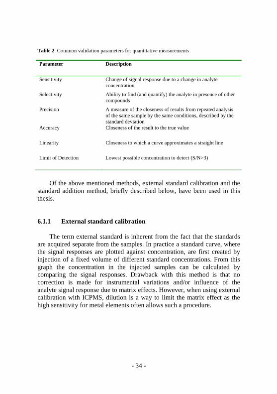
Table 2. Common validation parameters for quantitative measurements
Parameter Description
Sensitivity Change of signal response due to a change in analyte concentration
Selectivity Ability to find (and quantify) the analyte in presence of other compounds
Precision A measure of the closeness of results from repeated analysis of the same sample by the same conditions, described by the standard deviation
Accuracy Closeness of the result to the true value
Linearity Closeness to which a curve approximates a straight line
Limit of Detection Lowest possible concentration to detect (S/N>3)
Of the above mentioned methods, external standard calibration and the standard addition method, briefly described below, have been used in this thesis. 6.1.1 External standard calibration The term external standard is inherent from the fact that the standards are acquired separate from the samples. In practice a standard curve, where the signal responses are plotted against concentration, are first created by injection of a fixed volume of different standard concentrations. From this graph the concentration in the injected samples can be calculated by comparing the signal responses. Drawback with this method is that no correction is made for instrumental variations and/or influence of the analyte signal response due to matrix effects. However, when using external calibration with ICPMS, dilution is a way to limit the matrix effect as the high sensitivity for metal elements often allows such a procedure.
- 34 -

6.1.2 The standard addition method Working with quantification of compounds in complex matrices like biological fluids, matrix induces non-spectral interferences can be compensated for by using the standard addition calibration method. The method is accomplished by dividing the sample in to sub-samples of which all except one is spiked with different amount of the standard substance. Responses for all samples are acquired and plotted as a function of the added amount of standard where the un-spiked sample is set to zero. By extrapolation the unknown concentration can be determined where the calibration curve crosses the x-axis. If sample volume is sufficient to apply the standard addition procedure, this method provides accurate quantification in complex matrices. 6.2 Considerations in qualitative analysis The aim in qualitative analysis is to determine the presence and identity of a species in a given sample. This determination can however be defined at different levels of probability. Many studies of metal compounds are performed only by ICPMS analysis. But let us think for a while about what is really achieved by this technique. It is true that a compound that contains the metal-element analyzed for is detected, but in practice no more data can be extracted from this analysis. Even if the quantitative analysis of the metal can be performed without a species-specific standard, the identification of the compound requires a retention time of the species that correlates to that of a standard. Even so, the identification must still be considered as preliminary as, especially in complex samples, co-elution can occur. A strategy for a more reliable identification in this step could, when possible, be to identify the species with two different chromatographic modes. To confirm the identification from ICPMS by verifying the molecular mass and structure, molecular mass spectrometry has to be used. Achieving a molecular mass, combined with the compound retention time(s) using the same chromatographic set-up(s) as in the ICPMS analysis eventually leads to a confirmed identification. Further, by true MS/MS or in-source fragmentation, the mass spectrum achieved from the compound (now again compared to a species-specific standard) can be said to be unambiguously. Taking all of this in consideration it becomes quite clear that developing a method for unambiguous qualitative analysis in metallomics can be rather
- 35 -

time consuming and also requires both skilled scientists and well-equipped laboratories. The extensive laboratory work required for a full identification is probably a main reason why so far only one publication [58] has shown a positive identification of a platinum-binding biomolecule other than DNA in a real sample. 6.3 Summary of Papers In Paper I the standard addition method was used for determination of total platinum where PtCl4 was used as standard. Three different lysis methods were compared and in order to correct for the different matrix effects this method was selected for reliable data. Levels of CDDP were evaluated from transient data using the method from Paper II. As the matrix effects are usually much less when using a chromatographic step external calibration by use of the species-specific standard was applied. As the method was previously described and validated no further qualitative analysis were made than matching of the retention time. In Paper II the chromatographic method for the analysis of CDDP by HILIC-ICPMS was fully validated by common procedures for precision, linearity and limit-of-detection which overall showed a good performance. Discussing the quantification in ICPMS it is of importance to note that determining the concentration of a certain element in a final sample extract by the use of a species-unspecific standard can be considered as an easy task. However, to establish the analyte concentration in the original sample, factors like losses in sample pre-treatment procedures and the chromatographic step has to be considered. In this case the use of a species-specific standard becomes crucial. In Paper II recoveries of CDDP was evaluated for both the pre-treatment and chromatographic step which could then be used to correct the quantitative data acquired by the external calibration method. By using ESIMS in combination with ICPMS both CDDP and its hydrolysed metabolite could be qualitatively verified by their molecular masses and platinum and/or chlorine isotope patterns. In Paper V, total concentration of platinum and cupper were determined by external calibration after dilution of the sample in order to avoid matrix effects. From SEC and RPLC analysis of cell lysates no absolute quantification has so far been performed, but only estimates of the relative concentrations of the identified substances from the different analytical approaches was done. Certainly, this has to be more thoroughly
- 36 -

evaluated by further validation of the chromatographic performance by for example mass balance studies. However, it was possible to draw some indicative conclusion about the concentrations showing differences between sensitive and resistant cell lines. As discussed in 6.2 the differences between sensitive and resistant cell lines were observed by two different chromatographic modes (SEC and RPLC). By this approach we were able to; i) by SEC, define a molecular mass range for the differing peaks and ii) by RPLC, get an indication of their hydrophobicity and also the type of binding between the metal(s) and protein(s).
- 37 -
7. Concluding Remarks and Future Trends The key to success in use analytical results in order to understand biological functions lies within the control, optimization and validation of all steps in the analytical process. If the analytical performance is insufficient this can give rise to misleading results. This thesis describes some new and improved analytical methodologies for determining the Pt metallome in CDDP-exposed malignant cells. Taking the aims of this thesis in a summarized consideration we have; i) developed methods for measuring intact CDDP uptake in cell samples indicating a partly passive and/or endocytotic uptake of the drug, ii) demonstrated the importance of using methods to avoid artifacts by unwanted reactions during the sample preparation and chromatographic steps, iii) showed that by new strategies in selecting solvents for the mobile phase in the separation step achievements can be made in both sensitivity and robustness in ICPMS detection of metal species while preserving ESIMS performance for structure determination and iv) showed that we can use the developed methods to find differences between CDDP sensitive and resistant cells. By using tissue-specific tumor in-vitro grown cells and a CDDP exposure concentration of only 20 µM we have strived to mimic a “real” sample and clinical dosages to an extent not previously shown by that many studies. Certainly, we still have a long way to go before we have put all pieces together for a total understanding of CDDP’s mode of action, however some improvements definitely have been made. As mentioned, so far only experiments with in-vitro grown cells have been done but I am sure that we will approach towards analysis of tumor tissue in a quite near future. The obvious challenges with that work will be to enhance the sensitivity for determining CDDP and its metallome even at lower concentrations than what we have done so far. This will probably be accomplished by developing better methods for sample preparation considering the extraction and enrichment of the sought species of interest. Except for the usefulness to better understand CDDP’s mode of action and of cell resistance towards such drugs it is my hope that the developed methodologies also will be noticed and used in a more generic approach within metallomic work.
- 38 -

8. Acknowledgements / Tack Om det är något jag lärt mig av dessa underbara fem år är det att de som står först är viktigast! Martin, och våra tre underbara pojkar, Alexander, Samuel och Sebastian. Ni utgör fundamentet i hela mitt liv, älskar er så mycket. Utan er hade jag inte klarat det! Min lilla familj med stort hjärta, Mamma, Pappa och Lillasyster för att ni finns, utgör grunden för den jag är och för att ni trott på mig! Speciellt tack för all hjälp med barnpassning när det kört ihop sig. Sofi, min underbara rumskamrat, jag har verkligen uppskattat ditt sällskap, våra fester, allt skvaller och småprat. Lycka till med bebis och forskning! Jag förutsätter förstås en inbjudan till din kommande disputation. Kicki, tack för all hjälp med odling och exponering av celler. Utan dig hade mitt projekt inte varit någonting! Därefter kommer alla andra som bidragit till slutresultatet utan inbördes rangordning! Lars, Solomon, Dong, Daniel, Tom, Max och Andreas och Ida som alla bidragit till att göra tiden i TESA gruppen fantastisk rolig och minnesvärd. Jag hoppas att vi får tillfälle att ses igen. Veteranerna, Wolfgang Anders, Michael och Svante. William, och Lasse, som funnits, försvunnit och kommit tillbaka. Alla som kommit och gått i kromatografigruppen under åren. Speciellt stor kram till Petrus för trevliga diskussioner både på det privata och vetenskapliga planet. Åsa N-son Lindgren och andra i institutionsledningen för det otroliga gensvar jag fått genom delaktighet i institutionsarbetet. Det har varit mycket lärorikt och utvecklande på sidan av doktorandstudierna och har på något sätt gjort tiden vid kemiinstitutionen komplett.
- 39 -
Alla andra på kemiinstitutionen som på ett eller annat sätt gjort tiden som doktorand otroligt stimulerade och faktiskt inte en enda gång tråkig! Peter, för stimulerande diskussioner och för ditt oavkortade stöd för mitt projekt. Tack också för gott kaffe på ditt tjänsterum vid våra många möten! Calle Nilsson, tack för ditt stora stöd och uppmuntran, utan det tror jag faktiskt inte att jag skulle börjat som doktorand. Crister, tack för din ovärderliga insats som biträdande handledare, nu får vi se vem av oss som först blir docent! Britt och andra i FOIs ledning som stöttat genom att låta mig vara tjänstledig i fem långa år. Nu kommer jag tillbaka, med nuvunna kunskaper och nätverk som jag hoppas ni ska få nytta av. Orättvist sist kan man tycka, hittar man den som bestämmer, sköter korrespondensen, har pengar, erfarenhet och ett gott namn. Alltså, sist men som alla vet inte minst, Erik för din oändliga entusiasm, motivationsförmåga, vägledning och vänskap. Jag kan inte tänka mig att det kan finnas en bättre handledare än du! Tack för att du för fem år sedan valde just mig att få göra den här resan. Jag hoppas verkligen att vi på ett eller annat sätt får fortsätta vårt samarbete och av hela mitt hjärta önskar jag dig all lycka på livets alla plan i framtiden!
- 40 -

9. References [1] Peyrone M, Annual Chem. Pharm., (1845), 51, 1. [2] Werner A, Anorg. Chem., (1893), 3, 267. [3] Rosenberg B, Vancamp L, Krigas T, Nature (1965), 205, 698. [4] Rosenberg B, Vancamp L, Trosko JE, Mansour VH, Nature, (1969),
222, 385. [5] Rosenberg B, Vancamp L, Cancer Research, (1970), 30, 1799. [6] Petsko GA, Gen. Biol., (2002), 3, 1001. [7] Lippard SJ, Berg JM, “Principles of Bioinorganic Chemistry”,
University Science Books, Mill Valley, CA, (1994). [8] Alderden DA, Hall MD, Hambley TW, J.Chem.Educ., (2006), 83,
728. [9] Berners-Price SJ, Roncom L, Sadler PJ, Prog.Nucl.Magn.Reson.
Spectrosc., (2006), 49, 65. [10] Jennerwein M, Andrews PA, Drug. Metab. Dispos., (1995), 23, 178. [11] Berners-Price SJ, Appleton TG, “Platinum-based drugs in cancer
therapy“, Humana Press Totawa, NJ, (2000). [12] Takahara PM, Rosenzweig AC, Frederick CA, Lippard SJ, Nature,
(1995), 377, 649. [13] Todd RC, Lippard SJ, Metallomics, (2009), 1, 280. [14] Hall MD, Okabe M, Shen D-W, Liang X-J, Gottesman MM, Annu.
Rev. Pharmacol. Toxicol., (2008), 48, 495. [15] Gale GR, Morris CR, Atkins LM, Smith AB, Cancer Res. (1973), 33,
813. [16] Binks SP, Dobrota M, Biochem. Pharmacol., (1990), 40, 1329. [17] Liang X, Huang Y, Int. J. Biochem. Cell Biol., (2002), 34, 1248. [18] Liang XJ, Yin JJ, Zhou JW, Wang PC, Taylor B, Exp. Cell Res.,
(2004), 293, 283. [19] Mann SC, Andrews PA, Howell SB, Anticancer Res., (1998), 8,
1211. [20] Byfield JE, Calabro-Jones PM, Nature, (1981), 294, 281. [21] Liang XJ, Shen PW, Chen KG, Wincovitch SM, Garfield SH,
Gottesman MM, J. Cell Physiol., (2005), 202, 635. [22] Holzer AK, Howell SB, Cancer Res., (2006), 66, 10944. [23] Andrews PA, Velucy S, Mann SC, Howell SB, Cancer Res., (1988),
48, 68. [24] Andrews PA, Mann SC, Huynh HH, Albright KD, Cancer Res.,
(1991), 51, 3677.
- 41 -
[25] Dancis A, Haile D, Yuan DS, Klauser RD, J. Biol. Chem., (1994), 269, 25660.
[26] Ooi CE, Rabinovich E, Dancis A, Bonifacino JS, Klauser RD, EMBO J., (1996), 15, 3515.
[27] Petris MJ, Smith K, Lee J, Thiele DJ, J. Biol. Chem., (2003), 278, 9639.
[28] Kasherman Y, Sturup S, Gibson D, J. Med. Chem., (2009), 52, 4319. [29] Zhu GY, Chang P, Lippard SJ, Biochemistry, (2010), 49, 6177. [30] Ho V, Scharer OD, Environmental and Molecular Mutagenesis,
(2010), 51, 552. [31] Zhao Z, Tepperman K, Dorsey JG, Elder RC, J. Chromatogr. (1993),
615, 83 [32] Falter R, Wilbeen RD, Sci. Total Environ. (1999), 225, 167. [33] Bell DN, Liu JJ, Tingle MD, McKeage MJ, J. Chromatogr. B,
(2006), 837, 29. [34] Andersson A, Ehrsson H, J. Pharm. Biomed. Anal., (1995), 13, 639. [35] Verschragen M, Van der Bom K, Zwiers THU, Vander Vijgh WJF,
J. Chromatogr. B, Anal. Technol. Biomed. Life Sci., (2002), 772, 273. [36] El-Kateeb M, Appleton TG, Charled BG, Gahan LR, J. Pharm. Sci.,
(1999), 88, 319. [37] Heudi O, Cailleux A, Allain P, Chromatographia, (1997), 44, 19. [38] Hann S, Stefanka Z. Lenz K, Stingeder G, Anal. Bioanal. Chem.,
(2005), 381, 405. [39] Nygren Y, Hemström P, Åstot C, Naredi P, Björn E, J. Anal. At.
Spectrom., (2008), 23, 948. [40] Szpunar J, Makarov A, Pieper T, Keppler BK, Lobinski R, Anal.
Chim. Acta, (1999), 387, 135. [41] Esposito BP, Najjar R, Coord. Chem. Rev., (2002), 232, 137. [42a] Rudnev AV, Aleksenko SS, Semenova O, Hartinger CG, Timerbaev
AR, Keppler BK, J. Sep. Sci., (2005), 28, 121. [42b] Esteban-Fernandez D, Montes-Bayon M, Blanco Gonzales E,
Gomez-Gomez MM, Palacios MA, Sanz-Medel A, J. Anal. At. Spectrom., (2008), 23, 378.
[43] Begerow J, Turfeld M, Dunemann L, J. Anal. At. Spectrom., (1996), 11, 913.
[44] Tang X, Hayes II JW, Schroder L,Cacini W, Dorsey J, Elder RC, Tepperman K, Met.- Based Drugs, (1997), 4, 97.
[45] Stewart DJ, Benjamin RS, Luna M, Feun L, Caprioli R, Seifert W, Loo TL, Cancer Chemother. Pharmacol., (1982), 10, 51.
- 42 -

[46] Areberg J, Bjorkman S, Einarsson L, Frankenberg B, Lundquist H, Mattsson S, Norrgren K, Scheike O, Wallin R, Acta Oncol., (1999), 38, 221.
[47] Esteban-Fernandez D, Verdaguer JM, Ramiez-Camacho R, Palacios MA, Gomez-Gomez MM, J. Anal. Toxicol., (2008), 32, 140.
[48] Spingler B, Whittington DA, Lippard SJ, Inorg. Chem., (2001), 40, 5596.
[49] Chaney SG, Campbell SL, Temple B, Bassett E, Wu Y, Faldu M, J. Inorg. Biochem., (2004), 98, 1551.
[50] Blommaert FA, Michael C, van Dijk-Knijnenburg HCM, Schornagel JH, des Engelse L, Fichtinger-Schepman AMJ, Cancer Chemother. Pharmacol. (1996), 38, 273.
[51] Hann S, Zenker A, Galanski M, Bereuter TL, Stingeder G, Keppler BK, Fresenius J. Anal. Chem., (2001), 379, 581.
[52] Garcia Sar P, Montes-Bayon M, Agnado Ortiz L, Blanco-Gonzales E, Sierra LM, Sanz-Medel A, J. Anal. At. Spectrom., (2006), 21, 861.
[53] Pattanaik A, Bachowski G, Laib J, Lemkuil D, Shaw III CF, Petering DH, Hitchcock A, Savyan L, J. Biol Chem. (1992), 267, 16121.
[54] Knipp M, Karotki AV, Chesnov S, Natile G, Sadler PJ, Brabec V, Vasak M, J. Med. Chem., (2007), 50, 4075.
[55] Hagrman D, Goodisman J, Souid AK, J. Pharmacol. Exp. Ther., (2003), 308, 658.
[56] Dabrowiak JC, Goodisman J, Souid AK, Drug Metab. Dispos., (2002), 30, 1378.
[57a] Miao R, Yang G, Miao Y, Mei Y, Hong J, Zhao C, Zhu L, Rapid Commun. Mass Spectrom., (2005), 19, 1031.
[57b] Crider SE, Holbrook RJ, Franz KJ, Metallomics, (2010), 2, 74. [58] Ishikawa T, Ali-Osman F, J. Biol. Chem., (1993), 268, 20116. [59] Dill KA, Journal of Phys. Chem., (1987), 91, 1980. [60] Dorsey JG, Dill KA, Chemical Rewievs, (1989), 89, 331. [61] Tchapla A, Colin H, Guiochon G, Anal. Chem., (1984), 56, 621. [62] Tchapla A, Heron S, Colin H, Guiochon G, Anal. Chem. (1988), 60,
1443. [63] Horrath C, Melander W, Molnar I, J. Chromatogr., (1976), 125, 129. [64a] Hemstrom P, Irgum K, J. Sep. Sci., (2006), 29, 1784. [64b] “A Practical Guide to HILIC”, (2006), SeQuant AB, Umeå, Sweden [65] Alpert AJ, J. Chromatogr., (1990), 499, 177. [66] Szpunar J, Analyst, (2005), 130, 442.
- 43 -

[67] Lobinski R, Schaumloffel D, Szpunar J, Mass Spectrom. Rev., (2006), 25, 255.
[68] Montes-Bayon M, DeNicola K, Caruso JA, J. Chromatogr. A, (2003), 1000, 457.
[69] Gammelgaard B, Jensen BP, J. Anal. At. Spectrom., (2007), 22, 235. [70] Östman M, “Cisplatin reaction kinetics”, Batchelor thesis, Umeå
University, Sweden, (2010). [71] Poole CF, “The essence of chromatography”, Elsevier Science BV,
(2003). [72] McLafferty FW, Guan ZQ, Haupts K, Wood ID, Kelleher NL,
Journal of the American Chemical Society, (1998), 120, 4732. [73] Tanner SD, Baranov VI, Bandura DR, Spectrochim. Acta Part B,
(2002), 57, 1361. [74] Thompsson JJ, Houk RS, Anal. Chem., (1986), 57,1361. [75] Weir DG, Blades MW, J. Anal. At. Spectrom., (1994), 9, 1311. [76] Weir DG, Blades MW, J. Anal. At. Spectrom., (1994), 9, 1323. [77] Brotherton T, Barnes B, Vela N, Caruso J, J. Anal. At. Spectrom.,
(1987), 2, 389. [78] Cairns WRL, Ebdon L, Hill SJ, Fresenius. J. Anal. Chem., 1996, 355,
202. [79] Smith CJ, Wilson ID, bou-Shakra F, Payne R, Parry TC, Sinclair P,
Roberts DW, Anal. Chem., 2003, 75, 1463. [80] Bendahl L, Gammelgaard B, J. Anal. At. Spectrom., 2005, 20, 410.
[81] Zeleny J, J.Phys.Rev., 1917, 10,1. [82] Yamashita M, Fenn, JB, J. Phys. Chem., 1984, 88, 4451. [83] Wong SF, Meng CH, Fenn JB, J. Phys. Chem., (1988), 92, 546. [84] Alexandrov ML, Gall LN, Krasnov NV, Nikolaev VI, Pavlenko VA,
Shkurov, VA, Doklady Akademii Nauk Sssr, (1984), 277, 379., [85] Dole M, Mack LL, Hines RL, Journal of Chemical Physics, 1968,
49, 2240 [86] Iribarne JV, Thomson BA, Journal of Chemical Physics, 1976, 64,
2287-2294 [87] Burlinggame SJ, Boyd RK, Gaskell SJ, Anal. Chem., (1998), 70, 647. [88] Rosen AL, Hieftje GM, Spectrochim. Acta Part B, (2004), 59, 135. [89] Michalke B, Trend Analyt. Chem., (2002), 21, 154. [90] Montes-Bayon M, DeNicola K, Caruso JA, J. Chromatogr. A,
(2003), 1000, 457.
- 44 -