
Advanced infection prevention training CIP Consulting LLC.
133
Advanced infection prevention training CIP Consulting LLC
-
Upload
jordyn-banville -
Category
Documents
-
view
214 -
download
0
Transcript of Advanced infection prevention training CIP Consulting LLC.

Advanced infection prevention training
CIP Consulting LLC

Overview of Intermediate Infection Prevention Training
• Adult learning• Change Theory• Components of a successful Infection prevention
program• CDC Surveillance Definitions (“Big 4)• Outbreak investigation• Basic NHSN features• IP in the OR• Basic concepts of cleaning and disinfection• Hand hygiene

Clean hands are happy, healthy hands!!!!!
“Foam in Foam out”

Surveillance
• Surveillance should be based on sound epidemiological and statistical principles
• Surveillance methods continue to evolve and should be designed in accordance to current recommended practices and should consist of defined elements
• Surveillance plays an important role in identifying outbreaks, emerging infectious diseases, and bioterrorist events.
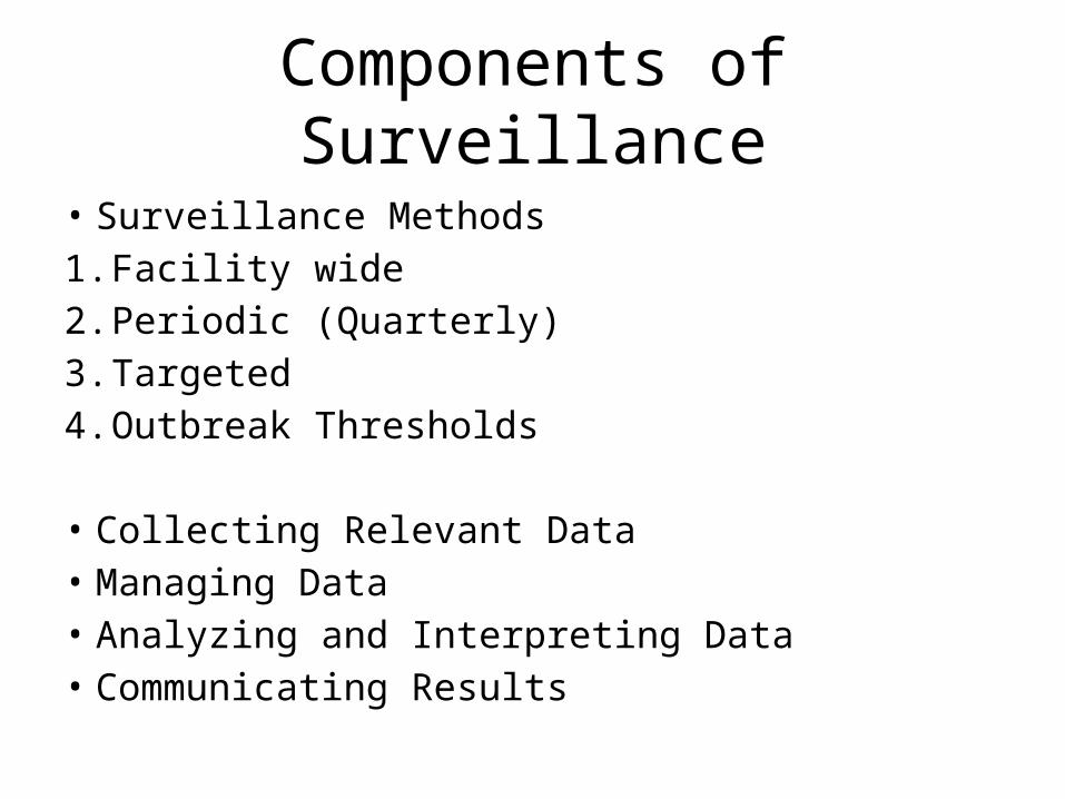
• Surveillance Methods1. Facility wide2. Periodic (Quarterly)3. Targeted4. Outbreak Thresholds
• Collecting Relevant Data• Managing Data• Analyzing and Interpreting Data• Communicating Results
Components of Surveillance
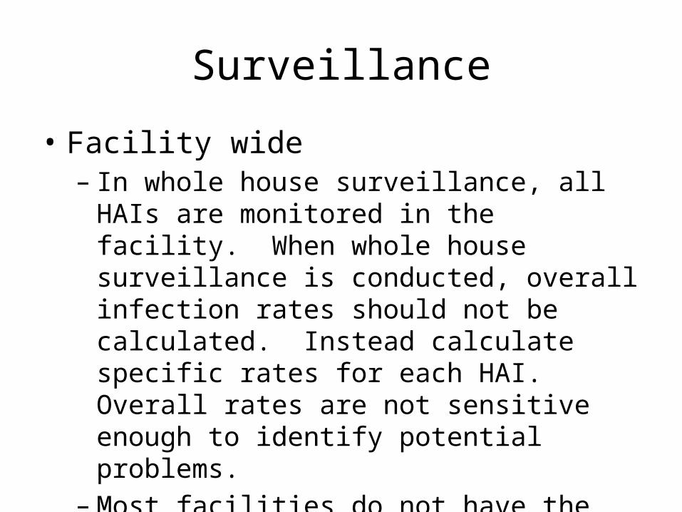
Surveillance
• Facility wide– In whole house surveillance, all HAIs are
monitored in the facility. When whole house surveillance is conducted, overall infection rates should not be calculated. Instead calculate specific rates for each HAI. Overall rates are not sensitive enough to identify potential problems.
– Most facilities do not have the resources to do this.

Surveillance
• Targeted– In the 1990s the CDC shifted away from whole
house to targeted surveillance. Targeted programs usually focus on high-risk, high-volume procedures or units.
– This give you the most bang for your buck!!

Surveillance
• Periodic– Monitoring a selected unit, device or procedure
for a specified time period.– Can be useful to monitor for changes in a stable
process.

IN GOD WE TRUST, ALL OTHERS BRING DATA

• Using Definitions for data collection– Determine the population or event to study– Determine the time period for observation– Write your definition or use an established one
e.g. CDC NHSN– Apply the definition consistently– Write or find a data collection tool
Collecting Relevant Data

Data Collection
• Concurrent or retrospective data collection– Advantages of concurrent surveillance:
• You can interview care gives• Observe patients and patient care• Implement immediate prevention and control
measures• Clusters and outbreaks can be identified quickly
– Disadvantages of concurrent surveillance:• Very time intensive• Incomplete records
Data Collection
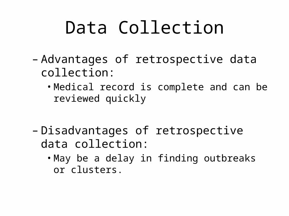
– Advantages of retrospective data collection:• Medical record is complete and can be reviewed
quickly
– Disadvantages of retrospective data collection:• May be a delay in finding outbreaks or clusters.

• Review your data collection for accuracy and effectiveness – Check for flaws in the data– Check your data sources (patient based, lab based,
post discharge surveillance letters, post op calls)– Validate if you make changes
• Sources of data
Collecting Relevant Data

• Record data systematically– Be consistent (data collection tool)– Flow sheet or line list– Can others look at the data and understand it
• Think about how you may want to manipulate or analyze the data later– Computer system– Software for analysis (Excel)
Managing Data

• Analyzing is the reason we do surveillance– Analyze promptly to identify needs for intervention
• Compare Data– Same definitions– Same patient population, risk group
• Proper denominator– Device Days– Patient Days– Surgical Cases
Analyzing Data

• Compare or Benchmark– Historically against your own rates– Against other hospitals of similar size– National Rates (Review NHSN report as a group)
• Interpretation and Significance– Use of statistics– Data interpretation pit falls– Reporting Data
Analyzing Data

Statistics
• Statistics can summarize and simplify large amounts of numerical data.
• Using statistics one can draw conclusions about data.
• Statistics can help communicate findings clearly and meaningfully to others.
• Statistics can not prove anything- estimates are normally presented in probabilistic terms (e.g. we are 95% sure ...)

Statistics
• Statistics may reveal underlying patterns in data not normally observable.
• If used correctly, statistics can separate the probable from the possible
• Statistics can not make bad data better - "garbage in, garbage out"

Statistics
• Infection Preventionist routinely use statistical methods to:– Prepare reports for committee– Identify problems or outbreaks– Monitor the impact of interventions– Identify areas for improvement

Statistics
• Some commonly used statistical methods in health care are:– Measure of central tendency
• Mean• Median• Mode
– Measures of Dispersion• Standard Deviation• Range• Variance

Statistics
– Measures of frequency• Incidence rate• Prevalence rate• Ratio• Proportion
• Statistical process control• Control Charts
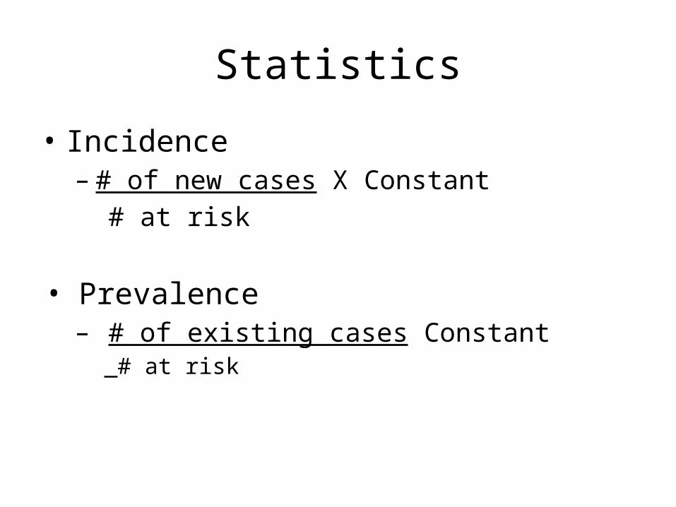
Statistics
• Incidence– # of new cases X Constant
# at risk
• Prevalence– # of existing cases Constant
# at risk

Pitfalls• A high rate does not necessarily indicate a
problem– Intensity of surveillance– Small denominator
• Sample size usually not less than 25• Surgical procedures for devices at least 50
Statistics

Practice
• Now let’s calculate the Mean, Median, Mode, and Range for the following:
– 7, 9, 6, 7, 8, 5– 31, 32, 35, 35, 37, 41, 42, 44, 52, 56– 2, 12, 4, 11, 3, 7, 10, 5, 9, 6

PracticePatient DOA DOD Date of + MRSA
resultMr. Jones 5/1 5/28 5/7
Mrs. Smith 5/9 5/15 5/9
Ms. Goldie 5/1 5/25 5/9
Joe Black 5/10 5/22 5/10
Mr. Chevy 5/12 6/25 5/13
Mrs. Ford 5/14 5/28 5/22
Mr. Dodge 5/15 5/27 5/22
Mr. Jones 5/1 5/28 5/22
Miss Prissy 5/21 6/5 5/25
Mr. Bill 5/25 6/10 5/27
Ms. Barn 5/30 6/7 6/2

PracticeDate # of patients Date # of patients Date # of patients
5/1 25 5/11 15 5/21 26
5/2 27 5/12 16 5/22 20
5/3 20 5/13 22 5/23 20
5/4 21 5/14 20 5/24 24
5/5 16 5/15 20 5/25 23
5/6 28 5/16 21 5/26 23
5/7 25 5/17 24 5/27 21
5/8 22 5/18 23 5/28 17
5/9 21 5/19 22 5/29 20
5/10 21 5/20 21 5/30 21
5/31 25

Practice
• Using the two previous slides, calculate the incidence of MRSA for the month of May
• What is the prevalence of MRSA on 6/1 with the patient days being 425?
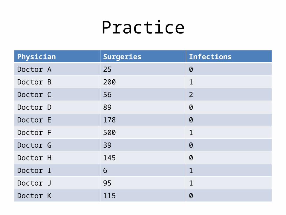
PracticePhysician Surgeries Infections
Doctor A 25 0
Doctor B 200 1
Doctor C 56 2
Doctor D 89 0
Doctor E 178 0
Doctor F 500 1
Doctor G 39 0
Doctor H 145 0
Doctor I 6 1
Doctor J 95 1
Doctor K 115 0


• Devices strongly correlated with infection– Urinary catheters– Central lines– Ventilators
# of device assoc infections x 1000# of device days
Device Related Data

• 4 BSI Infections• 120 patients• 1420 line days• 4500 Patient days• What is your rate?????
Central Line BSI Example

HA MRSA rate calculation
• HA MRSA definition is developed to identify an MRSA case as “new”: MRSA isolated from clinical or surveillance culture obtained after the third calendar day of admission to the unit in a patient that had no prior MRSA by culture, molecular test, or by history.
• # of new MRSA patients on the unit/month × 1,000 # of patient days on the unit/month = hospital-associated MRSA rate per 1,000 unit patient days
• Good references – 1. APIC MRSA Elimination guide2. CDC MDRO guidelines

• Communicate/Report Data • Look for trends (Analysis)• Implement Changes (Action plan)• Monitor, Track and report Effect of
Interventions
What do you do with the Data?

• What to report• How to report
– Chart• Pie Chart• Bar Charts
– Graph• Line Graph• Control Chart
Communicating Data

• Title• Time Period• Location• Values• Unit Labels• Definitions
Make Things Self-Explanatory
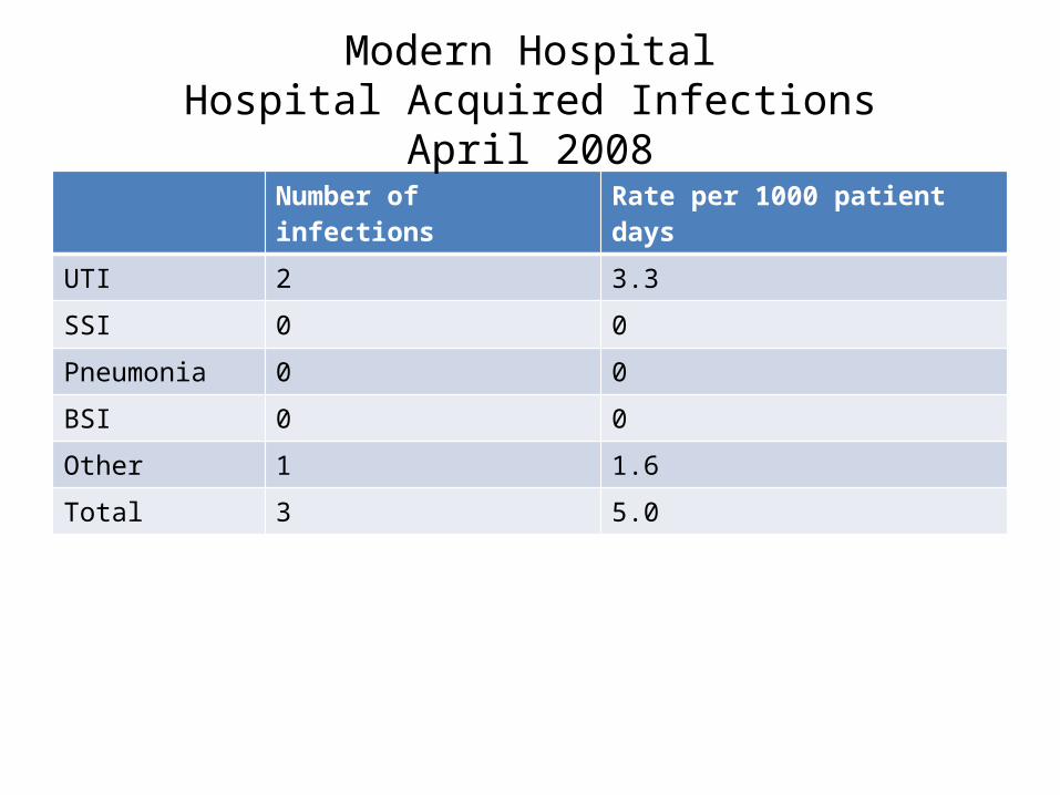
Number of infections Rate per 1000 patient days
UTI 2 3.3
SSI 0 0
Pneumonia 0 0
BSI 0 0
Other 1 1.6
Total 3 5.0
Modern HospitalHospital Acquired Infections
April 2008

62%10%
5%
8%
15%
Sites
UTIBSIPneumoSSIOther
Hospital Acquired Infections by site 2007
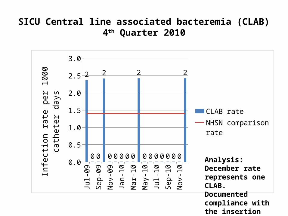
SICU Central line associated bacteremia (CLAB)4th Quarter 2010
Jul-0
9
Sep-
09
Nov
-09
Jan-
10
Mar
-10
May
-10
Jul-1
0
Sep-
10
Nov
-10
0.0
0.5
1.0
1.5
2.0
2.5
3.0
2
0 0
2
0 0 0 0 0
2
0 0 0 0 0 0 0
2
CLAB rateNHSN comparison rate
Infe
ction
rate
per
100
0 ca
thet
er
days
Analysis:December rate represents one CLAB. Documented compliance with the insertion bundle.
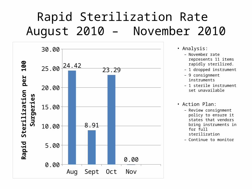
Rapid Sterilization Rate August 2010 – November 2010
Aug Sept Oct Nov0.00
5.00
10.00
15.00
20.00
25.00
30.00
24.42
8.91
23.29
0.00
Rapi
d St
erili
zatio
n pe
r 100
Sur
geri
es
• Analysis:– November rate
represents 11 items rapidly sterilized.
– 1 dropped instrument– 9 consignment
instruments– 1 sterile instrument set
unavailable
• Action Plan:– Review consignment
policy to ensure it states that vendors bring instruments in for full sterilization
– Continue to monitor

Surgical Site Infection Rate July 2010 – October 2010
July Aug Sept Oct0.00
0.20
0.40
0.60
0.80
1.00
1.20
1.40
1.60
0.00 0.00 0.00
1.47
Surg
ical
Site
Infe
ction
s pe
r 100
Sur
geri
es
• Analysis:– October rate
translates to 1 infection – see attached case review.
• Action Plan:– Continue
monthly monitoring and discussion of prevention measures

Lumbar Interbody Infection Rate
July 2010 – October 2010
July Aug Sept Oct0.00
0.10
0.20
0.30
0.40
0.50
0.60
0.70
0.80
0.90
1.00
0.00 0.00 0.00 0.00Surg
ical
Site
Infe
ction
s pe
r 100
Sur
geri
es
• Analysis:– No SSI
identified since surveillance began.
• Action Plan: Continue to do
surveillance and discuss prevention measures

January Fe
bM
arch
AprilM
ayJu
neJu
lyAug
Sep
OctNov
Dec02468
101214161820
Needle Sticks per 1000 visits
Needle Sticks per 1000 vis-its
Needle Sticks Injuries in ER 2007

Jan Feb Mar Apr May Jun Jul Aug Sep Oct Nov Dec0
2
4
6
8
10
12
14
16
Needle Stick Injuries
Needle Stick Injuries
Needle Stick Injuries

COMPARING THE RATESAdvanced Infection prevention Class
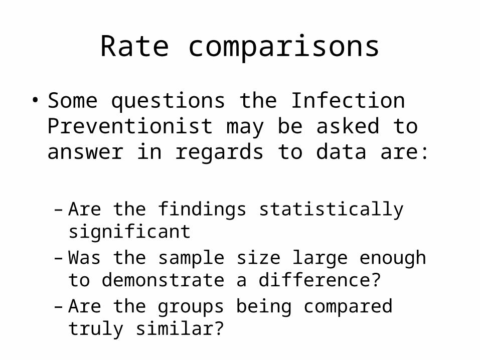
Rate comparisons
• Some questions the Infection Preventionist may be asked to answer in regards to data are:
– Are the findings statistically significant– Was the sample size large enough to demonstrate
a difference?– Are the groups being compared truly similar?

The Null Hypothesis
• When comparing SSI rates, the hypothesis being tested is that the rates are not different. This is called the null hypothesis.
• A statistical test can be used to test the
hypothesis and obtain a p-value

P-value
• What is "Statistical Significance" (p-value)?– The statistical significance of a result is the
probability that the observed relationship or a difference in a sample occurred by pure chance ("luck of the draw"), and that in the population from which the sample was drawn, no such relationship or differences exist. Using less technical terms, we could say that the statistical significance of a result tells us something about the degree to which the result is "true" (in the sense of being "representative of the population").

P-value
• More technically, the value of the p-value represents a decreasing index of the reliability of a result. P- values range from 0 – 1. The higher the p-value, the less we can believe that the observed relation between variables in the sample is a reliable indicator of the relation between the respective variables in the population.

P-value
• Typically, in many sciences, results that yield p .05 are considered borderline statistically significant, but remember that this level of significance still involves a pretty high probability of error (5%). Results that are significant at the p .01 level are commonly considered statistically significant, and p .005 or p .001 levels are often called "highly" significant.
P-value
• Let’s Practice• http://www.openepi.com/OE2.3/Menu/Open
EpiMenu.htm

• This is what adjusts for severity of illness. Should be procedure-specific. (Review NHSN SSI Data submission form)
• Based on 3 factors collected on all surgical patients:– Length of surgery– American Society of Anesthesiology (ASA) Score– Surgical wound classification
Surgical Site Risk Adjustment

Standard Infection Ratio (SIR)• What is a standardized infection ratio (SIR)?• The standardized infection ratio (SIR) is a summary measure used to track HAIs at a
national, state, or local level over time. The SIR adjusts for the fact that each healthcare facility treats different types of patients. For example, the experience with HAIs at a hospital with a large burn unit (a location where patients are more at risk of acquiring infections) cannot be directly compared to a facility without a burn unit.
• The method of calculating an SIR is similar to the method used to calculate the Standardized Mortality Ratio (SMR), a summary statistic widely used in public health to analyze mortality data. In HAI data analysis, the SIR compares the actual number of HAIs in a facility or state with the baseline U.S. experience (i.e., standard population), adjusting for several risk factors that have been found to be most associated with differences in infection rates.
• In other words, an SIR significantly greater than 1.0 indicates that more HAIs were observed than predicted, accounting for differences in the types of patients followed; conversely, an SIR of significantly less than 1.0 indicates that fewer HAIs were observed than predicted.
Reference - http://www.cdc.gov/hai/QA_stateSummary.html#6

FIRST STATE-SPECIFICHEALTHCARE-ASSOCIATED INFECTIONS SUMMARY DATA REPORT
January – June, 2009

SIR
SIR = Observed (O) HAIsExpected (predicted) (E) HAIs
To calculate O, sum the number of HAIs among a reporting entity
To calculate E, requires the use of the appropriate aggregate data from a standard population (NHSN)


REVIEW AND PRACTICE

Let’s Review!!
Question• You are assisting a new Infection preventionist
with setting up her Infection prevention program. She is new to the role and just moved to the area and accepted the position of ICP at a local medical surgical hospital…
• What documents should she review first?

Answer
• The following documents should be reviewed first.1. Infection prevention and surveillance plan. When
was it last reviewed? Was a risk assessment done? Did the risk assessment include an MDRO assessment?
2. TB control plan, when was the last TB risk assessment?
3. Blood borne pathogen exposure plan, when was it last reviewed? Is there a sharps injury prevention team?

Question
• How often does the infection prevention committee have to meet?
• What are some items on a good infection prevention agenda?

Collecting Data
Question• My IC surveillance plan states that I do
quarterly CA-UTI rates, how do I calculate this rate?????
Answers
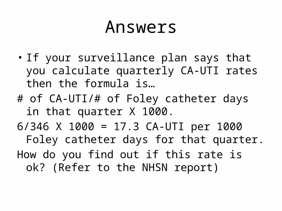
• If your surveillance plan says that you calculate quarterly CA-UTI rates then the formula is…
# of CA-UTI/# of Foley catheter days in that quarter X 1000.
6/346 X 1000 = 17.3 CA-UTI per 1000 Foley catheter days for that quarter.
How do you find out if this rate is ok? (Refer to the NHSN report)

Question
• What if my surveillance plan states that I do surgical site surveillance on patients that have had gallbladder surgery? How do I calculate the rates?

Answer
• You need to have a process in which you know the # of gallbladder surgeries for that month.
• Look at all those patients, have any of them returned for s/s of infection? If so, do the s/s match one of the CDC/NHSN HAI SSI definitions?
• # gallbladder patients found infected/# gallbladder surgeries X 100
• 1/22 X 100 = 4.5 infections per 100 surgeries for that month.
• Where do you find the comparison rate? (Review the NHSN report)
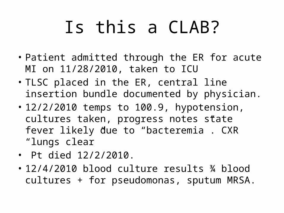
Is this a CLAB?
• Patient admitted through the ER for acute MI on 11/28/2010, taken to ICU
• TLSC placed in the ER, central line insertion bundle documented by physician.
• 12/2/2010 temps to 100.9, hypotension, cultures taken, progress notes state fever likely due to “bacteremia”. CXR “lungs clear”
• Pt died 12/2/2010.• 12/4/2010 blood culture results ¾ blood cultures +
for pseudomonas, sputum MRSA.

CLAB Case review…
• Fits Criteria 1 of CDC definition of CLAB – “patient has a recognized pathogen cultured from one or more blood cultures and the organism cultured from the blood is not related to an infection at another site”.
• What else is the ICP going to report?

Is this a CLAB?
• 86 year old patient admitted 11/18/2010 for colon resection.
• PICC line placed 11/21/2010, the PICC nurse documented use of the insertion bundle components.
• Transferred to Step down unit on 11/23/2010 with PICC.• POD 9 11/26/2010 temperatures up to 102, WBC 30,
progress notes report “sepsis”, cultures taken, transferred back to ICU and intubated.
• 12/2/2010 Enterococcus species in 2/4 blood culture bottles, moderate amount MSSA in sputum, urine negative.

CLAB?
• Fits Criteria 1 of CDC definition of CLAB – “patient has a recognized pathogen cultured from one or more blood cultures and the organism cultured from the blood is not related to an infection at another site”.

• During the month of February, the ICP finds that there were three hip wound infections among patients undergoing total hip replacement. Dr. A performed 28 cases, Dr. B performed 26 cases, and Dr. C performed 6 cases. What is the surgical site infection rate for total hips in February?– A. 3%– B. 4%– C. 5%– D. 20%
Surgical Site Infection Rate Example

Is this a VAP?Case Review
78 year old undernourished frail male patient admitted for Colon resection on 10/1/2010.
CXR on day of surgery “lungs have a hyper inflated appearance but noacute infiltrates”Remained intubated after surgeryVent settings FIO2 40, RR 10, TV 480, PS 8, Peep 5
10/6/2010 CXR “complete obliteration of L hemidiaphram consistent withconsolidation”.“Reduced lung sounds”Purulent sputumWBC 6.3Temperature of 102.0Increased vent settings to FI02 70, RR 16, TV 480, Peep 10.

VAP case review
10/7/2010 WBC 19.0Temperature of 100.4CXR “bibasilar densities same” “Rales”“Purulent sputum”Increased vent settings to FI02 80, RR 16, TV 480, Peep 10.
1. Vent round documentation indicates that all components of the VAP prevention bundle were done on 10/6, 10/7, and 10/12.
2. All final culture results negative3. IC reviewed case with ID physician who agreed the case fit the CDC VAP
Criteria 1 definition.

Is this a CA-UTI??
• Patient was admitted on 2/6/2011 for gallbladder surgery, Foley catheter placed on admit at 0600 am.
• Surgery went well and catheter was discontinued on 2/6/2011 at 9pm.
• The patient had nausea and vomiting and was not discharged until 2/8/2011, before discharge the patient complained to the physician about pain with urination, temps 99.5. The physician discharges the patient on Keflex, does not get a UA with culture.
• IS this a CA-UTI???
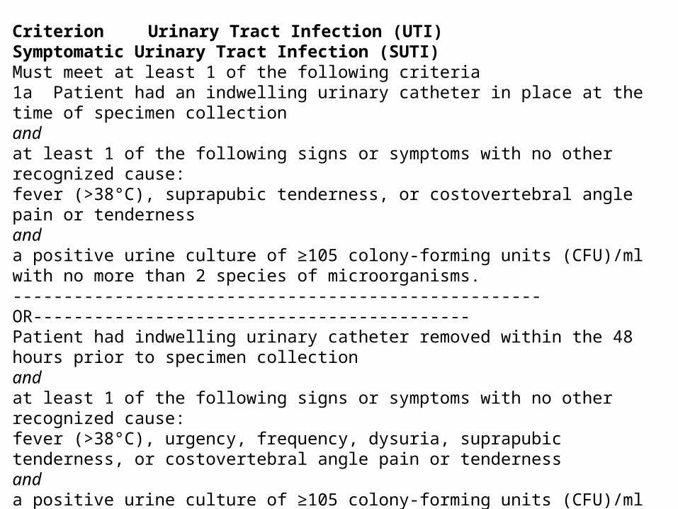
Criterion Urinary Tract Infection (UTI) Symptomatic Urinary Tract Infection (SUTI) Must meet at least 1 of the following criteria 1a Patient had an indwelling urinary catheter in place at the time of specimen collection and at least 1 of the following signs or symptoms with no other recognized cause: fever (>38°C), suprapubic tenderness, or costovertebral angle pain or tenderness and a positive urine culture of ≥105 colony-forming units (CFU)/ml with no more than 2 species of microorganisms. ----------------------------------------------------OR------------------------------------------- Patient had indwelling urinary catheter removed within the 48 hours prior to specimen collection and at least 1 of the following signs or symptoms with no other recognized cause: fever (>38°C), urgency, frequency, dysuria, suprapubic tenderness, or costovertebral angle pain or tenderness and a positive urine culture of ≥105 colony-forming units (CFU)/ml with no more than 2 species of microorganisms.
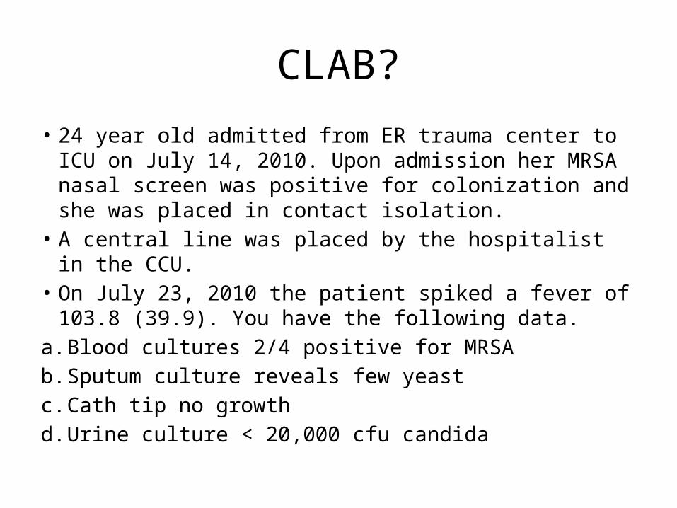
CLAB?
• 24 year old admitted from ER trauma center to ICU on July 14, 2010. Upon admission her MRSA nasal screen was positive for colonization and she was placed in contact isolation.
• A central line was placed by the hospitalist in the CCU.• On July 23, 2010 the patient spiked a fever of 103.8 (39.9).
You have the following data.a. Blood cultures 2/4 positive for MRSAb. Sputum culture reveals few yeastc. Cath tip no growthd. Urine culture < 20,000 cfu candida

CLAB?
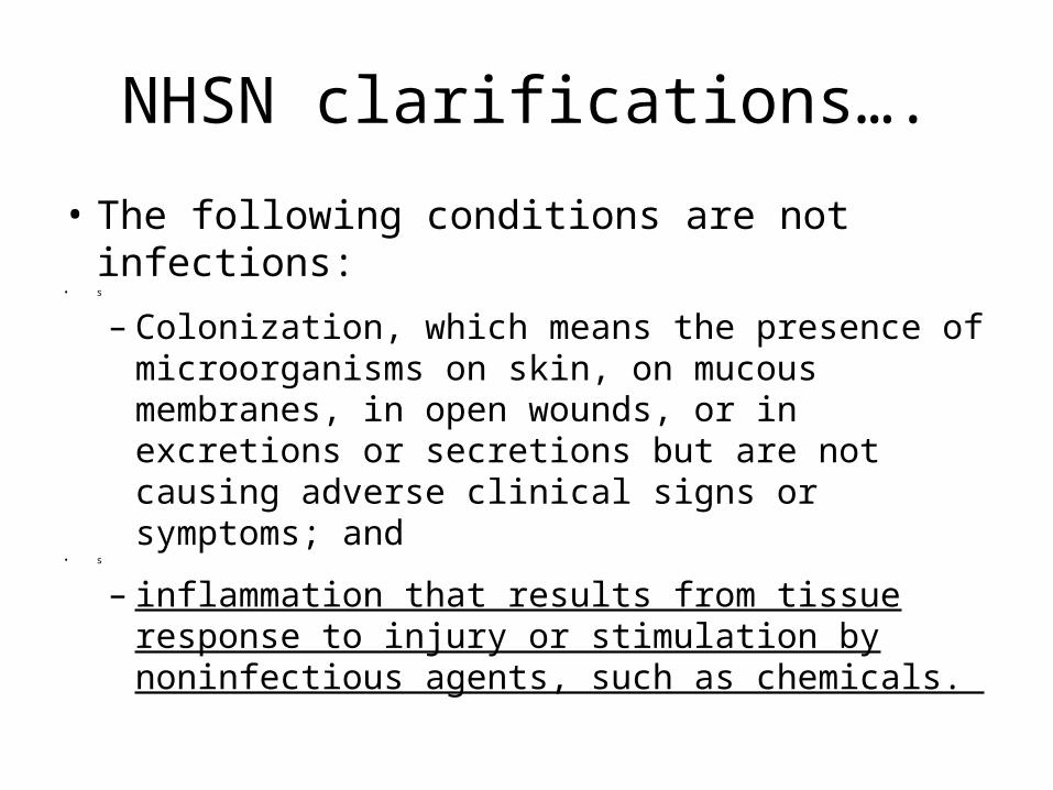
• The following conditions are not infections:
– Colonization, which means the presence of microorganisms on skin, on mucous membranes, in open wounds, or in excretions or secretions but are not causing adverse clinical signs or symptoms.
– It is a CLAB, meets criteria 1, recognized pathogen in a patient with a central line, 1 BC +, no other recognized cause.

CLAB/VAP or both???Don’t pull your hair out
• July 31, 2010, 62 year old male admitted to your hospital with chemical burns to face, oral cavity, nasal cavity and respiratory distress. He is immediately taken to the OR for trach placement.
• PICC placed 8/3/2011.• 9 days after admission (8/9/2010) you note the following;a. Rusty brown foul secretions from the trachb. Increased ventilator settings neededc. Rhonchi, breath sounds used to be “diminished”d. WBC increased to 13.9, temps up to 40 degrees Celsius.e. 8/9/2010 – CXR impression “persistent infiltrates”, 8/10/2010 “increasing
infiltrates”.f. 8/9/2011 “tracheal” culture = MRSA, E-coli.g. 8/9/2011 Blood culture 2/4 + for MRSAh. 8/10/2011 Blood culture 2/4 + MRSA

Identifying Hospital acquired pneumonia – 3 parts to PNU 1
1. Radiologic criteria• Patient with underlying pulmonary or cardiac
disease, must have two or more CXR with at least one of the following;
• One definitive CXR with the following criteria is acceptable in patients without underlying disease.
a. New or progressive and persistent infiltrate.b. Consolidationc. Cavitation

Signs and Symptoms
• At least one of the following;1. Fever (> 38˚ C or > 100.4˚ F) with no other
recognized cause…… Hmmmm, had MRSA in Blood cultures….
2. Leukopenia (< 4,000 WBC/mm, or leukocytosis > 12,000 WBC/mm)
3. Altered mental status with no other recognized cause

AND at least 2 of the following
1. New onset of purulent sputum, or change in character of the sputum, or increased respiratory secretions, or increased suctioning requirements.
2. New onset or worsening cough, or dyspnea, or tachypnea.
3. Rales or bronchial breath sounds4. Worsening gas exchange (O2 desats,
increased oxygen requirements, or increased ventilation demand.

VAP or CLAB continued…
• I do not think it could be called a pneumonia criteria 1, due to the MRSA in the blood cultures….
• Lets look at pneumonia criteria 2 a bit closer…


Read the fine print… “8”
• 8. “Care must be taken to determine the etiology of pneumonia in a patient with positive blood cultures and radiographic evidence of pneumonia, especially if the patient has invasive devices in place such as intravascular lines or an indwelling urinary catheter. In general, in an immunocompetent patient, blood cultures positive for coagulase-negative staphylococci, common skin contaminants, and yeasts will not be the etiologic agent of the pneumonia. “
• 9. Refer to threshold values for cultured specimens (Table 8). An endotracheal aspirate is not a minimally contaminated specimen. Therefore, an endotracheal aspirate does not meet the laboratory criteria.
NHSN clarifications….
• The following conditions are not infections: • s
– Colonization, which means the presence of microorganisms on skin, on mucous membranes, in open wounds, or in excretions or secretions but are not causing adverse clinical signs or symptoms; and
• s
– inflammation that results from tissue response to injury or stimulation by noninfectious agents, such as chemicals.

VAP or CLAB?• I think that the chemical burns that the patient was admitted with
would cause me to call this a CLAB criteria 1, not a VAP…• Patient with a central line• Recognized pathogen in ¼ blood cultures.• Did they use the insertion bundle on insertion?• Is this documented?• Has the dressing been changed per policy?• Curious – did they pull the PICC line?
• Thoughts?• May have been a clinical pneumonia diagnosis, but it did not fit
the CDC pneumonia surveillance definition that we must follow.
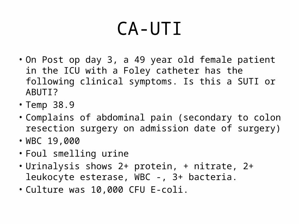
CA-UTI
• On Post op day 3, a 49 year old female patient in the ICU with a Foley catheter has the following clinical symptoms. Is this a SUTI or ABUTI?
• Temp 38.9• Complains of abdominal pain (secondary to colon resection
surgery on admission date of surgery)• WBC 19,000• Foul smelling urine• Urinalysis shows 2+ protein, + nitrate, 2+ leukocyte
esterase, WBC -, 3+ bacteria.• Culture was 10,000 CFU E-coli.

Identification and Categorization of SUTI Indwelling Catheter at the Time of Specimen Collection
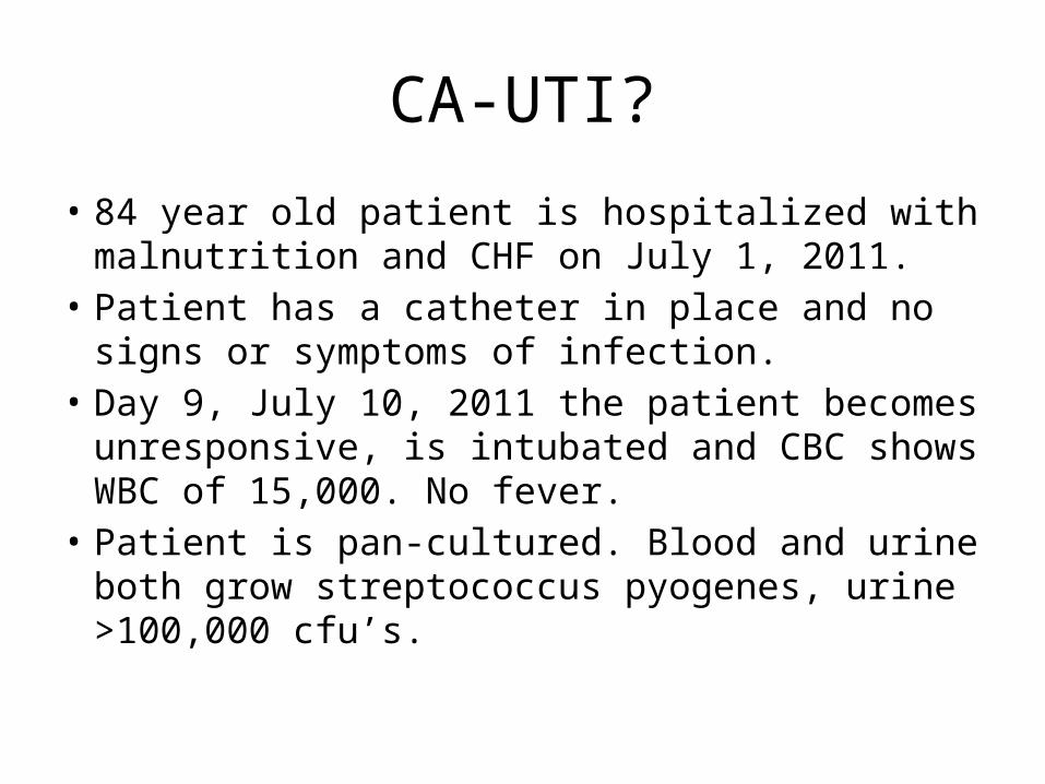
CA-UTI?
• 84 year old patient is hospitalized with malnutrition and CHF on July 1, 2011.
• Patient has a catheter in place and no signs or symptoms of infection.
• Day 9, July 10, 2011 the patient becomes unresponsive, is intubated and CBC shows WBC of 15,000. No fever.
• Patient is pan-cultured. Blood and urine both grow streptococcus pyogenes, urine >100,000 cfu’s.
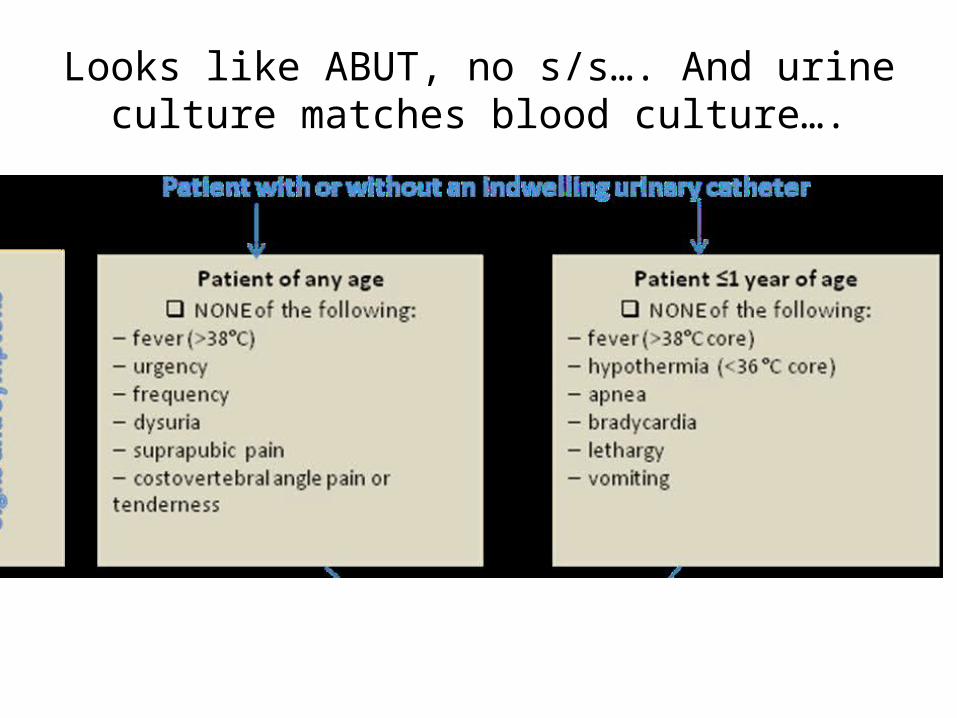
Looks like ABUT, no s/s…. And urine culture matches blood culture….

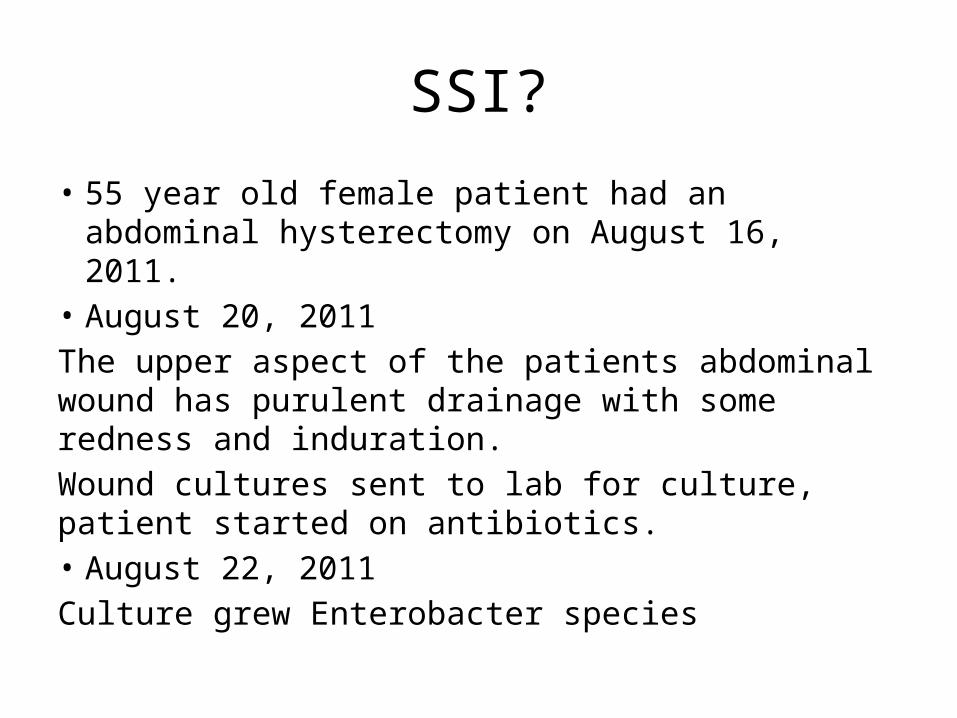
SSI?
• 55 year old female patient had an abdominal hysterectomy on August 16, 2011.
• August 20, 2011The upper aspect of the patients abdominal wound has purulent drainage with some redness and induration.Wound cultures sent to lab for culture, patient started on antibiotics.• August 22, 2011Culture grew Enterobacter species

A superficial incisional SSI (SIP or SIS) must meet the following criterion: Infection occurs within 30 days after the operative procedure and involves only skin and subcutaneous tissue of the incision and patient has at least 1 of the following: a. purulent drainage from the superficial incision b. organisms isolated from an aseptically obtained culture of fluid or tissue from the superficial incision c. at least 1 of the following signs or symptoms of infection: pain or tenderness, localized swelling, redness, or heat, and superficial incision is deliberately opened by surgeon and is culture positive or not cultured. A culture-negative finding does not meet this criterion. d. diagnosis of superficial incisional SSI by the surgeon or attending physician.
There are 2 specific types of superficial incisional SSI: Superficial incisional primary (SIP): a superficial incisional SSI that is identified in the primary incision in a patient who has had an operation with 1 or more incisions (eg, C-section incision or chest incision for coronary artery bypass graft with a donor site [CBGB]).
Superficial incisional secondary (SIS): a superficial incisional SSI that is identified in the secondary incision in a patient who has had an operation with more than 1 incision (eg, donor site [leg] incision for CBGB).
SSI?
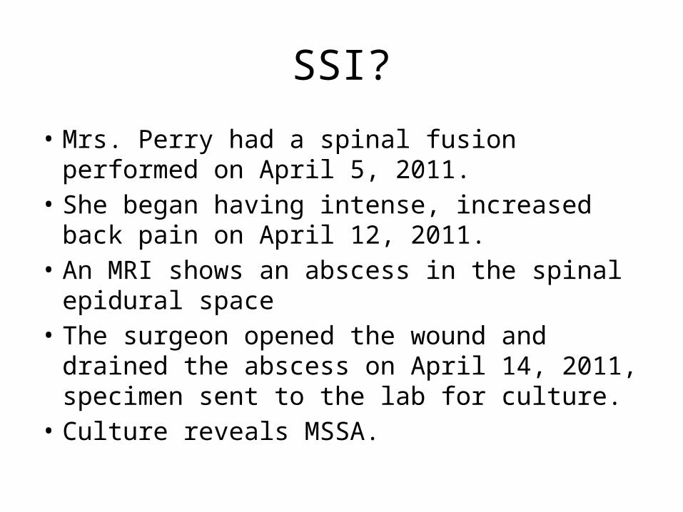
• Mrs. Perry had a spinal fusion performed on April 5, 2011.
• She began having intense, increased back pain on April 12, 2011.
• An MRI shows an abscess in the spinal epidural space
• The surgeon opened the wound and drained the abscess on April 14, 2011, specimen sent to the lab for culture.
• Culture reveals MSSA.
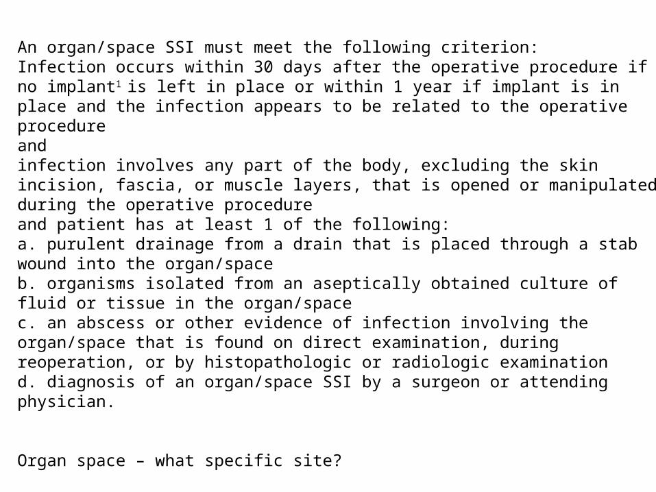
An organ/space SSI must meet the following criterion: Infection occurs within 30 days after the operative procedure if no implant1 is left in place or within 1 year if implant is in place and the infection appears to be related to the operative procedure and infection involves any part of the body, excluding the skin incision, fascia, or muscle layers, that is opened or manipulated during the operative procedure and patient has at least 1 of the following: a. purulent drainage from a drain that is placed through a stab wound into the organ/space b. organisms isolated from an aseptically obtained culture of fluid or tissue in the organ/space c. an abscess or other evidence of infection involving the organ/space that is found on direct examination, during reoperation, or by histopathologic or radiologic examination d. diagnosis of an organ/space SSI by a surgeon or attending physician.
Organ space – what specific site?

Organ/space Organ/space SSI. Indicate specific type: Specific sites are assigned to organ/space SSI to identify further the location of the infection.
BONE LUNG BRST MED CARD MEN DISC ORAL EAR OREP EMET OUTI ENDO SA ----- (Spinal abscess)EYE SINU GIT UR IAB VASC IC VCUF JNT
***Last class, question about Breast implant. Classify as deep or organ space?Superficial - skin and subcutaneous tissue Deep - deep soft tissues (eg, fascial and muscle layers) Organ space - organ/space SSI involves any part of the body, excluding the skin incision, fascia, or muscle layers, that is opened or manipulated during the operative procedure.

NHSN TRAINING SLIDESSSI/CAUTIBRIEF REVIEW OF NICU/CLAB

Healthcare Facility HAI Reporting to CMS via NHSN – Current and Proposed Requirements DRAFT (8/5/2011)
HAI Event Facility Type Reporting Start Date
CLABSI Acute Care HospitalsAdult, Pediatric, and Neonatal ICUs January 2011
CAUTI Acute Care HospitalsAdult and Pediatric ICUs January 2012
SSI Acute Care HospitalsColon and abdominal hysterectomy January 2012
I.V. antimicrobial start (proposed) Dialysis Facilities January 2012
Positive blood culture (proposed) Dialysis Facilities January 2012
Signs of vascular access infection (proposed) Dialysis Facilities January 2012
CLABSI Long Term Care Hospitals * October 2012
CAUTI Long Term Care Hospitals * October 2012
CAUTI Inpatient Rehabilitation Facilities October 2012
MRSA Bacteremia Acute Care Hospitals January 2013
C. difficile LabID Event Acute Care Hospitals January 2013
HCW Influenza Vaccination Acute Care Hospitals January 2013
HCW Influenza Vaccination OP Surgery, ASCs October 2013
SSI (proposed) Outpatient Surgery/ASCs January 2014
* Long Term Care Hospitals are called Long Term Acute Care Hospitals in NHSN
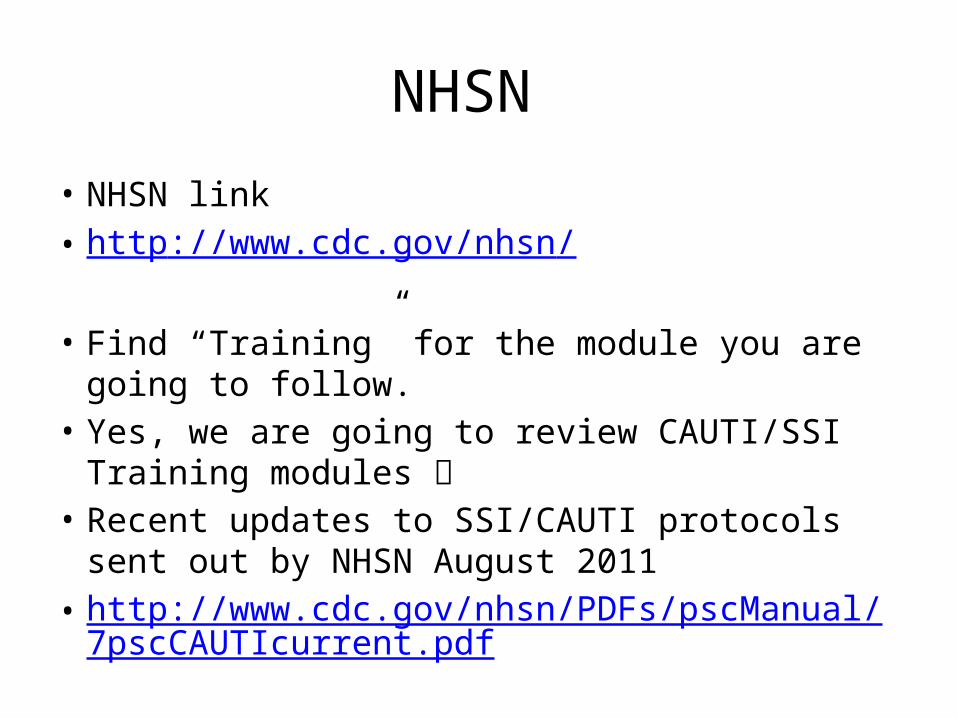
NHSN
• NHSN link• http://www.cdc.gov/nhsn/
• Find “Training” for the module you are going to follow.• Yes, we are going to review CAUTI/SSI Training
modules • Recent updates to SSI/CAUTI protocols sent out by
NHSN August 2011• http://www.cdc.gov/nhsn/PDFs/pscManual/7pscCAUT
Icurrent.pdf


REVIEW OF STERILIZATION PROCESSES
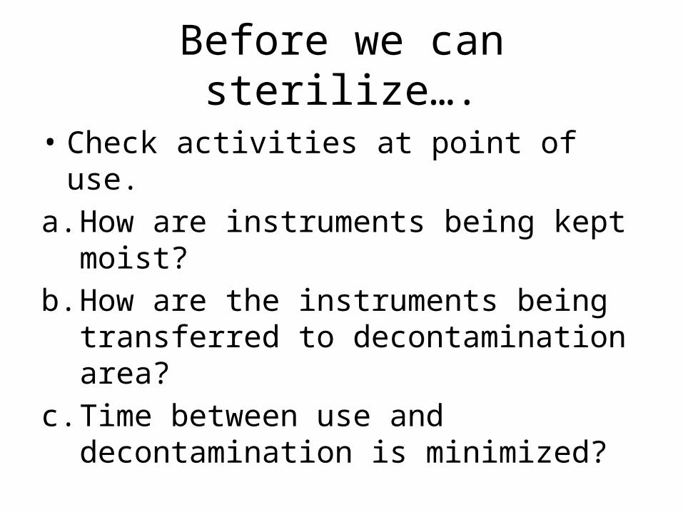
Before we can sterilize….
• Check activities at point of use.a. How are instruments being kept moist?b. How are the instruments being transferred to
decontamination area?c. Time between use and decontamination is
minimized?

Decontamination Room
• Negative air?• Temperature range (optimal 60-65 degrees)• Humidity ( optimal 30%-60%)• No Fans!• PPE?• Doors/pass through windows?• Enzymatic cleaner…..• Cleaning brushes• Manual cleaning or Automated washer?• Work flow• Hand washing

Instrument Preparation
• Inspection• Distribution of instruments in trays/peel packs• Single use devices• Chemical indicators are placed in each
package.• Sterilization wrap• Rigid containers
Sterilization
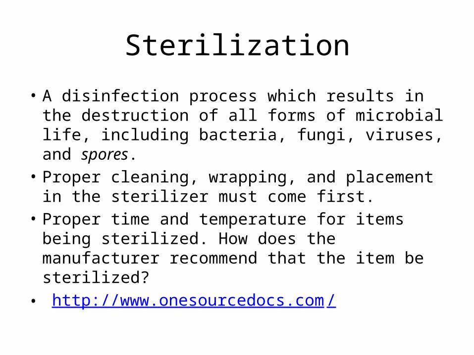
• A disinfection process which results in the destruction of all forms of microbial life, including bacteria, fungi, viruses, and spores.
• Proper cleaning, wrapping, and placement in the sterilizer must come first.
• Proper time and temperature for items being sterilized. How does the manufacturer recommend that the item be sterilized?
• http://www.onesourcedocs.com/
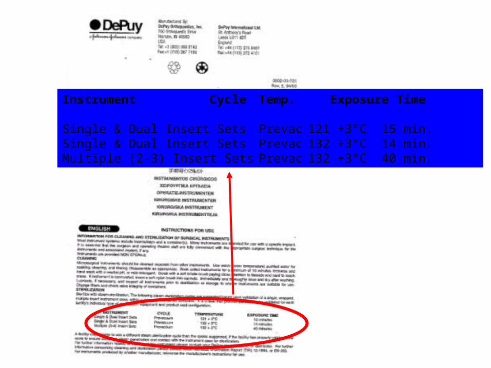
Instrument Cycle Temp. Exposure Time
Single & Dual Insert Sets Prevac 121 +3°C 15 min.Single & Dual Insert Sets Prevac 132 +3°C 14 min.Multiple (2-3) Insert Sets Prevac 132 +3°C 40 min.

Sterilizers
• Drain strainers • Sterilizer gaskets• When was the last time the sterilizer was
cleaned? What does the manufacturer recommend?

Methods of Sterilization –
• Steam1. Pre-Vacuum 2. Gravity 3. Flash• Hydrogen peroxide gas plasma• Ethylene Oxide• Peracetic acid

Prevac Sterilization
• Air is completely evacuated from the chamber by the vacuum. The steam injector helps eliminate the air out of the packages. Steam then penetrates the packages on all surfaces.

Gravity Sterilization
• Gravity pushes the air through the packages and down through the drain. Sterilization begins when steam passes the thermometer and reaches the desired temperature.

It is not called Flash Sterilization anymore…
“Immediate Use Sterilization”“Is broadly defined as the shortest possible time
between a sterilized item’s removal from the sterilizer and its aseptic transfer to the sterile field, it also implies that the sterilized item is
used for the procedure for which it was sterilized for and not stored for future use, or
held from one case to the another”

Immediate use sterilization….• 2010 Association for the Advancement of Medical
Instrumentation (AAMI) partnered with other healthcare organizations and regulatory agencies to develop a multi-society position statement to clear up confusion– Accreditation Association for Ambulatory Health Care (AAAHC)– Association for perioperative Registered Nurses (AORN)– Association for Professionals in Infection Control and
Epidemiology(APIC)– ASC Quality Collaboration (ASCQC)– International Association of Healthcare Central Service Materiel
Management (IAHCSMM)

Immediate use sterilization
• Multi-Society position statement also focuses on personnel requirements involved in reprocessing activities
• Personnel should:– Be knowledgeable– Exercise critical thinking and judgment– Implement standardized practices
• Supervising organization should:– Ensure appropriate training and education– Ensure staff competency– Ensure related resources are available

Immediate use sterilization should NOT be performed in the following situations
• Sterilization of Implants - except in a documented
emergency situation where no other option is available• For convenience or as a substitute for insufficient
instrument inventory• Sterilization of devices using cycles that have not been
validated for that device or sterilizer• Sterilization of devices that are sold sterile and
intended for single use only• Post-procedure decontamination of instruments used
on patients who may have Creutzfeldt-Jacob disease (CJD) or similar disorders

Follow manufacturer instructions for immediate use sterilization
• The device manufacturer’s written instructions for reprocessing any reusable device must be followed
• The cycle parameters required to achieve sterilization are determined by the design of the instrument, the characteristics of the load, the sterilizer capabilities, and the packaging (if used)
• Conflicting instructions (sterilizer manufacturer vs. device manufacturer)
Reference: AAMI position statement on Immediate use sterilization 2/2011

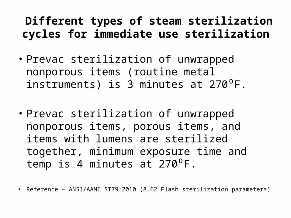
Different types of steam sterilization cycles for immediate use sterilization
• Gravity cycles for unwrapped nonporous items (routine metal instruments) is 3 minutes at 270⁰ - 275⁰
• Gravity cycles for unwrapped nonporous items, porous
items, and items with lumens are sterilized together, minimum exposure time and temp is 10 minutes at 270⁰ - 275⁰
• Reference – ANSI/AAMI ST79:2010 (8.62 Flash sterilization parameters)
Different types of steam sterilization cycles for immediate use sterilization
• Prevac sterilization of unwrapped nonporous items (routine metal instruments) is 3 minutes at 270⁰F.
• Prevac sterilization of unwrapped nonporous items, porous items, and items with lumens are sterilized together, minimum exposure time and temp is 4 minutes at 270⁰F.
• Reference – ANSI/AAMI ST79:2010 (8.62 Flash sterilization parameters)

Flash Sterilization Log
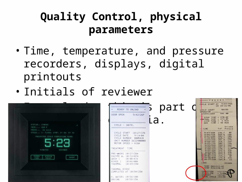
Quality Control, physical parameters
• Time, temperature, and pressure recorders, displays, digital printouts
• Initials of reviewer• Every load – this is part of load release criteria.

Quality control, chemical indicators
• Chemical indicators– Visual identification - processed vs. unprocessed
packs– Verify sterilant penetration– Verify air removal (prevacuum sterilizers – Bowie
Dick)– Pack monitoring– Load monitoring– Load release
• Six classes of chemical indicators• Each class has different performance specifications

Class 1 Chemical Indicators
• Class 1 – external, use on outside of every package unless the internal CI is visible.
• Class 1 – internal, some internal indicator strips
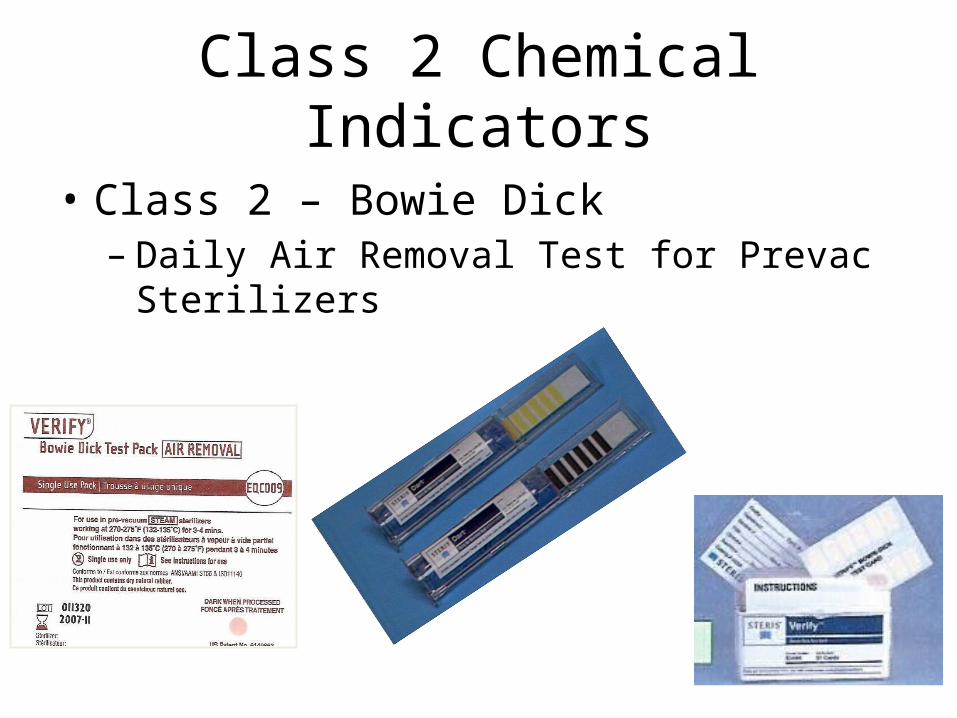
Class 2 Chemical Indicators
• Class 2 – Bowie Dick– Daily Air Removal Test for Prevac Sterilizers

Class 3 Chemical Indicators
• Internal indicators
• Not used in steam sterilization– Single-variable indicators
• Peracetic acid concentration• Hydrogen peroxide concentration
Class 4 Chemical Indicators
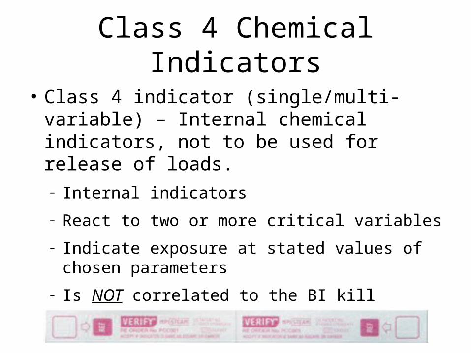
• Class 4 indicator (single/multi-variable) – Internal chemical indicators, not to be used for release of loads.– Internal indicators– React to two or more critical variables– Indicate exposure at stated values of chosen
parameters– Is NOT correlated to the BI kill

Class 5 Chemical Indicators
• Class 5 – Integrating indicators– Internal indicators and challenge packs– Respond to all critical parameters– Performance correlated to the BI – For gravity and pre-vacuum cycles– Monitors more of sterilization cycle than BI test

Class 6 Chemical Indicators
• Class 6 Emulating Indicators– Respond to all critical variables– Performance correlated to the sterilization cycle –
tighter tolerance– Monitors more of the total sterilization cycle than
other sterilization monitoring products

Process Control Device - PCD
• Process challenge device (PCD): Item designed to constitute a defined resistance to a sterilization process and used to assess performance of the process.

Quality control, chemical indicators
• Positioning of the indicators and process challenge devices– Should be placed flat in the area of the sterilizer
chamber and load that represent the greatest challenge (cold point) to the cycle
– Normally in the front, bottom section of the sterilizer, near the drain
– Should be identified by the sterilizer manufacturer

Quality control, biological indicators
• Is the type of BI being used appropriate for the cycles being processed?
• BI’s are incubated following the manufacturer instructions?
• Daily vs. Weekly• Every load containing an implantable device
Record keeping and Infection prevention visibility in sterile process areas of the facility.
• Lot number – sterilizer number, date of sterilization and cycle #.• Contents of the load detailed?• Patient identifier• Exposure time and temperature with initials of the operator?• BI test results• Bowie Dick test results• Chemical indicator response• Have a written mechanism for recall – “Suggested protocol for
management of positive biological indicator in steam sterilizer” – Table 12 CDC guideline for disinfection and sterilization in healthcare facilities, 2008.

In summary - routine load release
• The load was actually initiated.• The sterilization cycle was appropriate for items processed.• Every sterilizer load should be physically monitored (Time,
temp, pressure).• Every packaged item should be labeled externally with a
process indicator (Class 1).• And should contain an internal indicator (class 5 or 6).• If desired a PCD containing a BI, or a PCD containing a class 5
or class 6 (CI challenge pack) in the area of the chamber and load considered to be least favorable to sterilization.
• Reference ANSI/AAMI ST79:2010, Pages 106-107

Implant load
• Every load containing an implant should be monitored with a class 5 chemical indicator within a PCD that contains a BI.
• This may be used to release an implant load ONLY in an emergency! (do you have emergency release policy for implants?) “Annex L”
• Implants should be quarantined until the results of the BI are available. (“early readout or spore growth”)
• A class 6 emulating indicator within a PCD may be used as part of release criteria for loads containing implants.
• Reference ANSI/AAMI ST79:2010, Pages 103

Practice – what type of reprocessing?
• Bedside tables• Laryngoscope blades• Surgical instruments• Vaginal ultrasound probes• Endoscopes• Temporal Thermometers• Cardiac Catheters

Thank you!!!!!Infection prevention matters!!!!