Adsorption, metalation and magnetic properties of tetra ... · tetra-phenyl-porphyrin (2H-TPP)...
119
Transcript of Adsorption, metalation and magnetic properties of tetra ... · tetra-phenyl-porphyrin (2H-TPP)...

ii
iii
Abstract
Traditional semiconductor technology will reach a size limit within the next fewyears. A possible solution could be the use of organic molecules in technologicalapplications as single functional units in metal-organic based devices; the successof this approach strongly depends on the understanding of the behaviour of thesemolecules on metallic surfaces. The interaction with metallic substrates andthe interaction between the molecules themselves determine the electronic andmagnetic properties of the system, and it is thus of fundamental interest to studythese metal-organic interfaces both in the case of single molecules and layerstructures.
In this thesis, an extensive study of the electronic and magnetic properties oftetra-phenyl-porphyrin (2H-TPP) molecules adsorbed on metal surfaces is reported.
By means of scanning tunnelling microscopy (STM) we studied the adsorptiongeometry of these molecules on the Au(111), Ag(111) and Cu(100) surfaces. Byusing X-ray photoemission spectroscopy (XPS) and near-edge X-ray absorptionfine structure (NEXAFS) spectroscopy, a temperature-induced conformationaladaptation reaction of the 2H-TPP molecules adsorbed on the Au(111) and Ag(111)surfaces, upon annealing at 550 K, is described. A possible dehydrogenationreaction, with the formation of new C-C bonds, could explain the rotation of themolecule phenyl rings parallel to the surface plane and the associated increasing inthe molecule-substrate interaction.
In-situ metalation of porphyrins in ultra-high vacuum is obtained by two meth-ods: in the first one, the metalation of 2H-TPP on Ag(111) is achieved by directmetal evaporation (Mn, Rh and Fe) on the molecular layer; in the second casewe report the self-metalation of 2H-TPP through the coordination with a metalatom from the Fe(110) and Al(111) substrates. In addition, we investigated theeffects of metalation and temperature-induced conformational adaptation on themolecule-substrate interaction, by means of XPS and NEXAFS, in the case ofCoTPP on Ag(111).
The magnetic properties resulting from the metal coordination are studied byX-ray magnetic circular dichroism (XMCD). Here, a description of the magneticcoupling of a MnTPPCl single layer with a Fe(110) ferromagnetic substrate isdisclosed. Moreover, we focused on the study of the magnetic properties andexchange coupling of two layer of molecule and a ferromagnetic thin film. In thecase of a MnTPP layer on FeTPP/Fe(110) the magnetic coupling extends to thesecond layer of molecules, for which the magnetization is opposite with respect tothe substrate.

iv
v
Sommario
Le tradizionali tecnologie utilizzate nell’industria dei semiconduttori raggiun-geranno, entro breve tempo, il limite nella miniaturizzazione dei loro componenti.Una possibile alternativa potrebbe venire dall’utilizzo di molecole organiche comesingole unità funzionali in dispositivi metallo-organici; d’altra parte il successodi questo approccio dipende in maniera sostanziale dalla comprensione del com-portamento di queste molecole sulle superfici dei metalli. L’interazione con ilsubstrato metallico e la stessa interazione tra le molecole determinano le proprietàelettroniche e magnetiche di questi sistemi, ed è dunque di fondamentale interesselo studio di queste interfacce metallo-organiche sia nel caso di singole molecoleche di strutture più complesse.
In questa tesi è riportato uno studio dettagliato delle proprietà elettroniche emagnetiche di tetra-fenil-porfirine (2H-TPP) adsorbite su superfici metalliche.
Attraverso la microscopia a scansione a effetto tunnel (STM) è stata studiata lageometria di adsorbimento di queste molecole sulle superfici Au(111), Ag(111) eCu(100). Utilizzando le spettroscopie XPS (X-ray photoemission spectroscopy)e NEXAFS (near-edge X-ray absorption fine structure) è descritta la reazione diadattamento conformazionale delle 2H-TPP adsorbite sulle superfici Au(111) eAg(111) a seguito del processo di annealing a 550 K. Una possibile reazione dide-idrogenazione, con la formazione di nuovi legami C-C, può spiegare la rotazionedei gruppi fenili della molecola verso la superficie e l’aumento dell’interazionemolecola-substrato ad esso associato.
La metallazione in-situ delle porfirine in ultra-alto vuoto è ottenuta in due modi:nel primo, la metallazione delle 2H-TPP su Ag(111) è raggiunta con la direttaevaporazione del metallo (Mn, Rh e Fe) sullo strato di molecole; nel secondocaso, sulle superfici Fe(110) e Al(111) la metallazione avviene automaticamentetramite la coordinazione della 2H-TPP con un atomo della superficie. Inoltre,gli effetti della metallazione e dell’adattamento conformazionale sull’interazionemolecola-substrato sono stati studiati, tramite XPS e NEXAFS, nel caso di CoTPPsu Ag(111).
Le proprietà magnetiche risultanti dalla coordinazione della molecola con unatomo metallico sono state studiate per mezzo della tecnica XMCD (X-ray magnetic
circular dichroism). In particolare, viene descritto l’accoppiamento magnetico diun singolo strato di MnTPPCl con un substrato ferromagnetico Fe(110). Inoltre, cisi è focalizzati sullo studio delle proprietà magnetiche tra due strati di molecole eun film sottile ferromagnetico. Nel caso specifico di MnTPP su FeTPP/Fe(110)l’accoppiamento magnetico si estende al secondo strato di molecole, per il quale lamagnetizzazione è opposta rispetto al substrato.

vi
Contents
1 Introduction: Molecular magnetism 1
References . . . . . . . . . . . . . . . . . . . . . . . . . . . . . . . . . 11
2 Experimental background 13
2.1 STM . . . . . . . . . . . . . . . . . . . . . . . . . . . . . . . . . 132.1.1 Technique . . . . . . . . . . . . . . . . . . . . . . . . . . 132.1.2 Data analysis: a new procedure for drift correction in
scanning probe microscopy . . . . . . . . . . . . . . . . . 15Methods . . . . . . . . . . . . . . . . . . . . . . . . . . 15
2.1.3 Experimental setup . . . . . . . . . . . . . . . . . . . . . 212.2 XPS . . . . . . . . . . . . . . . . . . . . . . . . . . . . . . . . . 21
2.2.1 Technique . . . . . . . . . . . . . . . . . . . . . . . . . . 212.2.2 Experimental setup: the SuperESCA beamline . . . . . . 22
2.3 NEXAFS . . . . . . . . . . . . . . . . . . . . . . . . . . . . . . 232.3.1 Technique . . . . . . . . . . . . . . . . . . . . . . . . . . 232.3.2 Experimental setup: the ALOISA beamline . . . . . . . . 24
2.4 XMCD . . . . . . . . . . . . . . . . . . . . . . . . . . . . . . . 252.4.1 Technique . . . . . . . . . . . . . . . . . . . . . . . . . . 252.4.2 Experimental setup: the BACH beamline . . . . . . . . . 25
References . . . . . . . . . . . . . . . . . . . . . . . . . . . . . . . . . 28
3 Adsorption 29
3.1 Introduction . . . . . . . . . . . . . . . . . . . . . . . . . . . . . 293.2 Self-assembly on metal surfaces: STM of TPP monolayers on
Au(111), Ag(111) and Cu(100) . . . . . . . . . . . . . . . . . . . 303.2.1 2H-TPP on Ag(111) and MnTPPCl on Au(111) . . . . . . 303.2.2 NiTPP on Cu(100) . . . . . . . . . . . . . . . . . . . . . 31
3.3 Conformational adaptation of 2HTPP monolayer on Au(111) . . . 343.3.1 NEXAFS . . . . . . . . . . . . . . . . . . . . . . . . . . 353.3.2 XPS . . . . . . . . . . . . . . . . . . . . . . . . . . . . . 393.3.3 Experimental . . . . . . . . . . . . . . . . . . . . . . . . 42
vii
viii CONTENTS
3.4 Conformational adaptation: the case of 2HTPP on Ag(111) . . . . 42References . . . . . . . . . . . . . . . . . . . . . . . . . . . . . . . . . 51
4 Metalation of tetra-phenyl-porphyrins 53
4.1 Introduction . . . . . . . . . . . . . . . . . . . . . . . . . . . . . 534.2 Metal evaporation in UHV . . . . . . . . . . . . . . . . . . . . . 54
4.2.1 Mn-TPP and Rh-TPP on Ag(111) . . . . . . . . . . . . . 554.2.2 Fe-TPP on Ag(111) . . . . . . . . . . . . . . . . . . . . . 59
4.3 Metal atom picking-up from the substrate . . . . . . . . . . . . . 634.3.1 The case of Fe(110) . . . . . . . . . . . . . . . . . . . . 644.3.2 The case of Al(111) . . . . . . . . . . . . . . . . . . . . 67
2H-TPP on Al(111) . . . . . . . . . . . . . . . . . . . . . 67MnTPPCl on Al(111) . . . . . . . . . . . . . . . . . . . . 73
4.4 Effects of metalation on the molecule-substrate interaction . . . . 74References . . . . . . . . . . . . . . . . . . . . . . . . . . . . . . . . . 87
5 Magnetic properties of Metal Porphyrins 89
5.1 Introduction . . . . . . . . . . . . . . . . . . . . . . . . . . . . . 895.2 Monolayer magnetic coupling: the case of MnTPP-Cl on Fe(110) 905.3 Magnetic coupling of the second layer: MnTPP on FeTPP on Fe(110) 92
5.3.1 Results . . . . . . . . . . . . . . . . . . . . . . . . . . . 94References . . . . . . . . . . . . . . . . . . . . . . . . . . . . . . . . . 104
6 Conclusions and Outlook 105
A List of Publications 107
B Symbols and Abbreviations 109
Acknowledgements 111
Chapter 1
Introduction: Molecular magnetism
The miniaturization of electronic devices, starting from the 1950s, has progressedat a fast rate. The number of functional units integrated in single devices increasedexponentially with time, a trend known as Moore’s law [1], with a simultaneousreduction in their size and energy consumption. The semiconductor industry hasbeen characterized by the constant pursuit of creating smaller functional units,while the science interest went at the same pace towards smaller physical systems,with the advent of nanotechnology.
Traditionally, the production of such devices has been based on the so-calledtop-down approach [2], in which big structures are made smaller. This has beenachieved by the adaptation of traditional principles and techniques to a smaller scale,such as lithography using electron beam [3] or ultraviolet and X-ray radiation [4].However, despite the successful technological efforts adopted, in the last years,these methods reached overcoming technological limits, while their operating costsrapidly increased. Besides this, approaching the nano-scale, quantum effects playa dominant role and the same operational principles of these devices, once they areconstituted just by few atoms, are not more valid, and a switch to a new technologyis required [5].
An alternative approach consists in the assembly of small building blocks toproduce functional units, a method which is referred to as bottom-up [2, 6]. Organicmolecules represent one of the most promising class of materials in this sense andtheir use in nano-fabrication methods presents some key advantages [7]. Besidetheir small size, in the order of few nanometres, they are structurally identical andthe interaction and recognition between them can lead to the formation of self-assembled structures [8–13]. Moreover, through molecular synthesis chemistry,different functional groups can be substituted, allowing to introduce and controlnew properties of the molecule [14].
Organic molecules such as porphyrins and phthalocyanines can be depositedin the sub-monolayer regime on single crystal surfaces in ultra high vacuum and
1
2 CHAPTER 1. INTRODUCTION: MOLECULAR MAGNETISM
geometrically ordered structures can be obtained from this procedure [15]. Theunsaturated molecular macrocycle brings synthetic tailorability for these chemicalsystems thanks to the presence of different positions in which functional groups canbe substituted [16]. By varying the composition and geometry of these molecules,it is possible to control their structural, bonding, transport, and optical properties.
Taking advantage of the flexibility in choosing the substituents, in combinationwith the presence of active sites of interactions with metals, this kind of moleculescan be used to fabricate two-dimensional arrays of metallic centres with nanometricspacing, making them promising candidates for applications in emerging fieldslike spintronics and molecular electronics, as building blocks for molecular-baseddevices with controllable and tunable properties [17, 18], for novel metal–organicarchitectures in solid-state chemistry and in molecular engineering [13, 16, 19–23]as well as to form patterned surfaces for catalytic, magnetic, optoelectronic, andsensing materials [24–31].
Several metals can be efficiently coordinated to porphyrins by in-situ metalevaporation [25–27, 32–37], [[1]], or by simply picking-up substrate atoms orad-atoms which coordinate with the molecule macrocycle, forming a metallo-porphyrin [38–45], [[12]].
Recent developments in the field of surface magnetism, with the possible appli-cations of some paramagnetic metal-organic macrocycle molecules as switchableelements in molecular spintronic devices [46–48], have generated much interestinto the structural and magnetic properties of these molecules [20, 49–55, 55–71].
Experimental techniques such as X-ray photoelectron spectroscopy (XPS),near-edge X-ray absorption fine structure (NEXAFS) and X-ray magnetic circulardichroism (XMCD), due to their sensitivity and chemical selectivity, are ideal toprobe the electronic and magnetic properties of the metal-organic interface.
This thesis will address the study of tetra-phenyl-porphyrins on metal surfaces,focusing on the adsorption and metalation processes and the resulting electronicand magnetic properties of the system.
In chapter 2 the experimental techniques related to the reported results arebriefly described. In addition, in section 2.1.2 it will be presented a new approachfor the thermal drift correction in scanning probe microscopy images.
Chapter 3 addresses the adsorption and self-assembly of porphyrins on metalsurfaces in ultra high vacuum (UHV). Scanning tunnelling microscopy (STM) mi-crographs of tetra-phenyl-porphyrin (TPP) self-assembled monolayers on Au(111),Ag(111) and Cu(100) crystal surfaces are presented in section 3.2. In Sections3.3 and 3.4 the conformational adaptation reaction that the porphyrin monolayerundergoes upon annealing is described, in the case of adsorption on the Au(111)and Ag(111) surfaces, respectively.
The metalation process of tetra-phenyl-porphyrin in UHV is discussed in Chap-ter 4, for the two metalation processes investigated, namely metal evaporation
REFERENCES 3
(Section 4.2) and metal atom picking-up (Section 4.3). The effects of metalationand conformational adaptation on the molecule-substrate interaction is addressedin Section 4.4.
Chapter 5 is devoted to the investigation of the magnetic properties of tetra-phenyl-porphyrins arising after their metalation. In particular, the magnetic cou-pling of a single layer of TPP on a ferromagnetic substrate is reported in Section5.2, while in Section 5.3 the magnetic coupling between two layers of porphyrinsis reported for the first time.
Conclusions are summarized in Chapter 6 and a list of publications related tothis thesis is given in Appendix A.
References
[1] G.E. Moore. Cramming more components onto integrated circuits. Pro-
ceedings of the IEEE, 86(1):82–85, Jan 1998. ISSN 0018-9219. doi:10.1109/JPROC.1998.658762.
[2] Bo Cui. Recent Advances in Nanofabrication Techniques and Applications.InTech, 2011. doi: 10.5772/859.
[3] A E Grigorescu and C W Hagen. Resists for sub-20-nm electron beamlithography with a focus on HSQ: state of the art. Nanotechnology, 20(29):292001, jul 2009. doi: 10.1088/0957-4484/20/29/292001.
[4] Takashi Ito and Shinji Okazaki. Pushing the limits of lithography. Nature,406(6799):1027–1031, aug 2000. doi: 10.1038/35023233.
[5] Mike Mayberry. Expanding the technology envelope in an age of consolida-tion. IMEC Technology Forum, Brussels, May 22, 2013.
[6] Richard Feynman. There’s plenty of room at the bottom. 1959. URLhttp://calteches.library.caltech.edu/47/2/1960Bottom.pdf.
[7] Johannes V. Barth, Giovanni Costantini, and Klaus Kern. Engineering atomicand molecular nanostructures at surfaces. Nature, 437(7059):671–679, sep2005. doi: 10.1038/nature04166.
[8] Koji Suto, Soichiro Yoshimoto, and Kingo Itaya. Two-dimensional self-organization of phthalocyanine and porphyrin: dependence on the crystallo-graphic orientation of au. Journal of the American Chemical Society, 125(49):14976–7, 2003. doi: 10.1021/ja038857u.
4 REFERENCES
[9] Daniel Heim, David Ecija, Knud Seufert, Willi Auwärter, Claudia Aurisicchio,Chiara Fabbro, Davide Bonifazi, and Johannes V. Barth. Self-assembly offlexible one-dimensional coordination polymers on metal surfaces. J. Am.
Chem. Soc., 132(19):6783–6790, may 2010. doi: 10.1021/ja1010527.
[10] Florian Buchner, Ina Kellner, Wolfgang Hieringer, Andreas Görling, Hans-Peter Steinrück, and Hubertus Marbach. Ordering aspects and intramolecularconformation of tetraphenylporphyrins on ag(111). Phys. Chem. Chem. Phys.,12(40):13082, 2010. doi: 10.1039/c004551a.
[11] Sigrid Weigelt, Carsten Busse, Christian Bombis, Martin M. Knudsen, Kurt V.Gothelf, Thomas Strunskus, Christof Wöll, Mats Dahlbom, Bjørk Hammer,Erik Lægsgaard, Flemming Besenbacher, and Trolle R. Linderoth. Covalentinterlinking of an aldehyde and an amine on a au(111) surface in ultra-high vacuum. Angew. Chem. Int. Ed., 46(48):9227–9230, dec 2007. doi:10.1002/anie.200702859.
[12] Johannes V. Barth. Molecular architectonic on metal surfaces. Annu.
Rev. Phys. Chem., 58(1):375–407, may 2007. doi: 10.1146/an-nurev.physchem.56.092503.141259.
[13] Johannes V. Barth. Fresh perspectives for surface coordination chem-istry. Surface Science, 603(10-12):1533–1541, jun 2009. doi:10.1016/j.susc.2008.09.049.
[14] Leonhard Grill, Matthew Dyer, Leif Lafferentz, Mats Persson, Maike V.Peters, and Stefan Hecht. Nano-architectures by covalent assembly of molec-ular building blocks. Nature Nanotech, 2(11):687–691, oct 2007. doi:10.1038/nnano.2007.346.
[15] Willi Auwärter, David Écija, Florian Klappenberger, and Johannes V. Barth.Porphyrins at interfaces. Nature Chem, 7(2):105–120, jan 2015. doi:10.1038/nchem.2159.
[16] B D Berezin. Coordination Compounds of Porphyrins and Phthalocyanines.Wiley, 1981.
[17] Christof Wöll, editor. Physical and Chemical Aspects of Organic Electronics.Wiley edition, 2009.
[18] Pietro Gambardella, Sebastian Stepanow, Alexandre Dmitriev, Jan Honolka,Frank M. F. de Groot, Magalí Lingenfelder, Subhra Sen Gupta, D. D. Sarma,Peter Bencok, Stefan Stanescu, Sylvain Clair, Stéphane Pons, Nian Lin, Ari P.Seitsonen, Harald Brune, Johannes V. Barth, and Klaus Kern. Supramolecular
REFERENCES 5
control of the magnetic anisotropy in two-dimensional high-spin fe arraysat a metal interface. Nature Materials, 8(3):189–193, March 2009. ISSN1476-1122, 1476-4660. doi: 10.1038/nmat2376.
[19] J E Falk. Porphyrins and Metalloporphyrins: Their General, Physical and
Coordination Chemistry, and Laboratory Methods. Elsevier, 1964.
[20] M. Bernien, J. Miguel, C. Weis, Md. Ali, J. Kurde, B. Krumme, P. Panch-matia, B. Sanyal, M. Piantek, P. Srivastava, K. Baberschke, P. Oppeneer,O. Eriksson, W. Kuch, and H. Wende. Tailoring the nature of magneticcoupling of fe-porphyrin molecules to ferromagnetic substrates. Physical
Review Letters, 102(4), January 2009. ISSN 0031-9007, 1079-7114. doi:10.1103/PhysRevLett.102.047202.
[21] Mark Turner, Owain P. H. Vaughan, Georgios Kyriakou, David J. Watson,Lukas J. Scherer, Anthoula C. Papageorgiou, Jeremy K. M. Sanders, andRichard M. Lambert. Deprotection, tethering, and activation of a one-leggedmetalloporphyrin on a chemically active metal surface: NEXAFS, syn-chrotron XPS, and STM study of (sac)pmn(iii)cl on ag(100). Journal of
the American Chemical Society, 131(41):14913–14919, October 2009. ISSN0002-7863, 1520-5126. doi: 10.1021/ja904664e.
[22] Xuefei Huang, Koji Nakanishi, and Nina Berova. Porphyrins and metallo-porphyrins: Versatile circular dichroic reporter groups for structural stud-ies. Chirality, 12(4):237–255, 2000. ISSN 0899-0042, 1520-636X. doi:10.1002/(SICI)1520-636X(2000)12:4<237::AID-CHIR10>3.0.CO;2-6.
[23] Javier Méndez, M. Francisca López, and José A. Martín-Gago. On-surfacesynthesis of cyclic organic molecules. Chem. Soc. Rev., 40(9):4578, 2011.doi: 10.1039/c0cs00161a.
[24] Willi Auwärter, Florian Klappenberger, Alexander Weber-Bargioni, AgustinSchiffrin, Thomas Strunskus, Christof Wöll, Yan Pennec, Andreas Riemann,and Johannes V. Barth. Conformational adaptation and selective adatomcapturing of tetrapyridyl-porphyrin molecules on a copper (111) surface.Journal of the American Chemical Society, 129(36):11279–11285, September2007. ISSN 0002-7863, 1520-5126. doi: 10.1021/ja071572n.
[25] Florian Buchner, Veronika Schwald, Karmen Comanici, Hans-Peter Stein-rück, and Hubertus Marbach. Microscopic evidence of the metalation ofa free-base porphyrin monolayer with iron. Chemphyschem : a European
journal of chemical physics and physical chemistry, 8(2):241–3, 2007. doi:10.1002/cphc.200600698.
6 REFERENCES
[26] J.M. Gottfried, Ken Flechtner, Andreas Kretschmann, Thomas Lukasczyk,and H.P. Steinrück. Direct synthesis of a metalloporphyrin complex on asurface. Journal of the American Chemical Society, 128(17):5644–5, 2006.
[27] Florian Buchner, Ken Flechtner, Yun Bai, Elisabeth Zillner, Ina Kellner, Hans-Peter Steinrück, Hubertus Marbach, and J. Michael Gottfried. Coordinationof iron atoms by tetraphenylporphyrin monolayers and multilayers on ag(111)and formation of iron-tetraphenylporphyrin. Journal of Physical Chemistry
C, 112(39):15458–65, 2008. doi: 10.1021/jp8052955.
[28] T. Lukasczyk, K. Flechtner, L.R. Merte, N. Jux, F. Maier, J.M. Gottfried,and H.-P. Steinruck. Interaction of cobalt(II) tetraarylporphyrins with aag(111) surface studied with photoelectron spectroscopy. Journal of Physical
Chemistry C, 111(7):3090–8, 2007. doi: 10.1021/jp0652345.
[29] Willi Auwärter, Agustin Schiffrin, Alexander Weber Bargioni, Yan Pen-nec, Andreas Riemann, and Johannes V. Barth. Molecular nanoscienceand engineering on surfaces. IJNT, 5(9/10/11/12):1171, 2008. doi:10.1504/ijnt.2008.019836.
[30] W. Auwärter, A. Weber-Bargioni, A. Riemann, A. Schiffrin, O. Gröning,R. Fasel, and J. V. Barth. Self-assembly and conformation of tetrapyridyl-porphyrin molecules on ag(111). J. Chem. Phys., 124(19):194708, 2006. doi:10.1063/1.2194541.
[31] José I. Urgel, Martin Schwarz, Manuela Garnica, Daphné Stassen, DavideBonifazi, David Ecija, Johannes V. Barth, and Willi Auwärter. Controllingcoordination reactions and assembly on a cu(111) supported boron nitridemonolayer. J. Am. Chem. Soc., page 150217100353007, feb 2015. doi:10.1021/ja511611r.
[32] Giovanni Di Santo, Carla Castellarin-Cudia, Mattia Fanetti, Bidini Taleatu,Patrizia Borghetti, Luigi Sangaletti, Luca Floreano, Elena Magnano, Fed-erica Bondino, and Andrea Goldoni. Conformational adaptation and elec-tronic structure of 2h-tetraphenylporphyrin on ag(111) during fe metala-tion. The Journal of Physical Chemistry C, 115(10):4155–62, 2011. doi:10.1021/jp111151n.
[33] Giovanni Di Santo, Cristina Sfiligoj, Carla Castellarin-Cudia, Alberto Verdini,Albano Cossaro, Alberto Morgante, Luca Floreano, and Andrea Goldoni.Changes of the molecule-substrate interaction upon metal inclusion into aporphyrin. Chemistry - A European Journal, (18):12619–23, 2012. doi:10.1002/chem.201201640.
REFERENCES 7
[34] Min Chen, Xuefei Feng, Liang Zhang, Huanxin Ju, Qian Xu, Junfa Zhu,J. Michael Gottfried, Kurash Ibrahim, Haijie Qian, and Jiaou Wang. Directsynthesis of nickel(II) tetraphenylporphyrin and its interaction with a au(111)surface: A comprehensive study. The Journal of Physical Chemistry C, 114(21):9908–16, 2010. doi: 10.1021/jp102031m.
[35] Andreas Kretschmann, Marie-Madeleine Walz, Ken Flechtner, Hans-PeterSteinrück, and J Michael Gottfried. Tetraphenylporphyrin picks up zinc atomsfrom a silver surface. Chemical communications, (6):568–70, 2007. doi:10.1039/b614427f.
[36] A. Weber-Bargioni, J. Reichert, A.P. Seitsonen, W. Auwarter, A. Schiffrin,and J.V. Barth. Interaction of cerium atoms with surface-anchored porphyrinmolecules. Journal of Physical Chemistry C, 112(10):3453–5, March 2008.doi: 10.1021/jp076961i.
[37] David Écija, Marta Trelka, Christian Urban, Paula de Mendoza, Eva Mateo-Martí, Celia Rogero, José A. Martín-Gago, Antonio M. Echavarren, RobertoOtero, José M. Gallego, and Rodolfo Miranda. Molecular conformation,organizational chirality, and iron metalation of meso -tetramesitylporphyrinson copper(100). J. Phys. Chem. C, 112(24):8988–8994, jun 2008. doi:10.1021/jp801311x.
[38] Andrea Goldoni, Carlo A. Pignedoli, Giovanni Di Santo, Carla Castellarin-Cudia, Elena Magnano, Federica Bondino, Alberto Verdini, and DanielePasserone. Room temperature metalation of 2h-TPP monolayer on ironand nickel surfaces by picking up substrate metal atoms. ACS nano, 6(12):10800–7, 2012. doi: 10.1021/nn304134q.
[39] Sam Haq, Felix Hanke, Matthew S Dyer, Mats Persson, Patrizia Iavicoli,David B Amabilino, and Rasmita Raval. Clean coupling of unfunctionalizedporphyrins at surfaces to give highly oriented organometallic oligomers.Journal of the American Chemical Society, 133(31):12031–9, 2011. doi:10.1021/ja201389u.
[40] Felix Hanke, Sam Haq, Rasmita Raval, and Mats Persson. Heat-to-connect: surface commensurability directs organometallic one-dimensionalself-assembly. ACS nano, 5(11):9093–103, 2011. doi: 10.1021/nn203337v.
[41] K Diller, F Klappenberger, M Marschall, K Hermann, A Nefedov, Ch Wöll,and J V Barth. Self-metalation of 2h-tetraphenylporphyrin on cu(111): anx-ray spectroscopy study. The Journal of chemical physics, 136(1):014705,2012. doi: 10.1063/1.3674165.
8 REFERENCES
[42] K Diller, F Klappenberger, F Allegretti, a C Papageorgiou, S Fischer, A Wien-garten, S Joshi, K Seufert, D Écija, W Auwärter, and J V Barth. Investigatingthe molecule-substrate interaction of prototypic tetrapyrrole compounds: ad-sorption and self-metalation of porphine on cu(111). The Journal of chemical
physics, 138(15):154710, 2013. doi: 10.1063/1.4800771.
[43] Matthew S. Dyer, Abel Robin, Sam Haq, Rasmita Raval, Mats Persson, andJirí Klimeš. Understanding the interaction of the porphyrin macrocycle toreactive metal substrates: Structure, bonding, and adatom capture. ACS
Nano, 5(3):1831–1838, March 2011. ISSN 1936-0851, 1936-086X. doi:10.1021/nn102610k.
[44] Jan Nowakowski, Christian Wäckerlin, Jan Girovsky, Dorota Siewert,Thomas A Jung, and Nirmalya Ballav. Porphyrin metalation providing anexample of a redox reaction facilitated by a surface reconstruction. Chemical
communications, 49(23):2347–9, 2013. doi: 10.1039/c3cc39134e.
[45] Ruben González-Moreno, Carlos Sanchez-Sanchez, Marta Trelka, RobertoOtero, Albano Cossaro, Alberto Verdini, Luca Floreano, Marta Ruiz-Bermejo,Aran Garcia-Lekue, Jose Angel Martin-Gago, and Celia Rogero. Followingthe metalation process of protoporphyrin IX with metal substrate atoms atroom temperature. The Journal of Physical Chemistry C, 115(14):6849–54,2011. doi: 10.1021/jp200533a.
[46] Marco Affronte. Molecular nanomagnets for information technologies. Jour-
nal of Materials Chemistry, 19(12):1731, 2009. ISSN 0959-9428, 1364-5501.doi: 10.1039/b809251f.
[47] M Affronte, F Troiani, A Ghirri, A Candini, M Evangelisti, V Corradini,S Carretta, P Santini, G Amoretti, F Tuna, G Timco, and R E P Winpenny.Single molecule magnets for quantum computation. Journal of Physics
D: Applied Physics, 40:2999–3004, May 2007. ISSN 0022-3727. doi:10.1088/0022-3727/40/10/S01.
[48] Lapo Bogani and Wolfgang Wernsdorfer. Molecular spintronics using single-molecule magnets. Nature Materials, 7(3):179–186, March 2008. ISSN1476-1122, 1476-4660. doi: 10.1038/nmat2133.
[49] A. Lodi Rizzini, C. Krull, T. Balashov, J. J. Kavich, A. Mugarza, P. S.Miedema, P. K. Thakur, V. Sessi, S. Klyatskaya, M. Ruben, S. Stepanow,and P. Gambardella. Coupling single molecule magnets to ferromagneticsubstrates. Physical Review Letters, 107(17), October 2011. ISSN 0031-9007,1079-7114. doi: 10.1103/PhysRevLett.107.177205.
REFERENCES 9
[50] Klaus Baberschke. Magnetic switching of fe-porphyrin molecules adsorbedon surfaces: An XAFS and XMCD study. Journal of Physics: Conference
Series, 190:012012, November 2009. ISSN 1742-6596. doi: 10.1088/1742-6596/190/1/012012.
[51] M. Bernien, X. Xu, J. Miguel, M. Piantek, Ph. Eckhold, J. Luo, J. Kurde,W. Kuch, K. Baberschke, H. Wende, and P. Srivastava. Fe-porphyrin monolay-ers on ferromagnetic substrates: Electronic structure and magnetic couplingstrength. Physical Review B, 76(21), December 2007. ISSN 1098-0121,1550-235X. doi: 10.1103/PhysRevB.76.214406.
[52] Md. Ehesan Ali, Biplab Sanyal, and Peter M. Oppeneer. Tuning the magneticinteraction between manganese porphyrins and ferromagnetic co substratethrough dedicated control of the adsorption. The Journal of Physical Chem-
istry C, 113(32):14381–14383, August 2009. ISSN 1932-7447, 1932-7455.doi: 10.1021/jp902644q.
[53] A. Scheybal, T. Ramsvik, R. Bertschinger, M. Putero, F. Nolting, and T.A.Jung. Induced magnetic ordering in a molecular monolayer. Chemical
Physics Letters, 411(1-3):214–220, August 2005. ISSN 00092614. doi:10.1016/j.cplett.2005.06.017.
[54] H. Wende, M. Bernien, J. Luo, C. Sorg, N. Ponpandian, J. Kurde, J. Miguel,M. Piantek, X. Xu, Ph. Eckhold, W. Kuch, K. Baberschke, P. M. Panchmatia,B. Sanyal, P. M. Oppeneer, and O. Eriksson. Substrate-induced magneticordering and switching of iron porphyrin molecules. Nature Materials, 6(7):516–520, July 2007. ISSN 1476-1122, 1476-4660. doi: 10.1038/nmat1932.
[55] P.M. Oppeneer, P.M. Panchmatia, B. Sanyal, O. Eriksson, and Md.E. Ali.Nature of the magnetic interaction between fe-porphyrin molecules and ferro-magnetic surfaces. Progress in Surface Science, 84(1-2):18–29, March 2009.ISSN 00796816. doi: 10.1016/j.progsurf.2008.12.001.
[56] Christian Wäckerlin, Dorota Chylarecka, Armin Kleibert, Kathrin Müller,Cristian Iacovita, Frithjof Nolting, Thomas A. Jung, and Nirmalya Ballav.Controlling spins in adsorbed molecules by a chemical switch. Na-
ture Communications, 1(5):1–7, August 2010. ISSN 2041-1723. doi:10.1038/ncomms1057.
[57] Christian Wäckerlin, Kartick Tarafder, Dorota Siewert, Jan Girovsky, TatjanaHählen, Cristian Iacovita, Armin Kleibert, Frithjof Nolting, Thomas A. Jung,Peter M. Oppeneer, and Nirmalya Ballav. On-surface coordination chemistryof planar molecular spin systems: novel magnetochemical effects induced
10 REFERENCES
by axial ligands. Chemical Science, 3(11):3154, 2012. ISSN 2041-6520,2041-6539. doi: 10.1039/c2sc20828h.
[58] Dorota Chylarecka, Christian Wäckerlin, Timur K. Kim, Kathrin Müller,Frithjof Nolting, Armin Kleibert, Nirmalya Ballav, and Thomas A. Jung.Self-assembly and superexchange coupling of magnetic molecules on oxygen-reconstructed ferromagnetic thin film. The Journal of Physical Chemistry
Letters, 1(9):1408–1413, May 2010. ISSN 1948-7185, 1948-7185. doi:10.1021/jz100253c.
[59] D. Chylarecka, T. K. Kim, K. Tarafder, K. Müller, K. Gödel, I. Czekaj,C. Wäckerlin, M. Cinchetti, Md. E. Ali, C. Piamonteze, F. Schmitt, J.-P. Wüstenberg, C. Ziegler, F. Nolting, M. Aeschlimann, P. M. Oppeneer,N. Ballav, and T. A. Jung. Indirect magnetic coupling of manganese porphyrinto a ferromagnetic cobalt substrate †$. The Journal of Physical Chemistry
C, 115(4):1295–1301, February 2011. ISSN 1932-7447, 1932-7455. doi:10.1021/jp106822s.
[60] Taku Suzuki, Mitsunori Kurahashi, and Yasushi Yamauchi. Spin polarizationin molecular orbitals of copperphthalocyanine deposited on a magnetizedfe(100) substrate. The Journal of Physical Chemistry B, 106(31):7643–7646,August 2002. ISSN 1520-6106, 1520-5207. doi: 10.1021/jp0204760.
[61] C. Iacovita, M. Rastei, B. Heinrich, T. Brumme, J. Kortus, L. Limot, andJ. Bucher. Visualizing the spin of individual cobalt-phthalocyanine molecules.Physical Review Letters, 101(11), September 2008. ISSN 0031-9007, 1079-7114. doi: 10.1103/PhysRevLett.101.116602.
[62] S. Javaid, M. Bowen, S. Boukari, L. Joly, J.-B. Beaufrand, Xi Chen, Y. J.Dappe, F. Scheurer, J.-P. Kappler, J. Arabski, W. Wulfhekel, M. Alouani, andE. Beaurepaire. Impact on interface spin polarization of molecular bondingto metallic surfaces. Physical Review Letters, 105(7), August 2010. ISSN0031-9007, 1079-7114. doi: 10.1103/PhysRevLett.105.077201.
[63] E. Annese, F. Casolari, J. Fujii, and G. Rossi. Interface magnetic coupling offe-phthalocyanine layers on a ferromagnetic surface. Physical Review B, 87(5), feb 2013. doi: 10.1103/physrevb.87.054420.
[64] Charles Michael Drain, James D. Batteas, George W. Flynn, Tatjana Milic,Ning Chi, Dalia G. Yablon, and Heather Sommers. Designing supramolecu-lar porphyrin arrays that self-organize into nanoscale optical and magneticmaterials. Proceedings of the National Academy of Sciences of the United
States of America, 99(Suppl 2):6498–6502, 2002.
REFERENCES 11
[65] Wolfgang Hieringer, Ken Flechtner, Andreas Kretschmann, Knud Seufert,Willi Auwärter, Johannes V. Barth, Andreas Görling, Hans-Peter Steinrück,and J. Michael Gottfried. The surface trans effect: Influence of axial ligandson the surface chemical bonds of adsorbed metalloporphyrins. Journal of
the American Chemical Society, 133(16):6206–6222, April 2011. ISSN0002-7863, 1520-5126. doi: 10.1021/ja1093502.
[66] Ken Flechtner, Andreas Kretschmann, Hans-Peter Steinrück, and J. MichaelGottfried. NO-induced reversible switching of the electronic interactionbetween a porphyrin-coordinated cobalt ion and a silver surface. J. Am. Chem.
Soc., 129(40):12110–12111, oct 2007. doi: 10.1021/ja0756725.
[67] Keitaro Eguchi, Yasumasa Takagi, Takeshi Nakagawa, and ToshihikoYokoyama. Magnetic interactions of vanadyl phthalocyanine with ferro-magnetic iron, cobalt, and nickel surfaces. J. Phys. Chem. C, 118(31):17633–17637, aug 2014. doi: 10.1021/jp503851k.
[68] Cornelius Krull, Roberto Robles, Aitor Mugarza, and Pietro Gambardella.Site- and orbital-dependent charge donation and spin manipulation in electron-doped metal phthalocyanines. Nature Materials, 12(4):337–343, January2013. ISSN 1476-1122, 1476-4660. doi: 10.1038/nmat3547.
[69] Sebastian Stepanow, Alberto Lodi Rizzini, Cornelius Krull, Jerald Kavich,Julio C. Cezar, Flora Yakhou-Harris, Polina M. Sheverdyaeva, Paolo Moras,Carlo Carbone, Gustavo Ceballos, Aitor Mugarza, and Pietro Gambardella.Spin tuning of electron-doped metal–phthalocyanine layers. Journal of the
American Chemical Society, 136(14):5451–5459, April 2014. ISSN 0002-7863, 1520-5126. doi: 10.1021/ja501204q.
[70] A. P. Weber, A. N. Caruso, E. Vescovo, Md. E. Ali, K. Tarafder, S. Z. Janjua,J. T. Sadowski, and P. M. Oppeneer. Magnetic coupling of fe-porphyrinmolecules adsorbed on clean and c(2×2) oxygen-reconstructed co(100) in-vestigated by spin-polarized photoemission spectroscopy. Physical Review
B, 87(18), May 2013. ISSN 1098-0121, 1550-235X. doi: 10.1103/Phys-RevB.87.184411.
[71] Knud Seufert, Willi Auwärter, and Johannes V. Barth. Discriminative re-sponse of surface-confined metalloporphyrin molecules to carbon and nitro-gen monoxide. J. Am. Chem. Soc., 132(51):18141–18146, dec 2010. doi:10.1021/ja1054884.

12 REFERENCES
Chapter 2
Experimental background.
Techniques and data analysis
In this chapter will be briefly reviewed the experimental techniques used throughoutthis thesis in order to give an overview of their main features. Detailed informationon the principles and the methods are provided in the references. In addition,an approach for the correction of thermal drift in scanning probe microscopytechniques is derived.
2.1 STM
2.1.1 Technique
In scanning tunnelling microscopy (STM) a small metal tip is brought near enoughto a conducting surface so that the electron tunnelling through the vacuum betweenthem is finite and measurable.
For electronic states at the Fermi level, the surface-vacuum interface representsa potential barrier whose height is equal to the work function φ .
In first-order perturbation theory the tunnelling current is given by [1]:
I =2πe
h∑µ,ν
{ f (Eµ)[1− f (Eν)]− f (Eν)[1− f (Eµ)]}|Mµν |2δ (Eν +V −Eν)
(2.1)where f (E) is the Fermi distribution, V is the applied voltage, Mµν is the
tunnelling matrix element between states ψµ of the probe and ψν of the surface,and Eµ and Eν are their respective energies in the absence of tunnelling.
At high temperatures the negative term takes into account for inverse tunnelling.Since the experiments are performed at room temperature or below and at small
13
14 CHAPTER 2. EXPERIMENTAL BACKGROUND
voltages, in this contest the Fermi functions can be replaced by its zero-temperaturevalues; so in the limit of small applied voltages and temperatures the expressioncan be approximated by:
I =2π
he2V ∑
µ,ν
|Mµν |2δ (Eµ −EF)δ (Eν −EF) (2.2)
The evaluation of the matrix element |Mµν | is not simple, but Bardeen [2] hasshown that, under certain assumptions, it can be expressed as
Mµν =h2
2m
∫
dS ·(ψµ∇ψν −ψν∇ψν) (2.3)
where the integral is over any surface lying entirely within the barrier region.The actual atomic structure of the tip is generally not known and one must
therefore adopt a model. The ideal STM tip would consist of a mathematical pointsource of current at position rt ; in this case the tunnelling current for very smallvoltages reduces to [3, 4]
I ∝ ∑ν
|ψν(rt)|2δ (Eν −EF)≡ ρ(rt ,EF) (2.4)
This is nothing but local density of states (LDOS) of the sample at EF evaluatedin the absence of the tip, but at the position which the tip will occupy. Thus, withinthis model, STM can be simple interpreted as measuring the charge density fromthe states at the Fermi level of the bare surface, without referencing to the complextip-sample system.
Equation (2.4) remains valid, regardless of the tip size, so long as the thetunnelling matrix elements can be approximated by those of an s-wave tip wavefunction [3].
At larger voltages it seems reasonable to generalize (2.4) to an expression suchas
I ∼∫ EF+eV
EF
ρ(rt ,E)dE (2.5)
In this equation the energy dependence of the matrix elements and the tipdensity of states are not taken into account; moreover the finite voltage changesthe potential, and hence the wave-functions, outside the surface. Nevertheless thisapproximations are reasonable as long as the voltage is much smaller than the workfunction.
2.1. STM 15
2.1.2 Data analysis: a new procedure for drift correction in
scanning probe microscopy
Images obtained by scanning probe techniques are often found to be affected bydistortions due to thermal drift. This is especially true when the measurementsare performed at ambient temperature, turning very challenging the determinationof distances and angles. This thermal drift usually originates from a differentcoefficient of thermal expansion between the different part of the measuring deviceand the sample. Its effect is, in general, not negligible: considering a typicalcoefficient of thermal expansion of 10−5 K−1, for the dimension of an STM device,a change in temperature of just 1 K/h results in a thermal drift of the order of1.5 Å/min. Thus drift correction is of fundamental importance in order to performquantitative analysis and to determine precisely both distances and angles.
In order to reduce thermal drift, an useful approach is to place the scannerinside a cryostat or simply scan fast enough to make the thermal drift negligible [5–8]. However, these approaches not always can be applied, for example when itis required to investigate the system at room temperature. In these cases, driftcorrections can be performed by (i) measuring drift velocities (for example byatom tracking) [9–12] or (ii) through comparison to expected structures [13–15].In the former case, the approach is based on the comparison of two subsequentimages, in the latter cases the exact knowledge of the geometry and dimension ofthe expected structure is required.
The approach described here combines the two methods and relies on thecomparison of a periodic structure of known geometry (but unknown dimensions)between two counter-scanned images. Usually, in scanning probe microscopy, thesimultaneous measurement of an image in the forward and backward directionsis a common practice. In the imaging of periodic structures, by definition, is notpossible to apply any drift correction that relies on the tracking of a stationaryfeature, in the absence of defects on the surface. However, in this case, one can takeadvantage of the presence of a periodic structure in order to calculate the drift bythe comparison of the deformations between the two scanned images. In particular,in the following, an algorithm for the drift correction is derived. It relies on theanalysis of the Fourier transform (FFT) of the STM images and, through theircomparison, allows to calculate the correction factor that can be directly appliedby a scaling and shearing procedure on the same images.
Methods
STM images are typically obtained by using forward (F), backward (B), up anddown raster scanning. Considering Figure 2.1(a) the forward image (F) is con-structed by left to right in the fast scan direction and top to bottom in the slow scan
16 CHAPTER 2. EXPERIMENTAL BACKGROUND
(a) (b)
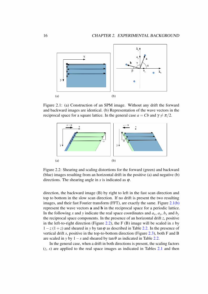
Figure 2.1: (a) Construction of an SPM image. Without any drift the forwardand backward images are identical. (b) Representation of the wave vectors in thereciprocal space for a square lattice. In the general case a =Cb and γ 6= π/2.
(a) (b)
Figure 2.2: Shearing and scaling distortions for the forward (green) and backward(blue) images resulting from an horizontal drift in the positive (a) and negative (b)directions. The shearing angle in x is indicated as ϕ .
direction, the backward image (B) by right to left in the fast scan direction andtop to bottom in the slow scan direction. If no drift is present the two resultingimages, and their fast Fourier transform (FFT), are exactly the same. Figure 2.1(b)represent the wave vectors a and b in the reciprocal space for a periodic lattice.In the following x and y indicate the real space coordinates and ax, ay, bx and by
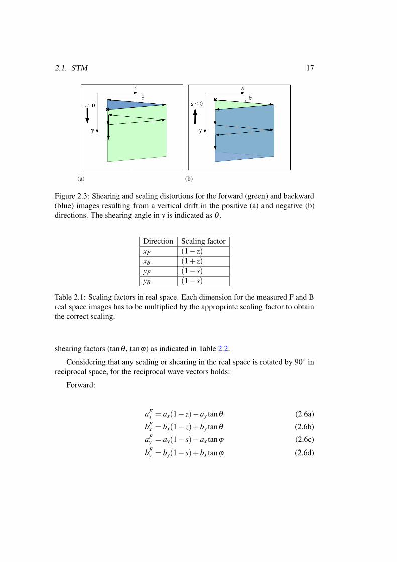
the reciprocal space components. In the presence of an horizontal drift z, positivein the left-to-right direction (Figure 2.2), the F (B) image will be scaled in x by1− z (1+ z) and sheared in y by tanϕ as described in Table 2.2. In the presence ofvertical drift s, positive in the top-to-bottom direction (Figure 2.3), both F and Bare scaled in y by 1− s and sheared by tanθ as indicated in Table 2.2.
In the general case, when a drift in both directions is present, the scaling factors(z, s) are applied to the real space images as indicated in Tables 2.1 and then
2.1. STM 17
(a) (b)
Figure 2.3: Shearing and scaling distortions for the forward (green) and backward(blue) images resulting from a vertical drift in the positive (a) and negative (b)directions. The shearing angle in y is indicated as θ .
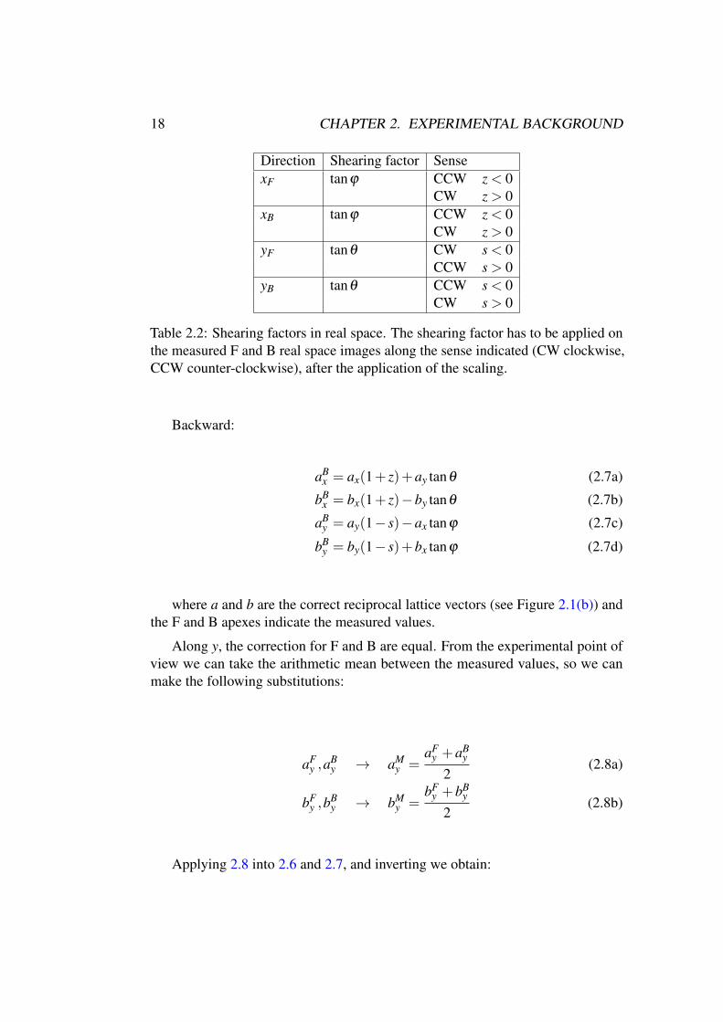
Direction Scaling factorxF (1− z)xB (1+ z)yF (1− s)yB (1− s)
Table 2.1: Scaling factors in real space. Each dimension for the measured F and Breal space images has to be multiplied by the appropriate scaling factor to obtainthe correct scaling.
shearing factors (tanθ , tanϕ) as indicated in Table 2.2.
Considering that any scaling or shearing in the real space is rotated by 90◦ inreciprocal space, for the reciprocal wave vectors holds:
Forward:
aFx = ax(1− z)−ay tanθ (2.6a)
bFx = bx(1− z)+by tanθ (2.6b)
aFy = ay(1− s)−ax tanϕ (2.6c)
bFy = by(1− s)+bx tanϕ (2.6d)
18 CHAPTER 2. EXPERIMENTAL BACKGROUND
Direction Shearing factor SensexF tanϕ CCW z < 0
CW z > 0xB tanϕ CCW z < 0
CW z > 0yF tanθ CW s < 0
CCW s > 0yB tanθ CCW s < 0
CW s > 0
Table 2.2: Shearing factors in real space. The shearing factor has to be applied onthe measured F and B real space images along the sense indicated (CW clockwise,CCW counter-clockwise), after the application of the scaling.
Backward:
aBx = ax(1+ z)+ay tanθ (2.7a)
bBx = bx(1+ z)−by tanθ (2.7b)
aBy = ay(1− s)−ax tanϕ (2.7c)
bBy = by(1− s)+bx tanϕ (2.7d)
where a and b are the correct reciprocal lattice vectors (see Figure 2.1(b)) andthe F and B apexes indicate the measured values.
Along y, the correction for F and B are equal. From the experimental point ofview we can take the arithmetic mean between the measured values, so we canmake the following substitutions:
aFy ,a
By → aM
y =aF
y +aBy
2(2.8a)
bFy ,b
By → bM
y =bF
y +bBy
2(2.8b)
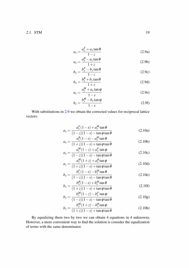
Applying 2.8 into 2.6 and 2.7, and inverting we obtain:
2.1. STM 19
ax =aF
x +ay tanθ
1− z(2.9a)
ax =aB
x −ay tanθ
1+ z(2.9b)
bx =bF
x −by tanθ
1− z(2.9c)
bx =bB
x +by tanθ
1+ z(2.9d)
ay =aM
y +ax tanϕ
1− s(2.9e)
by =bM
y −bx tanϕ
1− s(2.9f)
With substitutions in 2.9 we obtain the corrected values for reciprocal latticevectors:
ax =aF
x (1− s)+aMy tanθ
(1− z)(1− s)− tanϕ tanθ(2.10a)
ax =aB
x (1− s)−aMy tanθ
(1+ z)(1− s)+ tanϕ tanθ(2.10b)
ay =aM
y (1− z)+aFx tanϕ
(1− z)(1− s)− tanϕ tanθ(2.10c)
ay =aM
y (1+ z)+aBx tanϕ
(1+ z)(1− s)+ tanϕ tanθ(2.10d)
bx =bF
x (1− s)−bMy tanθ
(1− z)(1− s)− tanϕ tanθ(2.10e)
bx =bB
x (1− s)+bMy tanθ
(1+ z)(1− s)+ tanϕ tanθ(2.10f)
by =bM
y (1− z)−bFx tanϕ
(1− z)(1− s)− tanϕ tanθ(2.10g)
by =bM
y (1+ z)−bBx tanϕ
(1+ z)(1− s)+ tanϕ tanθ(2.10h)
By equalizing them two by two we can obtain 4 equations in 4 unknowns.However, a more convenient way to find the solution is consider the equalizationof terms with the same denominator.
20 CHAPTER 2. EXPERIMENTAL BACKGROUND
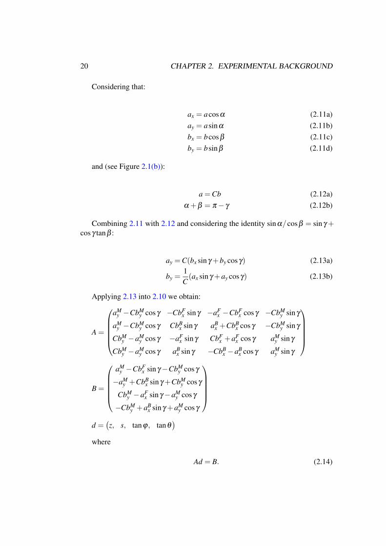
Considering that:
ax = acosα (2.11a)
ay = asinα (2.11b)
bx = bcosβ (2.11c)
by = bsinβ (2.11d)
and (see Figure 2.1(b)):
a =Cb (2.12a)
α +β = π − γ (2.12b)
Combining 2.11 with 2.12 and considering the identity sinα/cosβ = sinγ +cosγ tanβ :
ay =C(bx sinγ +by cosγ) (2.13a)
by =1C(ax sinγ +ay cosγ) (2.13b)
Applying 2.13 into 2.10 we obtain:
A =
aMy −CbM
y cosγ −CbFx sinγ −aF
x −CbFx cosγ −CbM
y sinγ
aMy −CbM
y cosγ CbBx sinγ aB
x +CbBx cosγ −CbM
y sinγ
CbMy −aM
y cosγ −aFx sinγ CbF
x +aFx cosγ aM
y sinγ
CbMy −aM
y cosγ aBx sinγ −CbB
x −aBx cosγ aM
y sinγ
B =
aMy −CbF
x sinγ −CbMy cosγ
−aMy +CbB
x sinγ +CbMy cosγ
CbMy −aF
x sinγ −aMy cosγ
−CbMy +aB
x sinγ +aMy cosγ
d =(
z, s, tanϕ, tanθ)
where
Ad = B. (2.14)
2.2. XPS 21
The solution of 2.14 gives the scaling and shearing factors that can be used asindicated in Tables 2.1 and 2.2.
For a square lattice (a = b and γ = π), 2.13 simplifies to:
ax = by (2.15a)
ay = bx (2.15b)
And:
A =
aMy −bF
x −aFx −bM
y
aMy bB
x aBx −bM
y
bMy −aF
x bFx aM
y
bMy aB
x −bBx aM
y
B =(
aMy −bF
x , bBx −aM
y , bMy −aF
x , aBx −bM
y
)
In Chapter 3 some applications of this approach are reported.
2.1.3 Experimental setup
The STM images reported in this thesis were measured by using an Omicron UHVroom temperature (RT) AFM–STM machine with Ape Research electronics. Thebias voltage refers to the sample and the images were recorded in constant currentmode. A chemically etched W tip was used as the STM probe. The tip was annealedat 700 K by electron bombardment in UHV to remove the native oxide. The STMdata were processed with the software Gwyddion (http://gwyddion.net).
2.2 XPS
2.2.1 Technique
Photoelectron spectroscopy (PES), and its X-ray variant X-ray photoelectron spec-troscopy (XPS), are experimental techniques consisting in the detection of anelectron emitted from a solid by photoelectric effect, in order to determine itsbinding energy and wave vector. Photoelectron effect was discovered by Hertz in1887 and later explained by Einstein in 1905, considering the particle nature oflight. Within this model a photon of energy hω impinging on a sample is absorbed
22 CHAPTER 2. EXPERIMENTAL BACKGROUND
and its energy transferred to an electron which is in turn emitted with a kineticenergy given by
EK = hω −φ −EB (2.16)
where φ is the work function of the solid and EB the electron binding energy.From the energy distribution and the wave vector of the electron, through someassumptions, it is possible to obtain the electronic dispersion curve in the solid.
Equation 2.16 can be derived within the so-called three steps model [16], inwhich the photoemission process is divided into three parts: excitation of theelectron in the solid, electron transfer toward the surface and propagation of theelectron from the surface to the vacuum. A fundamental assumption in the PEStheory is the sudden approximation in which the response of the system upon thecreation of the hole is instantaneous and there are no interactions between theemitted electron and the system itself [17].
The binding energy EB of an electron in an atom is characteristic of eachelement. This chemical sensitivity is one of the most striking features of XPSwhich can be used for the analysis of the elements in a sample. In particular,different types of bond determine the so-called chemical shift [18], that is, thedeviation of the binding energy from the free atom value. In this way is possibleto distinguish atoms depending on their chemical environment, for instance, non-equivalent atoms within a molecule or elements bonded to different atomic species.
Another important property of photoelectron spectroscopy is related to theelectron inelastic mean free path in solids. In the energy range of practical interestfor a typical XPS experiment, between 10 and 500 eV, it is less than 10 Å, meaningthat only photoelectrons excited within a depth of 10 Å from the surface can leavethe sample and be detected, making this technique extremely surface sensitive.
2.2.2 Experimental setup: the SuperESCA beamline
Part of the XPS spectra reported in this thesis have been measured at the Su-perESCA beamline at Elettra synchrotron. This beamline (the first one operatingat Elettra since 1993) combines high energy resolution with high flux of linearlypolarized photons in the 900 eV to 1500 eV energy range, thanks to a prefocusing-monochromator-refocusing scheme. The photoelectrons emitted from the sampleare collected with a system of 9 electrostatic lenses and brought to a PHOIBOShemispherical analyser from SPECS GmbH (150 mm mean radius), which isequipped with a delay line home-made detector [19], specifically designed to allowfast XPS acquisition.
2.3. NEXAFS 23
2.3 NEXAFS
2.3.1 Technique
The comprehension of the interaction of molecules with surfaces has been, andcontinue to be, of fundamental importance in a variety of fields and applications;it is just sufficient to think about catalysis of gasoline and ammonia or the tech-nological advancements in semiconductor materials. Near-edge X-ray absorptionfine structure (NEXAFS) is an experimental technique developed during 1980’sto specifically study low-Z molecules (mainly constituted by H, C, N, O and F)adsorbed on surfaces. The main capabilities of this technique are the detectionof specific bonds in molecules (e.g. C-H, C-C, C=C) and the determination ofthe bond lengths, but also the orientation of the molecule or functional groups onthe surface as well as the orbitals involved in chemisorption processes. Low-Zmolecules are particularly suitable to be studied with this technique due to theirstrong bond directionality which, combined with light polarization of NEXAFS,result in a strong dichroism for angle dependent measurements. These kind ofmolecules have also a strong dependence of the bond length on its hybridization;this, combined with a large back-scattering cross section of the low-Z atoms, pro-vides clear structure-sensitive resonances in the first 30 eV after the threshold [20].
Experimentally, the X-ray energy is scanned through a specific atomic speciesthreshold and the absorbed X-ray intensity is measured. In particular, as thephoton energy is increased from below the absorption threshold of the probed atom,photoelectrons are excited from core levels by the absorbed X-rays. The createdholes are then filled by Auger decay (which is dominant in the soft X-ray regionover fluorescence). As the photon energy is increased more, a photoemission peakis then observed. The intensity of the emitted primary Auger electrons, however,will follow the absorption cross-section of the atomic shell. In the so-called Augerelectron yield (AEY) modality, the recorded intensity of the Auger peak is a directmeasure of the X-ray absorption and, due to the small electron escape depth, is anhighly surface sensitive technique (∼1 nm).
As they leave the sample, primary Auger electrons create many scatteredsecondary electrons which dominate the total electron yield (TEY) intensity. Thisis the most simple detection modality and consists of collecting electrons of allenergies from the sample. The TEY cascade involves several scattering events andoriginates from an average depth, the electron sampling depth, of few nanometres.Electrons created deeper loose too much energy to overcome the work-functionbarrier of the sample, thus not contributing to the TEY intensity.
However, this detection technique suffers from the interference of photoemis-sion peaks entering in the measured intensity. In the partial electron yield (PEY)variant, only electrons with a kinetic energy larger than a set threshold value
24 CHAPTER 2. EXPERIMENTAL BACKGROUND
are detected; by suitably choosing this threshold, the presence of photoemissionpeaks in the detected kinetic energy window over the spanned energy range can beavoided [20].
The fine structure of NEXAFS arises when the photoelectrons coming fromthe probed element are excited into unoccupied orbitals, so when the X-ray energyis tuned close to the ionization potential and electrons are excited just few elec-tronvolts above the Fermi level. NEXAFS is element specific because the X-rayabsorption edges of different elements have different energies and is sensitive tothe bonding environment of the absorbing atom, which results in a chemical shiftsimilar to the case of XPS, but also with a considerably different fine-structureline-shape, thus allowing to achieve an higher sensitivity. Moreover, the major assetof NEXAFS is its polarization dependence. When the electric field of a linearlypolarized photon is aligned with the unoccupied orbitals in which electrons areexcited, the intensity increases, while when they are orthogonal it is quenched. Thisallows to determine the orientation of these empty orbitals and thus the orientationof functional groups and simple molecules on the surface. In the simple pictureof a diatomic molecule, the empty final states are constituted by Rydberg statesbelow the vacuum energy and a continuum above, as in the case of an isolated atom.In addition, there are unfilled molecular orbitals which, for neutral molecules,typically lies above the vacuum level. However, due to Coulomb electron-holeinteractions, the empty orbitals at lower energies are usually pulled down belowthe vacuum level, between the highest occupied molecular orbital (HOMO) andthe vacuum level (typically π∗ orbitals) while the others remains above the vacuumlevel (typically σ∗ orbitals) [20].
2.3.2 Experimental setup: the ALOISA beamline
NEXAFS spectra reported in this thesis were taken at the ALOISA beamline atElettra synchrotron. The spectra were measured in partial electron yield mode bymeans of a channeltron facing the sample, with an energy resolution of 80 meV [21].The low-energy secondary electrons have been filtered out by means of a negativelypolarized grid placed in front of the sample (230 V for the C edge and 370 V for theN edge). The orientation of the surface with respect to the linear polarization of thesynchrotron beam was changed by rotating the sample around the beam axis whilekeeping a constant grazing angle of 6◦. This scattering geometry allows to changethe linear polarization of the light from s to p without varying the illuminated areaon the sample [22]. The raw data were normalized to the total photon flux and,in the case of spectra taken on the molecular layer, divided by the clean substratesignal.
2.4. XMCD 25
2.4 XMCD
2.4.1 Technique
X-ray magnetic circular dichroism (XMCD) is a polarization effect in X-ray absorp-tion spectroscopy (XAS), initially proposed by Schütz and co-workers in 1987 [23],which allows to determine spin and angular momentum order in a sample. Theremarkable feature of XAS is the direct coupling of the X-ray electric field to thecharge and the X-ray angular momentum to the electron angular momentum andspin. The absorption processes are governed by electric dipole transitions that obeystrict selection rules on angular momentum conservation and couple different coreshells to specific valence shells. Spin orbit coupling in these final states allows tobe sensitive to the electronic spin in the absorption process.
Experimentally, the XMCD measurement technique is equivalent to NEXAFS,but, instead of linearly polarized photons, in order to be sensitive to the angularmomentum, circularly polarized light is sent to the sample. Left and right circularlypolarized photons possess opposite angular momenta which are transferred to thephotoelectron excited from the core shell. The photoelectron then possesses a welldefined angular momentum and, in a one-electron picture, one may view the emptyvalence shell as a detector of this momentum [24, 25].
The X-ray magnetic circular dichroism (XMCD) absorption intensity is definedas the intensity difference measured with left and right circularly polarized light.Specific sum rules links this intensity to the size of the orbital and spin momentaof the empty valence states [26–28]. Similarly to the case of NEXAFS, angledependent measurements in magnetic fields can determine the anisotropies of theorbital moment and of the spin density.
In the case of transition metals such as Fe, Co, and Ni, the absorption spectrafor XMCD are usually measured at the L-edge, in which the incoming photonexcite a 2p electron to a 3d state [29, 30].
2.4.2 Experimental setup: the BACH beamline
XMCD measurements reported in this thesis have been performed at the BACHbeamline at Elettra. The beamline works in the UV-soft x-ray photon energy range(from 35 eV to 1600 eV) with selectable light polarization (linear, circular andelliptical) [31]. The end-station is provided with a hemispherical electron energyanalyser Scienta 3000 at 60◦ from the incident photon beam in the horizontal plane,operating in the the partial electron yield mode.
26 REFERENCES
References
[1] Joseph A Stroscio and William J Kaiser. Scanning Tunneling Microscopy,volume 27. Academic Press, Inc., 1993.
[2] J. Bardeen. Tunnelling from a many-particle point of view. Phys. Rev. Lett.,6:57–59, Jan 1961. doi: 10.1103/PhysRevLett.6.57.
[3] J Tersoff and D R Hamann. Theory of the scanning tunneling microscope.Physical Review B, 31(2):805, 1985.
[4] J Tersoff and D R Hamann. Theory and Application for the Scanning Tunnel-ing Microscope. Physical Review Letters, 50(25):1998–2001, 1983.
[5] J. Wintterlin, J. Trost, S. Renisch, R. Schuster, T. Zambelli, and G. Ertl. Real-time {STM} observations of atomic equilibrium fluctuations in an adsorbatesystem: O/ru(0001). Surface Science, 394(1–3):159 – 169, 1997. ISSN0039-6028. doi: http://dx.doi.org/10.1016/S0039-6028(97)00604-3.
[6] O. M. Magnussen, W. Polewska, L. Zitzler, and R. J. Behm. In situ video-stmstudies of dynamic processes at electrochemical interfaces. Faraday Discuss.,121:43–52, 2002. doi: 10.1039/B200016B. URL http://dx.doi.org/10.
1039/B200016B.
[7] M. J. Rost, L. Crama, P. Schakel, E. van Tol, G. B. E. M. van Velzen-Williams, C. F. Overgauw, H. ter Horst, H. Dekker, B. Okhuijsen, M. Seynen,A. Vijftigschild, P. Han, A. J. Katan, K. Schoots, R. Schumm, W. van Loo,T. H. Oosterkamp, and J. W. M. Frenken. Scanning probe microscopes govideo rate and beyond. Review of Scientific Instruments, 76(5):053710, 2005.doi: http://dx.doi.org/10.1063/1.1915288.
[8] Carlo Dri, Friedrich Esch, Cristina Africh, and Giovanni Comelli. How toselect fast scanning frequencies for high-resolution fast stm measurementswith a conventional microscope. Measurement Science and Technology, 23(5):055402, 2012.
[9] V. Yu. Yurov and A. N. Klimov. Scanning tunneling microscope calibrationand reconstruction of real image: Drift and slope elimination. Review of
Scientific Instruments, 65(5), 1994.
[10] Rostislav V Lapshin. Automatic drift elimination in probe microscope imagesbased on techniques of counter-scanning and topography feature recognition.Measurement Science and Technology, 18(3):907, 2007.

REFERENCES 27
[11] Jens E.T. Andersen and P. Møller. Analysis and calibration of in situ scanningtunnelling microscopy images with atomic resolution influenced by surfacedrift phenomena. Surface and Coatings Technology, 67(3):213 – 220, 1994.ISSN 0257-8972. doi: http://dx.doi.org/10.1016/0257-8972(94)90121-X.
[12] John T. Woodward and Daniel K. Schwartz. Removing drift from scanningprobe microscope images of periodic samples. Journal of Vacuum Science
and Technology B, 16(1), 1998.
[13] J. F. Jo/rgensen, L. L. Madsen, J. Garnaes, K. Carneiro, and K. Schaumburg.Calibration, drift elimination, and molecular structure analysis. Journal of
Vacuum Science and Technology B, 12(3), 1994.
[14] K Henriksen and S L S Stipp. Image distortion in scanningprobe microscopy. American Mineralogist,, 87:5–16, 2002. doi:http://dx.doi.org/10.1116/1.587266.
[15] R. Staub, D. Alliata, and C. Nicolini. Drift elimination in the calibration ofscanning probe microscopes. Review of Scientific Instruments, 66(3), 1995.
[16] D P Woodruff and T A Delchar. Cambridge University Press, 1994.
[17] Stefan Hüfner. Photoelectron spectroscopy. Springer, 2003.
[18] C S Fadley, S B M Hagstrom, M P Klein, and D A Shirley. Chemical effectson core-electron binding energies in iodine and europium. The Journal of
Chemical Physics, 48:3779, 1968.
[19] G. Cautero, R. Sergo, L. Stebel, P. Lacovig, P. Pittana, M. Predon-zani, and S. Carrato. A two-dimensional detector for pump-and-probeand time resolved experiments. Nuclear Instruments and Methods in
Physics Research Section A: Accelerators, Spectrometers, Detectors and
Associated Equipment, 595(2):447 – 459, 2008. ISSN 0168-9002. doi:http://dx.doi.org/10.1016/j.nima.2008.06.046.
[20] Joachim Stohr. NEXAFS Spectroscopy. Springer, 1996.
[21] L. Floreano, G. Naletto, D. Cvetko, R. Gotter, M. Malvezzi, L. Marassi,A. Morgante, A. Santaniello, A. Verdini, F. Tommasini, and G. Tondello.Performance of the grating-crystal monochromator of the ALOISA beamlineat the elettra synchrotron. Review of Scientific Instruments, 70(10):3855,1999. ISSN 00346748. doi: 10.1063/1.1150001.
28 REFERENCES
[22] Luca Floreano, Albano Cossaro, Roberto Gotter, Alberto Verdini, GregorBavdek, Fabrizio Evangelista, Alessandro Ruocco, Alberto Morgante, andDean Cvetko. Periodic arrays of cu-phthalocyanine chains on au ( 110 ).Journal of Physical Chemistry C, (112):10794–10802, 2008.
[23] G Schütz, W Wagner, W Wilhelm, P Kienle, R Zeller, R Frahm, and G Mater-lik. Physical Review Letters, 58:737, 1987.
[24] Y Wu, J Stöhr, B D Hermsmeier, M G Samant, and D Weller. Physical
Review Letters, 69:2307, 1992.
[25] M Tischer, O Hjortstam, D Arvanitis, J H Dunn, F May, K Baberschke,J Trygg, J M Wills, B Johansson, and O Eriksson. Physical Review Letters,75:1602, 1995.
[26] B T Thole, P Carra, F Sette, and G van der Laan. X-ray circular dichroism asa probe of orbital magnetization. Physical Review Letters, 68:1943, 1992.
[27] P Carra, B T Thole, M Altarelli, and X Wang. X-ray circular dichroism andlocal magnetic fields. Physical Review Letters, 70:694, 1993.
[28] W L O’Brien and B P Tonner. Orbital and spin sum rules in x-ray magneticcircular dichroism. Physical Review B, 50:12673, 1994.
[29] C T Chen, Y U Idzerda, H J Lin, N V Smith, G Meigs, E Chaban, G H Ho,E Pellegrin, and F Sette. Experimental confirmation of the x-ray magneticcircular dichroism sum rules for iron and cobalt. Physical Review Letters, 75:152, 1995.
[30] J. P. Crocombette, B. T. Thole, and F. Jollet. The importance of the magneticdipole term in magneto-circular x-ray absorption dichroism for 3d transitionmetal compounds. Journal of Physics: Condensed Matter, 8(22):4095, 1996.
[31] M. Zangrando, M. Zacchigna, M. Finazzi, D. Cocco, R. Rochow, and F. Parmi-giani. Polarized high-brilliance and high-resolution soft x-ray source at elettra:The performance of beamline bach. Review of Scientific Instruments, 75(1),2004.
Chapter 3
Adsorption of porphyrins on crystal
surfaces
3.1 Introduction
The self-assembly of organic monolayers on metal surfaces is a pathway to producenano-sized metal-organic architectures and patterned surfaces not achievable byconventional methods [1–4].
In porphyrins, the presence of the unsaturated macrocycle brings about greatversatility for these chemical systems because of the presence of different positionsin which functional groups can be substituted to introduce new properties to themolecule [5, 6].
Moreover, the macrocycle can coordinate with a wide range of metal ions,forming metallo-porphyrins [7]. In this case, the interaction of the metal hostat the center of the molecule with the substrate plays a fundamental role in theadsorption behaviour, affecting chemical, electronic and magnetic properties of thesystem [8–11].
Taking advantage of the flexibility in choosing the substituents, in combinationwith the presence of active sites of interactions with metals, this kind of moleculescan be used to fabricate two-dimensional arrays of metallic centres with nano-metric spacing, rendering them suitable candidates as building blocks for novelmetal–organic architectures in solid-state chemistry and in molecular engineer-ing [5, 7–10, 12–14] as well as to form patterned surfaces for catalytic, magnetic,optoelectronic, and sensing materials [15–22].
Understanding and controlling the properties of such organic-inorganic inter-faces, constitutes a basic step toward the exploitation of new molecular-baseddevices with controllable and tunable properties [23].
29
30 CHAPTER 3. ADSORPTION
3.2 Self-assembly on metal surfaces: STM of TPP
monolayers on Au(111), Ag(111) and Cu(100)
Porphyrin molecules have the ability to self-assembly on crystal surfaces, consti-tuting long-range ordered structures. The periodic arrangement of the moleculesand the cell parameters of the formed super-lattice are of great importance in deter-mining the electronic properties of the resulting layer. In fact, the relative positionof the molecule with respect to the surface, as well as the relative distance betweenthe molecules themselves, besides being determinant for the molecule-substrateinteraction, is strictly related to process such as the metalation and the magneticexchange coupling.
STM can give precise information on the topology and geometry of the ad-sorbed porphyrin layers. In the following, STM measurements for a TPP monolayeradsorbed on Au(111), Ag(111) and Cu(100) are reported. All the measurementswere performed at RT. In this regime, thermal drift is usually not negligible and itscorrection is fundamental in order to perform a quantitative measurement. For thisreason, the micrograph images were processed by the drift correction algorithmdescribed in Section 2.1.2.
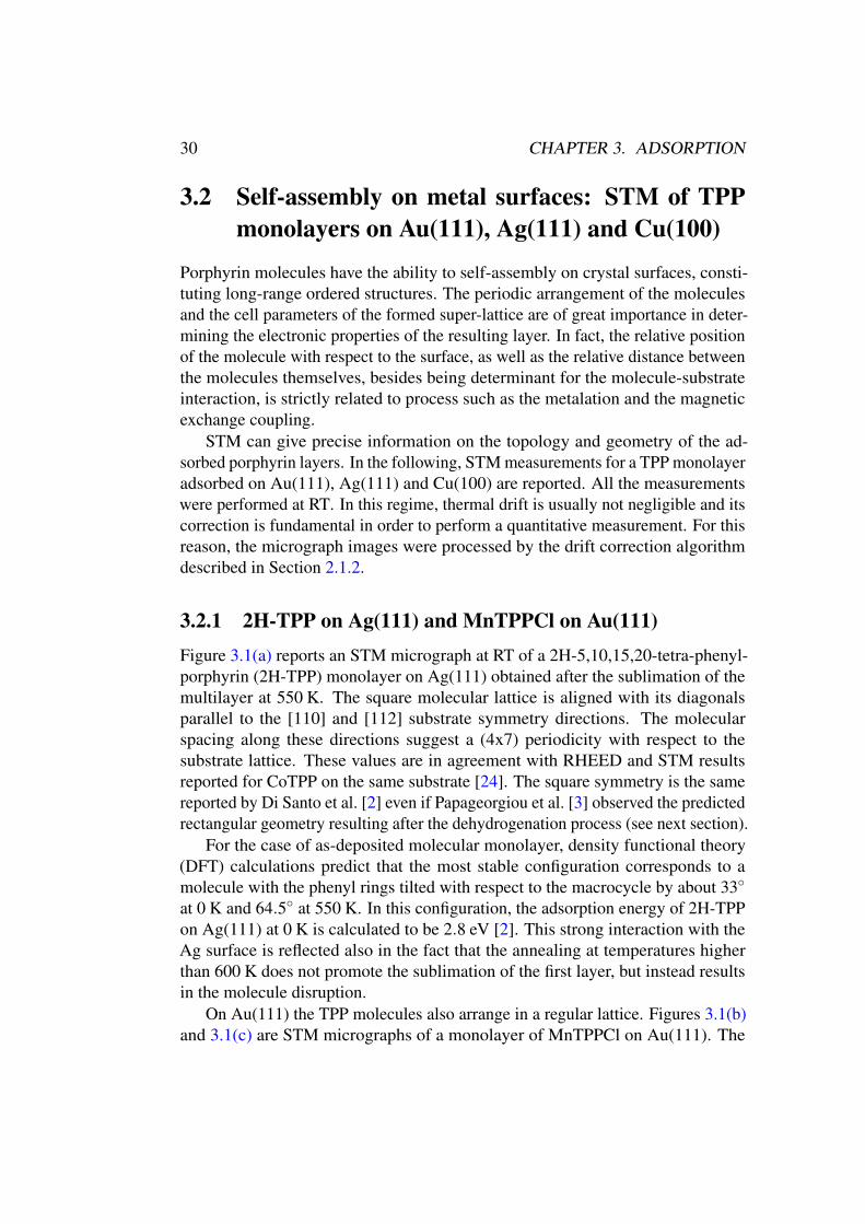
3.2.1 2H-TPP on Ag(111) and MnTPPCl on Au(111)
Figure 3.1(a) reports an STM micrograph at RT of a 2H-5,10,15,20-tetra-phenyl-porphyrin (2H-TPP) monolayer on Ag(111) obtained after the sublimation of themultilayer at 550 K. The square molecular lattice is aligned with its diagonalsparallel to the [110] and [112] substrate symmetry directions. The molecularspacing along these directions suggest a (4x7) periodicity with respect to thesubstrate lattice. These values are in agreement with RHEED and STM resultsreported for CoTPP on the same substrate [24]. The square symmetry is the samereported by Di Santo et al. [2] even if Papageorgiou et al. [3] observed the predictedrectangular geometry resulting after the dehydrogenation process (see next section).
For the case of as-deposited molecular monolayer, density functional theory(DFT) calculations predict that the most stable configuration corresponds to amolecule with the phenyl rings tilted with respect to the macrocycle by about 33◦
at 0 K and 64.5◦ at 550 K. In this configuration, the adsorption energy of 2H-TPPon Ag(111) at 0 K is calculated to be 2.8 eV [2]. This strong interaction with theAg surface is reflected also in the fact that the annealing at temperatures higherthan 600 K does not promote the sublimation of the first layer, but instead resultsin the molecule disruption.
On Au(111) the TPP molecules also arrange in a regular lattice. Figures 3.1(b)and 3.1(c) are STM micrographs of a monolayer of MnTPPCl on Au(111). The
3.2. SELF-ASSEMBLY 31
(a) (b)
(c)
Figure 3.1: (a) STM micrograph of 2H-TPP monolayer on Ag(111) at RT. Insetscale bar 4 nm (1 V, 0.03 nA). (a, b) STM micrographs of MnTPPCl monolayer onAu(111) at RT. (b) Inset scale bar 20 nm (0.8 V, 0.04 nA). (c) Inset scale bar 5 nm(1.1 V, 0.04 nA).
substrate reconstruction is still visible even after the molecular layer adsorption.The herringbone reconstruction is not modified by the presence of the molecules,as it happens instead, for example, in the case of the missing row reconstruction ofAu(110) which is modified by the adsorption of phthalocyanines [25]. Differentlyto the case of Ag, this indicates that on Au(111) the molecule-substrate interactionis weaker. This point will be further discussed in the next section, concerning theconformational adaptation of 2H-TPP.
3.2.2 NiTPP on Cu(100)
On surfaces with square unit cell, due to their D4h symmetry, TPP moleculesarrange in a square lattice. On these surfaces, in the case the molecule super-latticevectors are not parallel to those of the substrate lattice, there are two equivalentsymmetry directions resulting in the formation of two adsorption domains.
Cu has simple cubic crystal structure with a unit lattice of 361.49 pm and thus
32 CHAPTER 3. ADSORPTION
(a) (b)
(c)
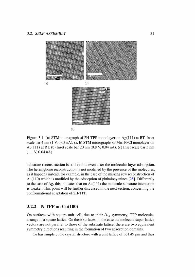
Figure 3.2: (a) STM micrograph of NiTPP monolayer on Cu(100). Inset 5 nm (0.2V, 0.035 nA). (b) 2D FFT of the STM image. (c) IFT after applying a pass filter.The two domains are clearly visible.
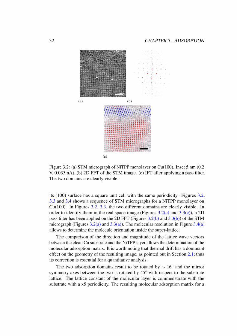
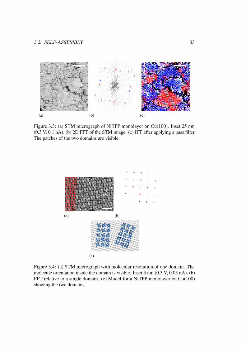
its (100) surface has a square unit cell with the same periodicity. Figures 3.2,3.3 and 3.4 shows a sequence of STM micrographs for a NiTPP monolayer onCu(100). In Figures 3.2, 3.3, the two different domains are clearly visible. Inorder to identify them in the real space image (Figures 3.2(c) and 3.3(c)), a 2Dpass filter has been applied on the 2D FFT (Figures 3.2(b) and 3.3(b)) of the STMmicrograph (Figures 3.2(a) and 3.3(a)). The molecular resolution in Figure 3.4(a)allows to determine the molecule orientation inside the super-lattice.
The comparison of the direction and magnitude of the lattice wave vectorsbetween the clean Cu substrate and the NiTPP layer allows the determination of themolecular adsorption matrix. It is worth noting that thermal drift has a dominanteffect on the geometry of the resulting image, as pointed out in Section 2.1; thusits correction is essential for a quantitative analysis.
The two adsorption domains result to be rotated by ∼ 16◦ and the mirrorsymmetry axes between the two is rotated by 45◦ with respect to the substratelattice. The lattice constant of the molecular layer is commensurate with thesubstrate with a x5 periodicity. The resulting molecular adsorption matrix for a
3.2. SELF-ASSEMBLY 33
(a) (b) (c)
Figure 3.3: (a) STM micrograph of NiTPP monolayer on Cu(100). Inset 25 nm(0.3 V, 0.1 nA). (b) 2D FFT of the STM image. (c) IFT after applying a pass filter.The patches of the two domains are visible.
(a) (b)
(c)
Figure 3.4: (a) STM micrograph with molecular resolution of one domain. Themolecule orientation inside the domain is visible. Inset 5 nm (0.3 V, 0.05 nA). (b)FFT relative to a single domain. (c) Model for a NiTPP monolayer on Cu(100)showing the two domains.
34 CHAPTER 3. ADSORPTION
NiTPP monolayer on Cu(100) is reported in the model of Figure 3.4(c) [[11]]. Inparticular, the first domain is aligned along the [4,3,0] and [3,-4,0] directions andthe second along the [3,4,0] and [4,-3,0] directions.
3.3 Conformational adaptation of 2HTPP monolayer
on Au(111)
2H-TPP molecules have been deposited on several metallic substrates, but Au [26–28] and Ag [3, 18, 29–33] are those on which it is possible to obtain a single layerwithout any modification of the macrocycle. In both cases, when deposited at roomtemperature (RT) or when the monolayer is obtained after the sublimation of amultilayer in the range 525 ÷ 580 K, the XPS analysis shows the presence of twounaffected N 1s peaks, indicating that, in the monolayer, the molecule macrocycleis still protonated. Actually, AuTPP and AgTPP can be synthesized and exist asstable compounds. The fact that the TPP molecule does not metalate on Au andAg substrates can be explained by the steric hindrance of these atoms, whose ionicradius is larger with respect to atoms such as Fe and Ni, thus not allowing thecoordination with the already stable and formed TPP molecule. Moreover, thethermodynamics related to the extraction of an atom from the surface could beunfavourable with respect to the sublimation or disruption of the molecule on thesurface.
However, a remarkable effect on the phenyls adsorption geometry was observedon the monolayer (or sub-monolayer) of 2H-TPPs, after annealing above 525 K.On the Ag(111) and Au(111) surfaces, the macrocycle of 2H-TPP deposited atRT has been shown to be adsorbed parallel to the substrate surface, adopting asaddle shape conformation [34], whereas its phenyl legs exhibit a tilt angle of about45◦÷55◦ with respect to the surface plane [33, 34]. Differently, after annealingabove 525 K, a temperature-induced conformational adaptation of the porphyrinmolecules on the surface of Ag(111) has been predicted and observed, resulting inthe rotation of the phenyl rings parallel to the substrate plane [3, 33].
In the following is presented a study of the electronic and geometric propertiesof 2H-tetra-phenyl-porphyrins monolayer adsorbed on Au(111) surface. Themolecule-surface interaction is studied for a monolayer as-deposited on the surfaceand after annealing up to 570 K. It will be shown that the annealing process resultsin a conformational adaptation of the molecule, with the rotation of the phenylrings towards the surface, similarly to the case of 2H-TPP on Ag(111) [33], and, inaddition, with a partial distortion of the macrocycle.
3.3. CONFORMATIONAL ADAPTATION ON AU(111) 35
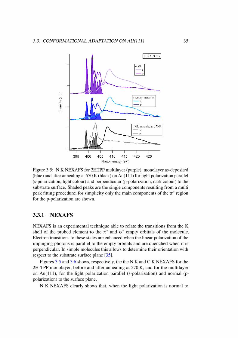
Figure 3.5: N K NEXAFS for 2HTPP multilayer (purple), monolayer as-deposited(blue) and after annealing at 570 K (black) on Au(111) for light polarization parallel(s-polarization, light colour) and perpendicular (p-polarization, dark colour) to thesubstrate surface. Shaded peaks are the single components resulting from a multipeak fitting procedure; for simplicity only the main components of the π∗ regionfor the p-polarization are shown.
3.3.1 NEXAFS
NEXAFS is an experimental technique able to relate the transitions from the Kshell of the probed element to the π∗ and σ∗ empty orbitals of the molecule.Electron transitions to these states are enhanced when the linear polarization of theimpinging photons is parallel to the empty orbitals and are quenched when it isperpendicular. In simple molecules this allows to determine their orientation withrespect to the substrate surface plane [35].
Figures 3.5 and 3.6 shows, respectively, the the N K and C K NEXAFS for the2H-TPP monolayer, before and after annealing at 570 K, and for the multilayeron Au(111), for the light polarization parallel (s-polarization) and normal (p-polarization) to the surface plane.
N K NEXAFS clearly shows that, when the light polarization is normal to
36 CHAPTER 3. ADSORPTION
the surface plane, π∗ features are enhanced, whereas, when it is parallel, they arecompletely suppressed. This indicates that the macrocycle, in which the N atomsare located, is parallel with respect to the substrate surface, both for the case ofthe monolayer and the multilayer. This first result tells us that (i) the moleculemacrocycle preserves its parallel orientation after the annealing process and (ii)the molecules in the multilayer are stacked parallel to each other, at least up to athickness of ∼6 monolayer (ML). The data fitting indicates a shift towards lowerenergies for the main π∗ features from the multilayer to the monolayer, with afurther decrease after the annealing process, as a consequence of the shift of thecore level (see Figure 3.8). In particular the first transition at 399.1 eV is shifted by0.2 eV after the annealing process. Actually, this peak does not completely vanishfor s-polarization, as can be seen by the presence of a bulge in the multilayer andthe monolayer as-deposited, while it disappears after the annealing. This featureis related to the pyrrolic nitrogen and it will be discussed later in the XPS section3.3.2.
The situation is quite different in the case of the C threshold. As reportedin [36], C K NEXAFS spectrum of TPP molecule is almost the superposition ofthe macrocycle and the phenyl (benzene) spectra. The most relevant feature relatedto the phenyl π∗ absorption component is the peak at ∼ 285.1 eV, whereas the firstπ∗ absorption peak at ∼ 284.2 eV is due to the macrocycle.
For both the multilayer and the monolayer, the macrocycle π∗ peaks followthe same behaviour of the N K edge NEXAFS, i.e. the feature is enhanced forout-of-plane polarization and is completely suppressed for in-plane polarization,again confirming that the macrocycle is adsorbed parallel to the substrate surface.
For the multilayer, applying the formula by Stöhr [35], the dichroic signal forthe peak at ∼ 285.1 eV indicates that the phenyl rings are rotated by an averageangle of ∼ 56.3◦ with respect to the macrocycle. This T-shaped geometry isexpected, considering that in gas-phase and in solution the meso-substituent tendsto align to an average angle in the range 50÷80◦, due to the steric interactionbetween the hydrogens atoms [37].
In the case of the monolayer as-deposited there is an increase in the dichroismof the phenyl moiety component with respect to the multilayer, indicating a rotationof the phenyl rings towards the surface, with an average aryl angle of ∼ 34.1◦ ;this effect is related to the interaction between the substrate and the π orbitals ofthe phenyl moieties [38].
After the annealing process this peak is almost completely suppressed for s-polarization, suggesting that in this case the phenyl rings lies flat on the surface.This behaviour is quite exceptional considering that the rotation of the phenyl ringsis inhibited by the steric interaction between the hydrogen atoms in the moleculeand they are expected to be rotated with respect to the macrocycle [16–19, 30, 39].
The reaction can be explained under the assumption that the molecules un-
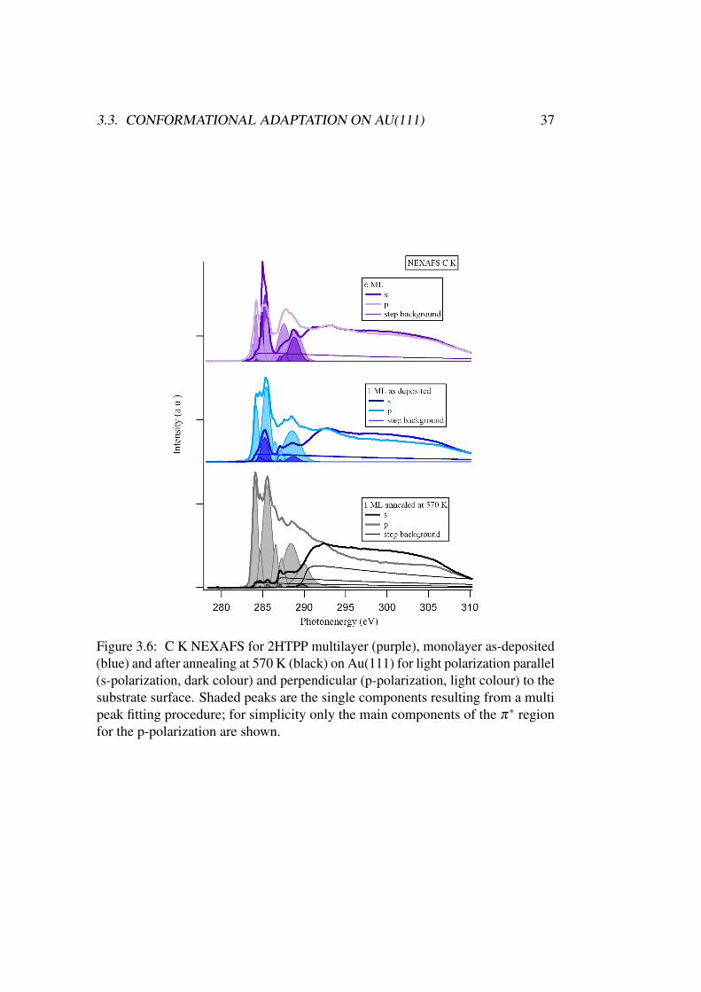
3.3. CONFORMATIONAL ADAPTATION ON AU(111) 37
Figure 3.6: C K NEXAFS for 2HTPP multilayer (purple), monolayer as-deposited(blue) and after annealing at 570 K (black) on Au(111) for light polarization parallel(s-polarization, dark colour) and perpendicular (p-polarization, light colour) to thesubstrate surface. Shaded peaks are the single components resulting from a multipeak fitting procedure; for simplicity only the main components of the π∗ regionfor the p-polarization are shown.
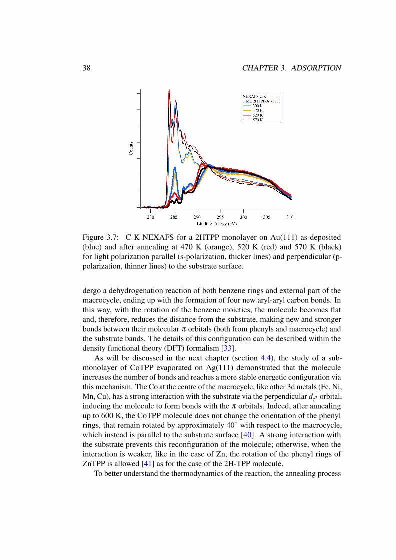
38 CHAPTER 3. ADSORPTION
Figure 3.7: C K NEXAFS for a 2HTPP monolayer on Au(111) as-deposited(blue) and after annealing at 470 K (orange), 520 K (red) and 570 K (black)for light polarization parallel (s-polarization, thicker lines) and perpendicular (p-polarization, thinner lines) to the substrate surface.
dergo a dehydrogenation reaction of both benzene rings and external part of themacrocycle, ending up with the formation of four new aryl-aryl carbon bonds. Inthis way, with the rotation of the benzene moieties, the molecule becomes flatand, therefore, reduces the distance from the substrate, making new and strongerbonds between their molecular π orbitals (both from phenyls and macrocycle) andthe substrate bands. The details of this configuration can be described within thedensity functional theory (DFT) formalism [33].
As will be discussed in the next chapter (section 4.4), the study of a sub-monolayer of CoTPP evaporated on Ag(111) demonstrated that the moleculeincreases the number of bonds and reaches a more stable energetic configuration viathis mechanism. The Co at the centre of the macrocycle, like other 3d metals (Fe, Ni,Mn, Cu), has a strong interaction with the substrate via the perpendicular dz2 orbital,inducing the molecule to form bonds with the π orbitals. Indeed, after annealingup to 600 K, the CoTPP molecule does not change the orientation of the phenylrings, that remain rotated by approximately 40◦ with respect to the macrocycle,which instead is parallel to the substrate surface [40]. A strong interaction withthe substrate prevents this reconfiguration of the molecule; otherwise, when theinteraction is weaker, like in the case of Zn, the rotation of the phenyl rings ofZnTPP is allowed [41] as for the case of the 2H-TPP molecule.
To better understand the thermodynamics of the reaction, the annealing process
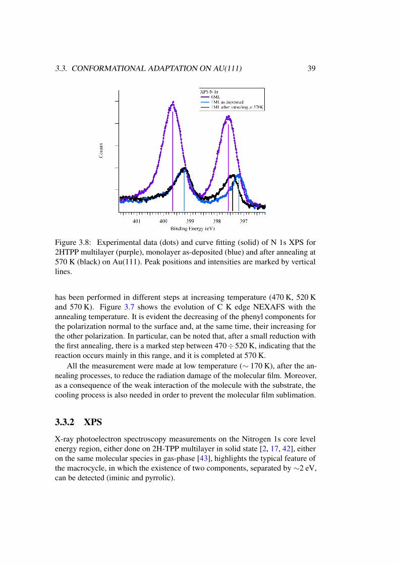
3.3. CONFORMATIONAL ADAPTATION ON AU(111) 39
Figure 3.8: Experimental data (dots) and curve fitting (solid) of N 1s XPS for2HTPP multilayer (purple), monolayer as-deposited (blue) and after annealing at570 K (black) on Au(111). Peak positions and intensities are marked by verticallines.
has been performed in different steps at increasing temperature (470 K, 520 Kand 570 K). Figure 3.7 shows the evolution of C K edge NEXAFS with theannealing temperature. It is evident the decreasing of the phenyl components forthe polarization normal to the surface and, at the same time, their increasing forthe other polarization. In particular, can be noted that, after a small reduction withthe first annealing, there is a marked step between 470÷520 K, indicating that thereaction occurs mainly in this range, and it is completed at 570 K.
All the measurement were made at low temperature (∼ 170 K), after the an-nealing processes, to reduce the radiation damage of the molecular film. Moreover,as a consequence of the weak interaction of the molecule with the substrate, thecooling process is also needed in order to prevent the molecular film sublimation.
3.3.2 XPS
X-ray photoelectron spectroscopy measurements on the Nitrogen 1s core levelenergy region, either done on 2H-TPP multilayer in solid state [2, 17, 42], eitheron the same molecular species in gas-phase [43], highlights the typical feature ofthe macrocycle, in which the existence of two components, separated by ∼2 eV,can be detected (iminic and pyrrolic).
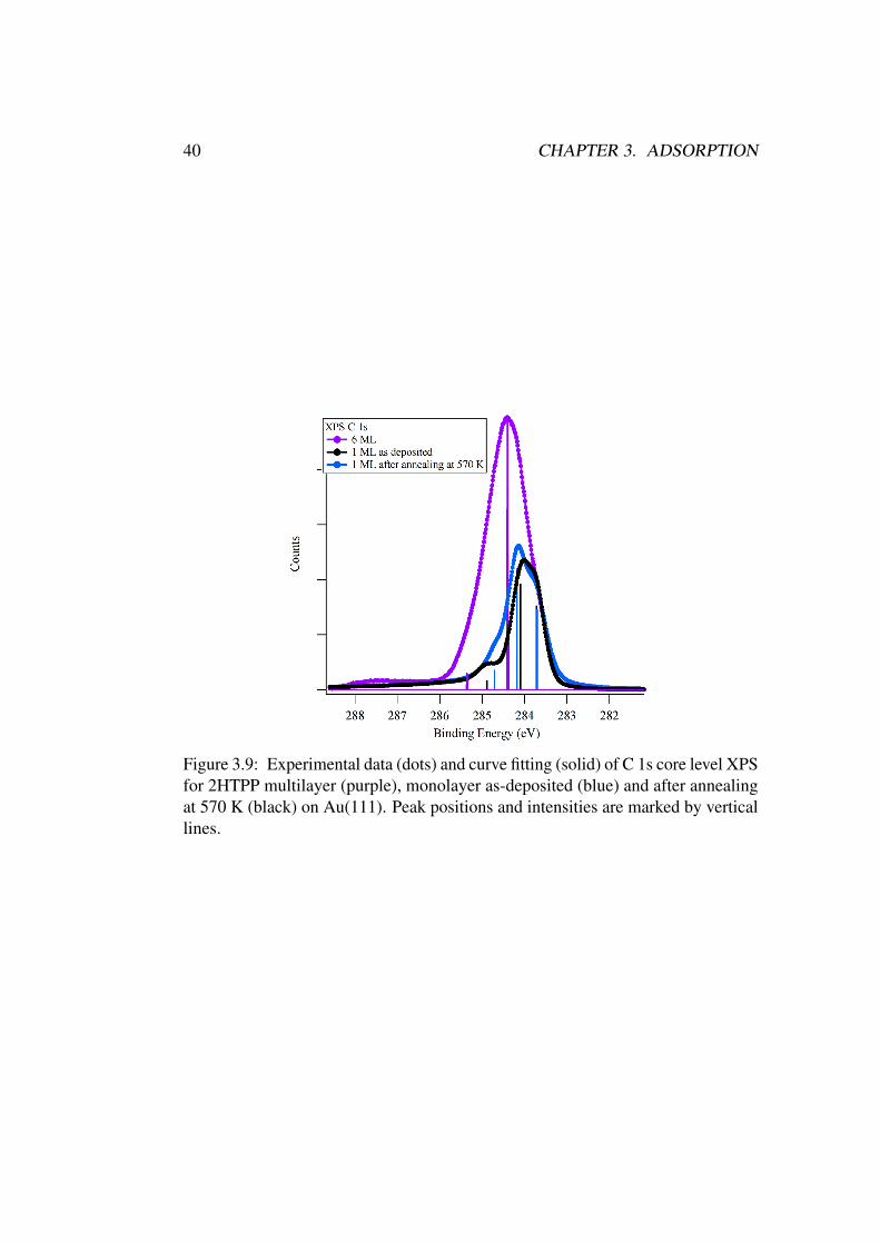
40 CHAPTER 3. ADSORPTION
Figure 3.9: Experimental data (dots) and curve fitting (solid) of C 1s core level XPSfor 2HTPP multilayer (purple), monolayer as-deposited (blue) and after annealingat 570 K (black) on Au(111). Peak positions and intensities are marked by verticallines.
3.3. CONFORMATIONAL ADAPTATION ON AU(111) 41
In Figure 3.8 the XPS spectrum for N 1s core level for 2H-TPP on Au(111) isreported. The component at higher binding energy (BE) corresponds to the pyrrolicN atom (=NH) while the iminic N component is shifted at lower binding energy.In the case of the multilayer they are positioned at 399.69 eV and 397.61 eVrespectively, with a relative distance of 2.08 eV.
In the case of C 1s, different components are expected which account forthe different species inside the molecule [44]. However since the chemical shiftbetween these components is small compared with the energy blurring due to thescreening of the molecular multilayer, the C 1s core level spectrum shows, in thiscase, not more than a single peak located at 284.4 eV (Figure 3.9).
Tuning the evaporation conditions it is possible to evaporate almost exactly1 ML of 2HTPP, and the coverage can be determined by measuring the intensityratio between the C 1s core level and the Au of the substrate.
In the monolayer as-deposited both N peaks undergo a rigid shift of 0.43 eVtowards lower binding energies (BE of 399.26 eV for pyrrolic and 397.18 eV foriminic N). This is attributed to the increased screening effect and to changes inthe surface dipole due to the interaction with the substrate, arising from the closervicinity to the surface. In this case, for the two N components both the shape andintensity remain the same, indicating that the macrocycle is still protonated and themolecule does not metalate, as it happens in the case of 2H-TPP on Ag(111) [33].It can be noted that the intensities of the two peaks are slightly different, with areduction in area of the iminic peak of ∼ 15% with respect to the pyrrolic one: thiscan be ascribed to a photoelectron diffraction effect as reported in [33]. In the C 1sXPS of the monolayer there is an overall shift of the spectra towards lower BE. Inthis case three components can be resolved, even if are not clearly assignable tothe different carbon atoms of the molecule.
After annealing at 570 K, the pyrrolic component does not change its position(399.26 eV), while the iminic N peak shifts at higher binding energy by 0.23 eV(BE=397.41 eV). The opposite shift is visible in the N K NEXAFS for the firstabsorption peak, (Figure 3.5) which corresponds to the transition from the iminicNitrogen 1s core level to the π∗ empty molecular orbitals. In the same figure, itcan be seen that, for the case of the multilayer and the monolayer as-deposited,the first iminic peak is actually still visible in s-polarization, while it is completelysuppressed after the annealing process. Considering that the molecule macrocycleis completely parallel to the surface, this indicates that the macrocycle is adoptinga saddle shape conformation [34], while it becomes more planar after the annealingprocess. At the same time, the main component of the C 1s orbital, shifts towardlower binding energy as a consequence of the rotation of the phenyl rings parallelto the substrate surface, with a change in the peak line-shape that becomes similarto that of a monolayer of Pc on Ag(111) [45].
The hypothesis of 2H-TPP modification is supported by recent analyses [3, 46],
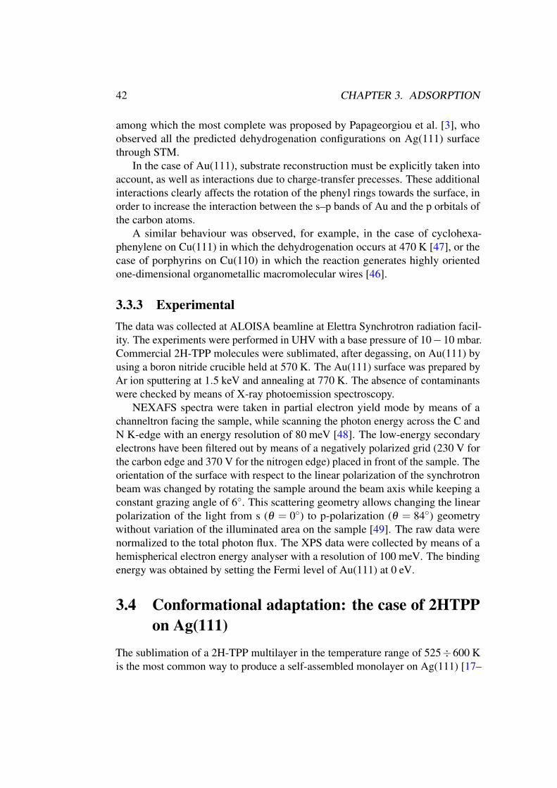
42 CHAPTER 3. ADSORPTION
among which the most complete was proposed by Papageorgiou et al. [3], whoobserved all the predicted dehydrogenation configurations on Ag(111) surfacethrough STM.
In the case of Au(111), substrate reconstruction must be explicitly taken intoaccount, as well as interactions due to charge-transfer precesses. These additionalinteractions clearly affects the rotation of the phenyl rings towards the surface, inorder to increase the interaction between the s–p bands of Au and the p orbitals ofthe carbon atoms.
A similar behaviour was observed, for example, in the case of cyclohexa-phenylene on Cu(111) in which the dehydrogenation occurs at 470 K [47], or thecase of porphyrins on Cu(110) in which the reaction generates highly orientedone-dimensional organometallic macromolecular wires [46].
3.3.3 Experimental
The data was collected at ALOISA beamline at Elettra Synchrotron radiation facil-ity. The experiments were performed in UHV with a base pressure of 10−10 mbar.Commercial 2H-TPP molecules were sublimated, after degassing, on Au(111) byusing a boron nitride crucible held at 570 K. The Au(111) surface was prepared byAr ion sputtering at 1.5 keV and annealing at 770 K. The absence of contaminantswere checked by means of X-ray photoemission spectroscopy.
NEXAFS spectra were taken in partial electron yield mode by means of achanneltron facing the sample, while scanning the photon energy across the C andN K-edge with an energy resolution of 80 meV [48]. The low-energy secondaryelectrons have been filtered out by means of a negatively polarized grid (230 V forthe carbon edge and 370 V for the nitrogen edge) placed in front of the sample. Theorientation of the surface with respect to the linear polarization of the synchrotronbeam was changed by rotating the sample around the beam axis while keeping aconstant grazing angle of 6◦. This scattering geometry allows changing the linearpolarization of the light from s (θ = 0◦) to p-polarization (θ = 84◦) geometrywithout variation of the illuminated area on the sample [49]. The raw data werenormalized to the total photon flux. The XPS data were collected by means of ahemispherical electron energy analyser with a resolution of 100 meV. The bindingenergy was obtained by setting the Fermi level of Au(111) at 0 eV.
3.4 Conformational adaptation: the case of 2HTPP
on Ag(111)
The sublimation of a 2H-TPP multilayer in the temperature range of 525÷600 Kis the most common way to produce a self-assembled monolayer on Ag(111) [17–
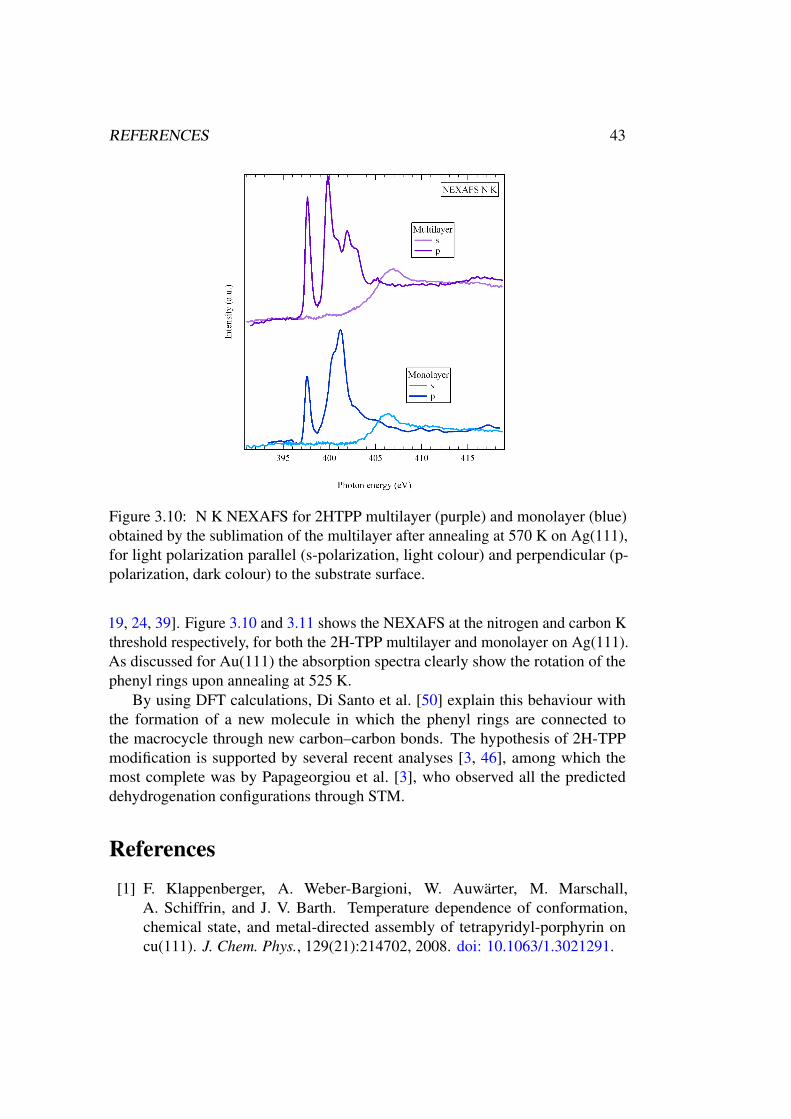
REFERENCES 43
Figure 3.10: N K NEXAFS for 2HTPP multilayer (purple) and monolayer (blue)obtained by the sublimation of the multilayer after annealing at 570 K on Ag(111),for light polarization parallel (s-polarization, light colour) and perpendicular (p-polarization, dark colour) to the substrate surface.
19, 24, 39]. Figure 3.10 and 3.11 shows the NEXAFS at the nitrogen and carbon Kthreshold respectively, for both the 2H-TPP multilayer and monolayer on Ag(111).As discussed for Au(111) the absorption spectra clearly show the rotation of thephenyl rings upon annealing at 525 K.
By using DFT calculations, Di Santo et al. [50] explain this behaviour withthe formation of a new molecule in which the phenyl rings are connected tothe macrocycle through new carbon–carbon bonds. The hypothesis of 2H-TPPmodification is supported by several recent analyses [3, 46], among which themost complete was by Papageorgiou et al. [3], who observed all the predicteddehydrogenation configurations through STM.
References
[1] F. Klappenberger, A. Weber-Bargioni, W. Auwärter, M. Marschall,A. Schiffrin, and J. V. Barth. Temperature dependence of conformation,chemical state, and metal-directed assembly of tetrapyridyl-porphyrin oncu(111). J. Chem. Phys., 129(21):214702, 2008. doi: 10.1063/1.3021291.
44 REFERENCES
Figure 3.11: C K NEXAFS for 2HTPP multilayer (purple) and monolayer (blue)obtained by the sublimation of the multilayer after annealing at 570 K on Ag(111),for light polarization parallel (s-polarization, light colour) and perpendicular (p-polarization, dark colour) to the substrate surface.
REFERENCES 45
[2] Giovanni Di Santo, Carla Castellarin-Cudia, Mattia Fanetti, Bidini Taleatu,Patrizia Borghetti, Luigi Sangaletti, Luca Floreano, Elena Magnano, Fed-erica Bondino, and Andrea Goldoni. Conformational adaptation and elec-tronic structure of 2h-tetraphenylporphyrin on ag(111) during fe metala-tion. The Journal of Physical Chemistry C, 115(10):4155–62, 2011. doi:10.1021/jp111151n.
[3] Anthoula C Papageorgiou, Sybille Fischer, Seung Cheol Oh, Ozge Saglam,Joachim Reichert, Alissa Wiengarten, Knud Seufert, Saranyan Vijayaragha-van, David Ecija, Willi Auwärter, Francesco Allegretti, Robert G Acres,Kevin C Prince, Katharina Diller, Florian Klappenberger, and Johannes VBarth. Self-terminating protocol for an interfacial complexation reaction invacuo by metal-organic chemical vapor deposition. ACS nano, 7(5):4520–6,2013. doi: 10.1021/nn401171z.
[4] David Écija, Marta Trelka, Christian Urban, Paula de Mendoza, Eva Mateo-Martí, Celia Rogero, José A. Martín-Gago, Antonio M. Echavarren, RobertoOtero, José M. Gallego, and Rodolfo Miranda. Molecular conformation,organizational chirality, and iron metalation of meso -tetramesitylporphyrinson copper(100). J. Phys. Chem. C, 112(24):8988–8994, jun 2008. doi:10.1021/jp801311x.
[5] B D Berezin. Coordination Compounds of Porphyrins and Phthalocyanines.Wiley, 1981.
[6] Alissa Wiengarten, Knud Seufert, Willi Auwärter, David Ecija, KatharinaDiller, Francesco Allegretti, Felix Bischoff, Sybille Fischer, David A. Duncan,Anthoula C. Papageorgiou, Florian Klappenberger, Robert G. Acres, Thien H.Ngo, and Johannes V. Barth. Surface-assisted dehydrogenative homocouplingof porphine molecules. Journal of the American Chemical Society, 136(26):9346–9354, 2014. doi: 10.1021/ja501680n. URL http://dx.doi.org/10.
1021/ja501680n. PMID: 24955656.
[7] J E Falk. Porphyrins and Metalloporphyrins: Their General, Physical and
Coordination Chemistry, and Laboratory Methods. Elsevier, 1964.
[8] M. Bernien, J. Miguel, C. Weis, Md. Ali, J. Kurde, B. Krumme, P. Panch-matia, B. Sanyal, M. Piantek, P. Srivastava, K. Baberschke, P. Oppeneer,O. Eriksson, W. Kuch, and H. Wende. Tailoring the nature of magneticcoupling of fe-porphyrin molecules to ferromagnetic substrates. Physical
Review Letters, 102(4), January 2009. ISSN 0031-9007, 1079-7114. doi:10.1103/PhysRevLett.102.047202.
46 REFERENCES
[9] Mark Turner, Owain P. H. Vaughan, Georgios Kyriakou, David J. Watson,Lukas J. Scherer, Anthoula C. Papageorgiou, Jeremy K. M. Sanders, andRichard M. Lambert. Deprotection, tethering, and activation of a one-leggedmetalloporphyrin on a chemically active metal surface: NEXAFS, syn-chrotron XPS, and STM study of (sac)pmn(iii)cl on ag(100). Journal of
the American Chemical Society, 131(41):14913–14919, October 2009. ISSN0002-7863, 1520-5126. doi: 10.1021/ja904664e.
[10] Xuefei Huang, Koji Nakanishi, and Nina Berova. Porphyrins and metallo-porphyrins: Versatile circular dichroic reporter groups for structural stud-ies. Chirality, 12(4):237–255, 2000. ISSN 0899-0042, 1520-636X. doi:10.1002/(SICI)1520-636X(2000)12:4<237::AID-CHIR10>3.0.CO;2-6.
[11] Ian J. Macdonald and Thomas J. Dougherty. Basic principles of photodynamictherapy. Journal of Porphyrins and Phthalocyanines (JPP), 05(02):105–129,February 2001. ISSN 1088-4246, 1099-1409. doi: 10.1002/jpp.328.
[12] Johannes V. Barth. Fresh perspectives for surface coordination chem-istry. Surface Science, 603(10-12):1533–1541, jun 2009. doi:10.1016/j.susc.2008.09.049.
[13] Javier Méndez, M. Francisca López, and José A. Martín-Gago. On-surfacesynthesis of cyclic organic molecules. Chem. Soc. Rev., 40(9):4578, 2011.doi: 10.1039/c0cs00161a.
[14] Pietro Gambardella, Sebastian Stepanow, Alexandre Dmitriev, Jan Honolka,Frank M. F. de Groot, Magalí Lingenfelder, Subhra Sen Gupta, D. D. Sarma,Peter Bencok, Stefan Stanescu, Sylvain Clair, Stéphane Pons, Nian Lin, Ari P.Seitsonen, Harald Brune, Johannes V. Barth, and Klaus Kern. Supramolecularcontrol of the magnetic anisotropy in two-dimensional high-spin fe arraysat a metal interface. Nature Materials, 8(3):189–193, March 2009. ISSN1476-1122, 1476-4660. doi: 10.1038/nmat2376.
[15] Willi Auwärter, Florian Klappenberger, Alexander Weber-Bargioni, AgustinSchiffrin, Thomas Strunskus, Christof Wöll, Yan Pennec, Andreas Riemann,and Johannes V. Barth. Conformational adaptation and selective adatomcapturing of tetrapyridyl-porphyrin molecules on a copper (111) surface.Journal of the American Chemical Society, 129(36):11279–11285, September2007. ISSN 0002-7863, 1520-5126. doi: 10.1021/ja071572n.
[16] Florian Buchner, Veronika Schwald, Karmen Comanici, Hans-Peter Stein-rück, and Hubertus Marbach. Microscopic evidence of the metalation ofa free-base porphyrin monolayer with iron. Chemphyschem : a European
REFERENCES 47
journal of chemical physics and physical chemistry, 8(2):241–3, 2007. doi:10.1002/cphc.200600698.
[17] J.M. Gottfried, Ken Flechtner, Andreas Kretschmann, Thomas Lukasczyk,and H.P. Steinrück. Direct synthesis of a metalloporphyrin complex on asurface. Journal of the American Chemical Society, 128(17):5644–5, 2006.
[18] Florian Buchner, Ken Flechtner, Yun Bai, Elisabeth Zillner, Ina Kellner, Hans-Peter Steinrück, Hubertus Marbach, and J. Michael Gottfried. Coordinationof iron atoms by tetraphenylporphyrin monolayers and multilayers on ag(111)and formation of iron-tetraphenylporphyrin. Journal of Physical Chemistry
C, 112(39):15458–65, 2008. doi: 10.1021/jp8052955.
[19] T. Lukasczyk, K. Flechtner, L.R. Merte, N. Jux, F. Maier, J.M. Gottfried,and H.-P. Steinruck. Interaction of cobalt(II) tetraarylporphyrins with aag(111) surface studied with photoelectron spectroscopy. Journal of Physical
Chemistry C, 111(7):3090–8, 2007. doi: 10.1021/jp0652345.
[20] W. Auwärter, A. Weber-Bargioni, A. Riemann, A. Schiffrin, O. Gröning,R. Fasel, and J. V. Barth. Self-assembly and conformation of tetrapyridyl-porphyrin molecules on ag(111). J. Chem. Phys., 124(19):194708, 2006. doi:10.1063/1.2194541.
[21] José I. Urgel, Martin Schwarz, Manuela Garnica, Daphné Stassen, DavideBonifazi, David Ecija, Johannes V. Barth, and Willi Auwärter. Controllingcoordination reactions and assembly on a cu(111) supported boron nitridemonolayer. J. Am. Chem. Soc., page 150217100353007, feb 2015. doi:10.1021/ja511611r.
[22] Willi Auwärter, Agustin Schiffrin, Alexander Weber Bargioni, Yan Pen-nec, Andreas Riemann, and Johannes V. Barth. Molecular nanoscienceand engineering on surfaces. IJNT, 5(9/10/11/12):1171, 2008. doi:10.1504/ijnt.2008.019836.
[23] Christof Wöll, editor. Physical and Chemical Aspects of Organic Electronics.Wiley edition, 2009.
[24] Willi Auwärter, Knud Seufert, Florian Klappenberger, Joachim Re-ichert, Alexander Weber-Bargioni, Alberto Verdini, Dean Cvetko, MartinaDell’Angela, Luca Floreano, Albano Cossaro, Gregor Bavdek, Alberto Mor-gante, Ari P. Seitsonen, and Johannes V. Barth. Site-specific electronic andgeometric interface structure of co-tetraphenyl-porphyrin layers on ag(111).Physical Review B, 81(24), June 2010. ISSN 1098-0121, 1550-235X. doi:10.1103/PhysRevB.81.245403.
48 REFERENCES
[25] M Panighel. Master thesis.
[26] Lieve G. Teugels, L. Gaby Avila-Bront, and S. J. Sibener. Chiral domainsachieved by surface adsorption of achiral nickel tetraphenyl- or octaethyl-porphyrin on smooth and locally kinked au(111). The Journal of Physical
Chemistry C, 115(6):2826–34, 2011. doi: 10.1021/jp1108398.
[27] Takashi Yokoyama, Shiyoshi Yokoyama, Toshiya Kamikado, and ShinroMashiko. Nonplanar adsorption and orientational ordering of porphyrinmolecules on au(111). The Journal of Chemical Physics, 115(8):3814, 2001.doi: 10.1063/1.1389276.
[28] Min Chen, Xuefei Feng, Liang Zhang, Huanxin Ju, Qian Xu, Junfa Zhu,J. Michael Gottfried, Kurash Ibrahim, Haijie Qian, and Jiaou Wang. Directsynthesis of nickel(II) tetraphenylporphyrin and its interaction with a au(111)surface: A comprehensive study. The Journal of Physical Chemistry C, 114(21):9908–16, 2010. doi: 10.1021/jp102031m.
[29] Willi Auwärter, Alexander Weber-Bargioni, Susan Brink, Andreas Riemann,Agustin Schiffrin, Mario Ruben, and Johannes V Barth. Controlled metalationof self-assembled porphyrin nanoarrays in two dimensions. Chemphyschem:
a European journal of chemical physics and physical chemistry, 8(2):250–4,2007. doi: 10.1002/cphc.200600675.
[30] Andreas Kretschmann, Marie-Madeleine Walz, Ken Flechtner, Hans-PeterSteinrück, and J Michael Gottfried. Tetraphenylporphyrin picks up zinc atomsfrom a silver surface. Chemical communications, (6):568–70, 2007. doi:10.1039/b614427f.
[31] Tatyana E Shubina, Hubertus Marbach, Ken Flechtner, Andreas Kretschmann,Norbert Jux, Florian Buchner, Hans-Peter Steinrück, Timothy Clark, andJ Michael Gottfried. Principle and mechanism of direct porphyrin metalation:joint experimental and theoretical investigation. Journal of the American
Chemical Society, 129(30):9476–83, 2007. doi: 10.1021/ja072360t.
[32] Andrea Goldoni, Carlo A. Pignedoli, Giovanni Di Santo, Carla Castellarin-Cudia, Elena Magnano, Federica Bondino, Alberto Verdini, and DanielePasserone. Room temperature metalation of 2h-TPP monolayer on ironand nickel surfaces by picking up substrate metal atoms. ACS nano, 6(12):10800–7, 2012. doi: 10.1021/nn304134q.
[33] Giovanni Di Santo, Stephan Blankenburg, Carla Castellarin-Cudia, Mat-tia Fanetti, Patrizia Borghetti, Luigi Sangaletti, Luca Floreano, Alberto
REFERENCES 49
Verdini, Elena Magnano, Federica Bondino, Carlo A. Pignedoli, Manh-Thuong Nguyen, Roberto Gaspari, Daniele Passerone, and Andrea Goldoni.Supramolecular engineering through temperature-induced chemical mod-ification of 2h-tetraphenylporphyrin on ag(111): Flat phenyl conforma-tion and possible dehydrogenation reactions. Chemistry - A European
Journal, 17(51):14354–14359, December 2011. ISSN 09476539. doi:10.1002/chem.201102268.
[34] Jens Brede, Mathieu Linares, Stefan Kuck, Jörg Schwöbel, AlessandroScarfato, Shih-Hsin Chang, Germar Hoffmann, Roland Wiesendanger, RoyLensen, Paul H J Kouwer, Johan Hoogboom, Alan E Rowan, Martin Bröring,Markus Funk, Sven Stafström, Francesco Zerbetto, and Roberto Lazzaroni.Dynamics of molecular self-ordering in tetraphenyl porphyrin monolay-ers on metallic substrates. Nanotechnology, 20(27):275602, 2009. doi:10.1088/0957-4484/20/27/275602.
[35] Joachim Stohr. NEXAFS Spectroscopy. Springer, 1996.
[36] C. Castellarin Cudia, P. Vilmercati, R. Larciprete, C. Cepek, G. Zampieri,L. Sangaletti, S. Pagliara, A. Verdini, A. Cossaro, L. Floreano, A. Morgante,L. Petaccia, S. Lizzit, C. Battocchio, G. Polzonetti, and A. Goldoni. Electronicstructure and molecular orientation of a zn-tetra-phenyl porphyrin multilayeron si(111). Surface Science, 600(18):4013–4017, September 2006. ISSN00396028. doi: 10.1016/j.susc.2006.01.115.
[37] Harry L. Anderson. Building molecular wires from the colours of life:conjugated porphyrin oligomers. Chemical Communications, (23):2323–2330, 1999. ISSN 13597345, 1364548X. doi: 10.1039/a904209a.
[38] Francesca Moresco, Gerhard Meyer, Karl-Heinz Rieder, Jiang Ping, Hao Tang,and Christian Joachim. TBPP molecules on copper surfaces: a low tempera-ture scanning tunneling microscope investigation. Surface Science, 499(1):94–102, February 2002. ISSN 00396028. doi: 10.1016/S0039-6028(01)01803-9.
[39] Yun Bai, Florian Buchner, Ina Kellner, Martin Schmid, Florian Vollnhals,Hans-Peter Steinrück, Hubertus Marbach, and J Michael Gottfried. Adsorp-tion of cobalt (II) octaethylporphyrin and 2h-octaethylporphyrin on ag(111):new insight into the surface coordinative bond. New Journal of Physics,11(12):125004, December 2009. ISSN 1367-2630. doi: 10.1088/1367-2630/11/12/125004.
[40] Giovanni Di Santo, Cristina Sfiligoj, Carla Castellarin-Cudia, Alberto Verdini,Albano Cossaro, Alberto Morgante, Luca Floreano, and Andrea Goldoni.
50 REFERENCES
Changes of the molecule-substrate interaction upon metal inclusion into aporphyrin. Chemistry - A European Journal, (18):12619–23, 2012. doi:10.1002/chem.201201640.
[41] Carla Castellarin-Cudia, Patrizia Borghetti, Giovanni Di Santo, MattiaFanetti, Rosanna Larciprete, Cinzia Cepek, Paolo Vilmercati, Luigi San-galetti, Alberto Verdini, Albano Cossaro, Luca Floreano, Alberto Mor-gante, and Andrea Goldoni. Substrate influence for the zn-tetraphenyl-porphyrin adsorption geometry and the interface-induced electron transfer.ChemPhysChem, 11(10):2248–2255, June 2010. ISSN 14394235, 14397641.doi: 10.1002/cphc.201000017.
[42] Michael Gottfried and Hubertus Marbach. Surface-confined coordinationchemistry with porphyrins and phthalocyanines: Aspects of formation, elec-tronic structure, and reactivity. Zeitschrift für Physikalische Chemie, 223(1-2):53–74, 2009. doi: 10.1524/zpch.2009.6024.
[43] Patrizia Borghetti, Giovanni Di Santo, Carla Castellarin-Cudia, MattiaFanetti, Luigi Sangaletti, Elena Magnano, Federica Bondino, and AndreaGoldoni. Adsorption geometry, conformation, and electronic structureof 2h-octaethylporphyrin on ag(111) and fe metalation in ultra high vac-uum. The Journal of Chemical Physics, 138(14):144702, 2013. doi:10.1063/1.4798934.
[44] C. Cudia-Castellarin, P. Vilmercati, R. Larciprete, C. Cepek, G. Zampieri,L. Sangaletti, S. Pagliara, A. Verdini, A. Cossaro, L. Floreano, A. Mor-gante, L. Petaccia, S. Lizzit, C. Battocchio, G. Polzonetti, and A. Goldoni.Electronic structure and molecular orientation of a zn-tetra-phenyl por-phyrin multilayer on si(111). Surface Science, 600(18):4013–7, 2006. doi:10.1016/j.susc.2006.01.115.
[45] Ingo Kröger, Benjamin Stadtmüller, Christoph Stadler, Johannes Ziroff, MarioKochler, Andreas Stahl, Florian Pollinger, Tien-Lin Lee, Jörg Zegenhagen,Friedrich Reinert, and Christian Kumpf. Submonolayer growth of copperphthalocyanine on ag(111). New Journal of Physics, 12(8):083038, August2010. ISSN 1367-2630. doi: 10.1088/1367-2630/12/8/083038.
[46] Sam Haq, Felix Hanke, Matthew S Dyer, Mats Persson, Patrizia Iavicoli,David B Amabilino, and Rasmita Raval. Clean coupling of unfunctionalizedporphyrins at surfaces to give highly oriented organometallic oligomers.Journal of the American Chemical Society, 133(31):12031–9, 2011. doi:10.1021/ja201389u.
REFERENCES 51
[47] Jinming Cai, Pascal Ruffieux, Rached Jaafar, Marco Bieri, Thomas Braun,Stephan Blankenburg, Matthias Muoth, Ari P. Seitsonen, Moussa Saleh,Xinliang Feng, Klaus Müllen, and Roman Fasel. Atomically precise bottom-up fabrication of graphene nanoribbons. Nature, 466(7305):470–473, July2010. ISSN 0028-0836, 1476-4687. doi: 10.1038/nature09211.
[48] L. Floreano, G. Naletto, D. Cvetko, R. Gotter, M. Malvezzi, L. Marassi,A. Morgante, A. Santaniello, A. Verdini, F. Tommasini, and G. Tondello.Performance of the grating-crystal monochromator of the ALOISA beamlineat the elettra synchrotron. Review of Scientific Instruments, 70(10):3855,1999. ISSN 00346748. doi: 10.1063/1.1150001.
[49] Luca Floreano, Albano Cossaro, Roberto Gotter, Alberto Verdini, GregorBavdek, Fabrizio Evangelista, Alessandro Ruocco, Alberto Morgante, andDean Cvetko. Periodic arrays of cu-phthalocyanine chains on au ( 110 ).Journal of Physical Chemistry C, (112):10794–10802, 2008.
[50] Giovanni DiSanto, Stephan Blankenburg, Carla Castellarin-Cudia, Mat-tia Fanetti, Patrizia Borghetti, Luigi Sangaletti, Luca Floreano, AlbertoVerdini, Elena Magnano, Federica Bondino, Carlo A. Pignedoli, Manh-Thuong Nguyen, Roberto Gaspari, Daniele Passerone, and Andrea Goldoni.Supramolecular engineering through temperature-induced chemical mod-ification of 2h-tetraphenylporphyrin on ag(111): Flat phenyl conforma-tion and possible dehydrogenation reactions. Chemistry - A European
Journal, 17(51):14354–14359, December 2011. ISSN 09476539. doi:10.1002/chem.201102268.

52 REFERENCES
Chapter 4
Metalation of
tetra-phenyl-porphyrins
4.1 Introduction
In-situ metalation of porphyrin molecules in UHV is of great interest for the pro-duction of metal-organic systems in a controlled environment. These complexes,the so-called metallo-porphyrins, are typically constituted by a metal ion com-plexed to the four nitrogen atoms in the center of the conjugated macrocycle.These molecules play a key role in several oxidative catalytic processes, includingbiological systems. Iron-porphyrins, for example, may undergo reversible redoxreactions and are very sensitive toward oxidation. Generally, the inclusion of metalatoms in porphyrin molecules is obtained via chemical reaction in solution, andtypically this requires a ligand coordinated to the metal atom in order to reduceits reactivity and prevent oxidation. The possibility to produce in-situ stable puremetallo-porphyrins without any ligand and to characterize them is an intriguingchallenge of fundamental interest.
Furthermore, the capability of this type of molecules to bind various metalions opens the discussion on the processes involved in the metalation as well ason how the presence of this central metal atom modifies the interaction with thesubstrate. The achievement of a complete metalation can be regarded as a test casefor successful preparation of well-defined and controlled arrays of molecular-basedmagnets.
Several metals can be efficiently coordinated to 2H-TPP adsorbed on Ag(111)and Au(111) by in-situ metal evaporation: Fe [1–3], Co [4, 5], Ni [6], Zn [7],Ce [8], Mn and Rh [[1]] can be easily inserted in the macrocycle to form metallo-porphyrins. The metals can be coordinated either by direct evaporation on the2H-TPP monolayer or multilayer, or by deposition of metal ad-atoms on the
53
54 CHAPTER 4. METALATION OF TETRA-PHENYL-PORPHYRINS
substrate before the molecules evaporation.Recently it was shown that on some reactive substrates with a small ionic radius,
like the first row 3d transition metals, the 2H-TPP monolayer metalates simply bypicking-up substrate atoms or ad-atoms; this happens on Fe and Ni [9], Cu [10–14]or Al [[12]] (2p metal) substrates. In the case of Ni and Fe, experimental resultsand DFT calculations demonstrate that the molecules metalate at room temperatureby incorporating a surface metal ad-atom when a monolayer is deposited on thesesubstrates, while for the case of Cu an annealing to 420 K is required to completethe metalation [12].
Finally, given the difficulty in evaporating some heavy metals (such us Rh, W,Ir or Ru), a useful procedure to metalate the 2H-TPP in UHV has been shown byPapageorgiou et al. [15]. They demonstrated the formation of Ru(II)-tetra-phenyl-porphyrin (RuTPP) on Ag(111) after the exposure of 2H-TPP to the carbonylprecursor molecules Ru3(CO)12 containing the metal, followed by a thermal treat-ment to complete the reaction.
Photoemission spectroscopy is extremely sensitive to the redox-reaction oc-curring at the macrocycle which leads to the metalation process. The evidence ofthe metal coordination comes from the changes of the N 1s XPS spectrum of theporphyrin when a metal ion is bound at the centre of the macrocycle. In particular,the spectrum of the 2H-TPP has two easily resolved components because of thetwo N species: the one at higher binding energy is assigned to the two pyrrolic Natoms (-NH-), while the lower BE peak corresponds to the two iminic ones (-N=);in the metallo-porphyrin, instead, the four N atoms are equivalent, and just onepeak is expected.
4.2 Metal evaporation in UHV
Recently, it was demonstrated that, by metal evaporation in UHV, metal ions canbe incorporated into adsorbed free-base tetrapyrrole molecules, like porphyrinsor phthalocyanines. This metalation process is a redox reaction resulting in theoxidation of the metal and a reduction of the porphyrin ligand. With this method itis possible to metalate the free-base porphyrins, both in the cases of monolayer andmultilayer, and various metals can be coordinated to the center of the macrocycle [1–8]
Potentially, this method can be employed to synthesize those metallo-porphyrinswhose extreme reactivity prevents its existence as a free compound and thus doesnot allow a direct evaporation. The unfavourable oxidation states of the metal ion,although needed for the existence of such compounds, can be stabilized by the thepresence of a surface: the interaction with the underlying substrate, acting as anelectron donor, may allow their existence.
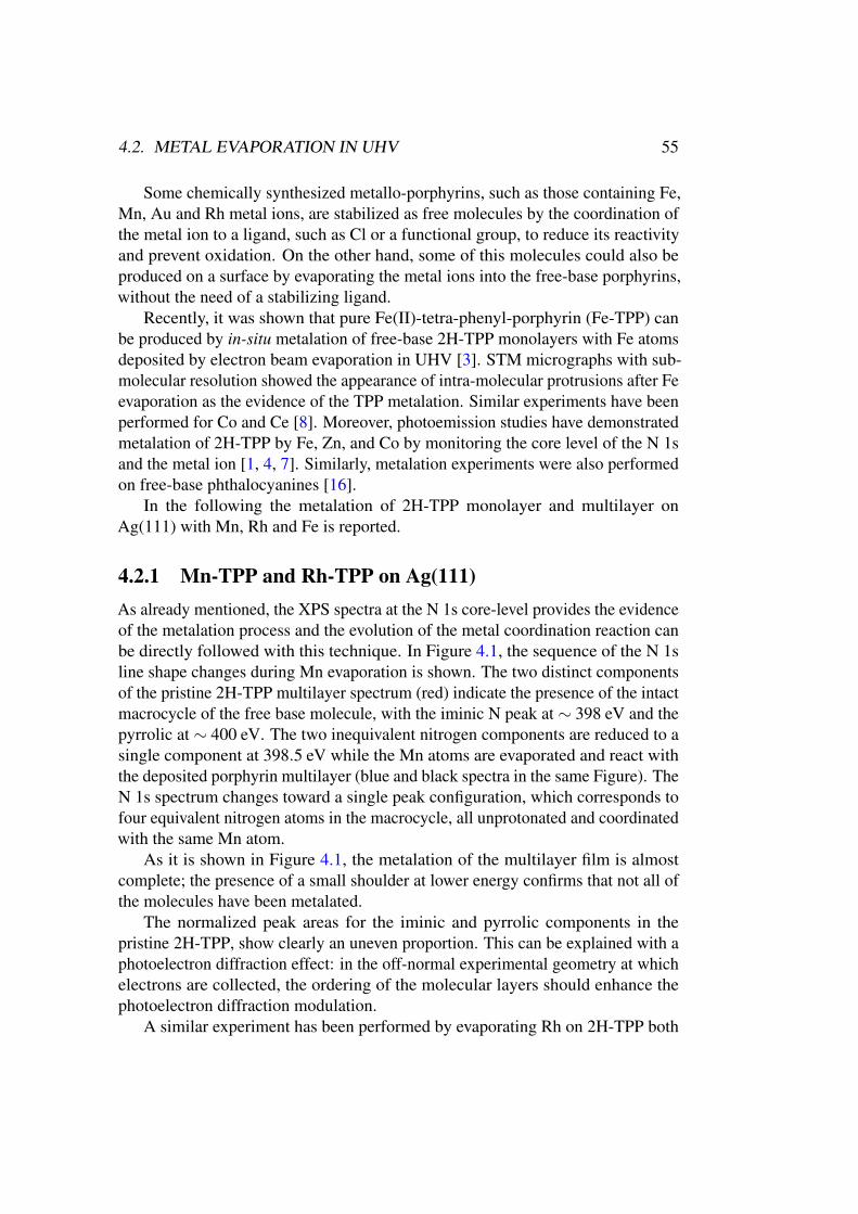
4.2. METAL EVAPORATION IN UHV 55
Some chemically synthesized metallo-porphyrins, such as those containing Fe,Mn, Au and Rh metal ions, are stabilized as free molecules by the coordination ofthe metal ion to a ligand, such as Cl or a functional group, to reduce its reactivityand prevent oxidation. On the other hand, some of this molecules could also beproduced on a surface by evaporating the metal ions into the free-base porphyrins,without the need of a stabilizing ligand.
Recently, it was shown that pure Fe(II)-tetra-phenyl-porphyrin (Fe-TPP) canbe produced by in-situ metalation of free-base 2H-TPP monolayers with Fe atomsdeposited by electron beam evaporation in UHV [3]. STM micrographs with sub-molecular resolution showed the appearance of intra-molecular protrusions after Feevaporation as the evidence of the TPP metalation. Similar experiments have beenperformed for Co and Ce [8]. Moreover, photoemission studies have demonstratedmetalation of 2H-TPP by Fe, Zn, and Co by monitoring the core level of the N 1sand the metal ion [1, 4, 7]. Similarly, metalation experiments were also performedon free-base phthalocyanines [16].
In the following the metalation of 2H-TPP monolayer and multilayer onAg(111) with Mn, Rh and Fe is reported.
4.2.1 Mn-TPP and Rh-TPP on Ag(111)
As already mentioned, the XPS spectra at the N 1s core-level provides the evidenceof the metalation process and the evolution of the metal coordination reaction canbe directly followed with this technique. In Figure 4.1, the sequence of the N 1sline shape changes during Mn evaporation is shown. The two distinct componentsof the pristine 2H-TPP multilayer spectrum (red) indicate the presence of the intactmacrocycle of the free base molecule, with the iminic N peak at ∼ 398 eV and thepyrrolic at ∼ 400 eV. The two inequivalent nitrogen components are reduced to asingle component at 398.5 eV while the Mn atoms are evaporated and react withthe deposited porphyrin multilayer (blue and black spectra in the same Figure). TheN 1s spectrum changes toward a single peak configuration, which corresponds tofour equivalent nitrogen atoms in the macrocycle, all unprotonated and coordinatedwith the same Mn atom.
As it is shown in Figure 4.1, the metalation of the multilayer film is almostcomplete; the presence of a small shoulder at lower energy confirms that not all ofthe molecules have been metalated.
The normalized peak areas for the iminic and pyrrolic components in thepristine 2H-TPP, show clearly an uneven proportion. This can be explained with aphotoelectron diffraction effect: in the off-normal experimental geometry at whichelectrons are collected, the ordering of the molecular layers should enhance thephotoelectron diffraction modulation.
A similar experiment has been performed by evaporating Rh on 2H-TPP both
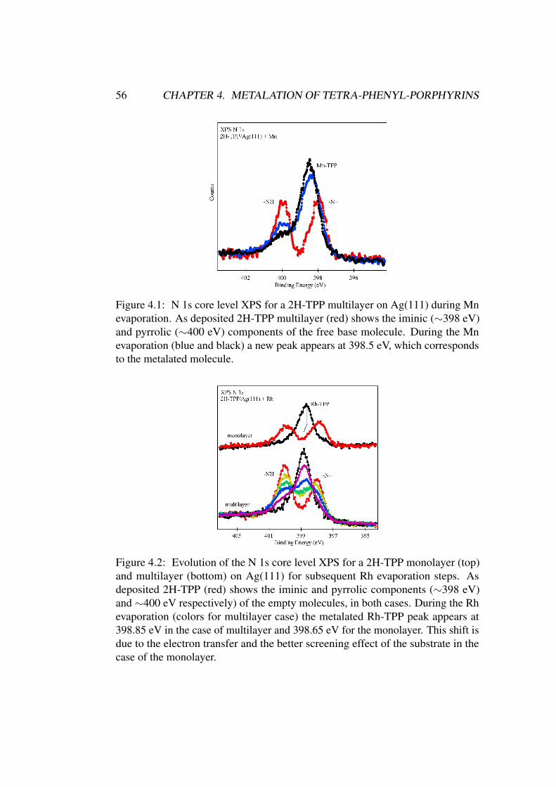
56 CHAPTER 4. METALATION OF TETRA-PHENYL-PORPHYRINS
Figure 4.1: N 1s core level XPS for a 2H-TPP multilayer on Ag(111) during Mnevaporation. As deposited 2H-TPP multilayer (red) shows the iminic (∼398 eV)and pyrrolic (∼400 eV) components of the free base molecule. During the Mnevaporation (blue and black) a new peak appears at 398.5 eV, which correspondsto the metalated molecule.
Figure 4.2: Evolution of the N 1s core level XPS for a 2H-TPP monolayer (top)and multilayer (bottom) on Ag(111) for subsequent Rh evaporation steps. Asdeposited 2H-TPP (red) shows the iminic and pyrrolic components (∼398 eV)and ∼400 eV respectively) of the empty molecules, in both cases. During the Rhevaporation (colors for multilayer case) the metalated Rh-TPP peak appears at398.85 eV in the case of multilayer and 398.65 eV for the monolayer. This shift isdue to the electron transfer and the better screening effect of the substrate in thecase of the monolayer.
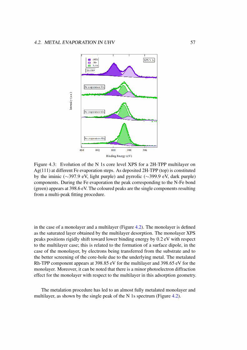
4.2. METAL EVAPORATION IN UHV 57
Figure 4.3: Evolution of the N 1s core level XPS for a 2H-TPP multilayer onAg(111) at different Fe evaporation steps. As deposited 2H-TPP (top) is constitutedby the iminic (∼397.9 eV, light purple) and pyrrolic (∼399.9 eV, dark purple)components. During the Fe evaporation the peak corresponding to the N-Fe bond(green) appears at 398.6 eV. The coloured peaks are the single components resultingfrom a multi-peak fitting procedure.
in the case of a monolayer and a multilayer (Figure 4.2). The monolayer is definedas the saturated layer obtained by the multilayer desorption. The monolayer XPSpeaks positions rigidly shift toward lower binding energy by 0.2 eV with respectto the multilayer case; this is related to the formation of a surface dipole, in thecase of the monolayer, by electrons being transferred from the substrate and tothe better screening of the core-hole due to the underlying metal. The metalatedRh-TPP component appears at 398.85 eV for the multilayer and 398.65 eV for themonolayer. Moreover, it can be noted that there is a minor photoelectron diffractioneffect for the monolayer with respect to the multilayer in this adsorption geometry.
The metalation procedure has led to an almost fully metalated monolayer andmultilayer, as shown by the single peak of the N 1s spectrum (Figure 4.2).
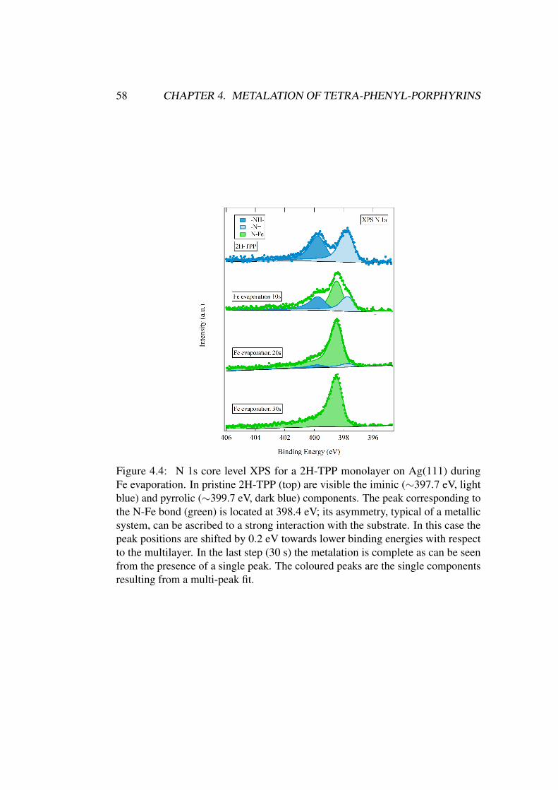
58 CHAPTER 4. METALATION OF TETRA-PHENYL-PORPHYRINS
Figure 4.4: N 1s core level XPS for a 2H-TPP monolayer on Ag(111) duringFe evaporation. In pristine 2H-TPP (top) are visible the iminic (∼397.7 eV, lightblue) and pyrrolic (∼399.7 eV, dark blue) components. The peak corresponding tothe N-Fe bond (green) is located at 398.4 eV; its asymmetry, typical of a metallicsystem, can be ascribed to a strong interaction with the substrate. In this case thepeak positions are shifted by 0.2 eV towards lower binding energies with respectto the multilayer. In the last step (30 s) the metalation is complete as can be seenfrom the presence of a single peak. The coloured peaks are the single componentsresulting from a multi-peak fit.
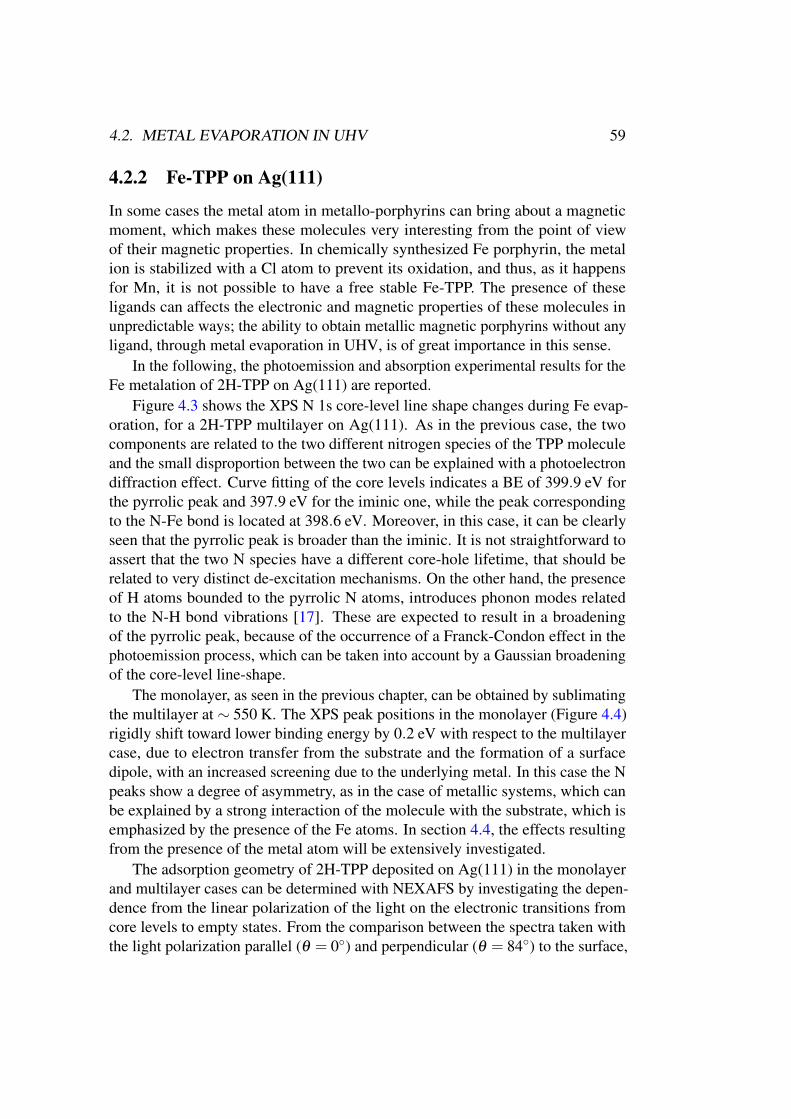
4.2. METAL EVAPORATION IN UHV 59
4.2.2 Fe-TPP on Ag(111)
In some cases the metal atom in metallo-porphyrins can bring about a magneticmoment, which makes these molecules very interesting from the point of viewof their magnetic properties. In chemically synthesized Fe porphyrin, the metalion is stabilized with a Cl atom to prevent its oxidation, and thus, as it happensfor Mn, it is not possible to have a free stable Fe-TPP. The presence of theseligands can affects the electronic and magnetic properties of these molecules inunpredictable ways; the ability to obtain metallic magnetic porphyrins without anyligand, through metal evaporation in UHV, is of great importance in this sense.
In the following, the photoemission and absorption experimental results for theFe metalation of 2H-TPP on Ag(111) are reported.
Figure 4.3 shows the XPS N 1s core-level line shape changes during Fe evap-oration, for a 2H-TPP multilayer on Ag(111). As in the previous case, the twocomponents are related to the two different nitrogen species of the TPP moleculeand the small disproportion between the two can be explained with a photoelectrondiffraction effect. Curve fitting of the core levels indicates a BE of 399.9 eV forthe pyrrolic peak and 397.9 eV for the iminic one, while the peak correspondingto the N-Fe bond is located at 398.6 eV. Moreover, in this case, it can be clearlyseen that the pyrrolic peak is broader than the iminic. It is not straightforward toassert that the two N species have a different core-hole lifetime, that should berelated to very distinct de-excitation mechanisms. On the other hand, the presenceof H atoms bounded to the pyrrolic N atoms, introduces phonon modes relatedto the N-H bond vibrations [17]. These are expected to result in a broadeningof the pyrrolic peak, because of the occurrence of a Franck-Condon effect in thephotoemission process, which can be taken into account by a Gaussian broadeningof the core-level line-shape.
The monolayer, as seen in the previous chapter, can be obtained by sublimatingthe multilayer at ∼ 550 K. The XPS peak positions in the monolayer (Figure 4.4)rigidly shift toward lower binding energy by 0.2 eV with respect to the multilayercase, due to electron transfer from the substrate and the formation of a surfacedipole, with an increased screening due to the underlying metal. In this case the Npeaks show a degree of asymmetry, as in the case of metallic systems, which canbe explained by a strong interaction of the molecule with the substrate, which isemphasized by the presence of the Fe atoms. In section 4.4, the effects resultingfrom the presence of the metal atom will be extensively investigated.
The adsorption geometry of 2H-TPP deposited on Ag(111) in the monolayerand multilayer cases can be determined with NEXAFS by investigating the depen-dence from the linear polarization of the light on the electronic transitions fromcore levels to empty states. From the comparison between the spectra taken withthe light polarization parallel (θ = 0◦) and perpendicular (θ = 84◦) to the surface,
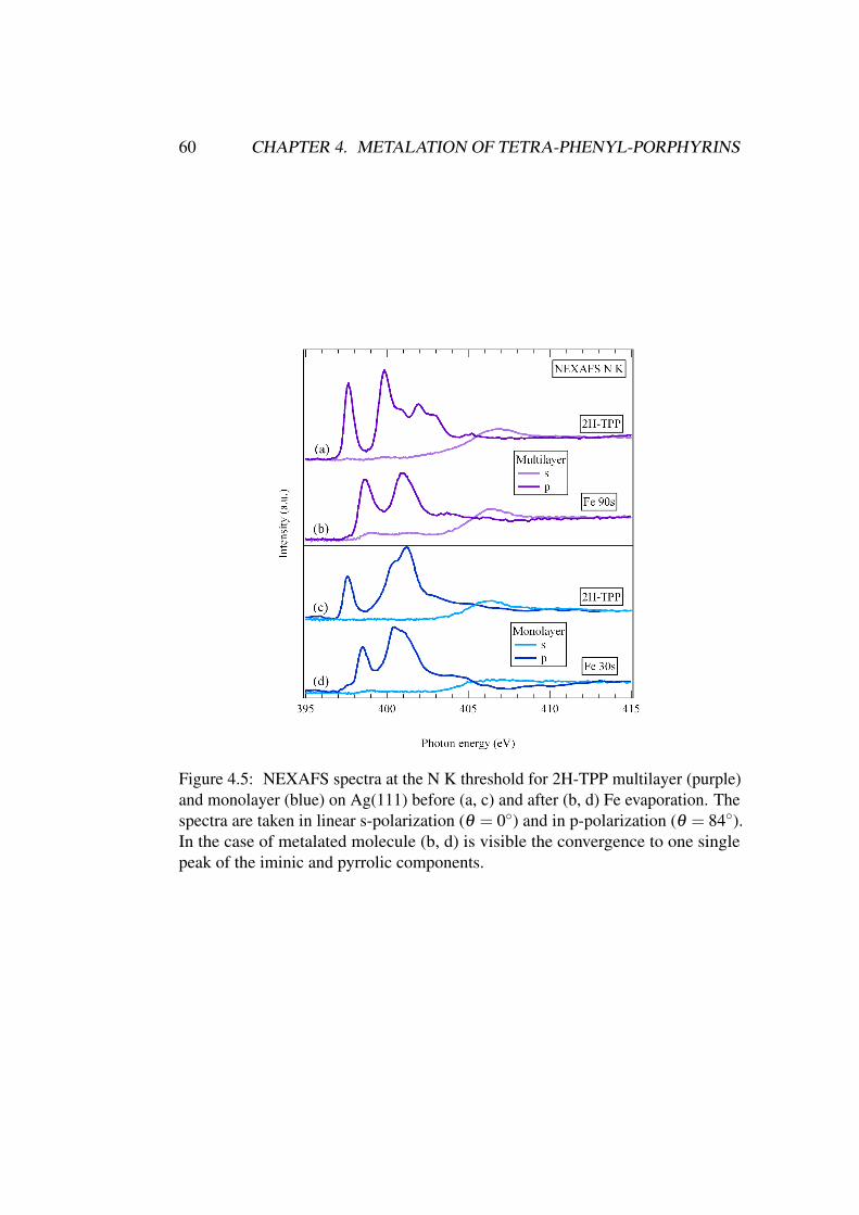
60 CHAPTER 4. METALATION OF TETRA-PHENYL-PORPHYRINS
Figure 4.5: NEXAFS spectra at the N K threshold for 2H-TPP multilayer (purple)and monolayer (blue) on Ag(111) before (a, c) and after (b, d) Fe evaporation. Thespectra are taken in linear s-polarization (θ = 0◦) and in p-polarization (θ = 84◦).In the case of metalated molecule (b, d) is visible the convergence to one singlepeak of the iminic and pyrrolic components.
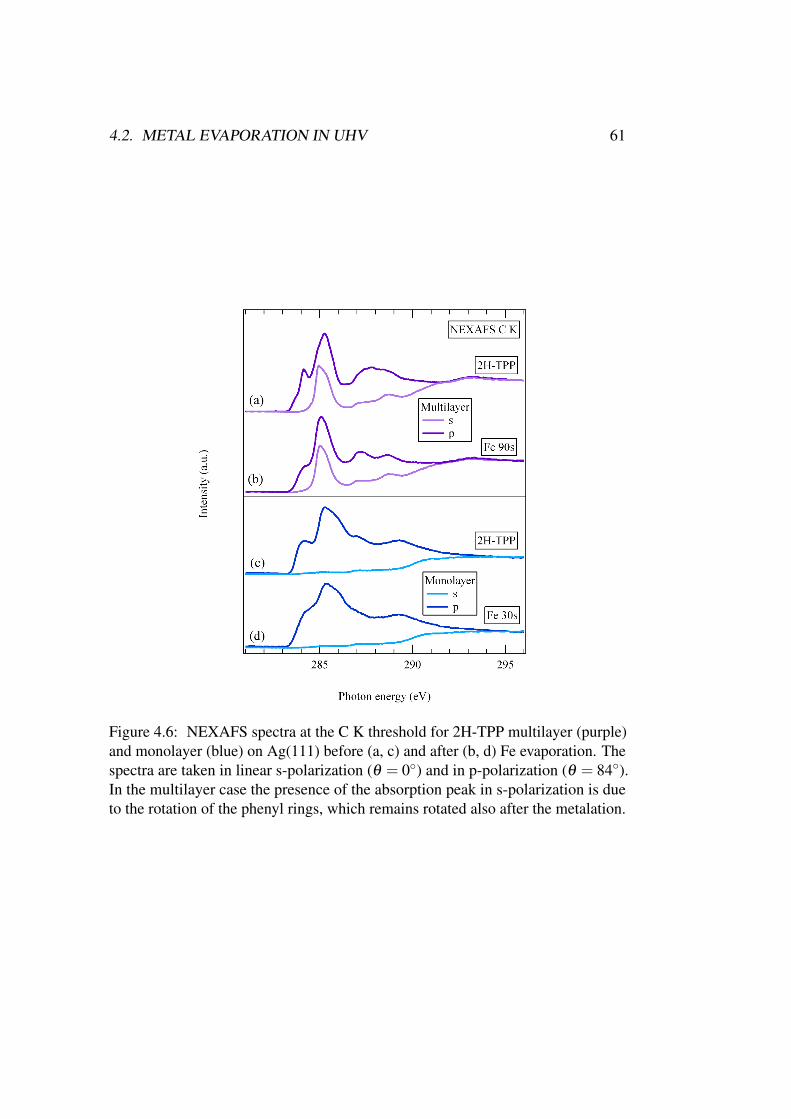
4.2. METAL EVAPORATION IN UHV 61
Figure 4.6: NEXAFS spectra at the C K threshold for 2H-TPP multilayer (purple)and monolayer (blue) on Ag(111) before (a, c) and after (b, d) Fe evaporation. Thespectra are taken in linear s-polarization (θ = 0◦) and in p-polarization (θ = 84◦).In the multilayer case the presence of the absorption peak in s-polarization is dueto the rotation of the phenyl rings, which remains rotated also after the metalation.
62 CHAPTER 4. METALATION OF TETRA-PHENYL-PORPHYRINS
it is possible to extract the information about the geometry of the system. As inthe case of 2H-TPP on Au(111) in the previous chapter, the comparison betweenNEXAFS and XPS is able to clarify the electronic structure of the molecular film.
Figure 4.5 shows the N K-edge NEXAFS spectrum of the 2H-TPP monolayerand multilayer, before and and after Fe evaporation. The multilayer spectrum issimilar to the 2H-TPP gas-phase spectrum, indicating that the interaction betweenthe molecules is weak. In this case, the complete quenching of the π∗ statesin s-polarization, indicates a flat adsorption of the macrocycle. The Fe atomcoordination at the center of the porphyrin macrocycle, is confirmed by the N Kedge absorption spectra: the first two π∗ peaks of the 2H-TPP NEXAFS spectrumconverge to a single peak located at a photon energy of 398.6 eV. This can beobserved also for the monolayer, even if in this case the absorption thresholds, bothin the case of free and metalated molecule, occurs at lower photon energies. Inthis case the first π∗ absorption peak, related to the iminic transition, appears at397.7 eV, equal to the binding energy of the N 1s iminic component measured inphotoemission. The correspondence with the photoemission BE is found also forthe first π∗ transition in the metalated case. Again, this agrees with a metal-likebehaviour of the monolayer as shown by core level XPS.
The geometry assumed by the whole molecule, in particular by the phenyl ringswith respect to the macrocycle, can be determined by the analysis of the absorptionspectra at the C K edge (Figure 4.6).
As seen in the previous chapter, the first structure at ∼ 284 eV is assigned to themacrocycle, while the absorption peak at ∼ 285 eV is mainly due to the π∗ stateslocated in the phenyl rings [18]. As in the case of the N K threshold, the first π∗
transition due to the macrocycle disappears in s-polarization, indicating again thatthe macrocycle lies flat with respect to the substrate surface plane, while the phenylpeak is still present, thus indicating a non-flat orientation of the meso-substituentrings. In the multilayer, the intermolecular interactions are mainly due to the weakVan der Waals forces and, therefore, the phenyl rings have an orientation similar tothe molecules in gas phase, in which they tend to stay at ∼ 70÷80◦ with respectto the macrocycle, as a consequence of the hydrogen steric interactions.
On the contrary, the monolayer showed a flat molecular adsorption geome-try with a planar conformation for the phenyl groups, as clearly seen from thesuppression of all the π∗ peaks in the N K and C K NEXAFS s-polarization spectra.
The interface adsorption geometry and the multilayer molecular orientationis directly affected by the metalation process, and a distortion of the macrocyclewith a change in the electronic structure may occur. In the case of C K NEXAFS(Figure 4.6), the Fe coordination does not affect the orientation of the macrocyclecarbon atoms, as can be seen by the complete quenching of the absorption peakin s-polarization, indicating that the macrocycle is flat as in the case of the empty2H-TPP. For the N absorption spectra (Figure 4.5), instead, the peak at 398.6 eV is
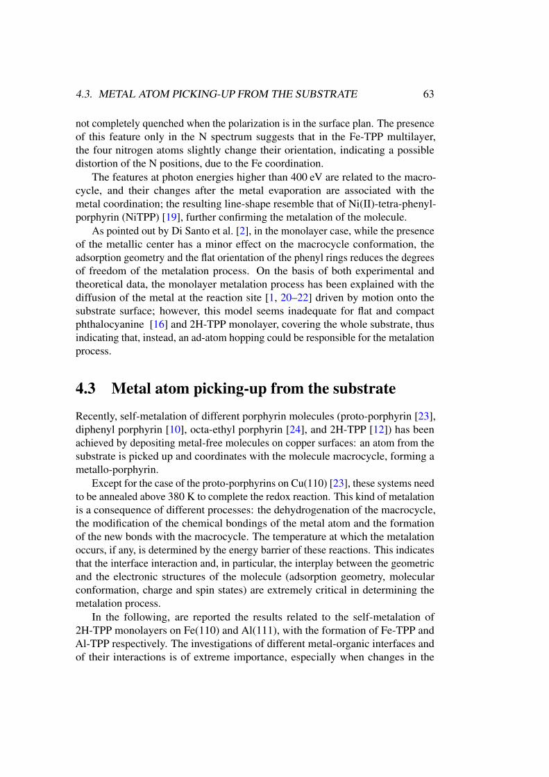
4.3. METAL ATOM PICKING-UP FROM THE SUBSTRATE 63
not completely quenched when the polarization is in the surface plan. The presenceof this feature only in the N spectrum suggests that in the Fe-TPP multilayer,the four nitrogen atoms slightly change their orientation, indicating a possibledistortion of the N positions, due to the Fe coordination.
The features at photon energies higher than 400 eV are related to the macro-cycle, and their changes after the metal evaporation are associated with themetal coordination; the resulting line-shape resemble that of Ni(II)-tetra-phenyl-porphyrin (NiTPP) [19], further confirming the metalation of the molecule.
As pointed out by Di Santo et al. [2], in the monolayer case, while the presenceof the metallic center has a minor effect on the macrocycle conformation, theadsorption geometry and the flat orientation of the phenyl rings reduces the degreesof freedom of the metalation process. On the basis of both experimental andtheoretical data, the monolayer metalation process has been explained with thediffusion of the metal at the reaction site [1, 20–22] driven by motion onto thesubstrate surface; however, this model seems inadequate for flat and compactphthalocyanine [16] and 2H-TPP monolayer, covering the whole substrate, thusindicating that, instead, an ad-atom hopping could be responsible for the metalationprocess.
4.3 Metal atom picking-up from the substrate
Recently, self-metalation of different porphyrin molecules (proto-porphyrin [23],diphenyl porphyrin [10], octa-ethyl porphyrin [24], and 2H-TPP [12]) has beenachieved by depositing metal-free molecules on copper surfaces: an atom from thesubstrate is picked up and coordinates with the molecule macrocycle, forming ametallo-porphyrin.
Except for the case of the proto-porphyrins on Cu(110) [23], these systems needto be annealed above 380 K to complete the redox reaction. This kind of metalationis a consequence of different processes: the dehydrogenation of the macrocycle,the modification of the chemical bondings of the metal atom and the formationof the new bonds with the macrocycle. The temperature at which the metalationoccurs, if any, is determined by the energy barrier of these reactions. This indicatesthat the interface interaction and, in particular, the interplay between the geometricand the electronic structures of the molecule (adsorption geometry, molecularconformation, charge and spin states) are extremely critical in determining themetalation process.
In the following, are reported the results related to the self-metalation of2H-TPP monolayers on Fe(110) and Al(111), with the formation of Fe-TPP andAl-TPP respectively. The investigations of different metal-organic interfaces andof their interactions is of extreme importance, especially when changes in the
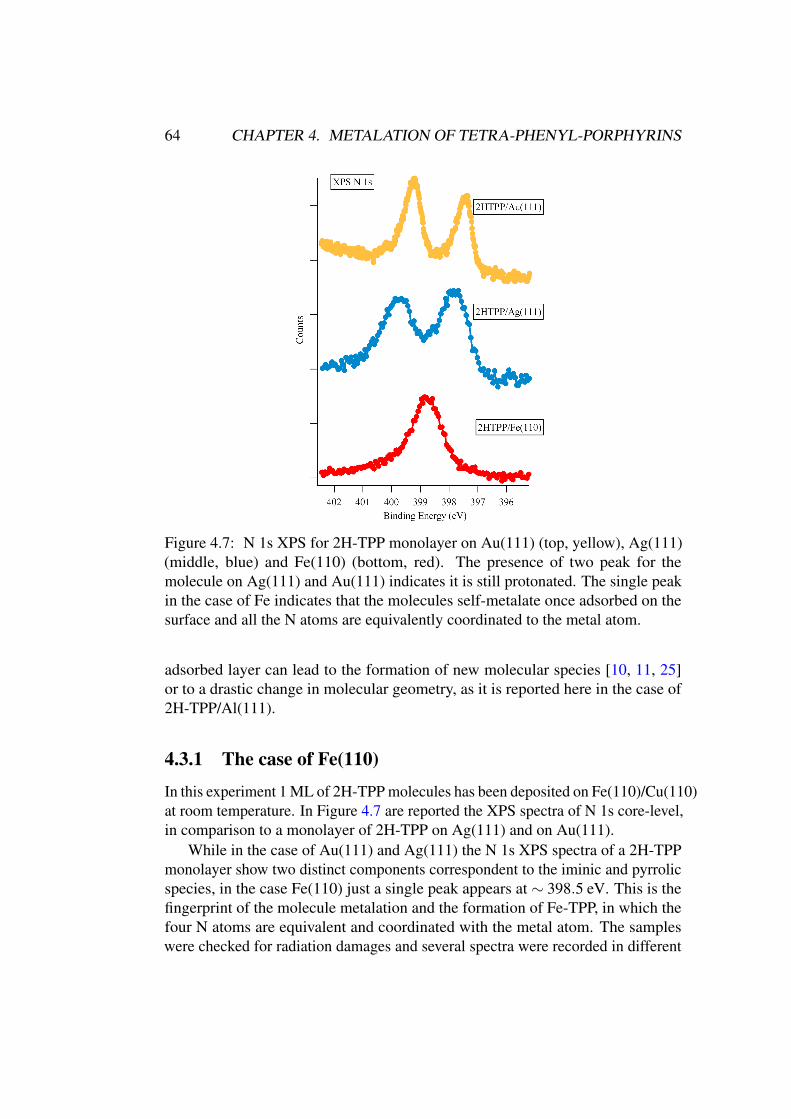
64 CHAPTER 4. METALATION OF TETRA-PHENYL-PORPHYRINS
Figure 4.7: N 1s XPS for 2H-TPP monolayer on Au(111) (top, yellow), Ag(111)(middle, blue) and Fe(110) (bottom, red). The presence of two peak for themolecule on Ag(111) and Au(111) indicates it is still protonated. The single peakin the case of Fe indicates that the molecules self-metalate once adsorbed on thesurface and all the N atoms are equivalently coordinated to the metal atom.
adsorbed layer can lead to the formation of new molecular species [10, 11, 25]or to a drastic change in molecular geometry, as it is reported here in the case of2H-TPP/Al(111).
4.3.1 The case of Fe(110)
In this experiment 1 ML of 2H-TPP molecules has been deposited on Fe(110)/Cu(110)at room temperature. In Figure 4.7 are reported the XPS spectra of N 1s core-level,in comparison to a monolayer of 2H-TPP on Ag(111) and on Au(111).
While in the case of Au(111) and Ag(111) the N 1s XPS spectra of a 2H-TPPmonolayer show two distinct components correspondent to the iminic and pyrrolicspecies, in the case Fe(110) just a single peak appears at ∼ 398.5 eV. This is thefingerprint of the molecule metalation and the formation of Fe-TPP, in which thefour N atoms are equivalent and coordinated with the metal atom. The sampleswere checked for radiation damages and several spectra were recorded in different
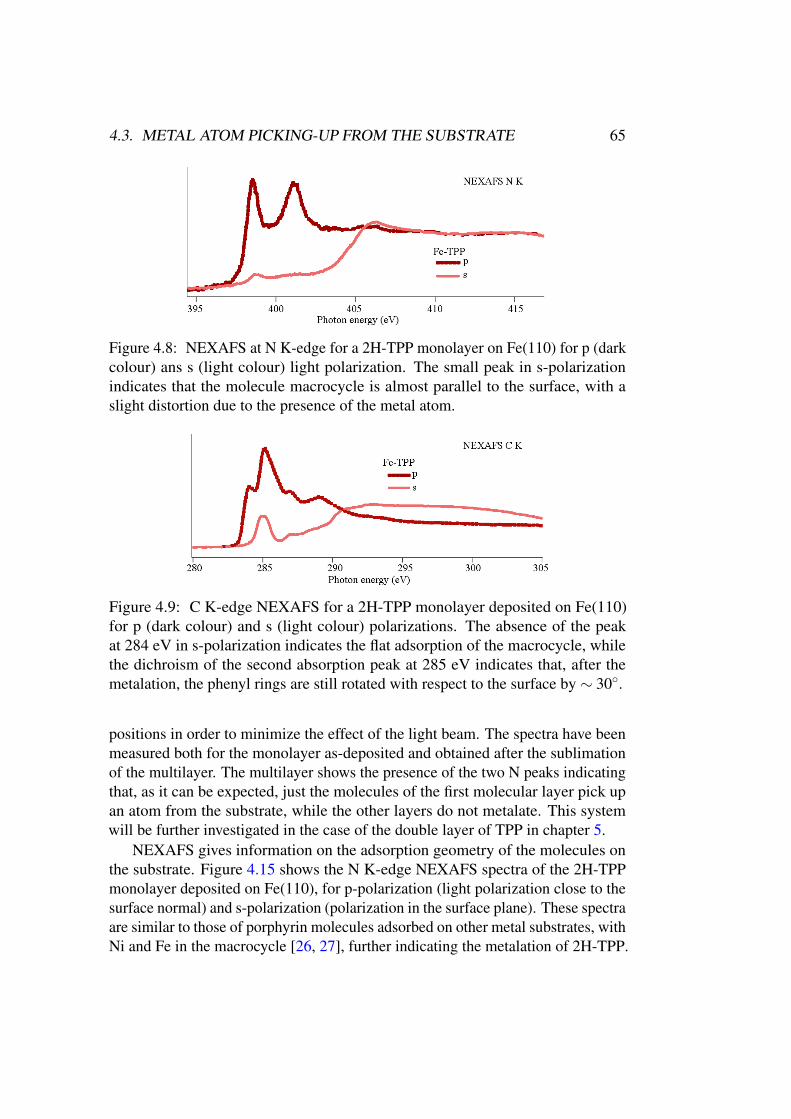
4.3. METAL ATOM PICKING-UP FROM THE SUBSTRATE 65
Figure 4.8: NEXAFS at N K-edge for a 2H-TPP monolayer on Fe(110) for p (darkcolour) ans s (light colour) light polarization. The small peak in s-polarizationindicates that the molecule macrocycle is almost parallel to the surface, with aslight distortion due to the presence of the metal atom.
Figure 4.9: C K-edge NEXAFS for a 2H-TPP monolayer deposited on Fe(110)for p (dark colour) and s (light colour) polarizations. The absence of the peakat 284 eV in s-polarization indicates the flat adsorption of the macrocycle, whilethe dichroism of the second absorption peak at 285 eV indicates that, after themetalation, the phenyl rings are still rotated with respect to the surface by ∼ 30◦.
positions in order to minimize the effect of the light beam. The spectra have beenmeasured both for the monolayer as-deposited and obtained after the sublimationof the multilayer. The multilayer shows the presence of the two N peaks indicatingthat, as it can be expected, just the molecules of the first molecular layer pick upan atom from the substrate, while the other layers do not metalate. This systemwill be further investigated in the case of the double layer of TPP in chapter 5.
NEXAFS gives information on the adsorption geometry of the molecules onthe substrate. Figure 4.15 shows the N K-edge NEXAFS spectra of the 2H-TPPmonolayer deposited on Fe(110), for p-polarization (light polarization close to thesurface normal) and s-polarization (polarization in the surface plane). These spectraare similar to those of porphyrin molecules adsorbed on other metal substrates, withNi and Fe in the macrocycle [26, 27], further indicating the metalation of 2H-TPP.
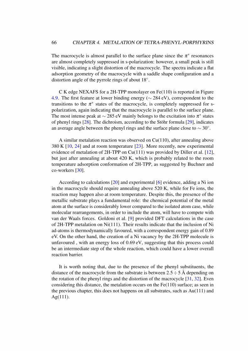
66 CHAPTER 4. METALATION OF TETRA-PHENYL-PORPHYRINS
The macrocycle is almost parallel to the surface plane since the π∗ resonancesare almost completely suppressed in s-polarization: however, a small peak is stillvisible, indicating a slight distortion of the macrocycle. The spectra indicate a flatadsorption geometry of the macrocycle with a saddle shape configuration and adistortion angle of the pyrrole rings of about 18◦.
C K edge NEXAFS for a 2H-TPP monolayer on Fe(110) is reported in Figure4.9. The first feature at lower binding energy (∼ 284 eV), correspondent to thetransitions to the π∗ states of the macrocycle, is completely suppressed for s-polarization, again indicating that the macrocycle is parallel to the surface plane.The most intense peak at ∼ 285 eV mainly belongs to the excitation into π∗ statesof phenyl rings [28]. The dichroism, according to the Stöhr formula [29], indicatesan average angle between the phenyl rings and the surface plane close to ∼ 30◦.
A similar metalation reaction was observed on Cu(110), after annealing above380 K [10, 24] and at room temperature [23]. More recently, new experimentalevidence of metalation of 2H-TPP on Cu(111) was provided by Diller et al. [12],but just after annealing at about 420 K, which is probably related to the roomtemperature adsorption conformation of 2H-TPP, as suggested by Buchner andco-workers [30].
According to calculations [20] and experimental [6] evidence, adding a Ni ionin the macrocycle should require annealing above 520 K, while for Fe ions, thereaction may happen also at room temperature. Despite this, the presence of themetallic substrate plays a fundamental role: the chemical potential of the metalatom at the surface is considerably lower compared to the isolated atom case, whilemolecular rearrangements, in order to include the atom, will have to compete withvan der Waals forces. Goldoni et al. [9] provided DFT calculations in the caseof 2H-TPP metalation on Ni(111). Their results indicate that the inclusion of Niad-atoms is thermodynamically favoured, with a correspondent energy gain of 0.89eV. On the other hand, the creation of a Ni vacancy by the 2H-TPP molecule isunfavoured , with an energy loss of 0.69 eV, suggesting that this process couldbe an intermediate step of the whole reaction, which could have a lower overallreaction barrier.
It is worth noting that, due to the presence of the phenyl substituents, thedistance of the macrocycle from the substrate is between 2.5÷5 Å depending onthe rotation of the phenyl rings and the distortion of the macrocycle [31, 32]. Evenconsidering this distance, the metalation occurs on the Fe(110) surface; as seen inthe previous chapter, this does not happens on all substrates, such as Au(111) andAg(111).
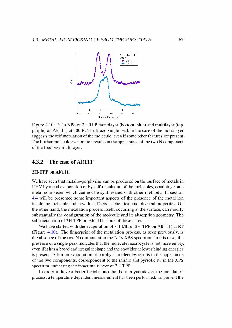
4.3. METAL ATOM PICKING-UP FROM THE SUBSTRATE 67
Figure 4.10: N 1s XPS of 2H-TPP monolayer (bottom, blue) and multilayer (top,purple) on Al(111) at 300 K. The broad single peak in the case of the monolayersuggests the self metalation of the molecule, even if some other features are present.The further molecule evaporation results in the appearance of the two N componentof the free base multilayer.
4.3.2 The case of Al(111)
2H-TPP on Al(111)
We have seen that metallo-porphyrins can be produced on the surface of metals inUHV by metal evaporation or by self-metalation of the molecules, obtaining somemetal complexes which can not be synthesized with other methods. In section4.4 will be presented some important aspects of the presence of the metal ioninside the molecule and how this affects its chemical and physical properties. Onthe other hand, the metalation process itself, occurring at the surface, can modifysubstantially the configuration of the molecule and its absorption geometry. Theself-metalation of 2H-TPP on Al(111) is one of these cases.
We have started with the evaporation of ∼1 ML of 2H-TPP on Al(111) at RT(Figure 4.10). The fingerprint of the metalation process, as seen previously, isthe absence of the two N component in the N 1s XPS spectrum. In this case, thepresence of a single peak indicates that the molecule macrocycle is not more empty,even if it has a broad and irregular shape and the shoulder at lower binding energiesis present. A further evaporation of porphyrin molecules results in the appearanceof the two components, correspondent to the iminic and pyrrolic N, in the XPSspectrum, indicating the intact multilayer of 2H-TPP.
In order to have a better insight into the thermodynamics of the metalationprocess, a temperature dependent measurement has been performed. To prevent the
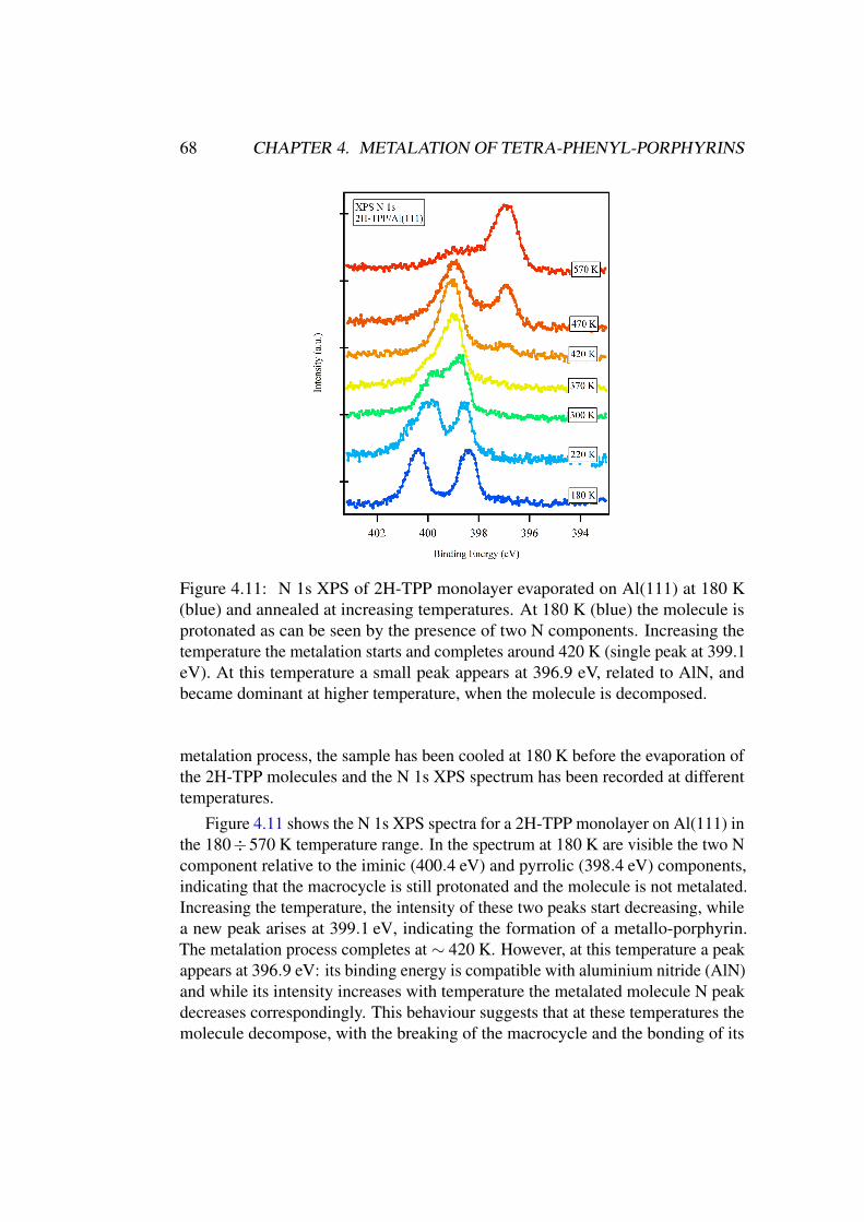
68 CHAPTER 4. METALATION OF TETRA-PHENYL-PORPHYRINS
Figure 4.11: N 1s XPS of 2H-TPP monolayer evaporated on Al(111) at 180 K(blue) and annealed at increasing temperatures. At 180 K (blue) the molecule isprotonated as can be seen by the presence of two N components. Increasing thetemperature the metalation starts and completes around 420 K (single peak at 399.1eV). At this temperature a small peak appears at 396.9 eV, related to AlN, andbecame dominant at higher temperature, when the molecule is decomposed.
metalation process, the sample has been cooled at 180 K before the evaporation ofthe 2H-TPP molecules and the N 1s XPS spectrum has been recorded at differenttemperatures.
Figure 4.11 shows the N 1s XPS spectra for a 2H-TPP monolayer on Al(111) inthe 180÷570 K temperature range. In the spectrum at 180 K are visible the two Ncomponent relative to the iminic (400.4 eV) and pyrrolic (398.4 eV) components,indicating that the macrocycle is still protonated and the molecule is not metalated.Increasing the temperature, the intensity of these two peaks start decreasing, whilea new peak arises at 399.1 eV, indicating the formation of a metallo-porphyrin.The metalation process completes at ∼ 420 K. However, at this temperature a peakappears at 396.9 eV: its binding energy is compatible with aluminium nitride (AlN)and while its intensity increases with temperature the metalated molecule N peakdecreases correspondingly. This behaviour suggests that at these temperatures themolecule decompose, with the breaking of the macrocycle and the bonding of its
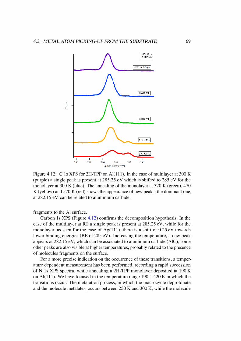
4.3. METAL ATOM PICKING-UP FROM THE SUBSTRATE 69
Figure 4.12: C 1s XPS for 2H-TPP on Al(111). In the case of multilayer at 300 K(purple) a single peak is present at 285.25 eV which is shifted to 285 eV for themonolayer at 300 K (blue). The annealing of the monolayer at 370 K (green), 470K (yellow) and 570 K (red) shows the appearance of new peaks; the dominant one,at 282.15 eV, can be related to aluminium carbide.
fragments to the Al surface.Carbon 1s XPS (Figure 4.12) confirms the decomposition hypothesis. In the
case of the multilayer at RT a single peak is present at 285.25 eV, while for themonolayer, as seen for the case of Ag(111), there is a shift of 0.25 eV towardslower binding energies (BE of 285 eV). Increasing the temperature, a new peakappears at 282.15 eV, which can be associated to aluminium carbide (AlC); someother peaks are also visible at higher temperatures, probably related to the presenceof molecules fragments on the surface.
For a more precise indication on the occurrence of these transitions, a temper-ature dependent measurement has been performed, recording a rapid successionof N 1s XPS spectra, while annealing a 2H-TPP monolayer deposited at 190 Kon Al(111). We have focused in the temperature range 190÷420 K in which thetransitions occur. The metalation process, in which the macrocycle deprotonateand the molecule metalates, occurs between 250 K and 300 K, while the molecule
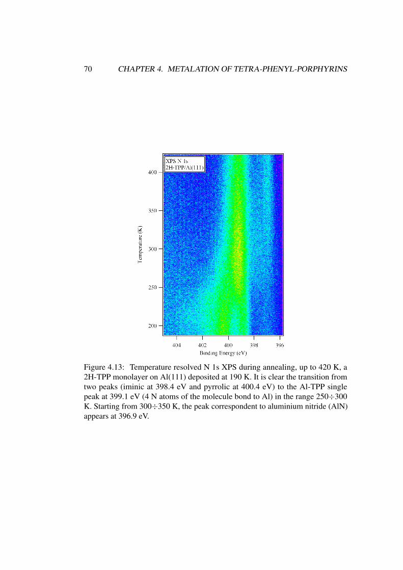
70 CHAPTER 4. METALATION OF TETRA-PHENYL-PORPHYRINS
Figure 4.13: Temperature resolved N 1s XPS during annealing, up to 420 K, a2H-TPP monolayer on Al(111) deposited at 190 K. It is clear the transition fromtwo peaks (iminic at 398.4 eV and pyrrolic at 400.4 eV) to the Al-TPP singlepeak at 399.1 eV (4 N atoms of the molecule bond to Al) in the range 250÷300K. Starting from 300÷350 K, the peak correspondent to aluminium nitride (AlN)appears at 396.9 eV.
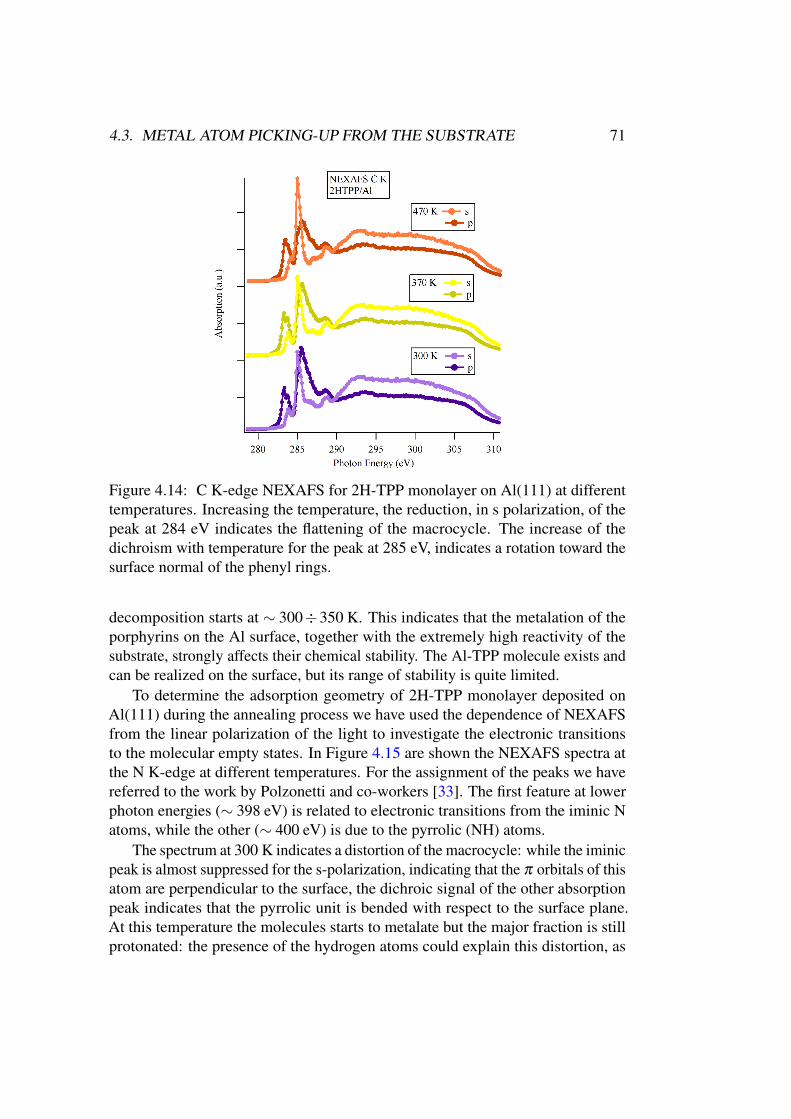
4.3. METAL ATOM PICKING-UP FROM THE SUBSTRATE 71
Figure 4.14: C K-edge NEXAFS for 2H-TPP monolayer on Al(111) at differenttemperatures. Increasing the temperature, the reduction, in s polarization, of thepeak at 284 eV indicates the flattening of the macrocycle. The increase of thedichroism with temperature for the peak at 285 eV, indicates a rotation toward thesurface normal of the phenyl rings.
decomposition starts at ∼ 300÷350 K. This indicates that the metalation of theporphyrins on the Al surface, together with the extremely high reactivity of thesubstrate, strongly affects their chemical stability. The Al-TPP molecule exists andcan be realized on the surface, but its range of stability is quite limited.
To determine the adsorption geometry of 2H-TPP monolayer deposited onAl(111) during the annealing process we have used the dependence of NEXAFSfrom the linear polarization of the light to investigate the electronic transitionsto the molecular empty states. In Figure 4.15 are shown the NEXAFS spectra atthe N K-edge at different temperatures. For the assignment of the peaks we havereferred to the work by Polzonetti and co-workers [33]. The first feature at lowerphoton energies (∼ 398 eV) is related to electronic transitions from the iminic Natoms, while the other (∼ 400 eV) is due to the pyrrolic (NH) atoms.
The spectrum at 300 K indicates a distortion of the macrocycle: while the iminicpeak is almost suppressed for the s-polarization, indicating that the π orbitals of thisatom are perpendicular to the surface, the dichroic signal of the other absorptionpeak indicates that the pyrrolic unit is bended with respect to the surface plane.At this temperature the molecules starts to metalate but the major fraction is stillprotonated: the presence of the hydrogen atoms could explain this distortion, as
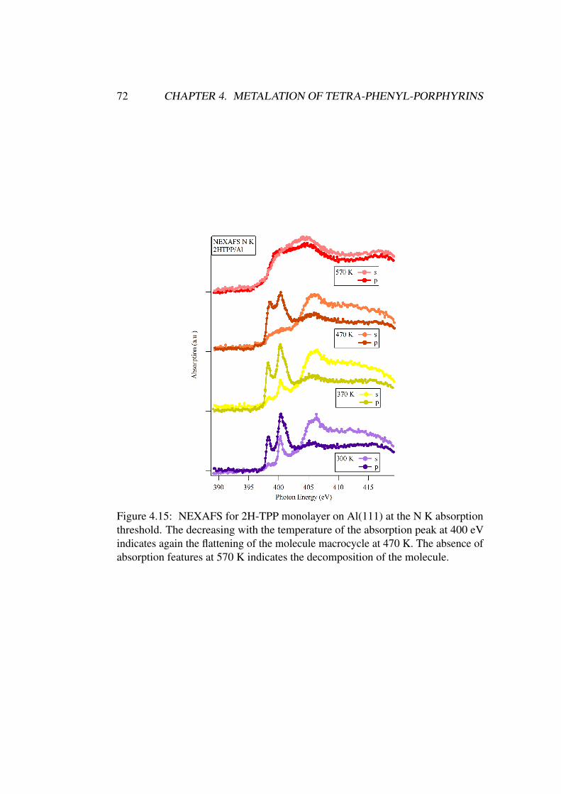
72 CHAPTER 4. METALATION OF TETRA-PHENYL-PORPHYRINS
Figure 4.15: NEXAFS for 2H-TPP monolayer on Al(111) at the N K absorptionthreshold. The decreasing with the temperature of the absorption peak at 400 eVindicates again the flattening of the molecule macrocycle at 470 K. The absence ofabsorption features at 570 K indicates the decomposition of the molecule.
4.3. METAL ATOM PICKING-UP FROM THE SUBSTRATE 73
a consequence of their steric hindrance. Actually, increasing the temperature,together with the metalation of the molecules, the peak intensity for s-polarizationdecreases; at 470 K, when the metalation is completed, the macrocycle is flat andparallel to the substrate surface. A second hypothesis is that the peak at 400 eVcorresponds to a π∗ transition for the metalated molecule and its decreasing inintensity is due to a different step of the metalation process. In this case, initiallythe N atoms in the macrocycle could be distorted toward an Al atom of the surface,and once the thermal energy is sufficient to overcome the energy barrier, the atomleaves the surface and the macrocycle metalates adopting a flat conformation. Inthe spectrum at 570 K the molecular features are no longer visible as a consequenceof the porphyrin decomposition.
Carbon K-edge NEXAFS (Figure 4.14) confirms the macrocycle distortion at300 K and its flattening at higher temperatures, as it is visible by the decreasingwith temperature of the peak at 284 eV in s-polarization. Furthermore, a change inthe dichroism of the second absorption peak (285 K), related to the phenyl rings, isvisible. This indicates that, with the increasing of temperature, the phenyl ringsrotate perpendicularly to the macrocycle and the substrate. Probably this adaptationis accompanied by a increasing in the molecule-substrate distance, which couldalso explain the successive flattening of the macrocycle, as a consequence of theextraction of an Al atom from the substrate, after an initial phase in which themacrocycle is bent toward the surface.
MnTPPCl on Al(111)
We have seen that the metalation process occurring at the Al surface can modifysubstantially the absorption geometry of the porphyrin molecules. To exclude theeffects of the metal picking-up on the molecular configuration and to focus on thesubstrate influence we have deposited pre-synthesized metallo-porphyrins on theAl substrate. In particular a multilayer and monolayer of Mn(III)-tetra-phenyl-porphyrin-Cl (MnTPPCl) has been evaporated on Al(111) single crystal at RT andthen annealed at 570 K, as in the case of 2H-TPP.
Figures 4.16(a) and 4.16(b) reports C 1s and N 1s core level XPS, respectively,for monolayer and multilayer of MnTPPCl on Al(111). The appearance of thealuminium carbide and nitride peaks after the annealing process demonstrates thatthe decomposition of the molecules happens also in the absence of the on-surfacemetalation process. The annealing process at 520÷550 K of 2H-TPP on Ag(111)and Au(111) promotes the dehydrogenation reaction of the macrocycle with theformation of new C-C bonds and the flattening of the phenyl rings; anyway, inthe latter case, the molecules, due to the weak interaction with the substrate, arealso subjected to sublimation, leaving the Au surface. In the case of Al(111)instead, due to the high reactivity of this substrate, the annealing process at the
74 CHAPTER 4. METALATION OF TETRA-PHENYL-PORPHYRINS
same temperature, both in the case of self-metalating porphyrin (2H-TPP) andpre-synthesized metallo-porphyrin (MnTPPCl), results in the decomposition of themolecule.
For comparison, Figures 4.17(a) and 4.17(b) reports the NEXAFS at the Cand N K threshold, respectively, for MnTPPCl on Al(111) as deposited at 300 Kand after annealing at 570 K. It is interesting to note that, while the C absorp-tion spectrum at 300 K is quite similar to the case of 2H-TPP, in the case of NK NEXAFS the presynthesized metallo-porphyrin deposited at 300 K has a flatmacrocycle: while the 2H-TPP NEXAFS at this temperature shows a distortionof the macrocycle towards the surface, in this case, the presence of the Mn atominside the macrocycle stabilize the molecular framework, as it happens, instead,in the case of 2H-TPP at 470 K, when the molecule is metalated, forming Al-TPP.This evidence corroborates the hypothesis of an initial macrocycle distortion in thecase of 2H-TPP just before the metal coordination, with the bending of the N atomstoward an Al atom on the surface, and the subsequent flattening once the metalion is coordinated. Moreover, it confirms that, on Al(111), the occurrence of themetalation process on the substrate strongly modifies the geometry of the porphyrinmolecule, affecting its chemical stability. Finally, it can be noted that, while theN K NEXAFS at 570 K has no recognisable features, the C K absorption edgestill shows some defined peaks; at this temperature XPS reveals that the moleculesundergo a decomposition, so, as pointed out in the case of 2H-TPP, these could berelated to molecule fragments on the surface.
4.4 Effects of metalation on the molecule-substrate
interaction
In TPP molecules, the phenyl groups and their orientation with respect to themacrocycle affect the distance between the molecule and the substrate, which, inturn, determines the charge transfer properties of the system. Moreover, the orien-tation of the meso-substituents play a crucial role in the mechanism of metalationof the molecule and the formation of metal-organic complexes.
As seen in the previous sections, the phenyl rings of 2H-TPP molecule onAu(111) and Ag(111) adopt a flat conformation when annealed above 525 K. Inthis case, the occurrence of a dehydrogenation process with the formation of newC-C bonds results in a lower distance between the molecule and the substrate, ina configuration similar to a phthalocyanine [21], in which the molecule-substratedistance is less than 3 Å. It has been observed [34] that the presence of the metalin the TPP macrocycle does not affect the supramolecular order in the monolayerdeposited at room temperature. At this point, it is interesting to investigate the
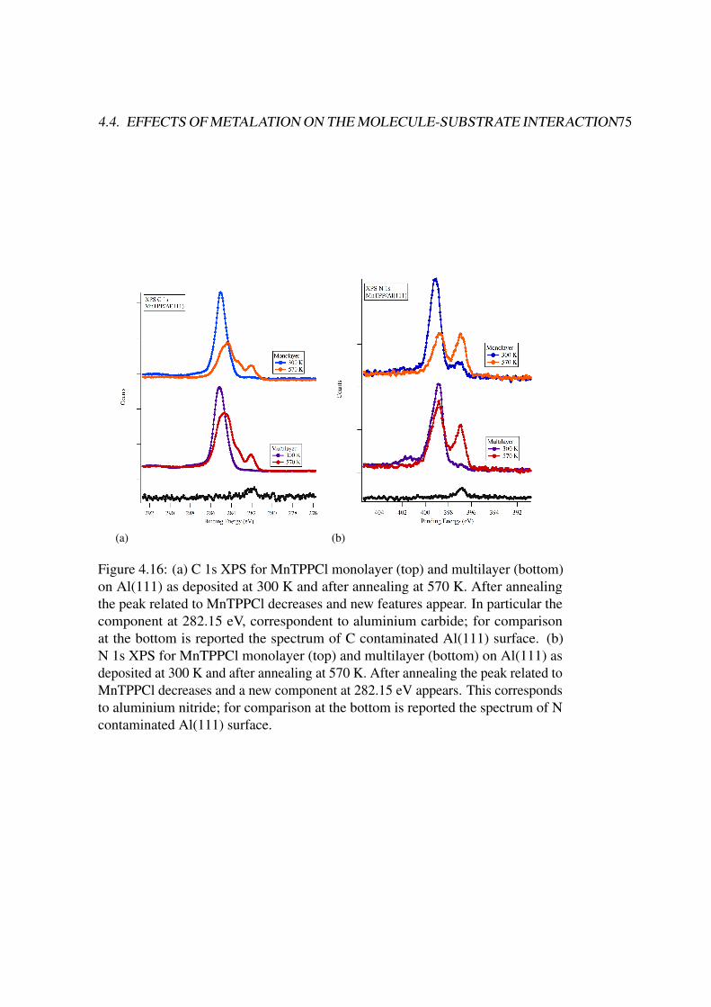
4.4. EFFECTS OF METALATION ON THE MOLECULE-SUBSTRATE INTERACTION75
(a) (b)
Figure 4.16: (a) C 1s XPS for MnTPPCl monolayer (top) and multilayer (bottom)on Al(111) as deposited at 300 K and after annealing at 570 K. After annealingthe peak related to MnTPPCl decreases and new features appear. In particular thecomponent at 282.15 eV, correspondent to aluminium carbide; for comparisonat the bottom is reported the spectrum of C contaminated Al(111) surface. (b)N 1s XPS for MnTPPCl monolayer (top) and multilayer (bottom) on Al(111) asdeposited at 300 K and after annealing at 570 K. After annealing the peak related toMnTPPCl decreases and a new component at 282.15 eV appears. This correspondsto aluminium nitride; for comparison at the bottom is reported the spectrum of Ncontaminated Al(111) surface.
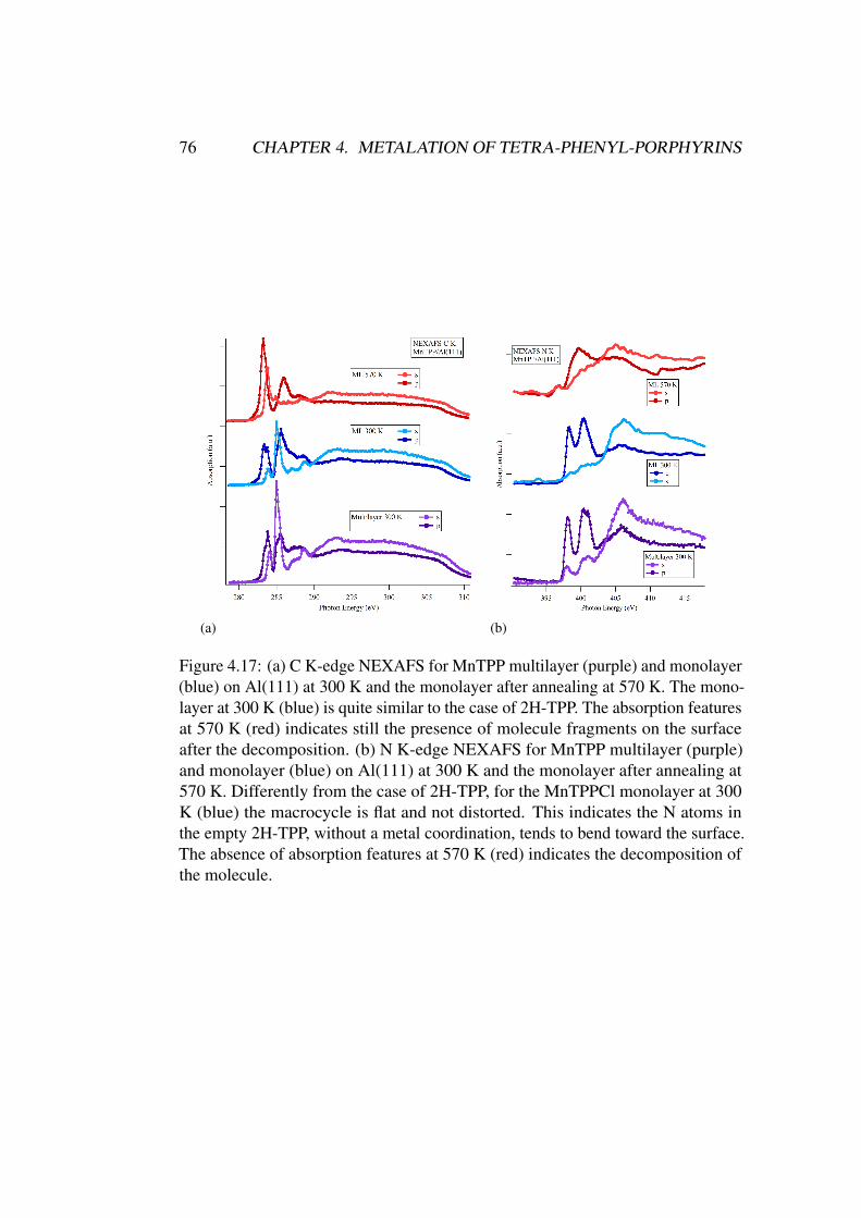
76 CHAPTER 4. METALATION OF TETRA-PHENYL-PORPHYRINS
(a) (b)
Figure 4.17: (a) C K-edge NEXAFS for MnTPP multilayer (purple) and monolayer(blue) on Al(111) at 300 K and the monolayer after annealing at 570 K. The mono-layer at 300 K (blue) is quite similar to the case of 2H-TPP. The absorption featuresat 570 K (red) indicates still the presence of molecule fragments on the surfaceafter the decomposition. (b) N K-edge NEXAFS for MnTPP multilayer (purple)and monolayer (blue) on Al(111) at 300 K and the monolayer after annealing at570 K. Differently from the case of 2H-TPP, for the MnTPPCl monolayer at 300K (blue) the macrocycle is flat and not distorted. This indicates the N atoms inthe empty 2H-TPP, without a metal coordination, tends to bend toward the surface.The absence of absorption features at 570 K (red) indicates the decomposition ofthe molecule.
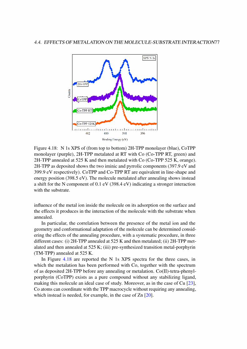
4.4. EFFECTS OF METALATION ON THE MOLECULE-SUBSTRATE INTERACTION77
Figure 4.18: N 1s XPS of (from top to bottom) 2H-TPP monolayer (blue), CoTPPmonolayer (purple), 2H-TPP metalated at RT with Co (Co-TPP RT, green) and2H-TPP annealed at 525 K and then metalated with Co (Co-TPP 525 K, orange).2H-TPP as deposited shows the two iminic and pyrrolic components (397.9 eV and399.9 eV respectively). CoTPP and Co-TPP RT are equivalent in line-shape andenergy position (398.5 eV). The molecule metalated after annealing shows insteada shift for the N component of 0.1 eV (398.4 eV) indicating a stronger interactionwith the substrate.
influence of the metal ion inside the molecule on its adsorption on the surface andthe effects it produces in the interaction of the molecule with the substrate whenannealed.
In particular, the correlation between the presence of the metal ion and thegeometry and conformational adaptation of the molecule can be determined consid-ering the effects of the annealing procedure, with a systematic procedure, in threedifferent cases: (i) 2H-TPP annealed at 525 K and then metalated; (ii) 2H-TPP met-alated and then annealed at 525 K; (iii) pre-synthesized transition metal-porphyrin(TM-TPP) annealed at 525 K.
In Figure 4.18 are reported the N 1s XPS spectra for the three cases, inwhich the metalation has been performed with Co, together with the spectrumof as deposited 2H-TPP before any annealing or metalation. Co(II)-tetra-phenyl-porphyrin (CoTPP) exists as a pure compound without any stabilizing ligand,making this molecule an ideal case of study. Moreover, as in the case of Cu [23],Co atoms can coordinate with the TPP macrocycle without requiring any annealing,which instead is needed, for example, in the case of Zn [20].
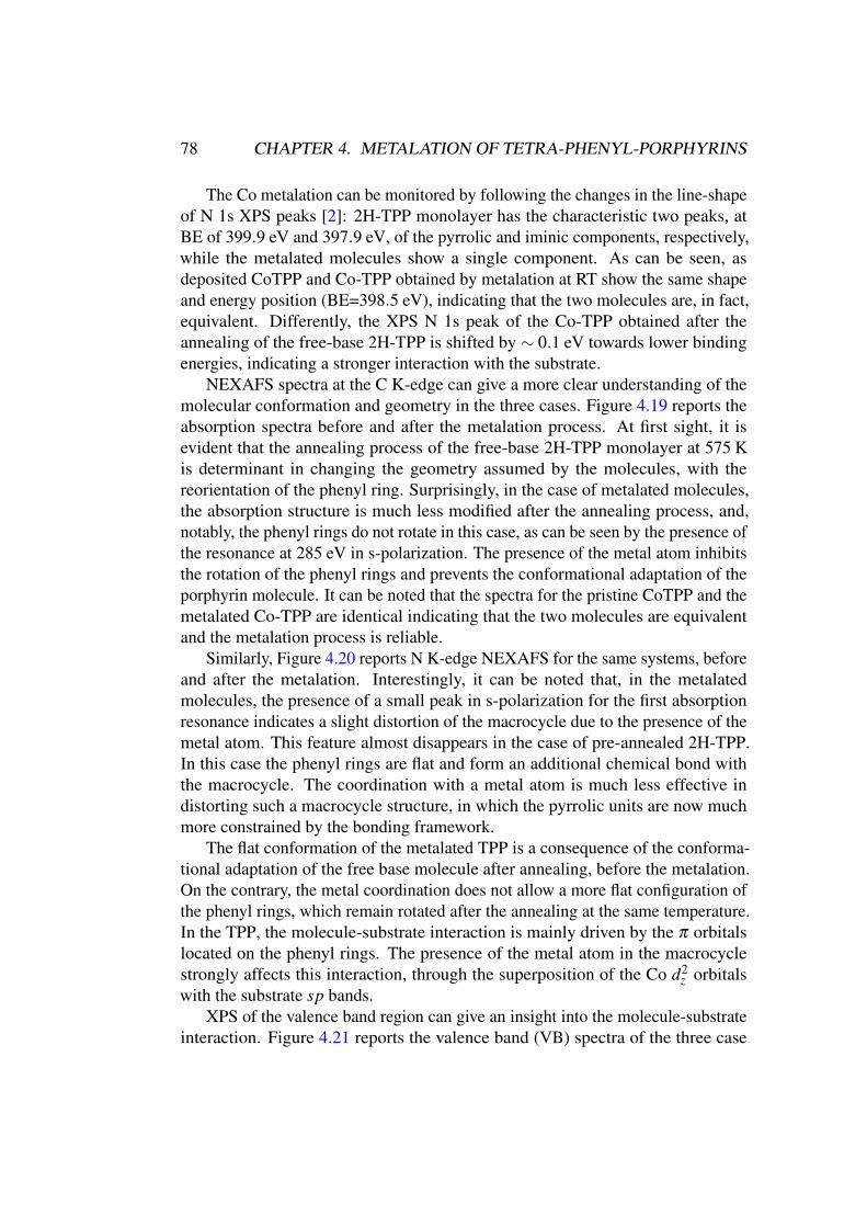
78 CHAPTER 4. METALATION OF TETRA-PHENYL-PORPHYRINS
The Co metalation can be monitored by following the changes in the line-shapeof N 1s XPS peaks [2]: 2H-TPP monolayer has the characteristic two peaks, atBE of 399.9 eV and 397.9 eV, of the pyrrolic and iminic components, respectively,while the metalated molecules show a single component. As can be seen, asdeposited CoTPP and Co-TPP obtained by metalation at RT show the same shapeand energy position (BE=398.5 eV), indicating that the two molecules are, in fact,equivalent. Differently, the XPS N 1s peak of the Co-TPP obtained after theannealing of the free-base 2H-TPP is shifted by ∼ 0.1 eV towards lower bindingenergies, indicating a stronger interaction with the substrate.
NEXAFS spectra at the C K-edge can give a more clear understanding of themolecular conformation and geometry in the three cases. Figure 4.19 reports theabsorption spectra before and after the metalation process. At first sight, it isevident that the annealing process of the free-base 2H-TPP monolayer at 575 Kis determinant in changing the geometry assumed by the molecules, with thereorientation of the phenyl ring. Surprisingly, in the case of metalated molecules,the absorption structure is much less modified after the annealing process, and,notably, the phenyl rings do not rotate in this case, as can be seen by the presence ofthe resonance at 285 eV in s-polarization. The presence of the metal atom inhibitsthe rotation of the phenyl rings and prevents the conformational adaptation of theporphyrin molecule. It can be noted that the spectra for the pristine CoTPP and themetalated Co-TPP are identical indicating that the two molecules are equivalentand the metalation process is reliable.
Similarly, Figure 4.20 reports N K-edge NEXAFS for the same systems, beforeand after the metalation. Interestingly, it can be noted that, in the metalatedmolecules, the presence of a small peak in s-polarization for the first absorptionresonance indicates a slight distortion of the macrocycle due to the presence of themetal atom. This feature almost disappears in the case of pre-annealed 2H-TPP.In this case the phenyl rings are flat and form an additional chemical bond withthe macrocycle. The coordination with a metal atom is much less effective indistorting such a macrocycle structure, in which the pyrrolic units are now muchmore constrained by the bonding framework.
The flat conformation of the metalated TPP is a consequence of the conforma-tional adaptation of the free base molecule after annealing, before the metalation.On the contrary, the metal coordination does not allow a more flat configuration ofthe phenyl rings, which remain rotated after the annealing at the same temperature.In the TPP, the molecule-substrate interaction is mainly driven by the π orbitalslocated on the phenyl rings. The presence of the metal atom in the macrocyclestrongly affects this interaction, through the superposition of the Co d2
z orbitalswith the substrate sp bands.
XPS of the valence band region can give an insight into the molecule-substrateinteraction. Figure 4.21 reports the valence band (VB) spectra of the three case
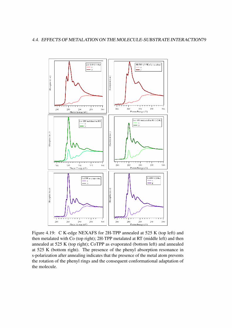
4.4. EFFECTS OF METALATION ON THE MOLECULE-SUBSTRATE INTERACTION79
Figure 4.19: C K-edge NEXAFS for 2H-TPP annealed at 525 K (top left) andthen metalated with Co (top right); 2H-TPP metalated at RT (middle left) and thenannealed at 525 K (top right); CoTPP as evaporated (bottom left) and annealedat 525 K (bottom right). The presence of the phenyl absorption resonance ins-polarization after annealing indicates that the presence of the metal atom preventsthe rotation of the phenyl rings and the consequent conformational adaptation ofthe molecule.
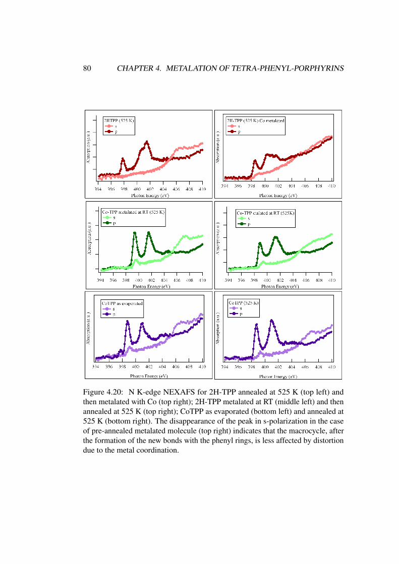
80 CHAPTER 4. METALATION OF TETRA-PHENYL-PORPHYRINS
Figure 4.20: N K-edge NEXAFS for 2H-TPP annealed at 525 K (top left) andthen metalated with Co (top right); 2H-TPP metalated at RT (middle left) and thenannealed at 525 K (top right); CoTPP as evaporated (bottom left) and annealed at525 K (bottom right). The disappearance of the peak in s-polarization in the caseof pre-annealed metalated molecule (top right) indicates that the macrocycle, afterthe formation of the new bonds with the phenyl rings, is less affected by distortiondue to the metal coordination.
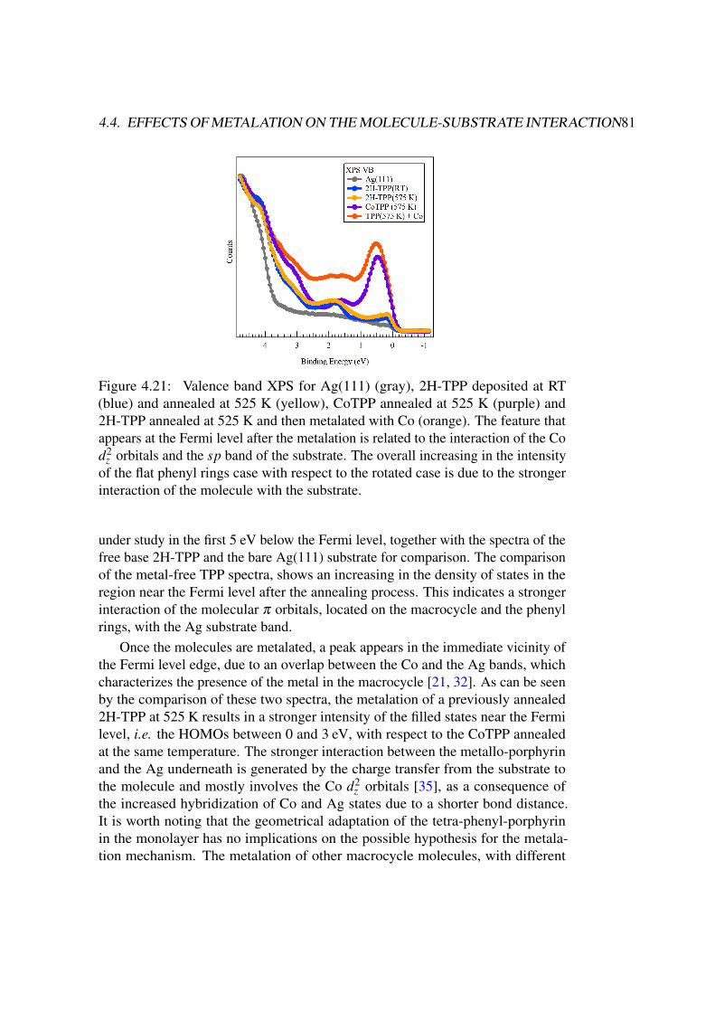
4.4. EFFECTS OF METALATION ON THE MOLECULE-SUBSTRATE INTERACTION81
Figure 4.21: Valence band XPS for Ag(111) (gray), 2H-TPP deposited at RT(blue) and annealed at 525 K (yellow), CoTPP annealed at 525 K (purple) and2H-TPP annealed at 525 K and then metalated with Co (orange). The feature thatappears at the Fermi level after the metalation is related to the interaction of the Cod2
z orbitals and the sp band of the substrate. The overall increasing in the intensityof the flat phenyl rings case with respect to the rotated case is due to the strongerinteraction of the molecule with the substrate.
under study in the first 5 eV below the Fermi level, together with the spectra of thefree base 2H-TPP and the bare Ag(111) substrate for comparison. The comparisonof the metal-free TPP spectra, shows an increasing in the density of states in theregion near the Fermi level after the annealing process. This indicates a strongerinteraction of the molecular π orbitals, located on the macrocycle and the phenylrings, with the Ag substrate band.
Once the molecules are metalated, a peak appears in the immediate vicinity ofthe Fermi level edge, due to an overlap between the Co and the Ag bands, whichcharacterizes the presence of the metal in the macrocycle [21, 32]. As can be seenby the comparison of these two spectra, the metalation of a previously annealed2H-TPP at 525 K results in a stronger intensity of the filled states near the Fermilevel, i.e. the HOMOs between 0 and 3 eV, with respect to the CoTPP annealedat the same temperature. The stronger interaction between the metallo-porphyrinand the Ag underneath is generated by the charge transfer from the substrate tothe molecule and mostly involves the Co d2
z orbitals [35], as a consequence ofthe increased hybridization of Co and Ag states due to a shorter bond distance.It is worth noting that the geometrical adaptation of the tetra-phenyl-porphyrinin the monolayer has no implications on the possible hypothesis for the metala-tion mechanism. The metalation of other macrocycle molecules, with different
82 REFERENCES
geometries, is indeed happening in a similar way [2, 5, 6, 16, 36]. Therefore, themetalation process can be explained by a simple ad-atom hopping instead of asubstrate surface-mediated diffusion to the reaction sites.
References
[1] Florian Buchner, Ken Flechtner, Yun Bai, Elisabeth Zillner, Ina Kellner, Hans-Peter Steinrück, Hubertus Marbach, and J. Michael Gottfried. Coordinationof iron atoms by tetraphenylporphyrin monolayers and multilayers on ag(111)and formation of iron-tetraphenylporphyrin. Journal of Physical Chemistry
C, 112(39):15458–65, 2008. doi: 10.1021/jp8052955.
[2] Giovanni Di Santo, Carla Castellarin-Cudia, Mattia Fanetti, Bidini Taleatu,Patrizia Borghetti, Luigi Sangaletti, Luca Floreano, Elena Magnano, Fed-erica Bondino, and Andrea Goldoni. Conformational adaptation and elec-tronic structure of 2h-tetraphenylporphyrin on ag(111) during fe metala-tion. The Journal of Physical Chemistry C, 115(10):4155–62, 2011. doi:10.1021/jp111151n.
[3] Florian Buchner, Veronika Schwald, Karmen Comanici, Hans-Peter Stein-rück, and Hubertus Marbach. Microscopic evidence of the metalation ofa free-base porphyrin monolayer with iron. Chemphyschem : a European
journal of chemical physics and physical chemistry, 8(2):241–3, 2007. doi:10.1002/cphc.200600698.
[4] J.M. Gottfried, Ken Flechtner, Andreas Kretschmann, Thomas Lukasczyk,and H.P. Steinrück. Direct synthesis of a metalloporphyrin complex on asurface. Journal of the American Chemical Society, 128(17):5644–5, 2006.
[5] Giovanni Di Santo, Cristina Sfiligoj, Carla Castellarin-Cudia, Alberto Verdini,Albano Cossaro, Alberto Morgante, Luca Floreano, and Andrea Goldoni.Changes of the molecule-substrate interaction upon metal inclusion into aporphyrin. Chemistry - A European Journal, (18):12619–23, 2012. doi:10.1002/chem.201201640.
[6] Min Chen, Xuefei Feng, Liang Zhang, Huanxin Ju, Qian Xu, Junfa Zhu,J. Michael Gottfried, Kurash Ibrahim, Haijie Qian, and Jiaou Wang. Directsynthesis of nickel(II) tetraphenylporphyrin and its interaction with a au(111)surface: A comprehensive study. The Journal of Physical Chemistry C, 114(21):9908–16, 2010. doi: 10.1021/jp102031m.
REFERENCES 83
[7] Andreas Kretschmann, Marie-Madeleine Walz, Ken Flechtner, Hans-PeterSteinrück, and J Michael Gottfried. Tetraphenylporphyrin picks up zinc atomsfrom a silver surface. Chemical communications, (6):568–70, 2007. doi:10.1039/b614427f.
[8] A. Weber-Bargioni, J. Reichert, A.P. Seitsonen, W. Auwarter, A. Schiffrin,and J.V. Barth. Interaction of cerium atoms with surface-anchored porphyrinmolecules. Journal of Physical Chemistry C, 112(10):3453–5, March 2008.doi: 10.1021/jp076961i.
[9] Andrea Goldoni, Carlo A. Pignedoli, Giovanni Di Santo, Carla Castellarin-Cudia, Elena Magnano, Federica Bondino, Alberto Verdini, and DanielePasserone. Room temperature metalation of 2h-TPP monolayer on ironand nickel surfaces by picking up substrate metal atoms. ACS nano, 6(12):10800–7, 2012. doi: 10.1021/nn304134q.
[10] Sam Haq, Felix Hanke, Matthew S Dyer, Mats Persson, Patrizia Iavicoli,David B Amabilino, and Rasmita Raval. Clean coupling of unfunctionalizedporphyrins at surfaces to give highly oriented organometallic oligomers.Journal of the American Chemical Society, 133(31):12031–9, 2011. doi:10.1021/ja201389u.
[11] Felix Hanke, Sam Haq, Rasmita Raval, and Mats Persson. Heat-to-connect: surface commensurability directs organometallic one-dimensionalself-assembly. ACS nano, 5(11):9093–103, 2011. doi: 10.1021/nn203337v.
[12] K Diller, F Klappenberger, M Marschall, K Hermann, A Nefedov, Ch Wöll,and J V Barth. Self-metalation of 2h-tetraphenylporphyrin on cu(111): anx-ray spectroscopy study. The Journal of chemical physics, 136(1):014705,2012. doi: 10.1063/1.3674165.
[13] K Diller, F Klappenberger, F Allegretti, a C Papageorgiou, S Fischer, A Wien-garten, S Joshi, K Seufert, D Écija, W Auwärter, and J V Barth. Investigatingthe molecule-substrate interaction of prototypic tetrapyrrole compounds: ad-sorption and self-metalation of porphine on cu(111). The Journal of chemical
physics, 138(15):154710, 2013. doi: 10.1063/1.4800771.
[14] Jan Nowakowski, Christian Wäckerlin, Jan Girovsky, Dorota Siewert,Thomas A Jung, and Nirmalya Ballav. Porphyrin metalation providing anexample of a redox reaction facilitated by a surface reconstruction. Chemical
communications, 49(23):2347–9, 2013. doi: 10.1039/c3cc39134e.
84 REFERENCES
[15] Anthoula C Papageorgiou, Sybille Fischer, Seung Cheol Oh, Ozge Saglam,Joachim Reichert, Alissa Wiengarten, Knud Seufert, Saranyan Vijayaragha-van, David Ecija, Willi Auwärter, Francesco Allegretti, Robert G Acres,Kevin C Prince, Katharina Diller, Florian Klappenberger, and Johannes VBarth. Self-terminating protocol for an interfacial complexation reaction invacuo by metal-organic chemical vapor deposition. ACS nano, 7(5):4520–6,2013. doi: 10.1021/nn401171z.
[16] Y. Bai, F. Buchner, M.T. Wendahl, I. Kellner, A. Bayer, H.-P. Steinruck,H. Marbach, and J.M. Gottfried. Direct metalation of a phthalocyaninemonolayer on ag(111) with coadsorbed iron atoms. Journal of Physical
Chemistry C, 112(15):6087–92, 2008. doi: 10.1021/jp711122w.
[17] Donald H. Aue, Hugh M. Webb, and Michael T. Bowers. Photoelectronspectrum and gas-phase basicity of manxine. evidence for a planar bridgeheadnitrogen. Journal of the American Chemical Society, 97(14):4136–4137, July1975. ISSN 0002-7863. doi: 10.1021/ja00847a049.
[18] Carla Castellarin-Cudia, Patrizia Borghetti, Giovanni Di Santo, MattiaFanetti, Rosanna Larciprete, Cinzia Cepek, Paolo Vilmercati, Luigi San-galetti, Alberto Verdini, Albano Cossaro, Luca Floreano, Alberto Mor-gante, and Andrea Goldoni. Substrate influence for the zn-tetraphenyl-porphyrin adsorption geometry and the interface-induced electron transfer.ChemPhysChem, 11(10):2248–2255, June 2010. ISSN 14394235, 14397641.doi: 10.1002/cphc.201000017.
[19] S A Krasnikov, N N Sergeeva, M M Brzhezinskaya, A B Preobrajenski, Y NSergeeva, N A Vinogradov, A A Cafolla, M O Senge, and A S Vinogradov.An x-ray absorption and photoemission study of the electronic structure ofni porphyrins and ni n-confused porphyrin. Journal of Physics: Condensed
Matter, 20(23):235207, June 2008. ISSN 0953-8984, 1361-648X. doi:10.1088/0953-8984/20/23/235207.
[20] Tatyana E Shubina, Hubertus Marbach, Ken Flechtner, Andreas Kretschmann,Norbert Jux, Florian Buchner, Hans-Peter Steinrück, Timothy Clark, andJ Michael Gottfried. Principle and mechanism of direct porphyrin metalation:joint experimental and theoretical investigation. Journal of the American
Chemical Society, 129(30):9476–83, 2007. doi: 10.1021/ja072360t.
[21] T. Lukasczyk, K. Flechtner, L.R. Merte, N. Jux, F. Maier, J.M. Gottfried,and H.-P. Steinruck. Interaction of cobalt(II) tetraarylporphyrins with aag(111) surface studied with photoelectron spectroscopy. Journal of Physical
Chemistry C, 111(7):3090–8, 2007. doi: 10.1021/jp0652345.
REFERENCES 85
[22] Willi Auwärter, Florian Klappenberger, Alexander Weber-Bargioni, AgustinSchiffrin, Thomas Strunskus, Christof Wöll, Yan Pennec, Andreas Riemann,and Johannes V. Barth. Conformational adaptation and selective adatomcapturing of tetrapyridyl-porphyrin molecules on a copper (111) surface.Journal of the American Chemical Society, 129(36):11279–11285, September2007. ISSN 0002-7863, 1520-5126. doi: 10.1021/ja071572n.
[23] Ruben González-Moreno, Carlos Sanchez-Sanchez, Marta Trelka, RobertoOtero, Albano Cossaro, Alberto Verdini, Luca Floreano, Marta Ruiz-Bermejo,Aran Garcia-Lekue, Jose Angel Martin-Gago, and Celia Rogero. Followingthe metalation process of protoporphyrin IX with metal substrate atoms atroom temperature. The Journal of Physical Chemistry C, 115(14):6849–54,2011. doi: 10.1021/jp200533a.
[24] Matthew S. Dyer, Abel Robin, Sam Haq, Rasmita Raval, Mats Persson, andJirí Klimeš. Understanding the interaction of the porphyrin macrocycle toreactive metal substrates: Structure, bonding, and adatom capture. ACS
Nano, 5(3):1831–1838, March 2011. ISSN 1936-0851, 1936-086X. doi:10.1021/nn102610k.
[25] Giovanni Di Santo, Stephan Blankenburg, Carla Castellarin-Cudia, Mat-tia Fanetti, Patrizia Borghetti, Luigi Sangaletti, Luca Floreano, AlbertoVerdini, Elena Magnano, Federica Bondino, Carlo A. Pignedoli, Manh-Thuong Nguyen, Roberto Gaspari, Daniele Passerone, and Andrea Goldoni.Supramolecular engineering through temperature-induced chemical mod-ification of 2h-tetraphenylporphyrin on ag(111): Flat phenyl conforma-tion and possible dehydrogenation reactions. Chemistry - A European
Journal, 17(51):14354–14359, December 2011. ISSN 09476539. doi:10.1002/chem.201102268.
[26] H. Wende, M. Bernien, J. Luo, C. Sorg, N. Ponpandian, J. Kurde, J. Miguel,M. Piantek, X. Xu, Ph. Eckhold, W. Kuch, K. Baberschke, P. M. Panchmatia,B. Sanyal, P. M. Oppeneer, and O. Eriksson. Substrate-induced magneticordering and switching of iron porphyrin molecules. Nature Materials, 6(7):516–520, July 2007. ISSN 1476-1122, 1476-4660. doi: 10.1038/nmat1932.
[27] Yang Li, Jie Xiao, Tatyana E. Shubina, Min Chen, Ziliang Shi, Martin Schmid,Hans-Peter Steinrück, J. Michael Gottfried, and Nian Lin. Coordination andmetalation bifunctionality of cu with 5,10,15,20-tetra(4-pyridyl)porphyrin:Toward a mixed-valence two-dimensional coordination network. Journal
of the American Chemical Society, 134(14):6401–6408, April 2012. ISSN0002-7863, 1520-5126. doi: 10.1021/ja300593w.
86 REFERENCES
[28] C. Cudia-Castellarin, P. Vilmercati, R. Larciprete, C. Cepek, G. Zampieri,L. Sangaletti, S. Pagliara, A. Verdini, A. Cossaro, L. Floreano, A. Mor-gante, L. Petaccia, S. Lizzit, C. Battocchio, G. Polzonetti, and A. Goldoni.Electronic structure and molecular orientation of a zn-tetra-phenyl por-phyrin multilayer on si(111). Surface Science, 600(18):4013–7, 2006. doi:10.1016/j.susc.2006.01.115.
[29] J. Stöhr and D. Outka. Determination of molecular orientations on surfacesfrom the angular dependence of near-edge x-ray-absorption fine-structurespectra. Physical Review B, 36(15):7891–7905, November 1987. ISSN0163-1829. doi: 10.1103/PhysRevB.36.7891.
[30] Florian Buchner, Jie Xiao, Elisabeth Zillner, Min Chen, Michael Röckert,Stefanie Ditze, Michael Stark, Hans-Peter Steinrück, J. Michael Gottfried,and Hubertus Marbach. Diffusion, rotation, and surface chemical bond ofindividual 2 h -tetraphenylporphyrin molecules on cu(111). The Journal of
Physical Chemistry C, 115(49):24172–7, 2011. doi: 10.1021/jp206675u.
[31] Michael Gottfried and Hubertus Marbach. Surface-confined coordinationchemistry with porphyrins and phthalocyanines: Aspects of formation, elec-tronic structure, and reactivity. Zeitschrift für Physikalische Chemie, 223(1-2):53–74, 2009. doi: 10.1524/zpch.2009.6024.
[32] Wolfgang Hieringer, Ken Flechtner, Andreas Kretschmann, Knud Seufert,Willi Auwärter, Johannes V. Barth, Andreas Görling, Hans-Peter Steinrück,and J. Michael Gottfried. The surface trans effect: Influence of axial ligandson the surface chemical bonds of adsorbed metalloporphyrins. Journal of
the American Chemical Society, 133(16):6206–6222, April 2011. ISSN0002-7863, 1520-5126. doi: 10.1021/ja1093502.
[33] G. Polzonetti, V. Carravetta, G. Iucci, A. Ferri, G. Paolucci, A. Goldoni,P. Parent, C. Laffon, and M.V. Russo. Electronic structure of platinumcomplex/zn-porphyrinato assembled macrosystems, related precursors andmodel molecules, as probed by x-ray absorption spectroscopy (NEXAFS):theory and experiment. Chemical Physics, 296(1):87–100, January 2004.ISSN 03010104. doi: 10.1016/j.chemphys.2003.09.036.
[34] Florian Buchner, Ina Kellner, Wolfgang Hieringer, Andreas Görling, Hans-Peter Steinrück, and Hubertus Marbach. Ordering aspects and intramolecularconformation of tetraphenylporphyrins on ag(111). Phys. Chem. Chem. Phys.,12(40):13082, 2010. doi: 10.1039/c004551a.
REFERENCES 87
[35] Mattia Fanetti, Arrigo Calzolari, Paolo Vilmercati, Carla Castellarin-Cudia,Patrizia Borghetti, Giovanni Di Santo, Luca Floreano, Alberto Verdini, Al-bano Cossaro, Ivana Vobornik, Emilia Annese, Federica Bondino, StefanoFabris, and Andrea Goldoni. Structure and molecule–substrate interaction ina co-octaethyl porphyrin monolayer on the ag(110) surface. The Journal of
Physical Chemistry C, 115(23):11560–11568, June 2011. ISSN 1932-7447,1932-7455. doi: 10.1021/jp2011233.
[36] Willi Auwärter, Alexander Weber-Bargioni, Susan Brink, Andreas Riemann,Agustin Schiffrin, Mario Ruben, and Johannes V Barth. Controlled metalationof self-assembled porphyrin nanoarrays in two dimensions. Chemphyschem:
a European journal of chemical physics and physical chemistry, 8(2):250–4,2007. doi: 10.1002/cphc.200600675.

88 REFERENCES
Chapter 5
Magnetic properties of Metal
Porphyrins
5.1 Introduction
Recently, magnetic coupling of self-assembled organic monolayers with metalsubstrates has raised increasing attention and the control of exchange couplingacross this molecule-substrate interface is a key issue in the field of molecularspintronics [1–5]. The interaction of spin-polarized electrons in the molecular andsurface orbitals, through the formation of interface states, could induce a magneticordering in paramagnetic molecules, even above room temperature. Recent devel-opments in the field of surface magnetism, with the possible applications of someparamagnetic 3d metal-organic macrocycle molecules as switchable elements inmolecular spintronic devices, have generated much interest into the structural andmagnetic properties of these molecules [6–26].
Considering metal-porphyrins and phthalocyanines, in order to stabilize theirmagnetic moment against thermal fluctuations, the deposition on ferromagneticfilms is required to efficiently allow the magnetic coupling of the metal molecularcentres with the surface magnetization. One fundamental question is in whichway the metallo-porphyrins interact with ferromagnetic surfaces. A number ofconsistent surface-magnetism experiments confirm that the coupling between thefirst molecular layer and the substrate originates from exchange interaction [10–13, 27–29].
In an unperturbed case, for a single layer of metallo-porphyrins, the 3d metalatom at the centre of the macrocycle would be at ∼ 0.25÷0.5 nm above an atomi-cally flat substrate. This is probably too far for a direct exchange coupling throughan overlap of the orbitals. Therefore, indirect exchange coupling through delo-calized electrons on the phenyl or ethyl substituents should be considered. On
89
90 CHAPTER 5. MAGNETIC PROPERTIES OF METAL PORPHYRINS
the other hand, a possible direct exchange coupling due to a distortion of themacrocycle or particular conformations of the molecule may be kept in mind. Itis worth noting that, on the Ag(111) substrate, the overlap between the dz2 orbitalof Fe or Co atoms at the centre of the macrocycle and the sp states of silver wasobserved by photoemission experiments [30].
A technique such as XMCD has several advantages over other techniques,because it is chemically selective (i.e. you know the probed chemical elementsince you are measuring a core level threshold) and it can be sensitive to just asingle layer of molecules. For example, this technique has been used to study themagnetic ordering induced on molecular monolayers of MnTPPCl and Fe(III)-octa-ethyl-porphyrin-Cl (FeOEPCl) deposited on a Co and Ni thin film substrates [8,10, 15], as well as one monolayer of CoTPP adsorbed on Ni film [13]. Thesestudies show that the CoTPP, MnTPPCl and the FeOEPCl molecules present aferromagnetic coupling to the substrate film, having a remanent magnetization inthe same direction of the substrate magnetization.
In the case of Co substrate, cooling the sample to 85 K did not get a largeincrease in the dichroic signal observed at RT [8], indicating that the magnetizationof iron ions in the molecule is close to saturation already at room temperature. Thisexcludes the dipole-dipole interaction as a mechanism of spin magnetic coupling,because, at such high temperature, the paramagnetic saturation would requireunreasonably large magnetic fields at the molecular site, much higher than thatused in these works.
Furthermore, in the work of Wende and co-workers [11] it is suggested that thesize of the XMCD for 1.5 ML is basically identical to the one of 1 ML. This seemsto indicate that the additional 0.5 ML does not contribute to any magnetic signal.Therefore, their conclusion is that only the first molecular monolayer couplesmagnetically to the ferromagnetic film, while the second layer is decoupled fromthe substrate.
5.2 Monolayer magnetic coupling: the case of MnTPP-
Cl on Fe(110)
We started investigating the magnetic properties of a single layer of porphyrinson a ferromagnetic substrate. In order to obtain a metal substrate with controlledmagnetic properties we have epitaxially grown an ultra-thin Fe film by electronbeam evaporation on a Cu(110) single crystal, previously cleaned by Ar ion sput-tering and annealing cycles. The thickness of the film has been chosen to beapproximately 10 ML in order to obtain an in-plane easy magnetization axes. AMnTPPCl multilayer has been evaporated in UHV on the Fe film, kept at room
5.2. MONOLAYER MAGNETIC COUPLING 91
temperature, by sublimating the molecules at 550 K. Then the sample has beenannealed at 570 K for 5 minutes to obtain a monolayer, as described in Chapter3. The sample has been magnetized in UHV by applying an external magneticfield of approximately 1 T and the measurements were made in remanence. X-rayabsorption spectroscopy measurements were performed at the BACH beamline atthe Elettra synchrotron.
Figure 5.1 shows absorption spectra at the Fe L2,3 edge for left and right circularpolarized light in grazing incidence geometry (angle of 60◦ between the photonwave vector and the surface normal), after the magnetization. The dichroismindicates that the Fe film is magnetized. The absence of a dichroic signal in theoff normal geometry confirms that the easy magnetization axis of the Fe film isparallel to the substrate surface.
At this point we have a ferromagnetic substrate with a precise magnetization,defined in direction and intensity by the XAS at the Fe edge, thus we can probe themagnetic coupling of the molecular layer adsorbed on it, in particular by lookingat the absorption threshold of the metallic atom inside the molecule macrocycle.
In figure 5.2 are reported the absorption spectra at grazing incidence of theMn L2,3 edge for the MnTPPCl monolayer on Fe/Cu(110). Quite surprisingly, thedichroic signal is opposite to that of the Fe film, indicating that the Mn atoms insidethe molecules have an anti-parallel magnetic coupling with the magnetization of thesubstrate, or, in other words, the coupling of the Mn spin is anti-ferromagnetic withrespect to the Fe substrate. A fundamental point to note here is that the evaporatedmolecule, namely Mn(III)TPPCl, has a Cl atom attached to the Mn ion to stabilizethe molecule (as already discussed in a previous section) and the Mn atom is in the+3 oxidation state. The presence of Cl on the surface is confirmed by XPS both inthe case of the evaporated multilayer and after the annealing process to obtain themonolayer. On the other hand, the line-shape of Mn does not correspond to Mn(III)oxidation state, but instead to Mn(II) [31], as can also be seen comparing it with theFigure 5.6, which reports the Mn absorption spectra for the Mn(II)TPP molecule.A possible explanation is that once the Mn(III)TPPCl molecule is adsorbed, the Clatom detach from Mn and remains on the surface, while the molecule, now in theform Mn(II)TPP, is stabilized by the presence of the substrate, as it happens forin-situ metalated MnTPP and FeTPP.
The presence of the Cl atom may be of fundamental importance in determiningthe magnetic properties of the molecular film, possibly playing a role similar tothat of NO for CoTPP and FeTPP [13, 19, 32]. In that case, on addition of NO, theformation of a nytrosil complex suppress the magnetic moment of the molecule,which can be restored by thermal dissociation with an annealing treatment. Pre-liminary DFT calculations [[13]] confirm the anti-ferromagnetic coupling with thesubstrate magnetization of the Mn atoms in the molecule layer. Moreover, thecoupling is predicted to be anti-parallel independently on the presence or absence
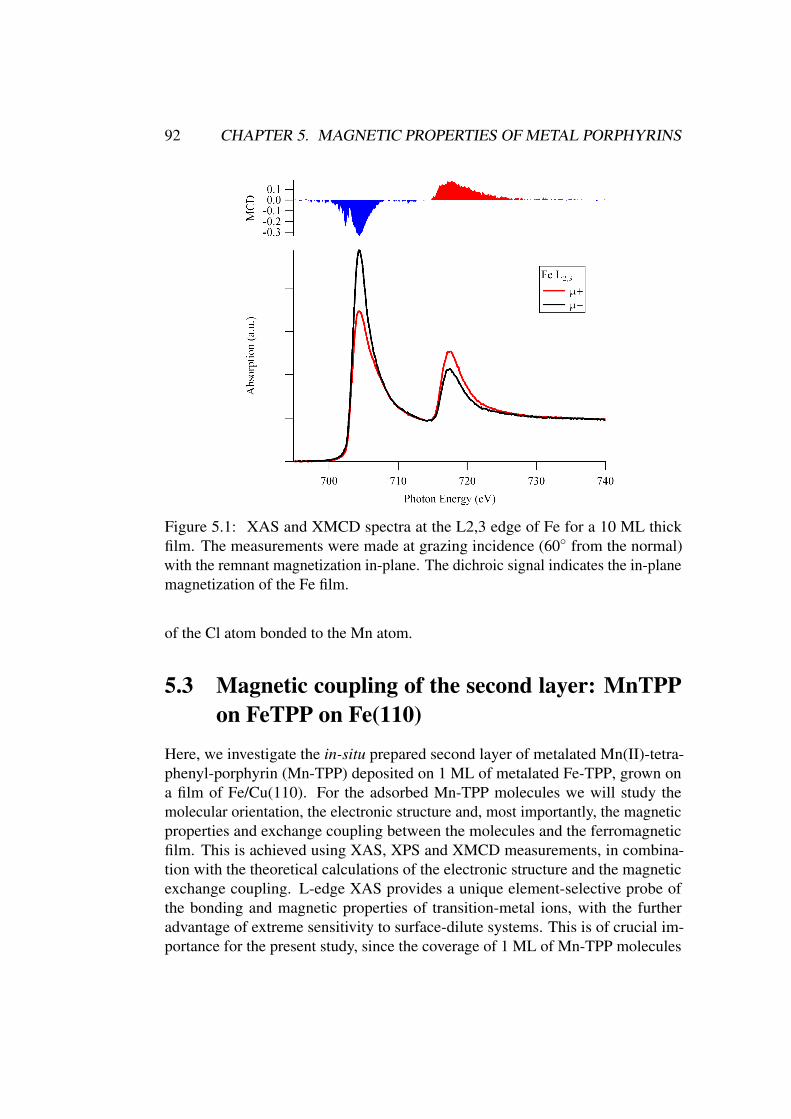
92 CHAPTER 5. MAGNETIC PROPERTIES OF METAL PORPHYRINS
Figure 5.1: XAS and XMCD spectra at the L2,3 edge of Fe for a 10 ML thickfilm. The measurements were made at grazing incidence (60◦ from the normal)with the remnant magnetization in-plane. The dichroic signal indicates the in-planemagnetization of the Fe film.
of the Cl atom bonded to the Mn atom.
5.3 Magnetic coupling of the second layer: MnTPP
on FeTPP on Fe(110)
Here, we investigate the in-situ prepared second layer of metalated Mn(II)-tetra-phenyl-porphyrin (Mn-TPP) deposited on 1 ML of metalated Fe-TPP, grown ona film of Fe/Cu(110). For the adsorbed Mn-TPP molecules we will study themolecular orientation, the electronic structure and, most importantly, the magneticproperties and exchange coupling between the molecules and the ferromagneticfilm. This is achieved using XAS, XPS and XMCD measurements, in combina-tion with the theoretical calculations of the electronic structure and the magneticexchange coupling. L-edge XAS provides a unique element-selective probe ofthe bonding and magnetic properties of transition-metal ions, with the furtheradvantage of extreme sensitivity to surface-dilute systems. This is of crucial im-portance for the present study, since the coverage of 1 ML of Mn-TPP molecules
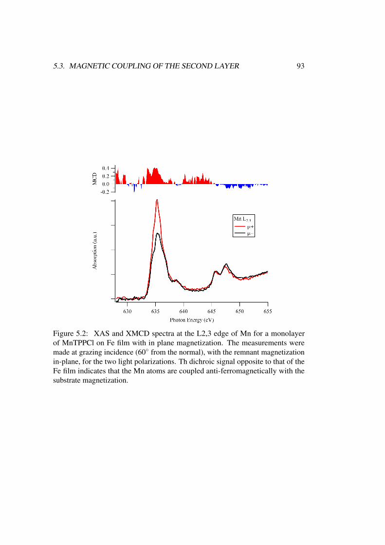
5.3. MAGNETIC COUPLING OF THE SECOND LAYER 93
Figure 5.2: XAS and XMCD spectra at the L2,3 edge of Mn for a monolayerof MnTPPCl on Fe film with in plane magnetization. The measurements weremade at grazing incidence (60◦ from the normal), with the remnant magnetizationin-plane, for the two light polarizations. Th dichroic signal opposite to that of theFe film indicates that the Mn atoms are coupled anti-ferromagnetically with thesubstrate magnetization.
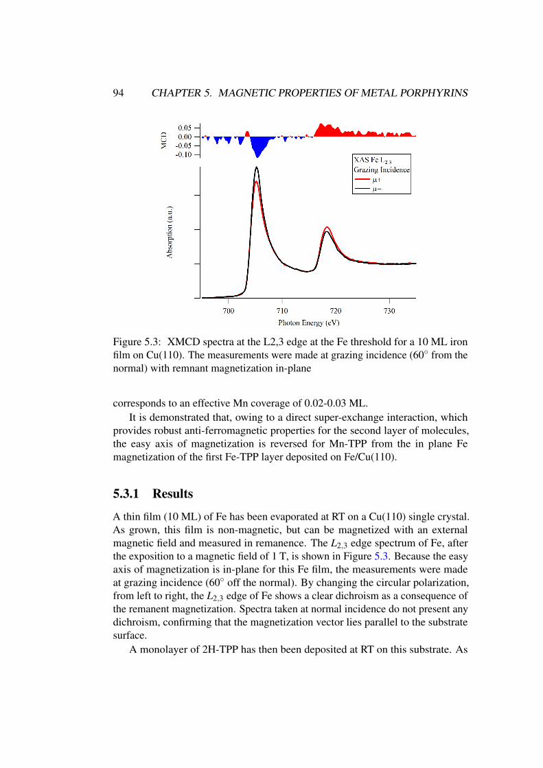
94 CHAPTER 5. MAGNETIC PROPERTIES OF METAL PORPHYRINS
Figure 5.3: XMCD spectra at the L2,3 edge at the Fe threshold for a 10 ML ironfilm on Cu(110). The measurements were made at grazing incidence (60◦ from thenormal) with remnant magnetization in-plane
corresponds to an effective Mn coverage of 0.02-0.03 ML.It is demonstrated that, owing to a direct super-exchange interaction, which
provides robust anti-ferromagnetic properties for the second layer of molecules,the easy axis of magnetization is reversed for Mn-TPP from the in plane Femagnetization of the first Fe-TPP layer deposited on Fe/Cu(110).
5.3.1 Results
A thin film (10 ML) of Fe has been evaporated at RT on a Cu(110) single crystal.As grown, this film is non-magnetic, but can be magnetized with an externalmagnetic field and measured in remanence. The L2,3 edge spectrum of Fe, afterthe exposition to a magnetic field of 1 T, is shown in Figure 5.3. Because the easyaxis of magnetization is in-plane for this Fe film, the measurements were madeat grazing incidence (60◦ off the normal). By changing the circular polarization,from left to right, the L2,3 edge of Fe shows a clear dichroism as a consequence ofthe remanent magnetization. Spectra taken at normal incidence do not present anydichroism, confirming that the magnetization vector lies parallel to the substratesurface.
A monolayer of 2H-TPP has then been deposited at RT on this substrate. As
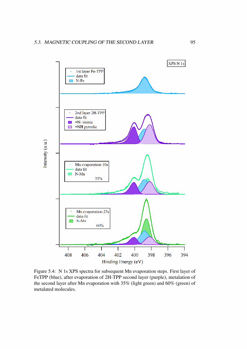
5.3. MAGNETIC COUPLING OF THE SECOND LAYER 95
Figure 5.4: N 1s XPS spectra for subsequent Mn evaporation steps. First layer ofFeTPP (blue), after evaporation of 2H-TPP second layer (purple), metalation ofthe second layer after Mn evaporation with 35% (light green) and 60% (green) ofmetalated molecules.
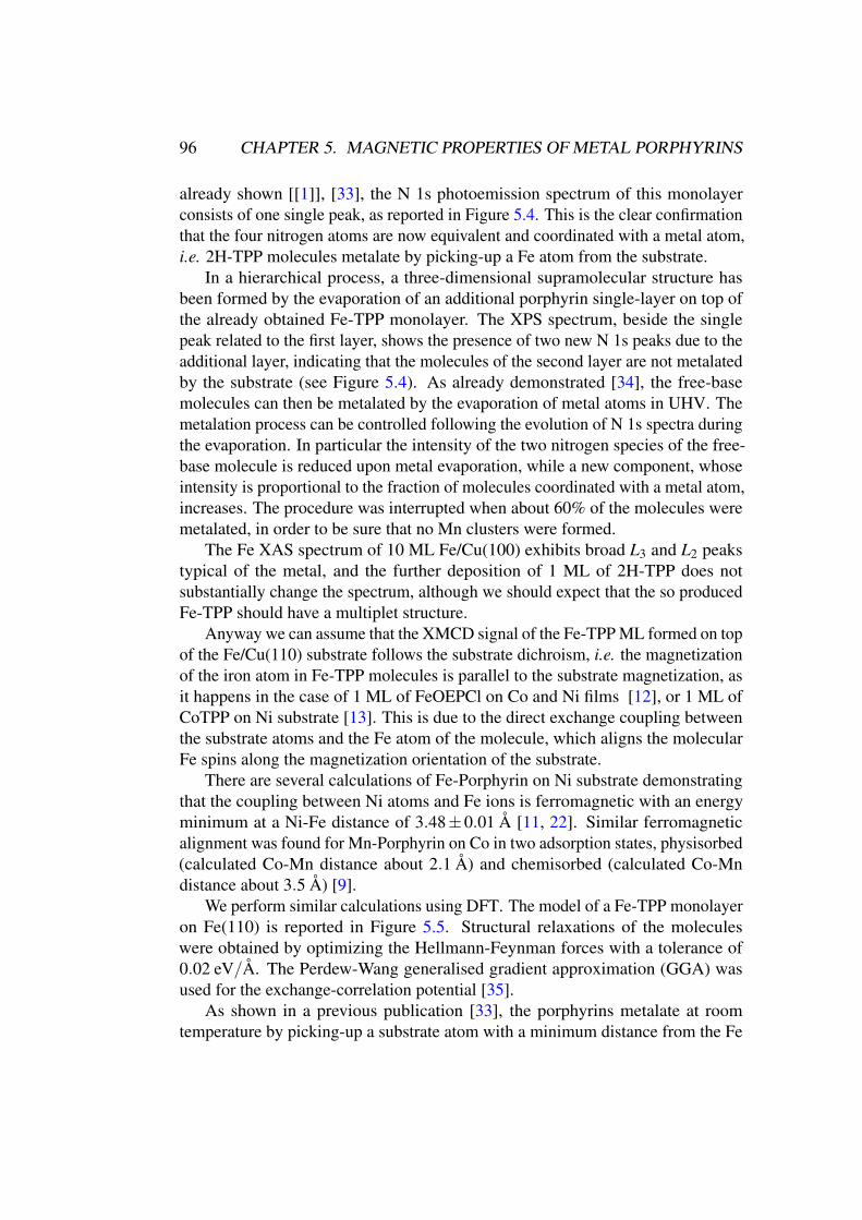
96 CHAPTER 5. MAGNETIC PROPERTIES OF METAL PORPHYRINS
already shown [[1]], [33], the N 1s photoemission spectrum of this monolayerconsists of one single peak, as reported in Figure 5.4. This is the clear confirmationthat the four nitrogen atoms are now equivalent and coordinated with a metal atom,i.e. 2H-TPP molecules metalate by picking-up a Fe atom from the substrate.
In a hierarchical process, a three-dimensional supramolecular structure hasbeen formed by the evaporation of an additional porphyrin single-layer on top ofthe already obtained Fe-TPP monolayer. The XPS spectrum, beside the singlepeak related to the first layer, shows the presence of two new N 1s peaks due to theadditional layer, indicating that the molecules of the second layer are not metalatedby the substrate (see Figure 5.4). As already demonstrated [34], the free-basemolecules can then be metalated by the evaporation of metal atoms in UHV. Themetalation process can be controlled following the evolution of N 1s spectra duringthe evaporation. In particular the intensity of the two nitrogen species of the free-base molecule is reduced upon metal evaporation, while a new component, whoseintensity is proportional to the fraction of molecules coordinated with a metal atom,increases. The procedure was interrupted when about 60% of the molecules weremetalated, in order to be sure that no Mn clusters were formed.
The Fe XAS spectrum of 10 ML Fe/Cu(100) exhibits broad L3 and L2 peakstypical of the metal, and the further deposition of 1 ML of 2H-TPP does notsubstantially change the spectrum, although we should expect that the so producedFe-TPP should have a multiplet structure.
Anyway we can assume that the XMCD signal of the Fe-TPP ML formed on topof the Fe/Cu(110) substrate follows the substrate dichroism, i.e. the magnetizationof the iron atom in Fe-TPP molecules is parallel to the substrate magnetization, asit happens in the case of 1 ML of FeOEPCl on Co and Ni films [12], or 1 ML ofCoTPP on Ni substrate [13]. This is due to the direct exchange coupling betweenthe substrate atoms and the Fe atom of the molecule, which aligns the molecularFe spins along the magnetization orientation of the substrate.
There are several calculations of Fe-Porphyrin on Ni substrate demonstratingthat the coupling between Ni atoms and Fe ions is ferromagnetic with an energyminimum at a Ni-Fe distance of 3.48±0.01 Å [11, 22]. Similar ferromagneticalignment was found for Mn-Porphyrin on Co in two adsorption states, physisorbed(calculated Co-Mn distance about 2.1 Å) and chemisorbed (calculated Co-Mndistance about 3.5 Å) [9].
We perform similar calculations using DFT. The model of a Fe-TPP monolayeron Fe(110) is reported in Figure 5.5. Structural relaxations of the moleculeswere obtained by optimizing the Hellmann-Feynman forces with a tolerance of0.02 eV/Å. The Perdew-Wang generalised gradient approximation (GGA) wasused for the exchange-correlation potential [35].
As shown in a previous publication [33], the porphyrins metalate at roomtemperature by picking-up a substrate atom with a minimum distance from the Fe
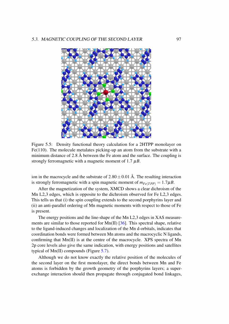
5.3. MAGNETIC COUPLING OF THE SECOND LAYER 97
Figure 5.5: Density functional theory calculation for a 2HTPP monolayer onFe(110). The molecule metalates picking-up an atom from the substrate with aminimum distance of 2.8 Å between the Fe atom and the surface. The coupling isstrongly ferromagnetic with a magnetic moment of 1.7 µB.
ion in the macrocycle and the substrate of 2.80±0.01 Å. The resulting interactionis strongly ferromagnetic with a spin magnetic moment of mFe(T PP) = 1.7µB.
After the magnetization of the system, XMCD shows a clear dichroism of theMn L2,3 edges, which is opposite to the dichroism observed for Fe L2,3 edges.This tells us that (i) the spin coupling extends to the second porphyrins layer and(ii) an anti-parallel ordering of Mn magnetic moments with respect to those of Feis present.
The energy positions and the line-shape of the Mn L2,3 edges in XAS measure-ments are similar to those reported for Mn(II) [36]. This spectral shape, relativeto the ligand-induced changes and localization of the Mn d-orbitals, indicates thatcoordination bonds were formed between Mn atoms and the macrocyclic N ligands,confirming that Mn(II) is at the centre of the macrocycle. XPS spectra of Mn2p core levels also give the same indication, with energy positions and satellitestypical of Mn(II) compounds (Figure 5.7).
Although we do not know exactly the relative position of the molecules ofthe second layer on the first monolayer, the direct bonds between Mn and Featoms is forbidden by the growth geometry of the porphyrins layers; a super-exchange interaction should then propagate through conjugated bond linkages,
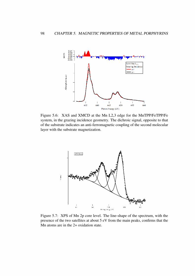
98 CHAPTER 5. MAGNETIC PROPERTIES OF METAL PORPHYRINS
Figure 5.6: XAS and XMCD at the Mn L2,3 edge for the MnTPP/FeTPP/Fesystem, in the grazing incidence geometry. The dichroic signal, opposite to thatof the substrate indicates an anti-ferromagnetic coupling of the second molecularlayer with the substrate magnetization.
Figure 5.7: XPS of Mn 2p core level. The line-shape of the spectrum, with thepresence of the two satellites at about 5 eV from the main peaks, confirms that theMn atoms are in the 2+ oxidation state.
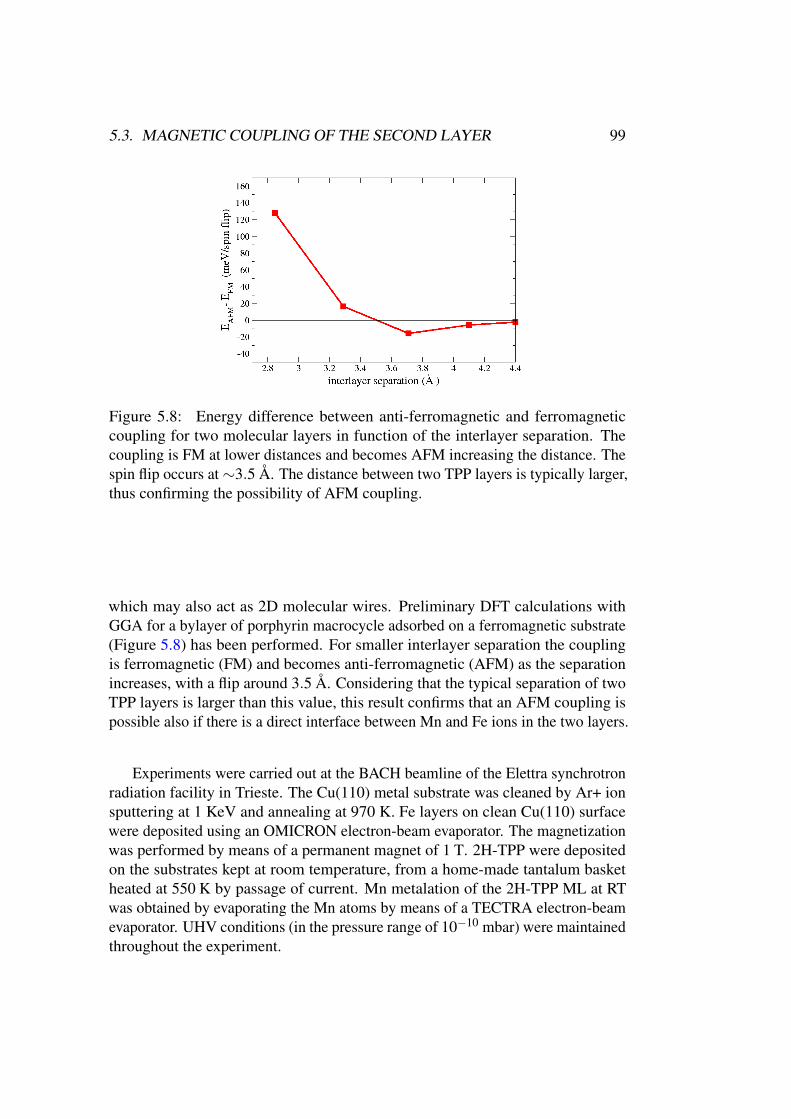
5.3. MAGNETIC COUPLING OF THE SECOND LAYER 99
Figure 5.8: Energy difference between anti-ferromagnetic and ferromagneticcoupling for two molecular layers in function of the interlayer separation. Thecoupling is FM at lower distances and becomes AFM increasing the distance. Thespin flip occurs at ∼3.5 Å. The distance between two TPP layers is typically larger,thus confirming the possibility of AFM coupling.
which may also act as 2D molecular wires. Preliminary DFT calculations withGGA for a bylayer of porphyrin macrocycle adsorbed on a ferromagnetic substrate(Figure 5.8) has been performed. For smaller interlayer separation the couplingis ferromagnetic (FM) and becomes anti-ferromagnetic (AFM) as the separationincreases, with a flip around 3.5 Å. Considering that the typical separation of twoTPP layers is larger than this value, this result confirms that an AFM coupling ispossible also if there is a direct interface between Mn and Fe ions in the two layers.
Experiments were carried out at the BACH beamline of the Elettra synchrotronradiation facility in Trieste. The Cu(110) metal substrate was cleaned by Ar+ ionsputtering at 1 KeV and annealing at 970 K. Fe layers on clean Cu(110) surfacewere deposited using an OMICRON electron-beam evaporator. The magnetizationwas performed by means of a permanent magnet of 1 T. 2H-TPP were depositedon the substrates kept at room temperature, from a home-made tantalum basketheated at 550 K by passage of current. Mn metalation of the 2H-TPP ML at RTwas obtained by evaporating the Mn atoms by means of a TECTRA electron-beamevaporator. UHV conditions (in the pressure range of 10−10 mbar) were maintainedthroughout the experiment.
100 REFERENCES
References
[1] Marco Affronte. Molecular nanomagnets for information technologies. Jour-
nal of Materials Chemistry, 19(12):1731, 2009. ISSN 0959-9428, 1364-5501.doi: 10.1039/b809251f.
[2] M Affronte, F Troiani, A Ghirri, A Candini, M Evangelisti, V Corradini,S Carretta, P Santini, G Amoretti, F Tuna, G Timco, and R E P Winpenny.Single molecule magnets for quantum computation. Journal of Physics
D: Applied Physics, 40:2999–3004, May 2007. ISSN 0022-3727. doi:10.1088/0022-3727/40/10/S01.
[3] Lapo Bogani and Wolfgang Wernsdorfer. Molecular spintronics using single-molecule magnets. Nature Materials, 7(3):179–186, March 2008. ISSN1476-1122, 1476-4660. doi: 10.1038/nmat2133.
[4] Pietro Gambardella, Sebastian Stepanow, Alexandre Dmitriev, Jan Honolka,Frank M. F. de Groot, Magalí Lingenfelder, Subhra Sen Gupta, D. D. Sarma,Peter Bencok, Stefan Stanescu, Sylvain Clair, Stéphane Pons, Nian Lin, Ari P.Seitsonen, Harald Brune, Johannes V. Barth, and Klaus Kern. Supramolecularcontrol of the magnetic anisotropy in two-dimensional high-spin fe arraysat a metal interface. Nature Materials, 8(3):189–193, March 2009. ISSN1476-1122, 1476-4660. doi: 10.1038/nmat2376.
[5] Christof Wöll, editor. Physical and Chemical Aspects of Organic Electronics.Wiley edition, 2009.
[6] A. Lodi Rizzini, C. Krull, T. Balashov, J. J. Kavich, A. Mugarza, P. S.Miedema, P. K. Thakur, V. Sessi, S. Klyatskaya, M. Ruben, S. Stepanow,and P. Gambardella. Coupling single molecule magnets to ferromagneticsubstrates. Physical Review Letters, 107(17), October 2011. ISSN 0031-9007,1079-7114. doi: 10.1103/PhysRevLett.107.177205.
[7] Klaus Baberschke. Magnetic switching of fe-porphyrin molecules adsorbedon surfaces: An XAFS and XMCD study. Journal of Physics: Conference
Series, 190:012012, November 2009. ISSN 1742-6596. doi: 10.1088/1742-6596/190/1/012012.
[8] M. Bernien, X. Xu, J. Miguel, M. Piantek, Ph. Eckhold, J. Luo, J. Kurde,W. Kuch, K. Baberschke, H. Wende, and P. Srivastava. Fe-porphyrin monolay-ers on ferromagnetic substrates: Electronic structure and magnetic couplingstrength. Physical Review B, 76(21), December 2007. ISSN 1098-0121,1550-235X. doi: 10.1103/PhysRevB.76.214406.
REFERENCES 101
[9] Md. Ehesan Ali, Biplab Sanyal, and Peter M. Oppeneer. Tuning the magneticinteraction between manganese porphyrins and ferromagnetic co substratethrough dedicated control of the adsorption. The Journal of Physical Chem-
istry C, 113(32):14381–14383, August 2009. ISSN 1932-7447, 1932-7455.doi: 10.1021/jp902644q.
[10] A. Scheybal, T. Ramsvik, R. Bertschinger, M. Putero, F. Nolting, and T.A.Jung. Induced magnetic ordering in a molecular monolayer. Chemical
Physics Letters, 411(1-3):214–220, August 2005. ISSN 00092614. doi:10.1016/j.cplett.2005.06.017.
[11] H. Wende, M. Bernien, J. Luo, C. Sorg, N. Ponpandian, J. Kurde, J. Miguel,M. Piantek, X. Xu, Ph. Eckhold, W. Kuch, K. Baberschke, P. M. Panchmatia,B. Sanyal, P. M. Oppeneer, and O. Eriksson. Substrate-induced magneticordering and switching of iron porphyrin molecules. Nature Materials, 6(7):516–520, July 2007. ISSN 1476-1122, 1476-4660. doi: 10.1038/nmat1932.
[12] M. Bernien, J. Miguel, C. Weis, Md. Ali, J. Kurde, B. Krumme, P. Panch-matia, B. Sanyal, M. Piantek, P. Srivastava, K. Baberschke, P. Oppeneer,O. Eriksson, W. Kuch, and H. Wende. Tailoring the nature of magneticcoupling of fe-porphyrin molecules to ferromagnetic substrates. Physical
Review Letters, 102(4), January 2009. ISSN 0031-9007, 1079-7114. doi:10.1103/PhysRevLett.102.047202.
[13] Christian Wäckerlin, Dorota Chylarecka, Armin Kleibert, Kathrin Müller,Cristian Iacovita, Frithjof Nolting, Thomas A. Jung, and Nirmalya Ballav.Controlling spins in adsorbed molecules by a chemical switch. Na-
ture Communications, 1(5):1–7, August 2010. ISSN 2041-1723. doi:10.1038/ncomms1057.
[14] Dorota Chylarecka, Christian Wäckerlin, Timur K. Kim, Kathrin Müller,Frithjof Nolting, Armin Kleibert, Nirmalya Ballav, and Thomas A. Jung.Self-assembly and superexchange coupling of magnetic molecules on oxygen-reconstructed ferromagnetic thin film. The Journal of Physical Chemistry
Letters, 1(9):1408–1413, May 2010. ISSN 1948-7185, 1948-7185. doi:10.1021/jz100253c.
[15] D. Chylarecka, T. K. Kim, K. Tarafder, K. Müller, K. Gödel, I. Czekaj,C. Wäckerlin, M. Cinchetti, Md. E. Ali, C. Piamonteze, F. Schmitt, J.-P. Wüstenberg, C. Ziegler, F. Nolting, M. Aeschlimann, P. M. Oppeneer,N. Ballav, and T. A. Jung. Indirect magnetic coupling of manganese porphyrinto a ferromagnetic cobalt substrate †$. The Journal of Physical Chemistry
102 REFERENCES
C, 115(4):1295–1301, February 2011. ISSN 1932-7447, 1932-7455. doi:10.1021/jp106822s.
[16] E. Annese, F. Casolari, J. Fujii, and G. Rossi. Interface magnetic coupling offe-phthalocyanine layers on a ferromagnetic surface. Physical Review B, 87(5), feb 2013. doi: 10.1103/physrevb.87.054420.
[17] Nirmalya Ballav, Christian Wäckerlin, Dorota Siewert, Peter M. Oppeneer,and Thomas A. Jung. Emergence of on-surface magnetochemistry. The
Journal of Physical Chemistry Letters, 4(14):2303–2311, July 2013. ISSN1948-7185. doi: 10.1021/jz400984k.
[18] Charles Michael Drain, James D. Batteas, George W. Flynn, Tatjana Milic,Ning Chi, Dalia G. Yablon, and Heather Sommers. Designing supramolecu-lar porphyrin arrays that self-organize into nanoscale optical and magneticmaterials. Proceedings of the National Academy of Sciences of the United
States of America, 99(Suppl 2):6498–6502, 2002.
[19] Ken Flechtner, Andreas Kretschmann, Hans-Peter Steinrück, and J. MichaelGottfried. NO-induced reversible switching of the electronic interactionbetween a porphyrin-coordinated cobalt ion and a silver surface. J. Am. Chem.
Soc., 129(40):12110–12111, oct 2007. doi: 10.1021/ja0756725.
[20] Keitaro Eguchi, Yasumasa Takagi, Takeshi Nakagawa, and ToshihikoYokoyama. Magnetic interactions of vanadyl phthalocyanine with ferro-magnetic iron, cobalt, and nickel surfaces. J. Phys. Chem. C, 118(31):17633–17637, aug 2014. doi: 10.1021/jp503851k.
[21] Cornelius Krull, Roberto Robles, Aitor Mugarza, and Pietro Gambardella.Site- and orbital-dependent charge donation and spin manipulation in electron-doped metal phthalocyanines. Nature Materials, 12(4):337–343, January2013. ISSN 1476-1122, 1476-4660. doi: 10.1038/nmat3547.
[22] P.M. Oppeneer, P.M. Panchmatia, B. Sanyal, O. Eriksson, and Md.E. Ali.Nature of the magnetic interaction between fe-porphyrin molecules and ferro-magnetic surfaces. Progress in Surface Science, 84(1-2):18–29, March 2009.ISSN 00796816. doi: 10.1016/j.progsurf.2008.12.001.
[23] Sebastian Stepanow, Alberto Lodi Rizzini, Cornelius Krull, Jerald Kavich,Julio C. Cezar, Flora Yakhou-Harris, Polina M. Sheverdyaeva, Paolo Moras,Carlo Carbone, Gustavo Ceballos, Aitor Mugarza, and Pietro Gambardella.Spin tuning of electron-doped metal–phthalocyanine layers. Journal of the
American Chemical Society, 136(14):5451–5459, April 2014. ISSN 0002-7863, 1520-5126. doi: 10.1021/ja501204q.
REFERENCES 103
[24] Christian Wäckerlin, Kartick Tarafder, Dorota Siewert, Jan Girovsky, TatjanaHählen, Cristian Iacovita, Armin Kleibert, Frithjof Nolting, Thomas A. Jung,Peter M. Oppeneer, and Nirmalya Ballav. On-surface coordination chemistryof planar molecular spin systems: novel magnetochemical effects inducedby axial ligands. Chemical Science, 3(11):3154, 2012. ISSN 2041-6520,2041-6539. doi: 10.1039/c2sc20828h.
[25] A. P. Weber, A. N. Caruso, E. Vescovo, Md. E. Ali, K. Tarafder, S. Z. Janjua,J. T. Sadowski, and P. M. Oppeneer. Magnetic coupling of fe-porphyrinmolecules adsorbed on clean and c(2×2) oxygen-reconstructed co(100) in-vestigated by spin-polarized photoemission spectroscopy. Physical Review
B, 87(18), May 2013. ISSN 1098-0121, 1550-235X. doi: 10.1103/Phys-RevB.87.184411.
[26] Johannes V. Barth, Giovanni Costantini, and Klaus Kern. Engineering atomicand molecular nanostructures at surfaces. Nature, 437(7059):671–679, sep2005. doi: 10.1038/nature04166.
[27] Taku Suzuki, Mitsunori Kurahashi, and Yasushi Yamauchi. Spin polarizationin molecular orbitals of copperphthalocyanine deposited on a magnetizedfe(100) substrate. The Journal of Physical Chemistry B, 106(31):7643–7646,August 2002. ISSN 1520-6106, 1520-5207. doi: 10.1021/jp0204760.
[28] C. Iacovita, M. Rastei, B. Heinrich, T. Brumme, J. Kortus, L. Limot, andJ. Bucher. Visualizing the spin of individual cobalt-phthalocyanine molecules.Physical Review Letters, 101(11), September 2008. ISSN 0031-9007, 1079-7114. doi: 10.1103/PhysRevLett.101.116602.
[29] S. Javaid, M. Bowen, S. Boukari, L. Joly, J.-B. Beaufrand, Xi Chen, Y. J.Dappe, F. Scheurer, J.-P. Kappler, J. Arabski, W. Wulfhekel, M. Alouani, andE. Beaurepaire. Impact on interface spin polarization of molecular bondingto metallic surfaces. Physical Review Letters, 105(7), August 2010. ISSN0031-9007, 1079-7114. doi: 10.1103/PhysRevLett.105.077201.
[30] Giovanni Di Santo, Cristina Sfiligoj, Carla Castellarin-Cudia, Alberto Verdini,Albano Cossaro, Alberto Morgante, Luca Floreano, and Andrea Goldoni.Changes of the molecule-substrate interaction upon metal inclusion into aporphyrin. Chemistry - A European Journal, (18):12619–23, 2012. doi:10.1002/chem.201201640.
[31] Peter L. Cook, Xiaosong Liu, Wanli Yang, and F. J. Himpsel. X-ray absorption spectroscopy of biomimetic dye molecules for solar
104 REFERENCES
cells. The Journal of Chemical Physics, 131(19):194701–, 2009. doi:http://dx.doi.org/10.1063/1.3257621.
[32] Wolfgang Hieringer, Ken Flechtner, Andreas Kretschmann, Knud Seufert,Willi Auwärter, Johannes V. Barth, Andreas Görling, Hans-Peter Steinrück,and J. Michael Gottfried. The surface trans effect: Influence of axial ligandson the surface chemical bonds of adsorbed metalloporphyrins. Journal of
the American Chemical Society, 133(16):6206–6222, April 2011. ISSN0002-7863, 1520-5126. doi: 10.1021/ja1093502.
[33] Andrea Goldoni, Carlo A. Pignedoli, Giovanni Di Santo, Carla Castellarin-Cudia, Elena Magnano, Federica Bondino, Alberto Verdini, and DanielePasserone. Room temperature metalation of 2h-TPP monolayer on ironand nickel surfaces by picking up substrate metal atoms. ACS nano, 6(12):10800–7, 2012. doi: 10.1021/nn304134q.
[34] Giovanni Di Santo, Carla Castellarin-Cudia, Mattia Fanetti, Bidini Taleatu,Patrizia Borghetti, Luigi Sangaletti, Luca Floreano, Elena Magnano, Fed-erica Bondino, and Andrea Goldoni. Conformational adaptation and elec-tronic structure of 2h-tetraphenylporphyrin on ag(111) during fe metala-tion. The Journal of Physical Chemistry C, 115(10):4155–62, 2011. doi:10.1021/jp111151n.
[35] John P. Perdew and Yue Wang. Accurate and simple analytic representation ofthe electron-gas correlation energy. Physical Review B, 45(23):13244–13249,June 1992. ISSN 0163-1829, 1095-3795. doi: 10.1103/PhysRevB.45.13244.
[36] Peter L. Cook, Xiaosong Liu, Wanli Yang, and F. J. Himpsel. X-ray ab-sorption spectroscopy of biomimetic dye molecules for solar cells. The
Journal of Chemical Physics, 131(19):194701, 2009. ISSN 00219606. doi:10.1063/1.3257621.
Chapter 6
Conclusions and Outlook
The interaction between tetra-phenyl-porphyrins and the substrate is stronglyrelated to their adsorption geometry on the surface. In Chapter 3 we have reportedhow these molecules are able to self-assembly on crystal surfaces producing long-range ordered structures. This is an interesting feature considering the ability ofthese molecules to coordinate to several metal ions: for example, it allows thefabrication of regular and equally spaced arrays of single metallic atoms on thesurface with a lateral extension of the order of few nanometres, which is otherwisenot achievable with conventional methods.
Moreover, the flexibility in the molecular chemical synthesis allows to tailor thestructure of these molecules, by the addition of functional groups able to introducenew useful properties. The notable effect exerted by the presence of the phenylrings in tetra-phenyl-porphyrins, beside its fundamental spacing action both fromthe surface and between the molecules, reveals in the conformational adaptationthat the molecule undergoes upon annealing. The rotation of the phenyl ringstowards the surface drives an essential modification of the chemical compositionof the molecule, as proven in the case of TPP adsorbed on Au(111) and Ag(111).
The main mechanisms involved in this interactions are the hybridization ofmolecular states upon adsorption and the charge transfer from the substrate. Thecoordination of the molecule macrocycle with a metal atom strongly affects thesemechanisms. As reported in Chapter 4, metal evaporation and picking-up ofa substrate atom are the methods that allow the creation of metallo-porphyrinson metal substrates. Moreover, the mechanism of molecule metalation and theformation of metal-organic complexes is in turn determined by the orientationof the meso-substituents in tetra-phenyl-porphyrins. This essentially changesthe molecule-substrate interaction as demonstrated in the case of TPP metalationwith Co, for which the correlation between the presence of the metal ion and thegeometry and conformational adaptation of the molecule is investigated.
As a consequence of the metalation process, the coordination of the macrocycle
105
106 CHAPTER 6. CONCLUSIONS AND OUTLOOK
with a metal ion can bring about a magnetic moment to the molecule. In Chapter 5it is studied the magnetic coupling of a single layer of TPP on a ferromagnetic sub-strate. Moreover, taking advantage of the molecular self-assembly, we have beenable to form a double layer square planar array of porphyrins at room temperature.Although individual molecules cannot be magnetized independently, this approachenables the fabrication of metal-organic molecular layers with stable magnetizationat room temperature.
Despite the experimental and theoretical challenges, the creation of devicesbased on organic molecules is undoubtedly promising, while the research efforts inthis sense are definitely of fundamental importance for a deeper comprehension ofthe world at the nano-scale.
Appendix A
List of Publications
Journal Articles
This thesis is based on part of the following peer-reviewed articles, which arereferred to in the text by double square brackets:
[[1]] M. Panighel, G. Di Santo, M. Caputo, A. Goldoni. Review of 2H-tetra-
phenyl-porphyrins metalation in ultra-high vacuum on metal surfaces.Journal of Physics: Conference Series 470, 012012 (2013). doi: 10.1088/1742-6596/470/1/012012
[[2]] M. Marsili, P. Umari, G. Di Santo, M. Caputo, M. Panighel, A. Goldoni,M. Kumar, M. Pedio. Solid state effects on the electronic structure of
H2OEP. Phys. Chem. Chem. Phys. 16, 27104 (2014) doi: 10.1039/c4cp03450c
[[3]] C. Struzzi, N.I. Verbitskiy, A.V. Fedorov, A. Nefedov, O. Frank, M. Kalbac,G. Di Santo, M. Panighel, A. Goldoni, J. Gärtner, W. Weber, M. Weinl,M. Schreck, Ch. Wöll, H. Sachdev, A. Grüneis, L. Petaccia. A multi-
spectroscopic and microscopic characterization of graphene on single
crystal Ir(111) films on Si(111) wafers. Carbon 81, 167 (2015)
doi: 10.1016/j.carbon.2014.09.045
[[4]] M. Caputo, M. Panighel, L. Petaccia, G. Di Santo, C. Struzzi, A. Goldoni.Metallic picene/C60 heterojunctions let continuous alkali metal dop-
ing of C60. Phys. Rev. B 90, 201401(R) (2014) doi: 10.1103/Phys-RevB.90.201401
[[5]] M. Caputo, M. Panighel, G. Di Santo, C. Struzzi, P. Parisse, L. Petaccia,L. Floreano, A. Verdini, B. Taleatu, C. Lal, A. Goldoni. Experimental
107
108 APPENDIX A. LIST OF PUBLICATIONS
study of pristine and alkali metal doped picene layers: confirmation of
the insulating phase in multilayer doped compounds. The Journal of
Physical Chemistry C 116, 19902 (2012). doi: 10.1021/jp306640z
[[6]] E. Ferrari, L. Galli, E. Miniussi, M. Morri , M. Panighel, M. Ricci, P. La-covig, S. Lizzit, A. Baraldi. Layer-dependent Debye temperature of
Ru(0001) by means of high-energy resolution core-level photoelectron
spectroscopy. Physical Review B 82, 195420 (2010). doi: 10.1103/Phys-RevB.82.195420
Other publications, based on the results of this thesis, are in preparation:
[[9]] M. Panighel, G. Di Santo, A. Goldoni. Drift corrections of scanning probe
microscopy images by Fourier analysis. (Section 2.1.2)
[[10]] M. Panighel, M. Caputo, G. Di Santo, L. Floreano, A. Verdini, A. Cossaro,A. Goldoni. Conformational adaptation of 2H-tetra-phenyl-porphyrin
on Au(111). (Section 3.3)
[[11]] G. Zamborlini, G. Di Santo, A. Goldoni, M. Panighel, V. Feyer, C. M. Schnei-der. Highly oriented self-assembly domains of Co and Ni-TPP on the Cu
(100) surface: multitechnique approach to study the electronic and geo-
metric structure. (Section 3.2.2)
[[12]] M. Panighel, M. Caputo, G. Di Santo, L. Floreano, A. Verdini, A. Cossaro, A.Goldoni. Conformational adaptation and metalation of 2H-tetra-phenyl-
porphyrin on Al(111). (Section 4.3.2)
[[13]] A. Goldoni, M. Panighel, M. Caputo, G. Di Santo, C. Castellarin-Cudia,M. Fanetti, F. Bondino, E. Magnano, N. Stojic, N. Binggeli. Anti-parallel
magnetic coupling of manganese tetra-phenyl-porphyrin to a ferromag-
netic iron substrate. (Section 5.2)
[[14]] M. Panighel, M. Caputo, G. Di Santo, C. Castellarin-Cudia, M. Fanetti,F. Bondino, E. Magnano, N. Stojic, N. Binggeli, A. Goldoni. Changes in
remanent magnetization by designing supramolecular metalated por-
phyrins arrays on 3d magnetic substrates. (Section 5.3)
Appendix B
Symbols and Abbreviations
2H-TPP 2H-5,10,15,20-tetra-phenyl-porphyrin
2H-OEP 2H-octa-ethyl-porphyrin
AFM anti-ferromagnetic
BE binding energy
CoTPP Co(II)-tetra-phenyl-porphyrin
DFT density functional theory
FeOEPCl Fe(III)-octa-ethyl-porphyrin-Cl
Fe-TPP Fe(II)-tetra-phenyl-porphyrin
FFT fast Fourier transform
FM ferromagnetic
HOMO highest occupied molecular orbital
LDOS local density of states
ML monolayer
Mn-TPP Mn(II)-tetra-phenyl-porphyrin
MnTPPCl Mn(III)-tetra-phenyl-porphyrin-Cl
NEXAFS Near-edge X-ray absorption fine structure
NiTPP Ni(II)-tetra-phenyl-porphyrin
109

110 APPENDIX B. SYMBOLS AND ABBREVIATIONS
RT room temperature
RuTPP Ru(II)-tetra-phenyl-porphyrin
TPP tetra-phenyl-porphyrin
UHV ultra high vacuum
STM scanning tunnelling microscopy
VB valence band
XAS X-ray absorption spectroscopy
XMCD X-ray magnetic circular dichroism
XPS X-ray photoelectron spectroscopy
Acknowledgements
The completion of my PhD would have never been possible without the support ofa number of people during these years.
My first thank goes to my PhD supervisor Alessandro Baraldi and my tutorAndrea Goldoni for the help and support they gave me during this time. I wouldespecially thank all my friends and, former and present, colleagues at Elettraand at the Micro and Nano Carbon Lab, in particular Giovanni Di Santo, MarcoCaputo, Lorenzo D’Arsiè, Giacomo Lovat, Claudia Struzzi, Cinzia Cepek andSilvio Modesti.
A special thanks goes to the staff of ALOISA, BACH and SuperESCA, andalso BAdElPh and Nanospectroscopy beamlines at Elettra for their support and thetime shared together.
In addition, I would like to acknowledge the contribution of Nadia Binggeli andNatasa Stojic and their group at ICTP for the state-of-the-art theoretical calculationscomplementing the experimental results of this thesis.
During my PhD, I had the opportunity to spend six months at the Institut Catalàde Nanociència i Nanotecnologia (ICN2) in Spain. I would especially like to thankmy tutor Aitor Mugarza and Gustavo Ceballos, Cornelius Krull, Stefano Schirone,Michele Gastaldo, Marc Maymò and Sylvie Godey for the very pleasant timetogether.
Finally, I want to thank my parents and my family, who sustained and allowedme in going through this path. A very special thanks goes to Chiara for his love,care and support along all these years together.
111