Adrenal glands
25
-
Upload
senzela-injilai -
Category
Documents
-
view
127 -
download
2
Transcript of Adrenal glands

Prepared by
Faiza Hameed Jan
The Adrenal glands
Adrenal glands
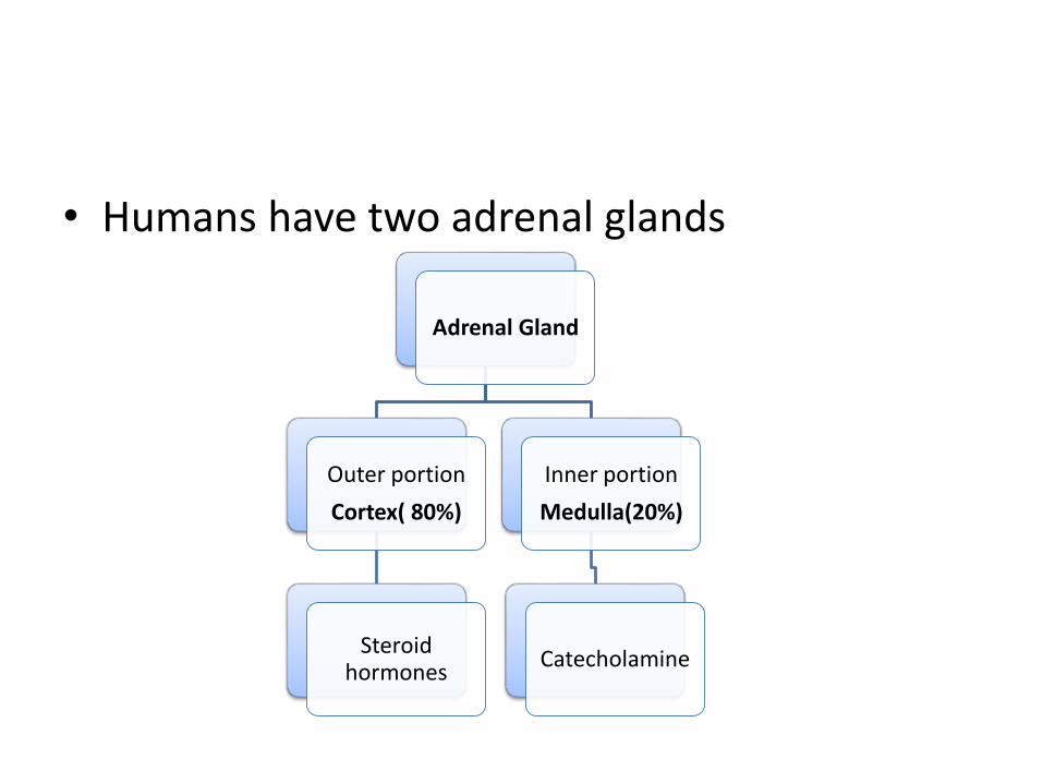
• Humans have two adrenal glands
Adrenal Gland
Outer portion
Cortex( 80%)
Steroid hormones
Inner portion
Medulla(20%)
Catecholamine
Anatomy of Adrenal gland
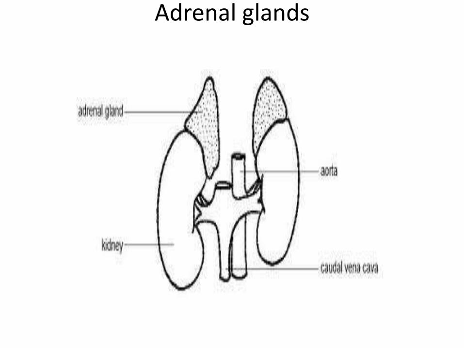
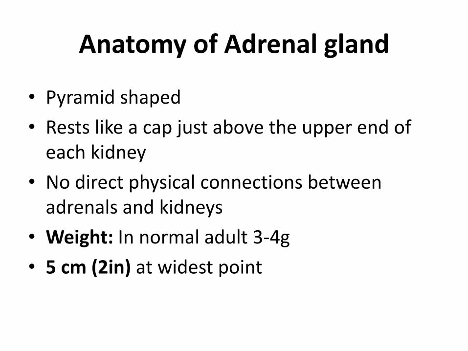
• Pyramid shaped
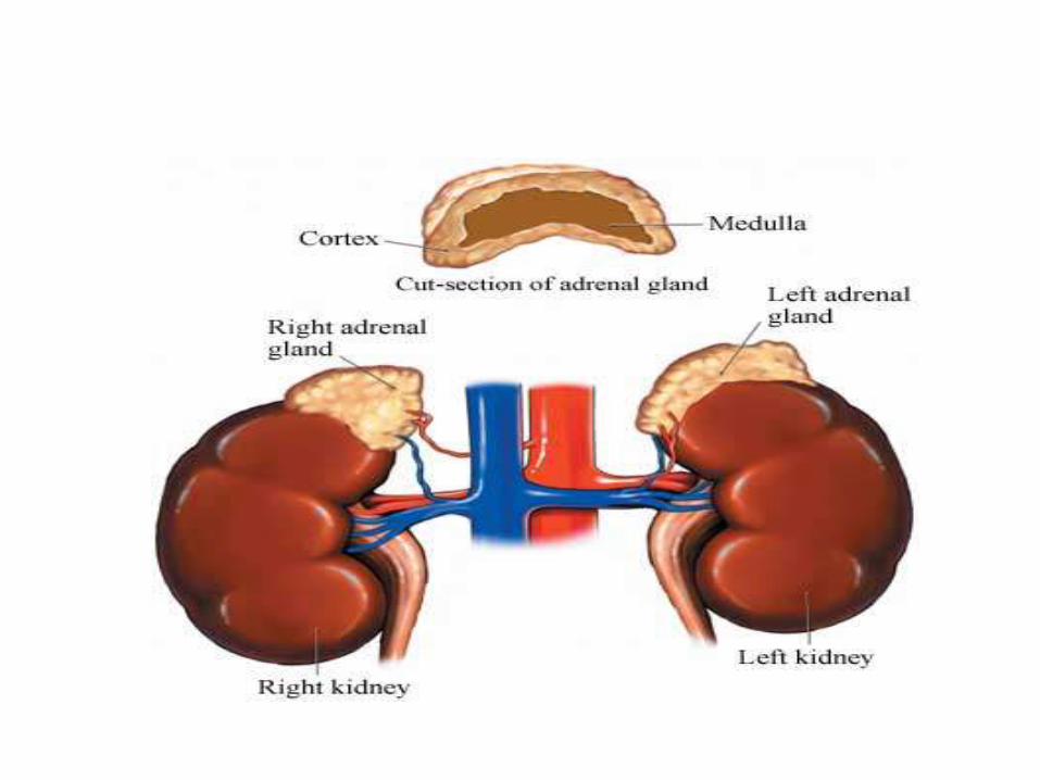
• Rests like a cap just above the upper end of each kidney
• No direct physical connections between adrenals and kidneys
• Weight: In normal adult 3-4g
• 5 cm (2in) at widest point
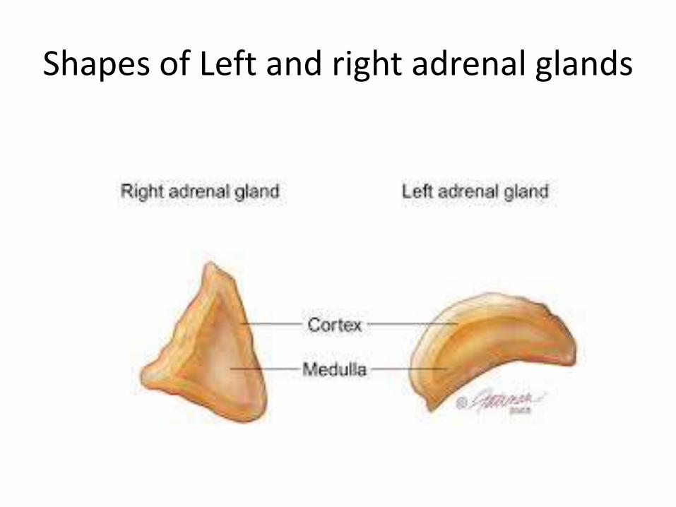
Shapes of Left and right adrenal glands
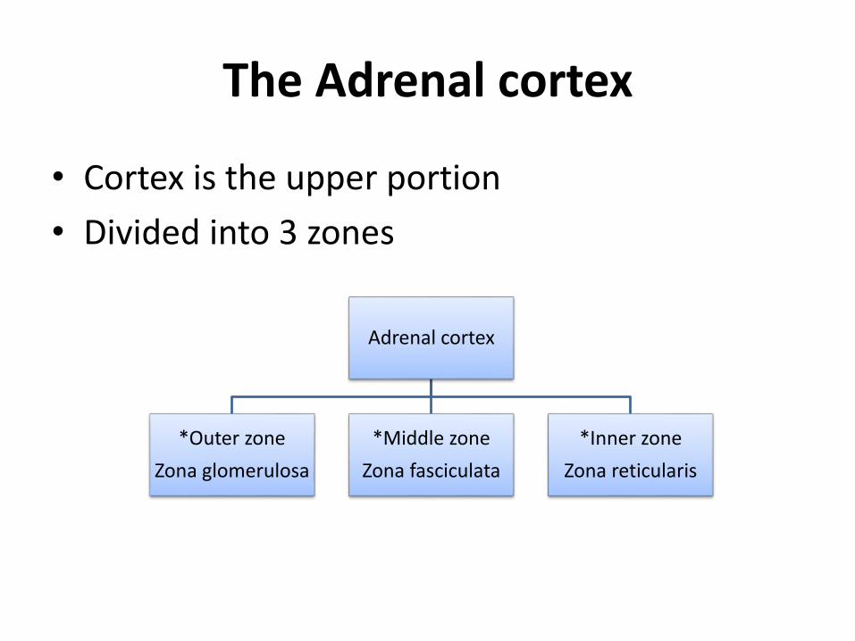
The Adrenal cortex
• Cortex is the upper portion
• Divided into 3 zones
Adrenal cortex
*Outer zone
Zona glomerulosa
*Middle zone
Zona fasciculata
*Inner zone
Zona reticularis
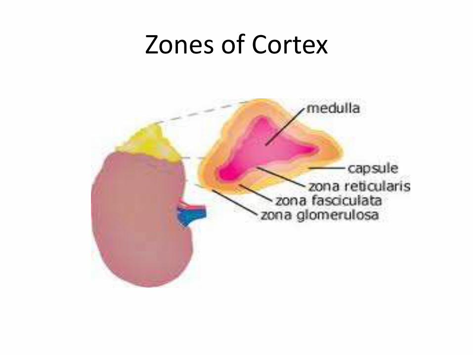
Zones of Cortex
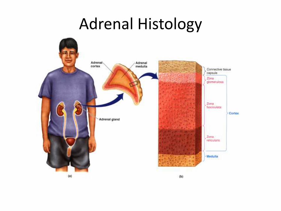
Adrenal Histology
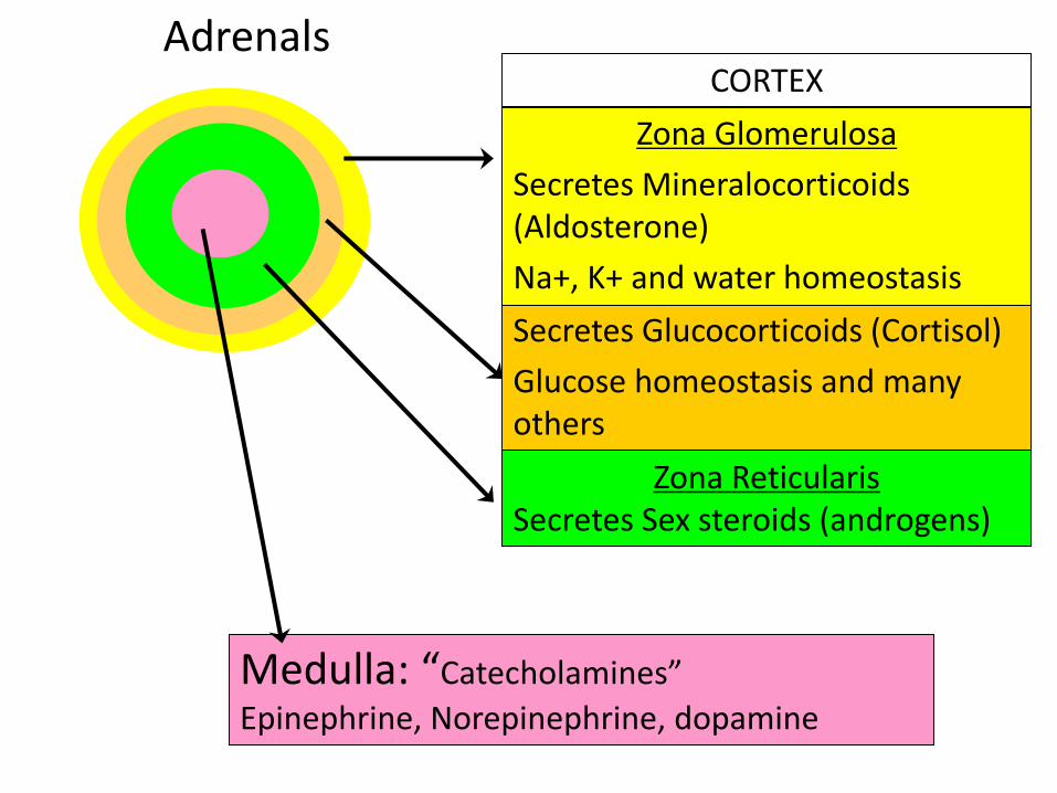
Adrenals
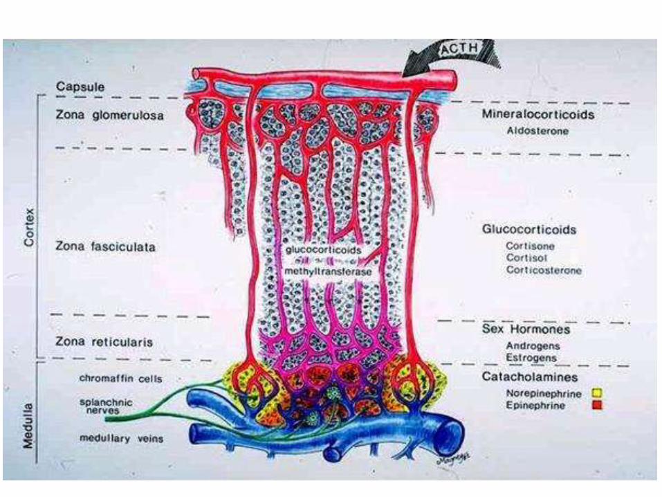
Zona ReticularisSecretes Sex steroids (androgens)
Zona FasciculataSecretes Glucocorticoids (Cortisol)
Glucose homeostasis and many others
Zona Glomerulosa
Secretes Mineralocorticoids(Aldosterone)
Na+, K+ and water homeostasis
Medulla: “Catecholamines”
Epinephrine, Norepinephrine, dopamine
CORTEX
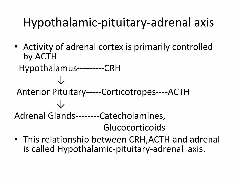
Hypothalamic-pituitary-adrenal axis
• Activity of adrenal cortex is primarily controlled by ACTH
Hypothalamus---------CRH↓
Anterior Pituitary-----Corticotropes----ACTH↓
Adrenal Glands--------Catecholamines, Glucocorticoids
• This relationship between CRH,ACTH and adrenal is called Hypothalamic-pituitary-adrenal axis.
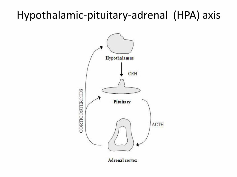
Hypothalamic-pituitary-adrenal (HPA) axis
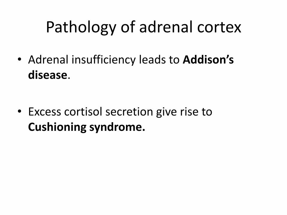
Pathology of adrenal cortex
• Adrenal insufficiency leads to Addison’sdisease.
• Excess cortisol secretion give rise to Cushioning syndrome.
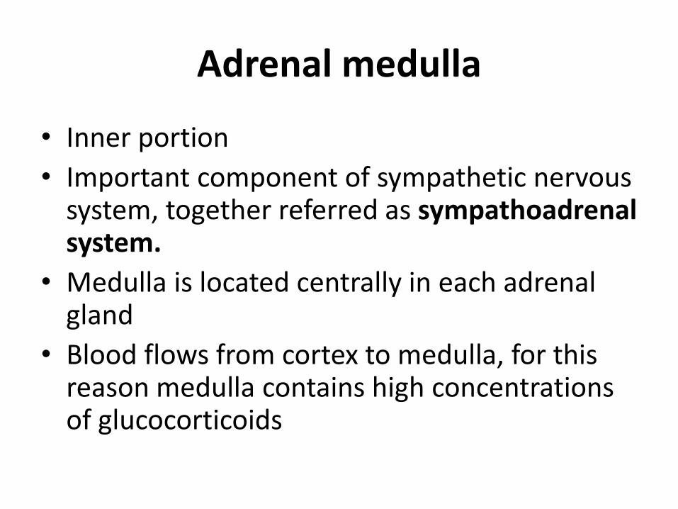
Adrenal medulla
• Inner portion
• Important component of sympathetic nervous system, together referred as sympathoadrenalsystem.
• Medulla is located centrally in each adrenal gland
• Blood flows from cortex to medulla, for this reason medulla contains high concentrations of glucocorticoids
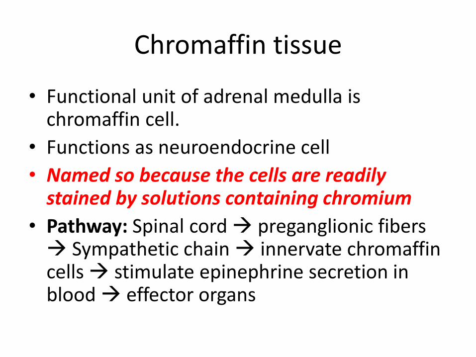
Chromaffin tissue
• Functional unit of adrenal medulla is chromaffin cell.
• Functions as neuroendocrine cell
• Named so because the cells are readily stained by solutions containing chromium
• Pathway: Spinal cord preganglionic fibers Sympathetic chain innervate chromaffincells stimulate epinephrine secretion in blood effector organs
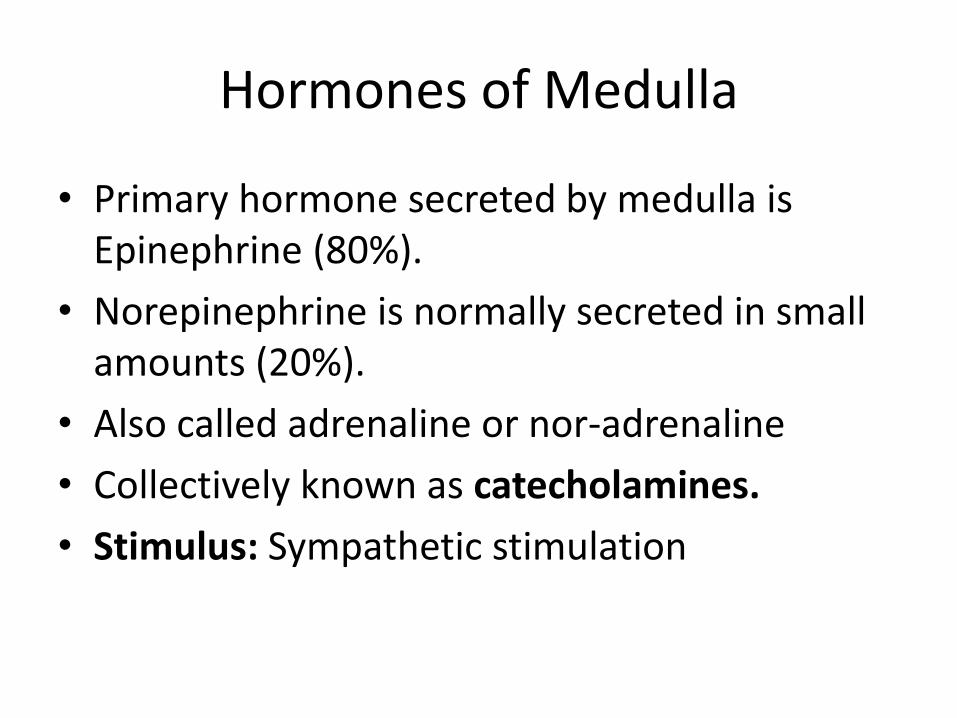
Hormones of Medulla
• Primary hormone secreted by medulla is Epinephrine (80%).
• Norepinephrine is normally secreted in small amounts (20%).
• Also called adrenaline or nor-adrenaline
• Collectively known as catecholamines.
• Stimulus: Sympathetic stimulation
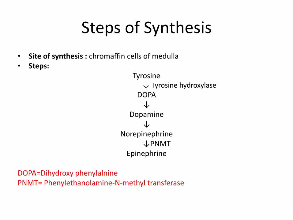
Steps of Synthesis
• Site of synthesis : chromaffin cells of medulla • Steps:
Tyrosine↓ Tyrosine hydroxylase
DOPA↓
Dopamine↓
Norepinephrine↓PNMT
Epinephrine
DOPA=Dihydroxy phenylalninePNMT= Phenylethanolamine-N-methyl transferase
• Epinephrine is then packaged and cocentratedwithin chromaffin cells into dense membrane bound vesicle called chromaffin granules.
• Chromaffin then release epinephrine form granules into blood.
Actions of Epinephrine
• Same actions as sympathetic nervous system.
• Triggers responses appropriate to “fight or flight” situations.
• Effects on cardiovascular system
Increases cardiac output
Increases heart rate
Increases strenght of cardiac contractions
Vasodilation in skeletal muscles
Vasoconstriction in skin and visceral organs ie.GIT.
• Effects on CNS
Increases mental alertness (advantageous in life-threatening situation.
• Effects on tissues
Relaxes smooth muscles of GIT, urinary bladder, airways of lung.
• Effects on metabolism
Increases glycogenolysis in liver and muscles
Increases lipolysis in adipose tissues (conversion of TAGs into fattyacids and glycerol).
Decreses insulin secretion.
Increases glucagon secretion.
In short, epinephrine increases body capability to perform vigorous muscle activity.

The End