Adenovirus Microbiology
32
ADENOVIRUS
Transcript of Adenovirus Microbiology
ADENOVIRUS
Introduction:Adenoviruses can replicate and produce disease in
respiratory, gastrointestinal and urinary tract and in the eye.
Many adenovirus infection are subclinical and virus persist in hosts for months.
One third of 51 serotypes are responsible for most cases of human adenovirus disease.
Whereas a few types serve as models for cancer induction in animal models.
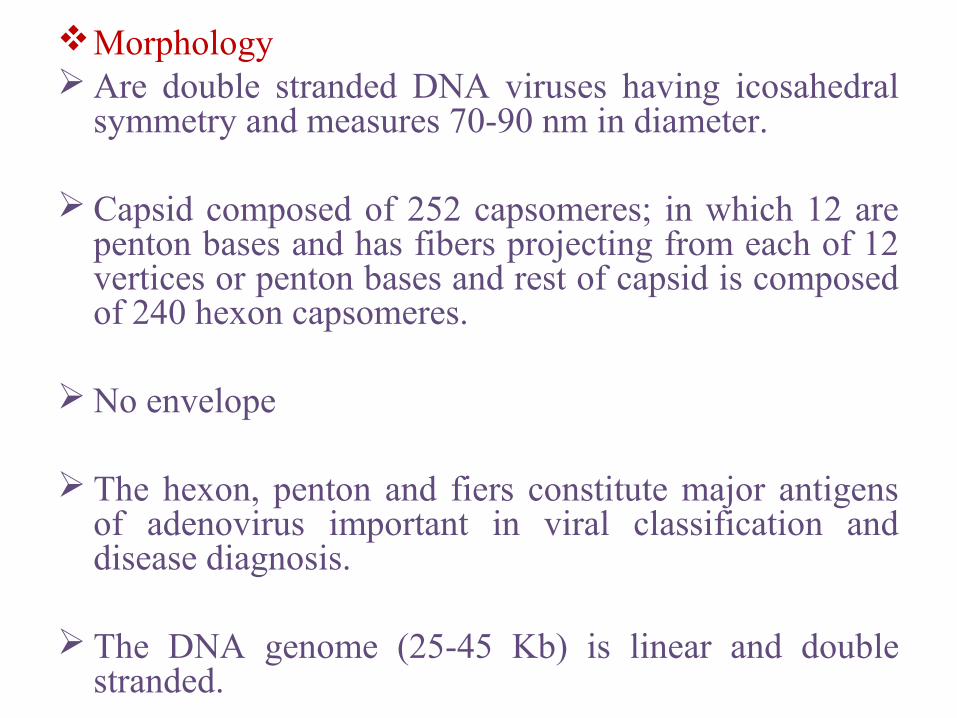
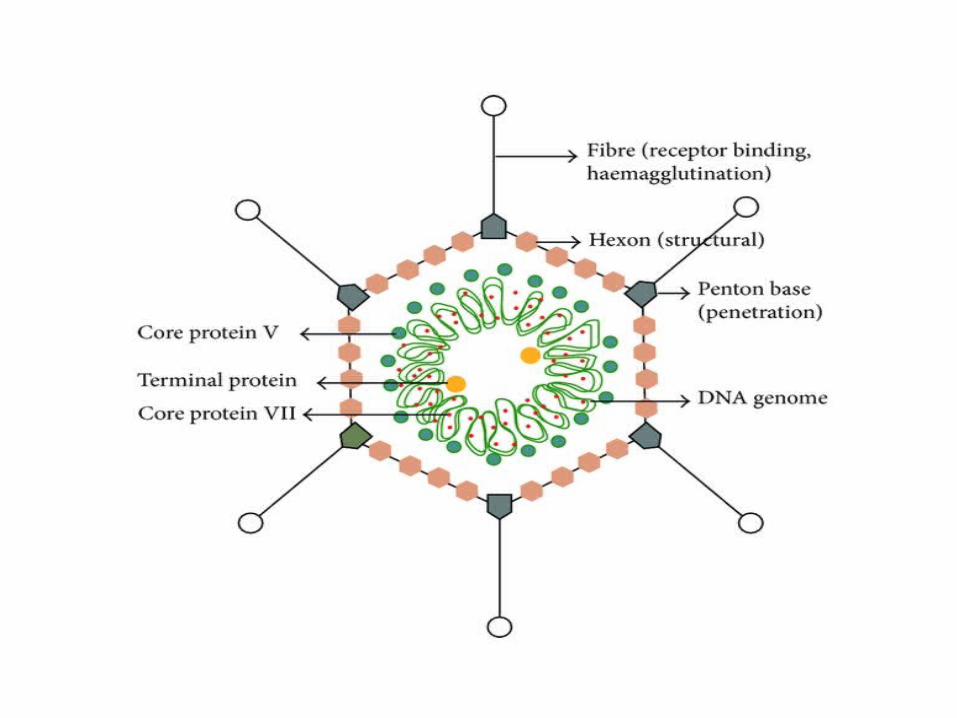
Morphology Are double stranded DNA viruses having icosahedral
symmetry and measures 70-90 nm in diameter.
Capsid composed of 252 capsomeres; in which 12 are penton bases and has fibers projecting from each of 12 vertices or penton bases and rest of capsid is composed of 240 hexon capsomeres.
No envelope
The hexon, penton and fiers constitute major antigens of adenovirus important in viral classification and disease diagnosis.
The DNA genome (25-45 Kb) is linear and double stranded.
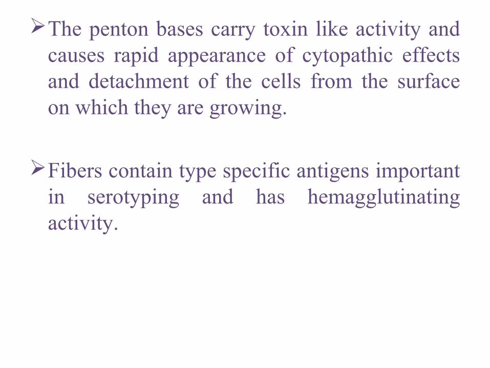
The penton bases carry toxin like activity and causes rapid appearance of cytopathic effects and detachment of the cells from the surface on which they are growing.
Fibers contain type specific antigens important in serotyping and has hemagglutinating activity.
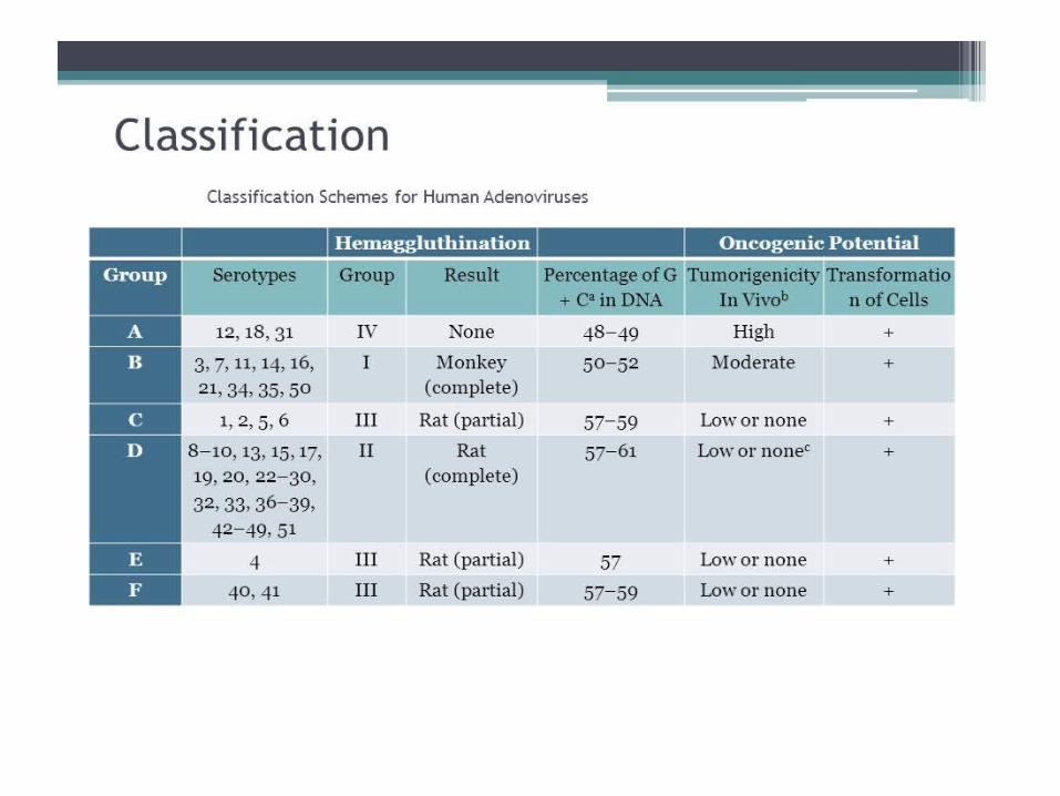
Classification: Family Adenoviridae is classified into two genera:
Mastadenovirus, the adenovirus of mammals.Aviadenovirus; adenovirus of birds
At least 51 distinct antigenic types have been isolated from humans and many other types from various animals.
Human adenovirus have been divided into six groups (A-F) on the basis of their genetic, physical, chemical and biological properties.
They are divided into serotypes on the basis of type specific antigens on penton bases and fibers.
Serotypes are identified by neutralization tests.
Pathogenesis: Infect and replicate in epithelial cells of respiratory
tract, gastrointestinal tract, urinary tract and the eye.
Do not spread beyond regional lymph nodes.
Group C persists as latent infections for years in adenoids and tonsils and are shed in feces for many months after initial infection.
Most adenovirus replicate in intestinal epithelium after ingestion but usually produce sub clinical infection than overt symptoms.
About 1/3 of human serotypes are commonly associated with human illnesses.
Single serotype may cause different clinical diseases and conversely more than one serotype can cause same clinical illness.
Adenoviruses 1-7 are more common types worldwide and account for most instances of adenovirus associated illness.
Responsible for 5% of respiratory diseases in young children but account for much less in adults.
Most infections are mild and self limited.
Occasionally case disease in other organs particularly eye and gastrointestinal tract.
o Respiratory Diseases: Typical symptoms: cough, nasal congestion, fever and sore
throat.
Symptoms most commonly manifested in infants and children and usually involves group C viruses.
Adenoviruses particularly 3, 7 and 21 are thought to be responsible for about 10-20% pneumonias in childhood.
Mortality rate of pneumonia 8-10% in very young.
Also are cause of acute respiratory disease syndrome among military recruits.
Characterised by fever, sorethroat, nasal congestion, cough and malaise; sometimes leading to pneumonia.

Occurs in epidemic form among young military recruits under conditions of fatigue, stress, and crowding soon after induction.
Disease is caused by serotype 4 and 7 and occasionally by type 3.
Outbreak of severe respiratory disease in 2007 caused by adenovirus 14 affects all age groups.
• Eye Infections: Mild occular involvement may be part of respiratory pharyngeal
syndrome.
Pharyngoconjunctival fever occurs as outbreak such as in children’s summer camps (swimming pool conjunctivitis) and caused by type 3 and 7.
Duration 1-2 weeks and complete recovery with no lasting sequelae is common outcome.
Epidemic keratoconjunctivitis more serious disease; occurs mainly in adults and is highly contagious.
Source of infection: sinks and hand towel.
Disease is characterises by acute conjunctivitis followed by keratitis that usually resolves in two weeks but may leave subepithelial opacities in the cornea for up to two years.
Caused by serotypes 8, 19 and 37.

o Gastrointestinal disease: Can often been isolated from feces but their relation to
intestinal disease has not been conclusively established.
However two serotypes (types 40 and 41) have been etiologically associated with infantile gastroenteritis.
May account for 5-15% cases of viral gastroenteritis in young children.
Adenoviruses 40 and 41 are abundantly present in diarrhoeal stools.
Enteric adenovirus are very difficult to cultivate.
Stool ELISA is often used for detection of these types.
o Other Diseases: Cause variety of casual and severe diseases in
immunocompromised patients.
Most common problem in transplant patients is respiratory diseases that may progress to pneumonia and may be fatal (usually types 1-7).
Adenovirus hepatitis in children having liver transplant.
AIDS patients mainly suffer from gastroenteritis due to adenovirus.
Types 11 and 21 may cause acute hemorrhagic cystitis in children esp. boys.
Laboratory Diagnosis:o Specimen: throat washings, stool, urine and eye for
pharyngoconjunctival fever.
Samples should be collected from affected sites as early as possible.
Duration of excretion of the virus varies among various clinical illness; 1-3 days from throat of adults with common cold, 3-5 days for throat, stool and eyes, for
pharyngoconjunctival fever, 2 weeks eye for keratoconjunctivitis, 3-6 weeks, throat and stool of children with respiratory
illness, 2-12 months in stool, throat, urine of immunocompromised
patients.
o Isolation Viral isolation in cell culture requires human cells.
Human epithelial cell lines such as Hep-2, HeLa and BK are sensitive but difficult to maintain.
Isolates grown in these cells are identified by noting cytopathic effects or by immunofluorescence staining using anti hexon antibody , heamagglutination inhibition test and neutralization test.
Shell vial techniques can also be employed for isolation of the virus.
Can also be detected by electron microscopy, ELISA or by latex agglutination tests.
o SerologyPaired sera should be used to detect rise in
antibody titre.Antibodies in patients sera can be detected by
ELISA, complement fixatation test, heamagglutination inhibition and neutralization test.
o Molecular techniques:PCR, restriction endonuclease digestion, DNA
hybridization.
o Treatment: no specific treatment
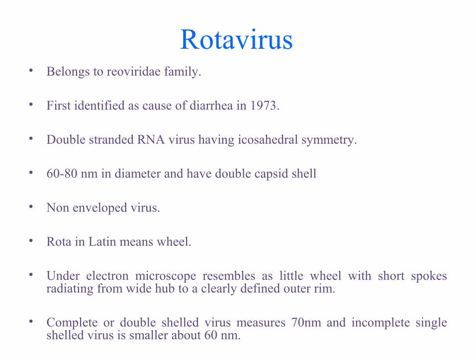
Rotavirus• Belongs to reoviridae family.
• First identified as cause of diarrhea in 1973.
• Double stranded RNA virus having icosahedral symmetry.
• 60-80 nm in diameter and have double capsid shell
• Non enveloped virus.
• Rota in Latin means wheel.
• Under electron microscope resembles as little wheel with short spokes radiating from wide hub to a clearly defined outer rim.
• Complete or double shelled virus measures 70nm and incomplete single shelled virus is smaller about 60 nm.

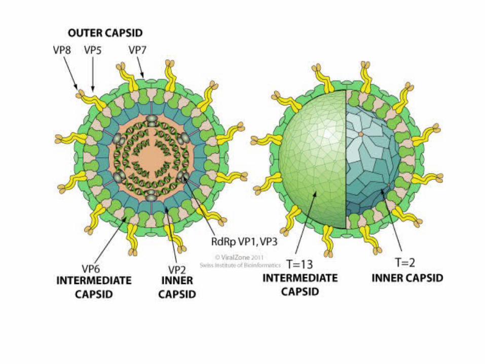
• Double stranded RNA as genome with 10-12 discrete segments.
• Contains nine structural proteins; core contains several enzymes.
• Major cause of infantile diarrhoea.
Classification and Antigenic Properties: Classified into 5 species (A-E) plus two tentative species (F and G)
based on antigenic epitopes on the internal structural protein VP6.
These can be detected by ELISA, immune electron microscopy.
Group A major human pathogen.
Outer capsid proteins VP4 and VP7 carry epitopes important in neutralizing activity with VP7 predominant antigen.
Group A have been divided into subgroups I and II by ELISA, CF or immune adherence agglutination and into serotypes (1,2,3 etc.) by neutralization tests.
Pathogenesis:The virus enters the body through the mouth.
Infects cells in the villi of small intestine (gastric and colonic mucosa are spared).
Multiply in the cytoplasm of the enterocytes and damage their transport mechanisms.
Rotavirus encoded protein NSP4, is viral enterotoxin and induces secretion by triggering signal transduction pathway.
Damaged cells may slough off into the lumen of the intestine and release large quantities of virus.
Viral excretion lasts from 2 to 12 days.Diarrhoea caused by rotavirus is due to
impaired sodium and glucose absorption as damaged cells on villi are replaced by nonabsorbing immature crypt cells.
It may take 3-8 weeks for normal function to be restored.
Clinical Findings: Cause major portion of diarrhoeal illness in infants and
children world wide but not in adults.
Incubation period:1-3 days
Typical symptoms include watery diarrhoea, fever, abdominal pain and vomiting leading to dehydration.
Stools usually greenish yellow or pale with no blood or mucus.
Group B rotavirus have been implicated in large outbreaks of severe gastroenteritis in adults in China.
Laboratory Diagnosis: The most widely available method for confirmation of
rotavirus infection is detection of rotavirus antigen in stool by enzyme-linked immunoassay (EIA) or by immune electron microscopy.
Polymerase chain reaction most sensitive method.
ELISA can be employed for detection of rise in antibody titre.
treatment: no specific treatment; supportive rehydration therapy
Prophylaxis:RV5 (RotaTeq); a live oral vaccine and RV1 (Rotarix), a live oral vaccine
Norwalk virus
• The Norwalk virus was first identified in 1968 after a severe outbreak of “winter vomiting” in a school in Norwalk Ohio.
• The descendents of Norwalk virus are now called noroviruses because these viruses change shape slightly about every 4 years so there is no virus that is exactly the same as the one discovered in 1968, but still related.
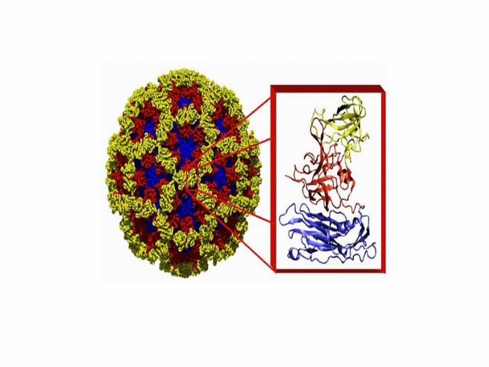
Belongs to Calciviridae family.
Single stranded RNA viruses having icosahedral symmetry.
Size: 27-40 nm in diameter, non enveloped virus. genome: single stranded, linear, positive sense, non segmented
RNA.
• Major cause of nonbacterial epidemic gastroenteritis.
• Epidemic nonbacterial gastroenteritis is characterised by:I. Absence of bacterial pathogens
II. Gastroenteritis with rapid onset and recovery and relatively mild systemic signs;
III. An epidemiological pattern of a highly communicable disease that spreads rapidly with no particular predilection in terms of age and geography.
Various descriptive terms used: epidemic viral gastroenteritis, winter vomiting disease, viral diarrhoea depending on predominant clinical feature.
• Mode of transmission: through food, indirect contact or direct contact.
• Incubation period: 24-48 hours
• The onset is rapid and clinical course is brief, lasting 12-48 hours.
• symptoms: diarrhoea, nausea, vomiting, low-grade fever, abdominal cramps, headache and malaise.
• Dehydration is the most common complication in young and adults.
• Most people will recover from this sickness in about 24-72 hours without any long term problems.
• Viral shedding persists for as long as 1 month.
• No sequelae have been reported.
Laboratory Diagnosis: Specimen: feces and vomitus
Electron Microscopy and Immunoelectron Microscopy can be used for detection of virus in stool samples.
ELISA is used to detect rise in antibody titre in patient serum.
Real-Time reverse transcriptase-polymerase chain reaction (RT-qPCR) is the most common way of testing samples for the virus in stool and vomitus and also in environmental samples (contaminated food and water).
treatment: no specific treatment; only supportive treatment
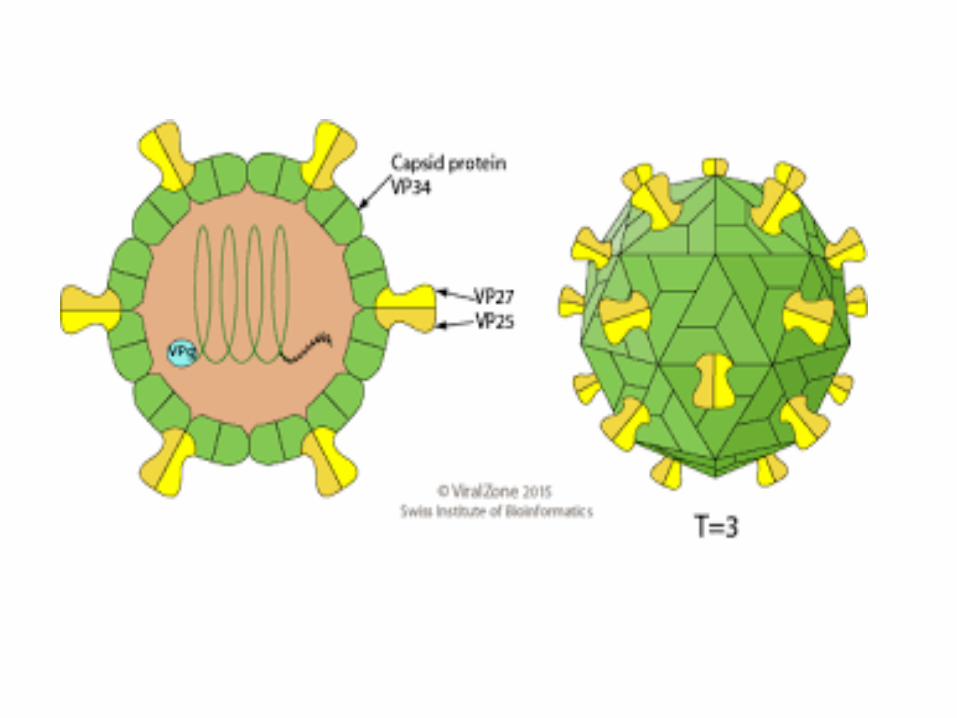
Astrovirus:
• 28-38nm in diameter, exhibit distinctive starlike morphology.
• Contain single stranded positive sense RNA.
• Astroviridae consists two genera.
• All human viruses are classified in Mamastrovirus genus.
• At least 8 serotypes of human viruses are recognised by IEM and neutralization.
Cause diarrhoeal illness and may shed in extraordinarily in large quantities in feces.
The virus is transmitted by fecal-oral route through contaminated food and water, person-person contact, or contaminated surfaces.
They are recognised as pathogens for infants and children, elderly institutionalized patients and immunocompromised persons.
Shed for prolonged periods in feces by immunocompromised hosts.