Adaptive radiotherapy for head and neck cancer
16
REVIEW Open Access Adaptive radiotherapy for head and neck cancer Howard E. Morgan and David J. Sher * Abstract Background: Although there have been dramatic improvements in radiotherapy for head and neck squamous cell carcinoma (HNSCC), including robust intensity modulation and daily image guidance, these advances are not able to account for inherent structural and spatial changes that may occur during treatment. Many sources have reported volume reductions in the primary target, nodal volumes, and parotid glands over treatment, which may result in unintended dosimetric changes affecting the side effect profile and even efficacy of the treatment. Adaptive radiotherapy (ART) is an exciting treatment paradigm that has been developed to directly adjust for these changes. Main body: Adaptive radiotherapy may be divided into two categories: anatomy-adapted (A-ART) and response- adapted ART (R-ART). Anatomy-adapted ART is the process of re-planning patients based on structural and spatial changes occurring over treatment, with the intent of reducing overdosage of sensitive structures such as the parotids, improving dose homogeneity, and preserving coverage of the target. In contrast, response-adapted ART is the process of re-planning patients based on response to treatment, such that the target and/or dose changes as a function of interim imaging during treatment, with the intent of dose escalating persistent disease and/or de- escalating surrounding normal tissue. The impact of R-ART on local control and toxicity outcomes is actively being investigated in several currently accruing trials. Conclusions: Anatomy-adapted ART is a promising modality to improve rates of xerostomia and coverage in individuals who experience significant volumetric changes during radiation, while R-ART is currently being studied to assess its utility in either dose escalation of radioresistant disease, or de-intensification of surrounding normal tissue following treatment response. In this paper, we will review the existing literature and recent advances regarding A-ART and R-ART. Keywords: Adaptive radiotherapy, Head and neck squamous cell carcinoma, IMRT, PET-guided radiotherapy, MRI- guided radiotherapy Background Over the past 20 years, the standard-of-care for radio- therapy of head and neck squamous cell carcinoma (HNSCC) has transitioned from 2D radiotherapy (RT) to 3D-conformal radiotherapy (3D-CRT) to intensity- modulated radiation therapy (IMRT) [1]. While IMRT has been shown to reduce normal tissue toxicities such as xerostomia [2], its dramatically improved conformal- ity over 2D and 3D-CRT means that anatomic changes in the patient due to weight loss or tumor reduction may have a dramatic impact on the delivered dose. Indeed, many patients will experience volumetric and spatial changes of the target volumes and organs-at-risk (OAR) during treatment, which may be due to some combination of treatment response, weight loss, inflam- mation, muscle atrophy, and radiation effects on normal tissues. These changes are of significant importance to the dose actually received by the patient, as they are not accounted for on the initial planning scan. For example, as patients progress through treatment, ra- diosensitive structures such as the parotids may migrate closer to high dose regions [3, 4] resulting in an unin- tended overdosage which has been associated with worse predicted xerostomia in small retrospective series [3], and target structures may develop dose inhomogeneities with © The Author(s). 2020 Open Access This article is distributed under the terms of the Creative Commons Attribution 4.0 International License (http://creativecommons.org/licenses/by/4.0/), which permits unrestricted use, distribution, and reproduction in any medium, provided you give appropriate credit to the original author(s) and the source, provide a link to the Creative Commons license, and indicate if changes were made. The Creative Commons Public Domain Dedication waiver (http://creativecommons.org/publicdomain/zero/1.0/) applies to the data made available in this article, unless otherwise stated. * Correspondence: [email protected] Department of Radiation Oncology, University of Texas Southwestern Medical Center, 2280 Inwood Rd, Dallas, TX 75390, USA Cancers of the Head & Neck Morgan and Sher Cancers of the Head & Neck (2020) 5:1 https://doi.org/10.1186/s41199-019-0046-z
Transcript of Adaptive radiotherapy for head and neck cancer

REVIEW Open Access
Adaptive radiotherapy for head and neckcancerHoward E. Morgan and David J. Sher*
Abstract
Background: Although there have been dramatic improvements in radiotherapy for head and neck squamous cellcarcinoma (HNSCC), including robust intensity modulation and daily image guidance, these advances are not ableto account for inherent structural and spatial changes that may occur during treatment. Many sources havereported volume reductions in the primary target, nodal volumes, and parotid glands over treatment, which mayresult in unintended dosimetric changes affecting the side effect profile and even efficacy of the treatment.Adaptive radiotherapy (ART) is an exciting treatment paradigm that has been developed to directly adjust for thesechanges.
Main body: Adaptive radiotherapy may be divided into two categories: anatomy-adapted (A-ART) and response-adapted ART (R-ART). Anatomy-adapted ART is the process of re-planning patients based on structural and spatialchanges occurring over treatment, with the intent of reducing overdosage of sensitive structures such as theparotids, improving dose homogeneity, and preserving coverage of the target. In contrast, response-adapted ART isthe process of re-planning patients based on response to treatment, such that the target and/or dose changes as afunction of interim imaging during treatment, with the intent of dose escalating persistent disease and/or de-escalating surrounding normal tissue. The impact of R-ART on local control and toxicity outcomes is actively beinginvestigated in several currently accruing trials.
Conclusions: Anatomy-adapted ART is a promising modality to improve rates of xerostomia and coverage inindividuals who experience significant volumetric changes during radiation, while R-ART is currently being studiedto assess its utility in either dose escalation of radioresistant disease, or de-intensification of surrounding normaltissue following treatment response. In this paper, we will review the existing literature and recent advancesregarding A-ART and R-ART.
Keywords: Adaptive radiotherapy, Head and neck squamous cell carcinoma, IMRT, PET-guided radiotherapy, MRI-guided radiotherapy
BackgroundOver the past 20 years, the standard-of-care for radio-therapy of head and neck squamous cell carcinoma(HNSCC) has transitioned from 2D radiotherapy (RT) to3D-conformal radiotherapy (3D-CRT) to intensity-modulated radiation therapy (IMRT) [1]. While IMRThas been shown to reduce normal tissue toxicities suchas xerostomia [2], its dramatically improved conformal-ity over 2D and 3D-CRT means that anatomic changesin the patient due to weight loss or tumor reductionmay have a dramatic impact on the delivered dose.
Indeed, many patients will experience volumetric andspatial changes of the target volumes and organs-at-risk(OAR) during treatment, which may be due to somecombination of treatment response, weight loss, inflam-mation, muscle atrophy, and radiation effects on normaltissues. These changes are of significant importance tothe dose actually received by the patient, as they are notaccounted for on the initial planning scan.For example, as patients progress through treatment, ra-
diosensitive structures such as the parotids may migratecloser to high dose regions [3, 4] resulting in an unin-tended overdosage which has been associated with worsepredicted xerostomia in small retrospective series [3], andtarget structures may develop dose inhomogeneities with
© The Author(s). 2020 Open Access This article is distributed under the terms of the Creative Commons Attribution 4.0International License (http://creativecommons.org/licenses/by/4.0/), which permits unrestricted use, distribution, andreproduction in any medium, provided you give appropriate credit to the original author(s) and the source, provide a link tothe Creative Commons license, and indicate if changes were made. The Creative Commons Public Domain Dedication waiver(http://creativecommons.org/publicdomain/zero/1.0/) applies to the data made available in this article, unless otherwise stated.
* Correspondence: [email protected] of Radiation Oncology, University of Texas SouthwesternMedical Center, 2280 Inwood Rd, Dallas, TX 75390, USA
Cancers of theHead & Neck
Morgan and Sher Cancers of the Head & Neck (2020) 5:1 https://doi.org/10.1186/s41199-019-0046-z

unintended cold spots which have been associated withworse local control in non-randomized cohorts [5, 6].Sophisticated image guidance technologies such as dailycone-beam computed tomography (CBCT) [7] can ac-count for setup errors between treatment days, but theycannot adjust for inherent changes in the volume andspatial location of the tumor and normal tissues.In response to this fundamental problem, adaptive
radiotherapy (ART) has been developed to address theseshortcomings. Adaptive radiotherapy is the process ofre-planning patients during treatment either in responseto a stimulus, such as weight loss or tumor shrinkage, orat pre-defined intervals over the course of radiation. Theprocess of re-planning allows the radiation plan to adjustto the changing tumor and normal tissue anatomy, redu-cing dose to sensitive structures such as the parotidglands, while minimizing dose inhomogeneity and inad-equate target coverage. In this scenario, ART can bereferred to as anatomy-adapted adaptive radiotherapy(A-ART), given ART is guided by structural changesoccurring over the course of radiation. In contrast, therehas been recent interest in utilizing diagnostic imagingduring treatment, such as PET/CT or MRI, to identifytreatment response in the primary tumor and/or nodalvolumes to guide dose escalation and de-escalation at-tempts. In this setting, ART can be referred to asresponse-adapted adaptive radiotherapy (R-ART), sinceART is guided by response to therapy. The purpose ofthis article is to review the existing literature on anat-omy- and response-adapted ART.
Main textA-ART: impact of volumetric and spatial changes duringradiotherapy on delivered doseSeveral structures have been shown to change size andshape over the course of radiation for HNSCC, mostnotably the primary tumor, involved nodes, and the par-otid glands. Many retrospective and prospective serieshave consistently reported decreased tumor size whichcan be detected as early as the first 2 weeks, with medianreported shrinkage rates ranging from 3 to 16%, 7 to48% and 6 to 66% reduction by the end of week 2 [8, 9],4 [10–16], and 7 [3, 9, 15, 17–28], respectively. Involvednodes can also shrink throughout treatment to similardegrees as the primary tumor [9, 13, 14, 16, 22–25, 28].Of note, tumor shrinkage during treatment is veryheterogenous even within studies, which is not surpris-ing given the known spectrum of radioresponsiveness inHNSCC. For example, one study reported a range of79.6% reduction to − 18.8% increase in primary tumorvolume by end of treatment among 34 patients receivingdefinitive chemoradiotherapy for HNSCC at the LoyolaUniversity Medical Center [16]. Another study reporteda range of 73% reduction to − 13% increase in the high
dose clinical target volume (CTV) by the end of treatmentin 15 patients receiving definitive chemoradiotherapy forHNSCC [3] in France. For any given patient with a signifi-cant volumetric change, though, there may be significantconsequences in delivered dose inhomogeneity [11, 15],potentially resulting in overdosage of normal tissues orunderdosage of the target structures [18, 29]. See Table 1for a summary of the volumetric changes of the primarytumor volume in HNSCC over the course of radiotherapy.In fact, there has been significant interest in the need
for ART to improve local control in the definitive treat-ment of HNSCC [18, 29]. If ART could correct for theseinhomogeneities, then cold spots would be minimizedand hypothetically improve in-field failure rates. No ran-domized data currently exists comparing oncologic out-comes of adaptive and non-adaptive plans to verify thisassertation. Retrospective data does appear to suggest abenefit. In a study of 317 patients receiving definitive oradjuvant radiation for HNSCC at UC Davis [5], 51 pa-tients who underwent A-ART per clinical discretionwere compared with those who were not re-planned,and there was a significantly higher rate of 2 year local-regional control with A-ART (88% vs 79%, p = 0.01). Ofnote, all of the local failures within the A-ART groupwere in-field of the primary PTV. In a separate propen-sity score matched analysis, 66 patients receiving defini-tive CRT for T3/T4 NPC with A-ART were matchedwith 66 patients without A-ART and found that 5 yearlocal-regional recurrence-free survival was higher inthose receiving A-ART (96.7% vs 88.1%, p = 0.022) [6],but with the major pattern of failure being distant me-tastases which did not differ significantly betweengroups. Both of these studies are limited by a lack ofstandardization of adaptive re-planning specificationsand their non-randomized study design. For example, iftumor response was used as a cue to initiate ART, thenthe use of it would likely select for patients more likelyto achieve a partial or complete response following com-pletion of treatment [13].With respect to OARs, the parotid glands are of par-
ticular importance in A-ART, as their radiosensitivity isclearly established, resulting in decreased salivary outputat low doses of radiation with associated xerostomia andreduced quality-of-life [30]. In 1999, Eisbruch and col-leagues [31] demonstrated that mean doses to the par-otid glands as low as 26 Gy can lead to irreversiblexerostomia. With the advent of IMRT, treatment planswere able to spare the parotid glands while still con-forming to the target and obtaining adequate coverage.Both contralateral parotid sparing as assessed in PAR-SPORT I [2] and bilateral superficial parotid sparingmethods as assessed in PARSPORT II [32, 33] haveshown promising results in regards to minimizing xeros-tomia following definitive RT for HNSCC. However, not
Morgan and Sher Cancers of the Head & Neck (2020) 5:1 Page 2 of 16
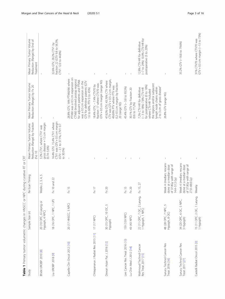
Table
1Prim
arytumor
volumetric
change
sin
HNSC
Cor
NPC
durin
gcurativeRT
orCRT
Stud
ySampleSize
(n)
Re-ScanTiming
MeanPrim
aryTumor
Volume
Redu
ction(Range
)by
Fractio
n(Fx)10
MeanPrim
aryTumor
Volume
Redu
ction(Range
)by
Fx20
MeanPrim
aryTumor
Volume
Redu
ction(Range
)by
Endof
Treatm
ent
BhideIJR
OBP
2010
[8]
20(10OPC
,6Laryng
orHypop
h,4NPC
)Weeks
2,3,4,5
3.2%
CTV1,whe
reCTV1was
grossdisease+1.5cm
margin
(0.74–5.6%
)
––
LiuIJR
OBP
2018
[9]
18(16OPC
,1NPC
,1UP)
Fx10
and22
16.4%
GTV,15.4%
CTV1whe
reCTV1was
GTV
+0.5cm
margin
(GTV
−2.1to
37.1%,C
TV10.7
to30.5%)
–32.6%
GTV,28.2%
CTV1by
fraction22
(GTV
9.8to
56.3%,
CTV112.0to
44.8%)
CapelleClin
Oncol
2012
[10]
20(17HNSC
C,3NPC
)Fx
15–
28.8%
GTV,16%
PTV60/66
whe
rePTV60was
a0.5cm
expansionon
CTV60
(involved
postop
region
s)foradjuvant
patientsandPTV66
was
a0.5cm
expansionon
the
GTV
forde
finitive
patients(GTV
1.6to
60%,PTV
0to
45%)
–
ChitapanaruxJRadiat
Res2015
[11]
17(17NPC
)Fx
17–
18.4%
GTV,−
7.4%
CTV70
byfraction17
whe
reCTV70
was
GTV
+0.5cm
margin(rang
eNS)
–
Dew
anAsian
PacJ2016
[12]
30(15OPC
,10OC,5
Hypop
h)Fx
20–
47.62%
GTV,43.76%
CTV
whe
reCTV
was
GTV
+0.5–2cm
margin,
49.69%
PTVwhe
rePTVwas
0.2–0.5cm
marginby
fraction
20(rang
eNS)
–
LeeCancerResTreat2016
(13)
159(159
NPC
)Fx
15–
43.4%
GTV
(−3.8to
93.5%)
–
LuChinMed
J2012
[14]
43(43NPC
)Fx
20–
30.1%
GTV
byFractio
n20
(0.6to
77.2%)
–
Mahmou
dTechno
lCancer
ResTreat2017
[15]
22(11OPC
,7OC,2
Laryng
,1Hypop
h,1NPC
)Fx
15,27
–7.2%
CTV-HRforde
finitive
(−3to
18%)7.8%
CTV-HR
forpo
stop
erative(0
to18%)
whe
reCTV-HR“in
clud
edtheregion
sand/or
subjacen
tlymph
node
chains
with
in2to
3cm
ofgrossdisease”
12.8%
CTV-HRforde
finitive
(−7to
29%)10.9%
CTV-HRfor
postop
erative(3
to20%)
Surucu
Techno
lCancerRes
Treat2016
[16]
48(28OPC
,7NPC
,5Hypop
h,8OC)
Week4(m
edian;rescans
done
atamed
iando
seof
37.8Gywith
rang
eof
14.4–51.5Gy)
–26.8%
GTV
(rang
eNS)
–
Surucu
Techno
lCancerRes
Treat2017
[27]
34(20OPC
,6OC,5
NPC
,3Hypop
h)Week4(m
edian;rescans
done
atamed
iando
seof
37.8Gywith
rang
eof
27.0–48.6Gy)
––
35.2%
GTV
(−18.8to
79.6%)
CastelliRadiat
Oncol
2015
[3]
15(11OPC
,2OC,1
Laryng
,1Hypop
h)Weekly
––
31%
CTV70
whe
reCTV70
was
GTV
+0.5cm
margin(−13
to73%)
Morgan and Sher Cancers of the Head & Neck (2020) 5:1 Page 3 of 16

Table
1Prim
arytumor
volumetric
change
sin
HNSC
Cor
NPC
durin
gcurativeRT
orCRT
(Con
tinued)
Stud
ySampleSize
(n)
Re-ScanTiming
MeanPrim
aryTumor
Volume
Redu
ction(Range
)by
Fractio
n(Fx)10
MeanPrim
aryTumor
Volume
Redu
ction(Range
)by
Fx20
MeanPrim
aryTumor
Volume
Redu
ction(Range
)by
Endof
Treatm
ent
LooClin
Oncol
2011
[17]
5(3
OPC
,1OC,1
Laryng
)Weekly
––
5.8%
CTV68
whe
reCTV68
“encom
passed
theGTV
and
high
-riskregion
s”(rang
eNS)
Beltran
JApp
lClin
Med
2012
[18]
16(7
OPC
,5OC,1
Hypop
h,1NPC
,2NS)
Fx15,25
––
13.25%
PTV2
whe
rePTV2
was
theCTV2(GTV
+high
-risk
region
s)+0.5cm
margin
(rang
eNS)
Fung
Med
Dosim
2012
[19]
10(10NPC
)Fx
21,31approx.
––
53.95%
CTV,36.19%
PTV
whe
rePTVwas
theCTV
+0.3cm
(rang
eNS)
JinRadiat
Oncol
2013
[20]
9(9
NPC
)Fx
23–
–9.4%
GTV
byfraction23
(rang
eNS)
(non
-sig
redu
ction)
Schw
artzRadiothe
rOncol
2013
[21]
22(22OPC
)Dailyscans,Weeklyrecalc
–5%
CTV
whe
reCTV
was
the
GTV
+high
-riskregion
s(−21
to13%)
8%CTV
(−6to
19%)
TanOncoTargetsTher
2013
[22]
20(20NPC
)Weekly
––
55.3%
GTV
(rang
eNS)
Fung
JRadiat
Res2014
[23]
30(30NPC
)Every2Fx
––
35.7%
GTV
(rang
eNS)
Huang
Radiat
Oncol
2015
[24]
19(19NPC
)Every5Fx
––
65.6%
GTV
(rang
eNS)
Kataria
BrJRadiol
[25]
36(21OPC
,5Laryng
,10
Hypop
h)Fx
23–
–34.0%
GTV
(rang
eNS)
ZhangRadiothe
rOncol
2016
[26]
13(13OPC
)Weekly
––
24.43%
CTV70
whe
reCTV70
was
GTV
+0.5cm
margin
(−12.6to
62.1%)
Rang
eof
med
iantumor
volume(GTV
/CTV
/PTV)
redu
ctions
oftheinclud
edstud
ies
3to
16%
7to
48%
6to
66%
Moststud
iesrepo
rted
aredu
ctionin
theprim
arytarget
volumeov
erthecourse
ofradiothe
rapy
.How
ever,the
stud
iesvarie
din
thede
finition
ofthetarget
volumerepo
rted
,with
somerepo
rtingtheGTV
,som
ethe
high
riskCTV
(with
varyingmargins),an
dsomethehigh
riskPT
V(alsowith
varyingmargins),makingcompa
rison
sacross
stud
iesdifficult.Notethat
forstud
iesthat
includ
edawiderang
eof
fractio
nsrepo
rted
attim
eof
re-scan,
themed
ianfractio
nwas
used
forcatego
rizinginto
theab
ovecolumns
(byFx
10,Fx20
,etc).In
rega
rdsto
BhideIJRO
BP20
10(8)somepa
tientsha
dindu
ctionchem
othe
rapy
priorto
defin
itive
CRT
,which
may
accoun
tforsomeof
thevaria
tionseen
betw
eenthevo
lumetric
chan
gesrepo
rted
bythisau
thor
andby
LiuIJRO
BP20
18(9).Overall,thetren
dseen
intheseserie
swas
forincreasing
tumor
volumeredu
ction
throug
hout
therap
ywith
med
ianredu
ctions
repo
rted
as3to
16%
byfractio
n10
,7to
48%
byfractio
n20
,and
6to
66%
byen
dof
treatm
ent
“-“informationwas
either
notavailableor
was
notdirectly
compa
rableto
othe
rvo
lumetric/dosim
etric
data
repo
rted
andthus
notinclud
edGTV
Gross
Tumor
Volume,
CTVClin
ical
Target
Volume,
PTVPlan
nedTarget
Volume,
HNSC
CHeadan
dNeckSq
uamou
sCellC
arcino
ma,OPC
Oroph
aryn
geal
Can
cer,OCOralC
avity
Can
cer,NPC
Nasop
haryng
ealC
ancer,
Laryng
Laryng
ealC
ancer,Hypop
hHyo
pharyn
geal
Can
cer,NSHeadan
dNeckSq
uamou
sCellC
arcino
ma,Site
Not
Specified
,SNSino
nasalC
ancer,UPHeadan
dNeckSq
uamou
sCellC
arcino
maof
Unk
nownPrim
ary,
Rang
eNSRa
ngeno
tstated
Morgan and Sher Cancers of the Head & Neck (2020) 5:1 Page 4 of 16

all patients who appear to have excellent sparing of theparotids on treatment planning have excellent rates ofxerostomia, as 38% receiving IMRT in PARSPORT I [2]and 21% in PARSPORT II still had grade 2 or greaterxerostomia by month 12 [32]. Whether this residualxerostomia is fully due to inherent differences in patientresponse to RT is unclear, but unrecognized (and there-fore unadjusted) changes in parotid dosimetry through-out treatment may partially contribute.Like the primary tumor and involved nodes, the par-
otid glands have also been consistently reported toshrink throughout treatment, a process that may start asearly as the first 2 weeks of treatment. The average vol-ume of the parotids has been reported to decrease asmuch as 14.7, 37, and 48% by the end of weeks 2 [8], 4[10–12, 14, 15, 18, 21, 24, 34], and 7 [3, 8, 11, 15, 17–21,23, 24, 34–37], meaning that the delivered dose could bemuch higher than expected by the original plan. Like thetarget volumes, there can also be wide heterogeneity inthe volume reduction of parotid glands. One study re-ported a range of 0.0 to 63.4% reduction by end of treat-ment [3] while another reported a range of 6.8 to 69.44%reduction by end of treatment [36]. This heterogeneitybetween patients likely contributes to the seeminglycontradictory findings between some small studies whichpredict a xerostomia reduction benefit of A-ART [3, 38]and some studies which do not [35, 39]. See Table 2 fora summary of volumetric and dosimetric changes of theparotids in HNSCC. Figure 1 is an example of a patientwho might benefit from A-ART.In addition to volume changes, the spatial orientation
of the parotid glands appears to shift during treatment,with a typical pattern of superior and medial displace-ment [3, 4, 8, 23, 24, 34] thought from shrinkage of thetarget tumor and associated weight loss. The implicationof this shift means that the parotid gland may migratecloser to high-dose regions, resulting in an unplannedoverdosage of this structure. In a retrospective cohort of15 locally advanced HNSCC (primarily oropharyngeal)receiving definitive CRT, Castelli and colleagues [3]noted that 74% of the parotid glands were an average of4.3 mm closer to the CTV by the end of treatment whencompared to the initial planning scan. This was associ-ated with an unplanned overdose of 59% of the parotidglands with an average mean dose increase of 3.7 Gy,with A-ART re-planning reducing the mean parotiddose by 5.1 Gy on average, with a predicted decrease inxerostomia risk of 11% based on a normal tissue compli-cation probability (NTCP) model [3]. Other studies havealso noted increased dose to the parotids with migrationmedially [24] towards the target [4]. Again, the degree ofmigration between patients is heterogenous with onestudy reporting parotid glands moving between 12mmcloser to the CTV to 6.2 mm away [3]. Given patients
appear to differ fairly broadly in the degree of targetshrinkage, parotid volume reduction, and parotid dis-placement, the subgroup of patients with minimal volu-metric and spatial changes over the treatment coursewould likely gain little from proactive adaptive re-planning. Therefore, the identification of a cohort of pa-tients who are most likely to benefit from A-ART is ofsignificant interest to identify the appropriate populationfor re-planning, as this process is currently labor-intensive. Figure 2 shows an example of the relativedosimetric improvement of A-ART.In addition to the parotid glands, the spinal cord and
brainstem are also of interest as hot spots may developin them over the course of radiotherapy, which may ex-ceed conventional dosimetric constraints, that have beenchosen to keep myelopathy and brainstem necrosis ratesnegligible. Most authors advocate for re-planning whenthese constraints (spinal cord max dose < 45–48 Gy andbrainstem max dose < 54 Gy) are breached during radio-therapy. However, studies do not consistently reportoverdosing of the spinal cord or brainstem with somereporting significant increases in the max dose through-out radiotherapy [8, 10, 11, 19, 24, 42] and some notingno change [20, 40, 43, 44]. In a prospective cohort studyof 22 patients with HNSCC where re-planning was trig-gered by underdosage of the target volumes (CTV cover-age < 95%), overdosage of the parotids (mean dose > 26Gy), or overdosage of the spinal cord (max dose > 45 Gy)[15], the spinal cord max dose reached the threshold fortriggering A-ART in only 3 of 22 patients, whereas par-otid gland overdosages occurred in 3 right and 5 left pa-rotids, and CTV undercoverage in 7 instances. Incontrast to the parotids, the volume and position of thespinal cord has not been shown to change over thecourse of radiotherapy [17, 45] which may partially ex-plain why dosimetric changes in the spinal cord and/orbrainstem are not as consistent or profound. However,in the studies that do report excess dose to the spinalcord and/or brainstem, dose variation can be quite highwith one study reporting a range of 0.2–15.4 Gy increasein the spinal cord max dose and 0.6–8.1 Gy increase inthe brainstem [42] and 2.1–9.9 Gy and 1.6–5.9 Gy re-spectively in another [11]. Note that a max dose increaseof 15 Gy in the spinal cord can be quite significant, asrates of myelopathy increase exponentially at higherdoses, with the estimated risk being < 1% at 54 Gy and <10% at 61 Gy [46]. In individuals who encounter hotspots in the spinal cord or brainstem during radiother-apy, re-planning has consistently been reported to de-crease these max doses back to within their pre-definedthresholds [11, 19, 42]. Though A-ART is beneficial innegating hot spots that may develop in the spinal cordand/or brainstem over the course of radiotherapy, theclinical significance of this is unclear as myelopathy rates
Morgan and Sher Cancers of the Head & Neck (2020) 5:1 Page 5 of 16

Table
2Parotid
volumetric
anddo
simetric
change
sin
HNSC
Cor
NPC
durin
gcurativeRT
orCRT
Stud
ySampleSize
(n)
Re-Scan
Timing
MeanParotid
Volume
Redu
ction(Range
)by
Fractio
n(Fx)10
MeanParotid
Volume
Redu
ction(Range
)by
Fx20
MeanParotid
Volume
Redu
ction(Range
)by
Endof
Treatm
ent
Dosim
etric:M
eanParotid
DoseChang
e(Range
)
BhideIJR
OBP
2010
[8]
20(10OPC
,6Laryng
orHypop
h,4NPC
)Weeks
2,3,4,5
14.7%
NS
–35%
NS
With
outART:2.8Gyincrease
ipsi
CapelleClin
Oncol
2012
[10]
20(17HNSC
C,3NPC
)Fx
15–
17.5%
NS(−1to
46%)
–With
ART:0.6Gyredu
ction
NS
LuChinMed
J2012
[14]
43(43NPC
)Fx
20–
35.5to
36.8%
*–
–
Beltran
JApp
lClin
Med
2012
[18]
16(7
OPC
,5OC,1
Hypop
h,1NPC
,2NS)
Fx15,25
–22%
NS
30%
NS
With
outART:4.7%
increase
Ipsi
6.1%
increase
Contra
Schw
artzRadiothe
rOncol
2013
[21]
22(22OPC
)Dailyscans,
Weeklyrecalc
–15%
NS(−19
to25%)
26%
NS(−8to
48%)
1ART
re-plan:1.3Gyredu
ction
ipsi
2ART
re-plans:4.1Gyredu
ction
ipsi
ChitapanaruxJRadiat
Res2015
[11]
17(17NPC
)Fx
17–
30.5%
ipsi24.3%
contra
–With
ART:1.1Gyredu
ction
Contra
Ipsilateraln
otsign
ificant
(0.9Gy)
Huang
Radiat
Oncol
2015
[24]
19(19NPC
)Every5Fx
–14.4to
15.8%
*38.0to
39.2%
*With
outART:3.09Gyto
5.6Gy
increase
*
Dew
anAsian
PacJ2016
[12]
30(15OPC
,10OC,
5Hypop
h)Fx
20–
33.65%
ipsi31.06%
contra
–With
ART:5.6Gyredu
ction
ipsi
Con
tralateralno
tsign
ificant
(2.7Gy)
ZhangJMed
Radiat
Sci
2017
[34]
39(39NPC
)Fx
10,20,30
–15.27%
ipsi
37.49%
ipsi34.55%
contra
–
Mahmou
dTechno
lCancer
ResTreat2017
[15]
22(11OPC
,7OC,2
Laryng
,1Hypop
h,1NPC
)
Fx15,27
–18.2to
19.0%
*forde
finitive
(3to
32%)10.0to
16.6%
*foradjuvant
(5to
44%)
30.1to
30.9%
*forde
finitive
(11to
52%)23.1to
25.3%
*foradjuvant
(3to
41%)
With
outART:
15.4to
16.4%
increase
*for
definitive
(−30
to76%)9.1
to10.4%
increase
*for
postop
(−25
to70%)
Neither
sign
ificant
given
largehe
teroge
neity
LooClin
Oncol
2011
[17]
5(3
OPC
,1OC,1
Laryng
)Weekly
––
30.2%
Ipsi(17.1to
55.8%)
17.5%
Contra(15.6to
48.5%)
With
outART:7.6Gy
increase
Ipsi(2.5to
19Gy)
7.3Gyincrease
Contra
(1.1to
11.6Gy)
Fung
Med
Dosim
2012
[19]
10(10NPC
)Fx
21,31
approx.
––
32.44to
33.31%
*With
ART:0.75Gyredu
ction
(righ
t,p=0.046)
1.25
Gy
redu
ction(left,p
=0.053)
Fung
JRadiat
Res2014
[23]
30(30NPC
)Every2Fx
––
47.54%
NS
–
Hun
terIJR
OBP
2013
[35]
18(18OPC
)Weekly
––
13.31%
NS
With
outART:0.92Gy(m
edian)
increase
NS(−4.9to
8.4Gy;no
tsig)
23/36parotid
shadan
increase
(2.2Gymed
ian)
JinRadiat
Oncol
2013
[5]
9(9
NPC
)Fx
23–
–38.4to
40.68%
*–
CastelliRadiat
Oncol
2015
[3]
15(11OPC
,2OC,1
Laryng
,1Hypop
h)Weekly
––
28.3%
NS
With
outART:67%
ofpts.:4.8Gy
increase
NS33%
ofpts.:3.9Gy
decrease
NSWith
ART:O
fthose
with
overdo
sedparotid
s:5.1G
y
Morgan and Sher Cancers of the Head & Neck (2020) 5:1 Page 6 of 16

Table
2Parotid
volumetric
anddo
simetric
change
sin
HNSC
Cor
NPC
durin
gcurativeRT
orCRT
(Con
tinued)
Stud
ySampleSize
(n)
Re-Scan
Timing
MeanParotid
Volume
Redu
ction(Range
)by
Fractio
n(Fx)10
MeanParotid
Volume
Redu
ction(Range
)by
Fx20
MeanParotid
Volume
Redu
ction(Range
)by
Endof
Treatm
ent
Dosim
etric:M
eanParotid
DoseChang
e(Range
)
decrease
NS(0.6to
12.2Gy)
YaoBiom
edResInt2015
[36]
50(50NPC
)Every5Fx
––
35%
NS(6.8to
69.4%)
With
outART:3.52Gy(11.38%)
increase
NS(−1.51
to30.57%
)Weigh
tloss
correlated
with
meanparotid
dose
ZhangRadiothe
rOncol
2016
[26]
13(13OPC
)Weekly
––
34.51%
ipsi(10to
57.6%)
27.98%
contra(−5.2to
57.3%)
With
outART:16/23
parotid
soverdo
sed:
4.1Gyincrease
NS
(0.5to
11.5Gy)3ART
re-plans:
3.1Gyredu
ction
NS6ART
re-plans:
3.3Gyredu
ction
NS
HuBM
CCancer2018
[40]
40(40NPC
)Med
ianFx
22–
–17.2%
Ipsi20%
Contra
With
ART:0.7Gyredu
ction
Ipsi
Con
tralateralno
tsign
ificant
(0.1Gy)
LeeIJR
OBP
2008
[4]
10(2
OPC
,5NPC
,1SN
,1OPC
+NPC
,1UP)
DailyMV-CT
––
–With
outART:3
Gy(11%
)NS(−6
to42%)Parotid
glands
migratin
gcloser
totarget
volumehadhigh
erchange
sin
meando
se
Fioren
tinoBr
JRadiol
2012
[41]
10(4
OPC
,5OC,1
Hypop
h)Daily
––
43.5%
Ipsi44.0%
Contra
–
Rang
eof
med
ianparotid
volumeredu
ctions
repo
rted
intheinclud
edstud
ies
15%
10to
37%
13to
48%
–
Allstud
iesrepo
rted
anaverag
ede
crease
inpa
rotid
volumeat
timeof
re-scan;
however,the
rewas
widehe
teroge
neity
betw
eenan
deven
with
instud
ies,with
afew
patie
ntsactually
having
anincrease
inpa
rotid
glan
dvo
lumeby
endof
treatm
ent.Th
iswas
associated
with
varia
bleredu
ctions
inmeanpa
rotid
dose
byad
aptiv
ere-plann
ingan
dsugg
ests
that
ART
may
notbe
approp
riate
forallp
atients.How
ever,A
RTdo
esap
pear
toredu
cemeanpa
rotid
dose
inpa
tientswho
sepa
rotid
sexpe
riencean
unintend
edov
erdo
sage
second
aryto
anatom
icchan
gesthroug
hout
treatm
ent,which
hasbe
enassociated
with
redu
cedpred
icted
xerostom
ia.H
owever,clin
ical
correlationisstilllackingbe
tweenART
andprospe
ctivetoxicity
data.N
otethat
ane
gativ
evo
lumetric
chan
gerepo
rted
abov
emeans
that
thisstructureincreasedin
size
(e.g.-1%
indicates
a1%
increase
invo
lume).A
nega
tivedo
simetric
chan
gemeans
that
itde
creasedin
thedo
sereceived
(e.g.-10
%indicatesa10
%de
crease
inmeanpa
rotid
dose)
“-“informationwas
either
notavailableor
was
notdirectly
compa
rableto
othe
rvo
lumetric/dosim
etric
data
repo
rted
andthus
notinclud
ed“N
S ”pa
rotid
side
was
notspecified
“*“parotid
side
(leftor
right)was
specified
;how
ever,ipsilaterala
ndcontralaterald
esigna
tionwereno
tspecified
“Ipsi“ipsilateralp
arotid
“Contra“con
tralateral
parotid
HNSC
CHeadan
dNeckSq
uamou
sCellC
arcino
ma,OPC
Oroph
aryn
geal
Can
cer,OCOralC
avity
Can
cer,NPC
Nasop
haryng
ealC
ancer,Laryng
Laryng
ealC
ancer,Hypop
hHyo
pharyn
geal
Can
cer,NSHeadan
dNeck
Squa
mou
sCellC
arcino
ma,Site
Not
Specified
,SNSino
nasalC
ancer,UPHeadan
dNeckSq
uamou
sCellC
arcino
maof
Unk
nownPrim
ary
Morgan and Sher Cancers of the Head & Neck (2020) 5:1 Page 7 of 16

remain very low in clinical practice. Further, most pa-tients appear to only have modest increases in the spinalcord max dose (2–4 Gy) [11, 24, 42] and with severalseries failing to show a significant overdosage of thisstructure [20, 40, 43, 44]; clinically significant deviationsmay only occur in a minority of patients.The effect of adaptive re-planning on the efficacy and
tolerability of postoperative radiation for HNSCC is lessclear, given the scarcity of data in this cohort. Some au-thors have advocated for A-ART in the postoperativesetting noting that the postsurgical graft can swell andcontract during radiation resulting in under-coverage[5]. However, other studies have questioned the utilityof A-ART in the postoperative setting. In a prospectivestudy of 20 patients with HNSCC who were re-plannedat fraction 15, the 7 patients receiving postoperative ra-diation only appeared to have incremental, minimallyimpactful, changes in dosimetry [10]. Another more re-cent study prospectively assessed 22 patients withHNSCC but performed CTs for dose recalculation atweeks 1, 3, and 6 to determine the need for A-ART[15]. In this study, 2 of the 11 patients receiving post-operative radiation were re-planned as indicated bycritical dosimetric changes by week 6, with one event
triggering A-ART by the development of a spinal cordhot spot and one by CTV underdosage. In contrast, 8of the 11 in the definitive group with bulky disease(gross tumor > 4 cm) encountered at least one eventtriggering A-ART which was significantly higher thanthe postoperative group (p = 0.03). Given that patientsreceiving definitive radiation had more weight loss(8.6% vs 4.9%, p < 0.001) and a trend towards morehigh risk CTV shrinkage (12.8% vs 10.9%, p > 0.05),the authors speculated that target shrinkage andweight loss may help explain the higher incidence ofA-ART triggers in the definitive group, though com-parisons are limited given the small sample size andlack of clinical correlation with A-ART trigger end-points. Therefore in the setting of postoperative radi-ation, because the disease has been resected and theprimary driver of anatomic change appears to beweight loss rather than tumor shrinkage, A-ART ap-pears to be needed less frequently in patients treatedwith adjuvant RT.Recently, MRI-guided radiotherapy has been evalu-
ated for its utility in A-ART [47, 48], with the idea thatadaptive scans utilizing MRI imaging can dramaticallyimprove visualization of soft tissue changes throughout
Fig. 1 Primary tumor, nodal, and parotid volumes decrease over the course of radiation. This patient is a 54 year-old man with p16-positivecT4N1M0 squamous cell carcinoma of the left tonsil who required adaptive radiotherapy over the course of radiation secondary to significanttumor response and weight loss during treatment noted on review of daily CBCTs. The primary tumor decreased by 25.0% from baseline (A1) toweek 5 (A2). The grossly involved nodes decreased by 48.6% from baseline (B1) to week 5 (B2). The left parotid decreased by 37.2% (cyan) andthe right parotid (blue) decreased by 41.9% from baseline (C1) to week 5 (C2). Note contraction of the lateral border of the bilateral parotids attime of re-simulation (C2)
Morgan and Sher Cancers of the Head & Neck (2020) 5:1 Page 8 of 16

treatment. In a prospective feasibility study at MD An-derson Cancer Center [48], five patients with locally ad-vanced HPV-positive OPC underwent definitive CRT,with intra-treatment MRI every 2 weeks. The primarygross tumor volume (GTV) volume was noted to de-crease on average by 44, 90, and 100% at weeks 2, 4,and 6, with the corresponding nodal volumes decreas-ing by 25, 60, and 80%. The high dose target volumewas reduced accordingly with these volumetric changes,resulting in an approximate reduction in the mean par-otid dose of 3.3 Gy with ART. Although NTCP model-ing only predicted a 1% xerostomia reduction at 6
months, the probability of predicted 6 month dysphagiawas reduced by 11% [48]. The high rates of radio-graphic complete response (CR) on MRI of the primarytumors in this study are congruent with a prior pilottrial by the same group where 15 out of 29 primary tu-mors had a CR detected on MRI imaging at 3–4 weeks[47]. A separate group reported 70% average GTV re-duction on MRI imaging by week 6 in eight patientswith OPC [49]. The MARTHA trial is an upcomingprospective trial attempting to assess if the use of dailyMRI imaging and weekly plan adaptation will benefitxerostomia in patients receiving RT for HNSCC [50].
Fig. 2 Adaptive re-planning reduces unplanned dose inhomogeneity and parotid gland overdose. These images are from the same case aspresented in Fig. 1. At time of initial simulation (a), anticipated coverage of the high dose planned target volume (PTV) was 98.5% receiving 70Gyand the mean dose of the left and right superficial parotids were 25.0 and 24.5 Gy, respectively. However, by week 5 (b), there was wide variationin dose within the high dose PTV with cold spots down to 88.0% and hot spots up to 113.4% of the prescription. In addition, the mean left andright superficial parotids doses increased to 32.2 Gy and 36.7 Gy, respectively. With adaptive re-planning (c), dose homogeneity was improvedwith cold spots only being 94.8% and hot spots only being 104.4% inside of the high dose PTV, with reduction of the mean right and leftsuperficial parotid dose back to 24.9 Gy and 24.6 Gy, respectively. The main benefit of A-ART in this case was sparing of the parotids, given therewas an unplanned overdose of an additional 7.2 Gy to the left and 12.2 Gy to the right parotids which was mitigated with adaptive re-planning
Morgan and Sher Cancers of the Head & Neck (2020) 5:1 Page 9 of 16

A-ART: practical considerations and implementationCurrently, the process of A-ART requires (1) identifying theappropriate patient, (2) re-simulation, (3) re-contouring, and(4) re-planning. Patients may be identified for A-ART byclinical variables (weight loss, tumor shrinkage, etc); regu-larly planned intervals; treatment response as assessed onCBCT scans, diagnostic CT or MRI scans; or dose re-calculations of cumulative dose delivered to the targets andOARs. Following identification, re-simulation of the patientshould occur promptly, which may require creation of anew aquaplastic/thermoplastic mask if the mask fit isinadequate. Then, re-contouring can be done via manual in-put from the physician, deformable image registration, orautomatic segmentation. Artificial intelligence methods ofauto-contouring are being developed to make this processmore efficient. If deformable image registration or an auto-matic method are used for re-contouring, it is recom-mended that the physician proofread these contours forerrors prior to approval. The plan is then re-planned andoptimized per physician discretion.One of the biggest obstacles with adaptive re-planning is
the time required to manually re-simulate, re-contour,and re-plan patients, which can be draining on a depart-ment’s resources; developing an optimal trigger for imple-menting A-ART is therefore a high priority to maximizeefficiency. At this time, no consensus exists on the mostappropriate timing regimen for A-ART during radiother-apy. Many centers perform adaptive re-planning based onclinical characteristics, such as weight loss, tumor shrink-age, changes in patient setup, and mask fitting. Other ap-proaches suggest performing A-ART at regular intervals(e.g. every 10 fractions), as reductions in target and parotidvolumes have been shown to occur as early as the first orsecond week which can result in significant dosimetricchanges [24, 26]. In a study assessing the timing of A-ART scans in 13 patients with OPC receiving definitiveradiation [26], weekly CT scans were performed andassessed for a dosimetric benefit of A-ART re-planning ateach interval. They found 3 re-plans (weeks 1, 2, and 5) tobe comparable with 6 weekly re-plans estimating a meanparotid gland benefit of 3.1 Gy with 3 re-plans comparedwith only 3.3 Gy if 6. The majority of the benefit appearedto be within the first 2 weeks, with the authors recom-mending A-ART at weeks 1, 2, and 5 [26]. In a separatestudy of 19 patients with NPC receiving definitive radi-ation with weekly CT scans, significant dosimetric varia-tions in the target, parotids, spinal cord, and brainstemwere noted mostly at fractions 5 and 15, with the authorsrecommending A-ART re-plans at these time points [24].Given the wide range of variability in anatomic changes
of the target structures and OARs between patientsthroughout radiation (as discussed in the previous sec-tion), we advocate that 1 A-ART regimen is not likelyapplicable to all patients. Some studies have attempted to
identify baseline and dosimetric factors influencing thelikelihood of a patient needing A-ART during their treat-ment course, with the most common factors identified be-ing: higher initial mean parotid gland dose [36, 38, 51],larger clinical target volumes (CTV) and bulkier disease[15, 38, 52], initial weight [52], and a faster rate of weightloss [36]. Most of these predictive variables have not beenvalidated. However, one study assessed initial mean par-otid dose > 22.2Gy as a cutoff value in a validation cohortof 43 patients, but the positive predictive value was only19% with a sensitivity of 80% [51], suggesting mostpatients meeting this criteria still would not benefit fromA-ART, resulting in significant clinic inefficiencies.Recent advances have looked at individualizing indica-
tions for A-ART by recalculating the cumulative dose ofthe target and OAR every day or every week to identifyactionable changes in dosimetry which may necessitatere-planning [53, 54]. In an initial pilot study of A-ARTat MDACC, Schwartz and colleagues [55] prospectivelyevaluated 22 patients with oropharyngeal SCC receivingdefinitive radiation with weekly CT dose recalculationsto prompt A-ART re-planning if target coverage waspoor or if OAR sparing was inadequate. All 22 patientshad at least 1 adaptive re-plan and 8 had 2 re-plans withthis approach. On dosimetric analysis, the ipsilateral par-otid dose was reduced by 1.3 Gy (p = 0.002) in those re-ceiving 1 re-plan and 4.1 Gy in those receiving 2 re-plans[21]. In a separate prospective study also utilizing weeklyCT scans in patients receiving definitive RT for HNSCC[56], patients were selected for A-ART if their re-calculated plan on their weekly CT scan yielded a PTV70or PTV60 receiving V95 < 95% or spinal cord receivingmax dose > 45Gy. This method resulted in 8 out of 10 pa-tients being re-planned with A-ART, with 41% of the re-plans triggered in the first 2 weeks. While these early stud-ies have predominantly used weekly CT scans, there hasbeen recent effort to improve efficiency by utilizingCBCTs used in the daily delivery of radiation to calculatethe cumulative dose received [53, 57–59] allowing theprompt identification of patients likely to benefit from A-ART. As technology and artificial intelligence advances,we anticipate that the identification of patients and theimplementation of A-ART will be significantly smootherand likely automated. Table 3 describes currently accruingand upcoming trials in A-ART.
Response-adapted adaptive radiotherapy (R-ART)In contrast to A-ART, in which the subsequent radiationre-plan essentially recapitulates the original radiation tar-gets and doses adapted to the new anatomy, response-adapted ART is the process of changing the radiationtargets and/or doses based on response to treatment.Whether the “response” is identified by CT, PET-CT, orMRI, the intent of R-ART is to either escalate the
Morgan and Sher Cancers of the Head & Neck (2020) 5:1 Page 10 of 16

radiotherapy dose to persistent disease or reduce the doseto responding disease, leading to improved tumor controland/or reduced normal tissue toxicity.Given that in-field recurrences are still a common
pattern-of-failure in HNSCC [62–64], further treatmentintensification is still needed in some patient popula-tions, with radiation dose escalation serving as one pos-sible paradigm. Response-adapted ART is an attractiveavenue for dose escalation, since persistent or refractorydisease during treatment may be directly targeted, effect-ively reducing the volume of disease being boosted. Forexample, PET-guided ART is under active investigation,with persistent radiotracer uptake early in treatmentthought to represent radioresistance. Both standardtracers such as [18F]Fluoro-2-deoxy-2-D-glucose (FDG)[65, 66] and more novel indicators of hypoxia such as[18F]Fluoroazomycin-arabinoside (FAZA) [61, 67] arebeing studied.
Oncologic outcomes with PET-based R-ART is lim-ited, but preliminary reports suggest that such a para-digm is feasible [68, 69]. In an initial phase I feasibilitytrial at Ghent University Hospital [68], the radiotherapydose was escalated to over 80 Gy to areas of persistentavidity on a PET-CT scan performed during week 2. Noacute dose-limiting toxicity was encountered. Althoughrandomized evidence is not yet available, a recent case-matched control study [70] compared 72 patients treatedon this study or similar subsequent trials receiving 70.2–85.5Gy to those receiving standard IMRT did not find astatistically significant difference in 5 year local control(82.3% vs 73.6%, p = 0.36). However, this retrospectiveanalysis did note increased chronic toxicity at higherdoses, with late grade > 3 dysphagia being 50% (vs 28%,p = 0.004) and with grade 4 mucosal ulcers occurring atthe site of dose escalation in 13% (9/72) of patients [70].The incidence of these late grade 4 mucosal ulcers was
Table 3 Currently accruing or upcoming clinical trials in anatomy-adapted adaptive radiotherapy (A-ART)
Clinical Trial Primary Investigator(Location)
Description Eligible Target Accrual(Actual or CurrentAccrual)
Status
Evaluation of theAutomatic DeformableRecontouring on theDaily MVCT for Headand Neck CancerAdaptive Radiotherapy(GIRAFE) [45, 60]
Laprie Anne(Institut UniversitaireDu Cancer Toulouse,Oncopole, France)
Prospective phase II trialevaluating the accuracyof deformable imageregistration on dailyMV-CTs. Deformableimage registration willbe compared to manualrecontouring on weeks3, 4, 5, and 6.Primary Outcome: Dicesimilarity coefficientImplication: ifdeformable imageregistration is reliable,may help streamlineA-ART and assist withidentification of thosewho would benefit
T3–4 and/ornode > 2 cmHNSCC receivingdefinitive RT
48 Not yet recruiting(as of July 25, 2019)
A Prospective Non-Inferiority Trial of theUse of AdaptiveRadiotherapy forHead and Neck CancerUndergoing RadiationTherapy [45]
Jillian Tsai, MD(Memorial SloanKettering CancerCenter)
Prospective trialcomparing LRFS inthose receiving ARTto historical controlswith the intent ofassessing non-inferiorityPrimary Outcome:LRFS at 2 years
HNSCC receivingdefinitive RT
65 [61] Active, not recruiting(as of May 27, 2019)
MRI-guided AdaptiveRadioTHerapy forreducing xerostomiAin Head and NeckCancer (MARTHA-trial) [50]
Panagiotis Balermpas, MD(University Hospital Zurich)
Prospective trial ofMRI-guided IGRT withdaily MRI imaging andweekly plan adaptation,with the objective ofevaluating xerostomiaby LENT-SOMAand salivary flowmeasurements atbaseline, 6, 12, and24 monthsPrimary Outcome:12 month grade 2 orworse xerostomia
Stages II-IVb HNSCCreceiving definitive RT
44 Not yet recruiting
Morgan and Sher Cancers of the Head & Neck (2020) 5:1 Page 11 of 16

correlated with both higher hotspots in the plan (84 Gywas an identified threshold) as well as continued smok-ing or alcohol use following therapy [71].It is still an open question whether dose-escalation
is a reasonable approach to improve locoregionalcontrol in HNSCC, especially in this new era of im-munotherapy. Although increased toxicity with doseescalation is anticipated, whether the potential for im-proved locoregional control counterbalances poten-tially serious mucosal complications remains to beseen. Fortunately, several randomized phase II trialsare currently attempting to answer this question. TheC-ART-2 study is a randomized phase II trial at theUniversity Hospital of Ghent comparing its institu-tional R-ART dose-escalation technique (R-ART basedon interim PET-CT during treatment) with standardchemoradiotherapy, with the primary endpoint beinglocoregional control [72]. ARTFORCE is a multi-institutional randomized phase II trial assessing ifPET-guided dose-escalation to 84Gy/35Fx improveslocoregional control in comparison to standard RT70Gy/35Fx. This study uses PET to develop the initialdose-escalation volume but the adaptation is actuallystrictly based on CT-only changes at week 2 [73, 74].In contrast to dose-escalation approaches to R-ART,
a separate treatment philosophy is to use interim diag-nostic imaging to guide dose de-intensification in re-sponders, with the goal to improve the acute andchronic toxicity profile of HNSCC RT. Preliminarywork on correlating interim PET-CT tumor response tolocal-regional failure free survival (LRFS) has demon-strated that, in general, patients who have a more pro-nounced metabolic response by mid-treatment scanappear to have better long-term locoregional control[75–77]. These non-intervention studies have generatedthe exciting concept that interim PET-CT can select ro-bust responders for dose de-intensification strategies,but no prospective data are yet available to prove theviability of this paradigm. An upcoming phase II feasi-bility study, entitled PEARL, will be assessing if dosede-intensification of surrounding normal tissues cansafely be executed with the use of an intra-treatmentPET/CT at 2 weeks to guide reduction in target vol-umes as the tumor responds [78].A separate approach is to harness the superior soft tis-
sue definition of MRI imaging to continuously adapt andshrink the high-dose treatment volume to MRI-visibledisease [47, 48]. Initial pilot trials utilizing intra-treatment MRI imaging in patients with OPC receivingdefinitive CRT have demonstrated high CR rates duringtreatment, with one reporting 51.7% CR of the primaryby week 3–4 [47], a second reporting 90 and 100% vol-ume reduction by weeks 4 and 6 [48], and a third report-ing 70% GTV reduction by week 6 [49]. These rates of
tumor shrinkage appear higher than what has been his-torically reported in separate studies using CT-basedintra-treatment scans (see Table 1). This has led tointerest of whether MRI-guided R-ART may help guidefurther shrinkage of high dose target volumes. However,some concern has been raised over whether limiting thetarget volumes to only the shrinking MRI-visible diseasemay hurt local control, as it has been hypothesized thatat least some of the tumor may be dissolving instead ofshrinking, leaving behind microscopic disease in areaspreviously occupied by the tumor. In a small study of 8patients with locally advanced OPC receiving definitiveCRT, fiducials were placed around the outer edge of theprimary tumors and patients had repeat MRIs done dur-ing weeks 3 and 6 of CRT. They found that the GTV asdetected on MRI reduced in size more than the displace-ment of the fiducials (absolute difference of 0.1 and 0.3cm at weeks 3 and 6, respectively) supporting the hy-pothesis that some of the tumor may be dissolving [49];this finding implies that the area previously occupied bythe tumor on baseline scans may still require low doseradiation sufficient to eradicate microscopic disease. TheMR-ADAPTOR trial is a currently accruing phase IIstudy that is assessing if weekly MRI imaging can besafely used to guide adaptation of the high risk targetvolumes whilst maintaining coverage of the areas previ-ously occupied by disease with at least 50.16 Gy, withthe primary endpoint being determination if 6 monthLRC is similar to standard non-adapted IMRT [37, 79].Table 4 details currently accruing and upcoming trialsregarding R-ART.
ConclusionsAlthough significant advances in radiation delivery andimage guidance have led to clear improvements inquality-of-life following head and neck radiotherapy,these methods do not account for volumetric and spatialchanges that occur throughout treatment, sometimes asearly as week 2. Anatomy-adapted adaptive radiotherapy(A-ART) offers a way to counteract these changes,achieving maintenance of target coverage and avoidingOAR overdosage, by re-simulating and re-planning pa-tients either in response to a clinical or dosimetric signalor at regularly timed intervals. Currently, there is noconsensus on the most appropriate way to incorporateA-ART into clinical practice. Noting the wide hetero-geneity in volume and spatial changes of targets andOARs across patients, A-ART may be futile for thosewith minimal anatomic change, while it could be instru-mental in dosimetric optimization in those with morepronounced changes. However, randomized evidence isnot yet available to confirm a clinical benefit. Given thetime burden required to re-plan patients and the lowyield of A-ART for a subgroup of patients without much
Morgan and Sher Cancers of the Head & Neck (2020) 5:1 Page 12 of 16

Table
4Currentlyaccruing
orup
comingclinicaltrialsin
respon
se-adaptiveadaptiveradiothe
rapy
(R-ART)
ClinicalTrial
Prim
aryInvestigator
(Location)
Descriptio
nEligible
Target
Accrual
(Actualo
rCurrent
Accrual)
Status
PEARL
PET-basedAdaptive
Radiothe
rapy
ClinicalTrial
(PEA
RL)[78]
Mererid
Evans
(VelindreCancerCen
tre,
Wales,U
nitedKing
dom)
Prospe
ctiveph
aseIIfeasibility
stud
yof
biolog
icaldo
seadaptatio
nusing
PET/CTat
baselineandat
2weeks
Prim
aryOutcome:PFSat
2years
P16-po
sitiveorop
haryng
ealSCC
T1–3
N0–1M0be
ingtreated
with
definitive
CRT
andno
n-sm
oker
for>2years
50Not
yetrecruitin
g(asof
May
2,2019)
Com
parison
ofAdaptiveDose
Paintin
gby
Num
berswith
Standard
Radiothe
rapy
for
HeadandNeckCancer
(C-ART-2)[72]
Wilfriedde
Neve,MDPh
D(University
Hospital,Ghe
nt,
Belgium)
Rand
omized
phaseIItrial
rand
omizingparticipantsto
adaptivedo
se-painting-by-
numbe
rsor
tostandard
radiation
Prim
aryOutcome:LC
at1year
SCCof
theoralcavity,o
roph
arynx,
hypo
pharynx,or
larynx
which
isT1–4
(orT3–4N0or
T1–4N1–3if
glottic)with
decision
forde
finitive
RTor
CRT
100(95)
Active,no
trecruitin
g(asof
May
21,2018)
Adaptive,Im
age-gu
ided
,Intensity-m
odulated
Radiothe
rapy
forHeadand
NeckCancerin
theRedu
ced
Volumes
ofElectiveNeck:a
Multicen
ter,Tw
o-arm,
Rand
omized
PhaseIITrial[80]
Wilfriedde
Neve,MDPh
D(University
Hospital,Ghe
nt,
B3elgium
)
Rand
omized
phaseIItrials
rand
omizingparticipantsto
standard
IMRT
orto
adaptive
radiothe
rapy
(with
2re-scans
with
either
CT,PET/CT,or
MRI
durin
gtreatm
ent)with
the
objectiveto
redu
ceelective
neck
volumes
basedon
tumor
respon
sePrim
aryOutcome:Redu
ctionof
acuteandlate
dysphagia
T1–4
N0–3HNSC
Creceiving
definitive
RT100(100)
Com
pleted
,not
yetpu
blishe
d(asof
Janu
ary27,2016)
Adaptiveandinno
vative
RadiationTreatm
entFO
Rim
provingCancertreatm
ent
outcom
e(ARTFO
RCE)
[73,74]
OlgaHam
ming-Vrieze,M
D(The
Nethe
rland
sCancer
Institu
te)
Rand
omized
phaseIItrial
rand
omizingparticipantsin
afactorial2
by2de
sign
tocisplatin
orcetuximab
and
standard
RT70Gy/35Fx
oradaptiveradiothe
rapy
70-84G
y/35Fx
boostin
gthe50%
SUVmax
inside
theGTV,w
ithre-scans
atweek2
Prim
aryOutcomes:2
year
grade
3+toxicity,2
year
LRFS
AJCC7StageIII/IV
SCCof
the
orop
harynx,oralcavity,or
hypo
pharynx
268
Recruitin
g(asof
Septem
ber
28,2017)
Magne
ticResonance-based
Respon
seAssessm
entand
DoseAdaptationin
Hum
anPapillomaVirusPo
sitive
Tumorsof
theOroph
arynx
treatedwith
Radiothe
rapy
(MR-ADAPTOR)
[37,79]
CliftonFuller,MDPh
D(M
DAnd
ersonCancer
Cen
ter)
PhaseIItrialu
sing
weeklyMRI
imagingto
assess
treatm
ent
respon
seandgu
idedo
sede
-intensificatio
nby
redu
cing
the
69.96GyPTVvolumeas
the
tumor
shrin
ks.N
otetheelective
volumes
willno
tchange
durin
gR-ART
andwillreceiveaminim
umof
50.16Gy.Stage2willrand
omize
participantsto
standard
IMRT
orMRI-guide
dRT.
Prim
aryOutcome:LRCat
6mon
ths
P16-po
sitiveT1–2
N0-2B
(AJCC7),
lymph
node
<3cm
,packyears
<10,receiving
definitive
RT
Stage1:15
Stage2:60
Total:75
Recruitin
g(asof
July26,2019)
Morgan and Sher Cancers of the Head & Neck (2020) 5:1 Page 13 of 16

anatomic change, the identification of individuals whowould most benefit is an area of active research. Per-haps, most promising is the development of automatedmethods for calculating cumulative dose received by thetargets and OARs to identify candidates for A-ART.Soon even clinical re-planning will be feasible based oneach CBCT [81], so that A-ART can be entirely auto-mated. In fact, if daily adaptive re-planning becomesmore automated and streamlined, planning target vol-ume (PTV) expansions currently used for setup uncer-tainty could be significantly reduced, minimizing normaltissue doses from day one.Response-adapted adaptive radiotherapy (R-ART) has
been the subject of more recent prospective investiga-tions and holds the promise of using novel technologiesto improve tumor control and/or the acute and late tol-erance of radiotherapy. Several trials utilizing R-ARTshould mature over the next several years and may helpdiscern whether such an approach is worth further pur-suit. In principle, response-adapted ART may furtherimprove the therapeutic ratio in a disease site whosenormal tissue structures are intrinsically entangled withthe targets for irradiation.
AbbreviationsA-ART: Anatomy-adapted Adaptive Radiotherapy; ART: AdaptiveRadiotherapy; CBCT: Cone-Beam CT; CR: Complete Response; CT: ComputedTomography; CTV: Clinical Target Volume; FAZA: [18F]Fluoroazomycin-arabinoside; FDG: [18F]Fluoro-2-deoxy-2-D-glucose; Fx: Fraction; GTV: GrossTumor Volume; HNSCC: Head and Neck Squamous Cell Carcinoma;Hypoph: Hyopharyngeal Cancer; IMRT: Intensity-Modulated Radiation;Laryng: Laryngeal Cancer; MRI: Magnetic Resonance Imaging;NPC: Nasopharyngeal Cancer; NPC: Nasopharyngeal Carcinoma; NS: Headand Neck Squamous Cell Carcinoma, Site Not Specified; OAR: Organ at Risk;OC: Oral Cavity Cancer; OPC: Oropharyngeal Cancer; PTV: Planned TargetVolumeR-ARTResponse-adapted Adaptive Radiotherapy; SN: SinonasalCancer; UP: Head and Neck Squamous Cell Carcinoma of Unknown Primary
AcknowledgmentsNot applicable.
Authors’ contributionsBoth authors wrote, read, and approved the final manuscript.
Authors’ informationNot applicable.
FundingThere was no funding required for this review article.
Availability of data and materialsNot applicable.
Ethics approval and consent to participateNot applicable.
Consent for publicationConsent for publication was provided by the individual presented in Figs. 1 and 2.Following informed consent, he signed the BioMed Central consent form.
Competing interestsThe authors declare that they have no competing interests.
Received: 20 August 2019 Accepted: 11 November 2019
References1. (NCCN) NCCN. NCCN clinical practice guidelines in oncology: head and
neck cancers (version 2.2019) 2019 [updated 6/28/2019. Available from:https://www.nccn.org/professionals/physician_gls/pdf/head-and-neck.pdf.
2. Nutting CM, Morden JP, Harrington KJ, Urbano TG, Bhide SA, Clark C, et al.Parotid-sparing intensity modulated versus conventional radiotherapy inhead and neck cancer (PARSPORT): a phase 3 multicentre randomisedcontrolled trial. Lancet Oncol. 2011;12(2):127–36.
3. Castelli J, Simon A, Louvel G, Henry O, Chajon E, Nassef M, et al. Impact ofhead and neck cancer adaptive radiotherapy to spare the parotid glandsand decrease the risk of xerostomia. Radiat Oncol. 2015;10:6.
4. Lee C, Langen KM, Lu W, Haimerl J, Schnarr E, Ruchala KJ, et al. Assessmentof parotid gland dose changes during head and neck cancer radiotherapyusing daily megavoltage computed tomography and deformable imageregistration. Int J Radiat Oncol Biol Phys. 2008;71(5):1563–71.
5. Chen AM, Daly ME, Cui J, Mathai M, Benedict S, Purdy JA. Clinical outcomesamong patients with head and neck cancer treated by intensity-modulatedradiotherapy with and without adaptive replanning. Head Neck. 2014;36(11):1541–6.
6. Luo Y, Qin Y, Lang J. Effect of adaptive replanning in patients with locallyadvanced nasopharyngeal carcinoma treated by intensity-modulatedradiotherapy: a propensity score matched analysis. Clin Transl Oncol. 2017;19(4):470–6.
7. Ciardo D, Alterio D, Jereczek-Fossa BA, Riboldi M, Zerini D, Santoro L, et al.Set-up errors in head and neck cancer patients treated with intensitymodulated radiation therapy: quantitative comparison between three-dimensional cone-beam CT and two-dimensional kilovoltage images. PhysMed. 2015;31(8):1015–21.
8. Bhide SA, Davies M, Burke K, McNair HA, Hansen V, Barbachano Y, et al.Weekly volume and dosimetric changes during chemoradiotherapy withintensity-modulated radiation therapy for head and neck cancer: aprospective observational study. Int J Radiat Oncol Biol Phys. 2010;76(5):1360–8.
9. Liu Q, Liang J, Zhou D, Krauss DJ, Chen PY, Yan D. Dosimetric evaluation ofincorporating patient geometric variations into adaptive plan optimizationthrough probabilistic treatment planning in head and neck cancers. Int JRadiat Oncol Biol Phys. 2018;101(4):985–97.
10. Capelle L, Mackenzie M, Field C, Parliament M, Ghosh S, Scrimger R.Adaptive radiotherapy using helical tomotherapy for head and neck cancerin definitive and postoperative settings: initial results. Clin Oncol (R CollRadiol). 2012;24(3):208–15.
11. Chitapanarux I, Chomprasert K, Nobnaop W, Wanwilairat S, Tharavichitkul E,Jakrabhandu S, et al. A dosimetric comparison of two-phase adaptiveintensity-modulated radiotherapy for locally advanced nasopharyngealcancer. J Radiat Res. 2015;56(3):529–38.
12. Dewan A, Sharma S, Dewan A, Srivastava H, Rawat S, Kakria A, et al. Impact ofadaptive radiotherapy on locally advanced head and neck Cancer - aDosimetric and volumetric study. Asian Pac J Cancer Prev. 2016;17(3):985–92.
13. Lee H, Ahn YC, Oh D, Nam H, Noh JM, Park SY. Tumor volume reductionrate during adaptive radiation therapy as a prognosticator fornasopharyngeal Cancer. Cancer Res Treat. 2016;48(2):537–45.
14. Lu N, Feng LC, Cai BN, Hou J, Wang YL, Xie CB. Clinical study on thechanges of the tumor target volume and organs at risk in helicaltomotherapy for nasopharyngeal carcinoma. Chin Med J. 2012;125(1):87–90.
15. Mahmoud O, Reis IM, Samuels MM, Elsayyad N, Bossart E, Both J, et al.Prospective pilot study comparing the need for adaptive radiotherapy inUnresected bulky disease and in postoperative patients with head and neckCancer. Technol Cancer Res Treat. 2017;16(6):1014–21.
16. Surucu M, Shah KK, Mescioglu I, Roeske JC, Small W Jr, Choi M, et al.Decision trees predicting tumor shrinkage for head and neck Cancer:implications for adaptive radiotherapy. Technol Cancer Res Treat. 2016;15(1):139–45.
17. Loo H, Fairfoul J, Chakrabarti A, Dean JC, Benson RJ, Jefferies SJ, et al.Tumour shrinkage and contour change during radiotherapy increase thedose to organs at risk but not the target volumes for head and neck cancerpatients treated on the TomoTherapy HiArt system. Clin Oncol (R CollRadiol). 2011;23(1):40–7.
Morgan and Sher Cancers of the Head & Neck (2020) 5:1 Page 14 of 16

18. Beltran M, Ramos M, Rovira JJ, Perez-Hoyos S, Sancho M, Puertas E, et al.Dose variations in tumor volumes and organs at risk during IMRT for head-and-neck cancer. J Appl Clin Med Phys. 2012;13(6):3723.
19. Fung WW, Wu VW, Teo PM. Dosimetric evaluation of a three-phase adaptiveradiotherapy for nasopharyngeal carcinoma using helical tomotherapy. MedDosim. 2012;37(1):92–7.
20. Jin X, Han C, Zhou Y, Yi J, Yan H, Xie C. A modified VMAT adaptiveradiotherapy for nasopharyngeal cancer patients based on CT-CT imagefusion. Radiat Oncol. 2013;8:277.
21. Schwartz DL, Garden AS, Shah SJ, Chronowski G, Sejpal S, Rosenthal DI,et al. Adaptive radiotherapy for head and neck cancer--dosimetric resultsfrom a prospective clinical trial. Radiother Oncol. 2013;106(1):80–4.
22. Tan W, Li Y, Han G, Xu J, Wang X, Li Y, et al. Target volume and positionvariations during intensity-modulated radiotherapy for patients withnasopharyngeal carcinoma. Onco Targets Ther. 2013;6:1719–28.
23. Fung WW, Wu VW, Teo PM. Developing an adaptive radiation therapystrategy for nasopharyngeal carcinoma. J Radiat Res. 2014;55(2):293–304.
24. Huang H, Lu H, Feng G, Jiang H, Chen J, Cheng J, et al. Determining appropriatetiming of adaptive radiation therapy for nasopharyngeal carcinoma duringintensity-modulated radiation therapy. Radiat Oncol. 2015;10:192.
25. Kataria T, Gupta D, Goyal S, Bisht SS, Basu T, Abhishek A, et al. Clinicaloutcomes of adaptive radiotherapy in head and neck cancers. Br J Radiol.2016;89(1062):20160085.
26. Zhang P, Simon A, Rigaud B, Castelli J, Ospina Arango JD, Nassef M, et al.Optimal adaptive IMRT strategy to spare the parotid glands inoropharyngeal cancer. Radiother Oncol. 2016;120(1):41–7.
27. Surucu M, Shah KK, Roeske JC, Choi M, Small W Jr, Emami B. Adaptive radiotherapyfor head and neck Cancer. Technol Cancer Res Treat. 2017;16(2):218–23.
28. Tan W, Wang Y, Yang M, Amos RA, Li W, Ye J, et al. Analysis of geometricvariation of neck node levels during image-guided radiotherapy fornasopharyngeal carcinoma: recommended planning margins. QuantImaging Med Surg. 2018;8(7):637–47.
29. Wang W, Yang H, Hu W, Shan G, Ding W, Yu C, et al. Clinical study of thenecessity of replanning before the 25th fraction during the course ofintensity-modulated radiotherapy for patients with nasopharyngealcarcinoma. Int J Radiat Oncol Biol Phys. 2010;77(2):617–21.
30. Lin A, Kim HM, Terrell JE, Dawson LA, Ship JA, Eisbruch A. Quality of lifeafter parotid-sparing IMRT for head-and-neck cancer: a prospectivelongitudinal study. Int J Radiat Oncol Biol Phys. 2003;57(1):61–70.
31. Eisbruch A, Ten Haken RK, Kim HM, Marsh LH, Ship JA. Dose, volume, andfunction relationships in parotid salivary glands following conformal andintensity-modulated irradiation of head and neck cancer. Int J Radiat OncolBiol Phys. 1999;45(3):577–87.
32. Miah AB, Schick U, Bhide SA, Guerrero-Urbano MT, Clark CH, Bidmead AM,et al. A phase II trial of induction chemotherapy and chemo-IMRT for headand neck squamous cell cancers at risk of bilateral nodal spread: theapplication of a bilateral superficial lobe parotid-sparing IMRT techniqueand treatment outcomes. Br J Cancer. 2015;112(1):32–8.
33. Miah AB, Gulliford SL, Morden J, Newbold KL, Bhide SA, Zaidi SH, et al.Recovery of salivary function: contralateral parotid-sparing intensity-modulatedradiotherapy versus bilateral superficial lobe parotid-sparing intensity-modulated radiotherapy. Clin Oncol (R Coll Radiol). 2016;28(9):e69–76.
34. Zhang Y, Lin C, Wu J, Jiang X, Lee SWY, Tam SY, et al. A longitudinalevaluation of early anatomical changes of parotid gland in intensitymodulated radiotherapy of nasopharyngeal carcinoma patients withparapharyngeal space involvement. J Med Radiat Sci. 2017;64(3):188–94.
35. Hunter KU, Fernandes LL, Vineberg KA, McShan D, Antonuk AE, Cornwall C,et al. Parotid glands dose-effect relationships based on their actuallydelivered doses: implications for adaptive replanning in radiation therapy ofhead-and-neck cancer. Int J Radiat Oncol Biol Phys. 2013;87(4):676–82.
36. Yao WR, Xu SP, Liu B, Cao XT, Ren G, Du L, et al. Replanning criteria andtiming definition for parotid protection-based adaptive radiation therapy innasopharyngeal carcinoma. Biomed Res Int. 2015;2015:476383.
37. Bahig H, Yuan Y, Mohamed ASR, Brock KK, Ng SP, Wang J, et al. Magneticresonance-based response assessment and dose adaptation in humanpapilloma virus positive tumors of the oropharynx treated with radiotherapy(MR-ADAPTOR): an R-IDEAL stage 2a-2b/Bayesian phase II trial. Clin TranslRadiat Oncol. 2018;13:19–23.
38. Castelli J, Simon A, Rigaud B, Lafond C, Chajon E, Ospina JD, et al. ANomogram to predict parotid gland overdose in head and neck IMRT.Radiat Oncol. 2016;11:79.
39. Pukala J, Staton R, Langen K. SU-E-J-201: What is the Importance of DoseRecalculation for Adaptive Radiotherapy Dose Assessment? Med Phys. 2012;39(6Part9):3699.
40. Hu YC, Tsai KW, Lee CC, Peng NJ, Chien JC, Tseng HH, et al. Whichnasopharyngeal cancer patients need adaptive radiotherapy? BMC Cancer.2018;18(1):1234.
41. Fiorentino A, Caivano R, Metallo V, Chiumento C, Cozzolino M, Califano G,et al. Parotid gland volumetric changes during intensity-modulatedradiotherapy in head and neck cancer. Br J Radiol. 2012;85(1018):1415–9.
42. Hansen EK, Bucci MK, Quivey JM, Weinberg V, Xia P. Repeat CT imaging andreplanning during the course of IMRT for head-and-neck cancer. Int J RadiatOncol Biol Phys. 2006;64(2):355–62.
43. Wu Q, Chi Y, Chen PY, Krauss DJ, Yan D, Martinez A. Adaptive replanningstrategies accounting for shrinkage in head and neck IMRT. Int J RadiatOncol Biol Phys. 2009;75(3):924–32.
44. Zhao L, Wan Q, Zhou Y, Deng X, Xie C, Wu S. The role of replanning infractionated intensity modulated radiotherapy for nasopharyngealcarcinoma. Radiother Oncol. 2011;98(1):23–7.
45. ClinicalTrials.gov. Evaluation of the automatic deformable recontouring onthe daily MVCT for head and neck cancer adaptive radiotherapy (GIRAFE)(NCT03953352). Bethesda: National Library of Medicine (US); 2019. [updated7/25/2019. Available from: https://clinicaltrials.gov/ct2/show/NCT03953352
46. Kirkpatrick JP, van der Kogel AJ, Schultheiss TE. Radiation dose-volume effectsin the spinal cord. Int J Radiat Oncol Biol Phys. 2010;76(3 Suppl):S42–9.
47. Ding Y, Hazle JD, Mohamed AS, Frank SJ, Hobbs BP, Colen RR, et al.Intravoxel incoherent motion imaging kinetics during chemoradiotherapyfor human papillomavirus-associated squamous cell carcinoma of theoropharynx: preliminary results from a prospective pilot study. NMR Biomed.2015;28(12):1645–54.
48. Mohamed ASR, Bahig H, Aristophanous M, Blanchard P, Kamal M, Ding Y,et al. Prospective in silico study of the feasibility and dosimetric advantagesof MRI-guided dose adaptation for human papillomavirus positiveoropharyngeal cancer patients compared with standard IMRT. Clin TranslRadiat Oncol. 2018;11:11–8.
49. Hamming-Vrieze O, van Kranen SR, Heemsbergen WD, Lange CAH, van denBrekel MWM, Verheij M, et al. Analysis of GTV reduction during radiotherapyfor oropharyngeal cancer: implications for adaptive radiotherapy. RadiotherOncol. 2017;122(2):224–8.
50. ClinicalTrials.gov. A prospective non-inferiority trial of the use of adaptiveradiotherapy for head and neck cancer undergoing radiation therapy(NCT03096808). Bethesda: National Library of Medicine (US); 2019. [updated5/27/2019. Available from: https://clinicaltrials.gov/ct2/show/NCT03096808
51. Brouwer CL, Steenbakkers RJ, van der Schaaf A, Sopacua CT, van Dijk LV,Kierkels RG, et al. Selection of head and neck cancer patients for adaptiveradiotherapy to decrease xerostomia. Radiother Oncol. 2016;120(1):36–40.
52. Brown E, Owen R, Harden F, Mengersen K, Oestreich K, Houghton W, et al.Predicting the need for adaptive radiotherapy in head and neck cancer.Radiother Oncol. 2015;116(1):57–63.
53. Vickress JR, Battista J, Barnett R, Yartsev S. Online daily assessment of dosechange in head and neck radiotherapy without dose-recalculation. J ApplClin Med Phys. 2018;19(5):659–65.
54. Wagenblast S, Kampfer S, Borm KJ, Combs SE, Pigorsch SU, Duma MN. CT-based dose recalculations in head and neck cancer radiotherapy:comparison of daily dose recalculations to less time-consuming approaches.Strahlenther Onkol. 2019;195(6):475–81.
55. Schwartz DL, Garden AS, Thomas J, Chen Y, Zhang Y, Lewin J, et al.Adaptive radiotherapy for head-and-neck cancer: initial clinical outcomesfrom a prospective trial. Int J Radiat Oncol Biol Phys. 2012;83(3):986–93.
56. Aly F, Miller AA, Jameson MG, Metcalfe PE. A prospective study of weeklyintensity modulated radiation therapy plan adaptation for head and neckcancer: improved target coverage and organ at risk sparing. Australas PhysEng Sci Med. 2019;42(1):43–51.
57. Hu CC, Huang WT, Tsai CL, Wu JK, Chao HL, Huang GM, et al. Practically acquiredand modified cone-beam computed tomography images for accurate dosecalculation in head and neck cancer. Strahlenther Onkol. 2011;187(10):633–44.
58. Lu W, Yan H, Gu X, Tian Z, Luo O, Yang L, et al. Reconstructing cone-beamCT with spatially varying qualities for adaptive radiotherapy: a proof-of-principle study. Phys Med Biol. 2014;59(20):6251–66.
59. Liu C, Kim J, Kumarasiri A, Mayyas E, Brown SL, Wen N, et al. An automateddose tracking system for adaptive radiation therapy. Comput Methods ProgBiomed. 2018;154:1–8.
Morgan and Sher Cancers of the Head & Neck (2020) 5:1 Page 15 of 16

60. Esteyrie V, Gleyzolle B, Lusque A, Graff P, Modesto A, Rives M, et al. The GIRAFEphase II trial on MVCT-based “volumes of the day” and “dose of the day”addresses when and how to implement adaptive radiotherapy for locallyadvanced head and neck cancer. Clin Transl Radiat Oncol. 2019;16:34–9.
61. Halmos GB, Bruine de Bruin L, Langendijk JA, van der Laan BF, Pruim J,Steenbakkers RJ. Head and neck tumor hypoxia imaging by 18F-fluoroazomycin-arabinoside (18F-FAZA)-PET: a review. Clin Nucl Med. 2014;39(1):44–8.
62. De Felice F, Thomas C, Barrington S, Pathmanathan A, Lei M, Urbano TG.Analysis of loco-regional failures in head and neck cancer after radicalradiation therapy. Oral Oncol. 2015;51(11):1051–5.
63. Chen AM, Chin R, Beron P, Yoshizaki T, Mikaeilian AG, Cao M. Inadequatetarget volume delineation and local-regional recurrence after intensity-modulated radiotherapy for human papillomavirus-positive oropharynxcancer. Radiother Oncol. 2017;123(3):412–8.
64. de Ridder M, Gouw ZAR, Sonke JJ, Navran A, Jasperse B, Heukelom J, et al.Recurrent oropharyngeal cancer after organ preserving treatment: patternof failure and survival. Eur Arch Otorhinolaryngol. 2017;274(3):1691–700.
65. Ling CC, Humm J, Larson S, Amols H, Fuks Z, Leibel S, et al. Towardsmultidimensional radiotherapy (MD-CRT): biological imaging and biologicalconformality. Int J Radiat Oncol Biol Phys. 2000;47(3):551–60.
66. Bentzen SM. Theragnostic imaging for radiation oncology: dose-painting bynumbers. Lancet Oncol. 2005;6(2):112–7.
67. Servagi-Vernat S, Differding S, Sterpin E, Hanin FX, Labar D, Bol A, et al.Hypoxia-guided adaptive radiation dose escalation in head and neckcarcinoma: a planning study. Acta Oncol. 2015;54(7):1008–16.
68. Duprez F, De Neve W, De Gersem W, Coghe M, Madani I. Adaptive dosepainting by numbers for head-and-neck cancer. Int J Radiat Oncol Biol Phys.2011;80(4):1045–55.
69. Berwouts D, Olteanu LA, Duprez F, Vercauteren T, De Gersem W, De NeveW, et al. Three-phase adaptive dose-painting-by-numbers for head-and-neckcancer: initial results of the phase I clinical trial. Radiother Oncol. 2013;107(3):310–6.
70. Berwouts D, Madani I, Duprez F, Olteanu AL, Vercauteren T, Boterberg T,et al. Long-term outcome of (18) F-fluorodeoxyglucose-positron emissiontomography-guided dose painting for head and neck cancer: matchedcase-control study. Head Neck. 2017;39(11):2264–75.
71. Olteanu LAM, Duprez F, De Neve W, Berwouts D, Vercauteren T, Bauters W,et al. Late mucosal ulcers in dose-escalated adaptive dose-paintingtreatments for head-and-neck cancer. Acta Oncol. 2018;57(2):262–8.
72. ClinicalTrials.gov. A Two-arm Phase II Randomized Study, ComparingAdaptive Biological Imaging - Voxel Intensity - Based Radiotherapy(Adaptive Dose Escalation) Versus Standard Radiotherapy for Head andNeck Cancer (C-ART-2) (NCT01341535). Bethesda: National Library ofMedicine (US); 2018. [updated 5/21/2018. Available from: https://clinicaltrials.gov/ct2/show/NCT01341535
73. Heukelom J, Hamming O, Bartelink H, Hoebers F, Giralt J, Herlestam T, et al.Adaptive and innovative radiation treatment FOR improving Cancertreatment outcomE (ARTFORCE); a randomized controlled phase II trial forindividualized treatment of head and neck cancer. BMC Cancer. 2013;13:84.
74. ClinicalTrials.gov. Phase III Randomized Study With Cisplatinum andConventional or Adaptive High Dose Radiotherapy for Advanced Head andNeck Cancer (ARTFORCE) (NCT01504815). Bethesda: National Library ofMedicine (US); 2017. [updated 9/28/2017. Available from: https://clinicaltrials.gov/ct2/show/NCT01504815
75. Min M, Lin P, Lee MT, Shon IH, Lin M, Forstner D, et al. Prognostic role ofmetabolic parameters of (18)F-FDG PET-CT scan performed during radiationtherapy in locally advanced head and neck squamous cell carcinoma. Eur JNucl Med Mol Imaging. 2015;42(13):1984–94.
76. Lin P, Min M, Lee M, Holloway L, Forstner D, Bray V, et al. Prognostic utilityof (18)F-FDG PET-CT performed prior to and during primary radiotherapy fornasopharyngeal carcinoma: index node is a useful prognostic imagingbiomarker site. Radiother Oncol. 2016;120(1):87–91.
77. Min M, Lin P, Lee M, Shon IH, Lin M, Forstner D, et al. 18F-FDG PET-CTperformed before and during radiation therapy of head and necksquamous cell carcinoma: are they independent or complementary to eachother? J Med Imaging Radiat Oncol. 2016;60(3):433–40.
78. ClinicalTrials.gov. MRI-guided Adaptive RadioTHerapy for reducingxerostomiA in Head and Neck Cancer (MARTHA-trial) (NCT03972072).Bethesda: National Library of Medicine (US); 2019. [updated 6/3/2019.Available from: https://clinicaltrials.gov/ct2/show/NCT03972072?cond=martha+trial&rank=1
79. ClinicalTrials.gov. ayesian Phase II Trial of Magnetic Resonance ImagingGuided Radiotherapy Dose Adaptation in Human Papilloma Virus PositiveOropharyngeal Cancer (MR-ADAPTOR) (NCT03224000). Bethesda: NationalLibrary of Medicine (US); 2019. [updated 7/26/2019. Available from: https://clinicaltrials.gov/ct2/show/NCT03224000
80. ClinicalTrials.gov. Adaptive, Image-guided, Intensity-modulated Radiotherapyfor Head and Neck Cancer in the Reduced Volumes of Elective Neck: aMulticenter, Two-arm, Randomized Phase II Trial (NCT01287390). Bethesda:National Library of Medicine (US); 2016. [updated 1/27/2016. Available from:https://clinicaltrials.gov/ct2/show/NCT01287390
81. Liang X, Chen L, Nguyen D, Zhou Z, Gu X, Yang M, et al. Generatingsynthesized computed tomography (CT) from cone-beam computedtomography (CBCT) using CycleGAN for adaptive radiation therapy. PhysMed Biol. 2019;64(12):125002.
Publisher’s NoteSpringer Nature remains neutral with regard to jurisdictional claims inpublished maps and institutional affiliations.
Morgan and Sher Cancers of the Head & Neck (2020) 5:1 Page 16 of 16