Acyclic Sesquiterpenes from the Fruit Pericarp of … · Sapindus saponaria Induce Ultrastructural...
11
Research Article Acyclic Sesquiterpenes from the Fruit Pericarp of Sapindus saponaria Induce Ultrastructural Alterations and Cell Death in Leishmania amazonensis Amanda Louzano Moreira, 1 Débora Botura Scariot, 1 Bruna Luíza Pelegrini, 1 Greisiele Lorena Pessini, 2 Tânia Ueda-Nakamura, 1 Celso Vataru Nakamura, 1 and Izabel Cristina Piloto Ferreira 1 1 Programa de P´ os-Graduac ¸˜ ao em Ciˆ encias Farmacˆ euticas, Universidade Estadual de Maring´ a, Avenida Colombo 5790, Jd. Universit´ ario, Maring´ a, PR, Brazil 2 Laborat´ orio de Inovac ¸˜ ao Tecnol´ ogica no Desenvolvimento de F´ armacos e Cosm´ eticos, Universidade Estadual de Maring´ a, Avenida Colombo 5790, Jd. Universit´ ario, Maring´ a, PR, Brazil Correspondence should be addressed to Izabel Cristina Piloto Ferreira; [email protected] Received 5 March 2017; Revised 29 June 2017; Accepted 19 July 2017; Published 22 August 2017 Academic Editor: Juntra Karbwang Copyright © 2017 Amanda Louzano Moreira et al. is is an open access article distributed under the Creative Commons Attribution License, which permits unrestricted use, distribution, and reproduction in any medium, provided the original work is properly cited. Previous studies reported antiprotozoal activities of Sapindus saponaria L. e aim of this work was the evaluation of antileishmanial activity and mechanism of action of extract and fractions of S. saponaria L. Hydroethanolic extract (EHA) obtained from fruit pericarps was fractionated using solid-phase extraction in a reversed phase, resulting in fractions enriched with saponins (SAP fraction) and acyclic sesquiterpene oligoglycosides (OGSA fraction). e activities of EHA, SAP, and OGSA were evaluated by antiproliferative assays with promastigote and intracellular amastigote forms. Cytotoxicity on macrophages and hemolytic activity were also analyzed. Morphological and ultrastructural changes in Leishmania amazonensis promastigotes were evaluated by electron microscopy. Flow cytometry was used to investigate mitochondrial dysfunction and phosphatidylserine exposure. OGSA was more selective for parasites than mammalian J774A1 macrophage cells, with selectivity indices of 3.79 and 7.35, respectively. Our results showed that only the OGSA fraction did not present hemolytic activity at its IC 50 for promastigote growth. Electron microscopy revealed changes in parasite flagellum, cell body shape, and organelle size, mainly mitochondria. Flow cytometry analysis indicated mitochondrial membrane and cell membrane dysfunction. OGSA showed antileishmanial activity, resulting in several changes to protozoa cells, including mitochondrial depolarization and early phosphatidylserine exposure, suggesting a possible apoptotic induction. 1. Introduction Sapindus saponaria L. (Sapindaceae) is a tropical tree species that is regularly found within the north, northeast, and central west areas of Brazil. Its height ranges from 4 to 9m, and it presents bright brown-colored fruits 2 cm in diameter [1]. S. saponaria L., easily accessible owing to its popularity, has diverse traditional uses in the treatment of diseases of the nervous, respiratory, and genitourinary systems, in addition to being used to heal skin illnesses and as a biocide. e fruits, bark, and root of this species are employed in folk medicine as an astringent, soothing, expectorant, diuretic, tonic, and antitussive agent [2]. Potential activities of Sapindus species, including antimicrobial, antidiabetic, molluscicidal, cytotoxic, fungicidal, and anti-inflammatory, are attributed to the presence of, among other substances, glycosides [3]. Fruits of S. saponaria L. possess various glycosides in their pericarps. ere are 30 saponins derived from the hedera- genin triterpene oleanolic acid and 63 acyclic sesquiterpene oligoglycosides, as shown by liquid chromatography with Hindawi Evidence-Based Complementary and Alternative Medicine Volume 2017, Article ID 5620693, 11 pages https://doi.org/10.1155/2017/5620693
Transcript of Acyclic Sesquiterpenes from the Fruit Pericarp of … · Sapindus saponaria Induce Ultrastructural...

Research ArticleAcyclic Sesquiterpenes from the Fruit Pericarp ofSapindus saponaria Induce Ultrastructural Alterations andCell Death in Leishmania amazonensis
Amanda LouzanoMoreira,1 Débora Botura Scariot,1
Bruna Luíza Pelegrini,1 Greisiele Lorena Pessini,2 Tânia Ueda-Nakamura,1
Celso Vataru Nakamura,1 and Izabel Cristina Piloto Ferreira1
1Programa de Pos-Graduacao em Ciencias Farmaceuticas, Universidade Estadual de Maringa,Avenida Colombo 5790, Jd. Universitario, Maringa, PR, Brazil2Laboratorio de Inovacao Tecnologica no Desenvolvimento de Farmacos e Cosmeticos, Universidade Estadual de Maringa,Avenida Colombo 5790, Jd. Universitario, Maringa, PR, Brazil
Correspondence should be addressed to Izabel Cristina Piloto Ferreira; [email protected]
Received 5 March 2017; Revised 29 June 2017; Accepted 19 July 2017; Published 22 August 2017
Academic Editor: Juntra Karbwang
Copyright © 2017 Amanda Louzano Moreira et al. This is an open access article distributed under the Creative CommonsAttribution License, which permits unrestricted use, distribution, and reproduction in any medium, provided the original work isproperly cited.
Previous studies reported antiprotozoal activities of Sapindus saponariaL.The aimof thisworkwas the evaluation of antileishmanialactivity and mechanism of action of extract and fractions of S. saponaria L. Hydroethanolic extract (EHA) obtained from fruitpericarps was fractionated using solid-phase extraction in a reversed phase, resulting in fractions enriched with saponins (SAPfraction) and acyclic sesquiterpene oligoglycosides (OGSA fraction). The activities of EHA, SAP, and OGSA were evaluatedby antiproliferative assays with promastigote and intracellular amastigote forms. Cytotoxicity on macrophages and hemolyticactivity were also analyzed.Morphological and ultrastructural changes in Leishmania amazonensis promastigotes were evaluated byelectron microscopy. Flow cytometry was used to investigate mitochondrial dysfunction and phosphatidylserine exposure. OGSAwas more selective for parasites than mammalian J774A1 macrophage cells, with selectivity indices of 3.79 and 7.35, respectively.Our results showed that only the OGSA fraction did not present hemolytic activity at its IC
50for promastigote growth. Electron
microscopy revealed changes in parasite flagellum, cell body shape, and organelle size, mainly mitochondria. Flow cytometryanalysis indicated mitochondrial membrane and cell membrane dysfunction. OGSA showed antileishmanial activity, resultingin several changes to protozoa cells, including mitochondrial depolarization and early phosphatidylserine exposure, suggesting apossible apoptotic induction.
1. Introduction
Sapindus saponaria L. (Sapindaceae) is a tropical tree speciesthat is regularly found within the north, northeast, andcentral west areas of Brazil. Its height ranges from 4 to 9m,and it presents bright brown-colored fruits 2 cm in diameter[1].
S. saponaria L., easily accessible owing to its popularity,has diverse traditional uses in the treatment of diseasesof the nervous, respiratory, and genitourinary systems, inaddition to being used to heal skin illnesses and as a biocide.
The fruits, bark, and root of this species are employedin folk medicine as an astringent, soothing, expectorant,diuretic, tonic, and antitussive agent [2]. Potential activitiesof Sapindus species, including antimicrobial, antidiabetic,molluscicidal, cytotoxic, fungicidal, and anti-inflammatory,are attributed to the presence of, among other substances,glycosides [3].
Fruits of S. saponariaL. possess various glycosides in theirpericarps. There are 30 saponins derived from the hedera-genin triterpene oleanolic acid and 63 acyclic sesquiterpeneoligoglycosides, as shown by liquid chromatography with
HindawiEvidence-Based Complementary and Alternative MedicineVolume 2017, Article ID 5620693, 11 pageshttps://doi.org/10.1155/2017/5620693

2 Evidence-Based Complementary and Alternative Medicine
UV-detection and MS (LC/UV/ESI-MS) and MS/MS frag-mentation studies [4].
Glycosides are also considered promising antiprotozoalagents, particularly against Leishmania spp. [5–9]. The pro-tozoa that cause leishmaniasis are transmitted by sandflies(insects of Lutzomyia spp. and Phlebotomus spp.) that areintermediate hosts of flagellated promastigote forms of theparasite [10, 11].
An understudied disease, leishmaniasis, is prevalent inAmerica, Africa, and Asia, mainly in developing countries,but also in Europe [12]. Despite its prevalence, few drugs havebeen applied to treat this illness, and high toxicity andpronounced side effects have been reported. Pentavalentantimonials, used as the first-line treatment for cutaneousleishmaniasis, are effective, but usually other limitations areobserved, such the necessity for long-term treatment and theresulting difficulty in patient adherence [13–15].
Currently, elucidating the chemical composition of plantextracts, which are easily accessed by low-income popula-tions, has become relevant to treating neglected diseases anddiscovering substances with new therapeutic mechanisms.Therefore, this study aimed to examine the antileishmanialactivity and mechanism of action of extracts and fractions ofS. saponaria L. on L. amazonensis parasites.
2. Materials and Methods
2.1. Obtaining the Hydroethanolic Extract, Acyclic Sesquiter-pene Oligoglycosides, and Saponins of S. saponaria L. Fruitsof S. saponaria L. were collected during maturation oncampus at the State University of Maringa on September 14,2012, and the vegetal material was identified by the Depart-ment of Botany, where the sample of this species wasdeposited as number HUM 11710.The fruits were washed andseparated into pericarps and seed. Pericarpswere subjected tomaceration with occasional stirring until exhaustion, usingan extraction solvent mixture of ethanol : water 9 : 1 (v/v).This solution was filtered, and the solvent evaporated underreduced pressure in a rotary evaporator at 40∘C. The portionnot volatilized in this process was lyophilized to become thehydroethanolic extract (EHA). To obtain two separate classesof glycosides, acyclic sesquiterpene oligoglycosides (calledfraction OGSA) and saponins (called SAP fraction), 1000mgof EHA was subjected to a solid-phase extraction in a car-tridge with a reverse phase of octadecylsilane (ODS, SupelcoSupelclean LC-18, 6mL, 1000 g) under reduced pressure on aSupelco system. The eluents used were 5mL aliquots of themixtures of ACN (chromatographic grade) : H
2O (purified
with a Milli-Q system) in proportions (v/v) of 2 : 8, 3 : 7, 4 : 6,5 : 5, 6 : 4, 7 : 3, and 10 : 0, in that order.
2.2. Glycoside Characterization by Mass Spectrometry withElectrospray Ionization (ESI-MS). Analyses were performedon a mass spectrometer model MICRO MASS QUATTROLC, equipped with triple quadrupole and electrospray ioniza-tion in negative mode.The samples were diluted in methanol(chromatographic grade) and inserted via direct injection(10 𝜇L), using nitrogen as the nebulization and desolvationgas. The capillary voltage values and cone extractor were
optimized for each sample. The MS/MS experiments wereperformed using argon as collision gas: collision energy was25–35 eV for saponins and 40–70 eV for OGSA. For equip-ment operation and collection and processing of data, Mass-lynx 3.3 software was used (Micro Mass).
2.3. Parasite and Macrophage Cultures. Promastigote formsof Leishmania amazonensis (MHOM/BR/75/Josefa) were cul-tivated at 25∘C inWarren’s medium, consisting of brain-heartinfusion medium and 10 𝜇g/mL each of hemin and folic acid(pH 7.2), plus 10% inactivated fetal bovine serum (FBS; GibcoInvitrogen, New York, NY, USA). The macrophage lineage(J774A1) was maintained in RPMI 1640 medium (GibcoInvitrogen Co., Grand Island, NY, USA) with L-glutamineand 10% FBS, at 37∘C in an atmosphere containing 5% CO
2.
2.4. In Vitro Antiproliferative Assays. A suspension contain-ing 1× 10⁶ parasites/mLof promastigote forms in the logarith-mic phase of growthwas added in 24-well culturemicroplateswith increasing concentrations of OGSA, SAP, and EHA (10,50, 100, 250, and 500 𝜇g/mL) previously solubilized indimethyl sulfoxide (DMSO) at a final concentration of 0.5%in all tests. For 72 h, these protozoa were maintained at 25∘Cin Warren’s medium supplemented with 10% FBS. Antileish-manial activity was determined by directly counting free-living parasites in a Neubauer chamber to find the concentra-tion that inhibited 50% (IC
50) and 90% (IC
90) of the parasite
growth compared to untreated control cells.Antileishmanial activity assays against intracellular amas-
tigote formswere performed according tomethods describedby Kaplum et al. [16]. Briefly, peritoneal macrophages wereisolated from BALB/c mice by washing the peritoneal cavitywith cold phosphate-buffered saline (PBS) supplementedwith 3% FBS (protocol number 029/2014 approved by theEthical Committee of the State University of Maringa). Asuspension of 5× 10⁵macrophages/mLwas plated in a 24-wellmicroplate and incubated for 2 h at 37∘C in a 5% CO
2atmo-
sphere. These cells were infected with promastigote forms inthe stationary growth phase at a 7 : 1 parasite :macrophageratio for 4 h at 34∘C and 5% CO
2. Afterwards, the infected
macrophages were treated with OGSA, SAP, or EHA (1, 5, 10,50, and 100 𝜇g/mL) and incubated for 48 h. The number ofintracellular amastigotes was counted under a light micro-scope after Giemsa staining, in at least 200 macrophages foreach condition.
2.5. In Vitro Cytotoxicity and Hemolysis Assays. J774A1 ma-crophage cells (5 × 10⁵ cells/mL) were added to each wellin 96-well microplates and incubated in a 5% CO
2atmo-
sphere at 37∘C to obtain a macrophage monolayer. Increasingconcentrations of OGSA (1, 5, 10, 50, 100, 250, or 500 𝜇g/mL)were added and incubated for 48 h in the same conditions. Acolorimetric MTT method was applied to verify cell viabilitythrough the metabolization of MTT (3-[4,5-dimethylthiazol-2-yl]-2,5-diphenyltetrazolium bromide formazan; 2mg/mL)to purple formazan crystals after 4 h of incubation in the dark.DMSO was used to solubilize formazan to facilitate analysison a microplate reader (Bio-Tek Power Wave XS) at 570 nm

Evidence-Based Complementary and Alternative Medicine 3
[17]. The percentage of viable macrophages was calculatedcompared to the untreated cells and the concentration thatresulted in 50% cell viability of the macrophages (CC
50) was
determined.Human blood (type A+) was defibrinated using glass
beads and a 6% erythrocyte suspension was prepared in PBS.Plant extracts and fractions were added to the erythrocytesuspension in a 96-well microplate (10, 50, 100, 500, or1000 𝜇g/mL) and incubated for 3 h at 37∘C. Next, the plateswere centrifuged for 3min at 1200 rpm, and 100𝜇L of thesupernatant was transferred to another sterile 96-well plate toread the absorbance at 550 nm in a microplate reader (Bio-Tek, model Power Wave XS). The nonionic surfactant TritonX-100 (1%), known to rupture lipid membranes, was used aspositive control, and the negative control was a suspension ofuntreated red blood cells.Thismethodmeasured hemoglobinrelease due to hemolysis.
2.6. Scanning Electron Microscopy. After treatment for 72 hat the IC
50or IC
90of OGSA, cells were fixed with 2.5%
glutaraldehyde in 0.1M cacodylate buffer for 3 h. Next, thecells were adhered to poly-L-Lysine coated plates, which weredehydrated in increasing percentages of ethanol from 30%to 100%. The samples were critical-point-dried in CO
2and
then coated with gold alloy. The analysis was performed on aShimadzu SS-550 scanning electron microscope to evaluatepossible morphological alterations in the cell surfaces uponOGSA treatment [18].
2.7. Transmission Electron Microscopy. Promastigote sampleswere treated with the IC
50or IC90of OGSA for 72 h at 25∘C.
Fixed samples were postfixed in a solution containing 1%osmium tetroxide, 5mM calcium chloride, and 0.8% potas-sium ferrocyanide. At the end of this process, these parasiteswere dehydrated in graded acetone, embedded with epoxyresin, and then polymerized at 60∘C. Ultrathin sections wereobtained and stained with 5% uranyl acetate and lead citrateand observed on JEOL JEM 1400 transmission electronmicroscope to analyze ultrastructural cell aspects [18].
2.8. Determination of Parasite Cell Volume by Flow Cytometry.Promastigote forms were treated with OGSA (100, 450, or900 𝜇g/mL) for 24 h at 25∘C. These treated parasites wereanalyzed using a BD FACSCalibur flow cytometer and 10,000events were interpreted by CellQuest Pro software, usingforward scatter (FSC) as a parameter. Histograms were ana-lyzed considering the median point. Actinomycin D (Sigma-Aldrich), a known antineoplasic drug that induces apoptosis,and consequently reduces cell volume, was used as a positivecontrol [19, 20].
2.9. Determination of Mitochondrial Transmembrane Poten-tial (ΔΨm) by Flow Cytometry. Promastigote forms weretreated with OGSA at 100, 450, or 900 𝜇g/mL for 24 h at 25∘Cand incubated with 1 𝜇L (5mg/mL in ethanol) rhodamine 123(Rh 123; Sigma-Aldrich, St. Louis, MO, USA) for 15min.Parasites treated with the protonophore carbonyl cyanide3-chlorophenylhydrazone (CCCP; Sigma-Aldrich; 100.0 𝜇M)
were used as a positive control. CCCP is a known mito-chondrial uncoupler that allows proton transportation tothe mitochondrial matrix, causing mitochondrial transmem-brane potential depolarization [21]. Ten thousand events wereanalyzed using a BD FACSCalibur flow cytometer, and thedata were analyzed on CellQuest Pro software [22]. Parasitesexhibiting mitochondrial depolarization were represented ina histogram, using FL-2 as a parameter.
2.10. Detection of Phosphatidylserine Exposure by FlowCytom-etry. Double staining for annexin-V-fluorescein isothio-cyanate (FITC) and propidium iodide (PI) was performedusing an annexin-V apoptosis detection kit according to themanufacturer’s instructions. Briefly, untreated and OGSA-treated parasites (100, 450, or 900𝜇g/mL for 24 h at 25∘C)were resuspended in 500 𝜇L binding buffer (140mM NaCl,5mM CaCl
2, and 10mM HEPES-Na, pH 4.7), and 5𝜇L of
FITC-conjugated Annexin-V was added. The labeled par-asites were maintained at 25∘C in the dark for 5min. Tocomplete the double staining, 400 𝜇L of binding buffer plus50 𝜇L PI were added. Fluorescence of 10,000 events wasanalyzed on a BD FACSCalibur cytometer and results wereinterpreted by CellQuest Pro software, using FL-1 and FL-2parameters. Miltefosine, an antineoplasic drug that inducesautophagy, was used as a positive control [23]. Late apoptoticprocesses were represented by PI-positive parasites in upperquadrants and apoptotic processes by FITC fluorescence inupper and lower-right quadrants [24].
2.11. Statistical Analysis. In the cellular experiments, theresults were expressed as the mean and standard deviationof at least three independent experiments. A Student’s 𝑡-testwas performed, and 𝑝 values less than 0.05 were regarded assignificant.
2.12. Ethics Statement. For the hemolytic assay, blood wasobtained from healthy volunteer donors according to theDeclaration of Helsinki (Ethical principles for medicalresearch involving human subjects) last reviewed in 2008.Thedonors received an explanation about the purpose of thestudy and provided their written consent before blood col-lection. The blood was collected by brachial vein punctureby a trained professional with appropriate material andmedical support. All procedures were conducted as describedin specific protocol approved by the “Comite de Etica emPesquisa com Seres Humanos of the Universidade Estad-ual de Maringa” (acceptance 293/2006 COPEP-UEM). Forthe assays that involved mice macrophages, BALB/c micewere obtained from the Central Animal Facility of theUniversidade Estadual de Maringa. All procedures werecarried out in accordance with the guidelines established bythe Committee on Ethics of Animal Experiments of theUniversidade Estadual de Maringa, as stated in the detailedprotocol approved for this experiment (acceptance 029/2014).
3. Results
3.1. Characterization of EHA, SAP, and OGSA Fractions.EHA, OGSA, and SAP solutions in methanol were inserted

4 Evidence-Based Complementary and Alternative Medicine
Table 1: Cytotoxic and antiparasitic effect of S. saponaria L. on J774A1 macrophages and L. amazonensis forms, respectively.
S. saponariasamples
CytotoxicityCC50(𝜇g/mL)
IC50intracellular
amastigotes (𝜇g/mL)
SIintracellularamastigotes
IC50promastigotes(𝜇g/mL)
EHA 81.66 ± 2.88 181 ± 8.12 0.45 153.70 ± 3.20
OGSA 383.33 ± 14.43 52.11 ± 7.63 7.35 100.92 ± 1.56
SAP 2.00 ± 0.7 13.98 ± 1.43 0.14 25.41 ± 2.88
via direct injection into the mass spectrometer in negativemode, resulting in mass spectra similar to those obtainedby Murgu & Rodrigues-Filho (2006). In the mass spectrumof EHA, peaks were observed from m/z of 400–1550. Theregion of acyclic sesquiterpene oligoglycosideswas formed byintense peaks from m/z 1100–1550, and each peak was sepa-rated by 42Da (equivalent to repeated losses COCH
2). The
region of saponins contained less intense peaks from m/z of650–1000, and some peaks were also separated by 42Da.
In the OGSA mass spectrum, there was a prevalentpresence of peaks above m/z values of 1100, confirming theenrichment of the OGSA fraction with acyclic sesquiterpeneoligoglycosides.The same result applied to themass spectrumof the SAP fraction, in which the extensive presence ofpeaks below the m/z of 1100 was observed, confirming theenrichment of the SAP fraction with saponins.
A peak from each spectrum of the fractions (m/z 1187 ofOGSA and m/z 923 of SAP) was selected for MS/MS exper-iments. Data provided by MS/MS confirmed the structureof the glycosides, as the sequence and molecular weight ofsugars and number and types of acyl groups were linked tothe substance.Data fragmentation of the ionm/z 1187 andm/z923 agreed with the fragmentation pattern of the referencesubstances, according to data obtained by Murgu (2002) inS. saponaria L., Wong et al. (1991) in Sapindus delavayi, andKasai et al. (1986) in Sapindus mukorossi.
3.2. Activity against L. amazonensis Forms, Cytotoxicity, andHemolysis. EHA, OGSA, and SAP dose-dependently inhib-ited promastigote forms during 72 h of treatment, exhibitingIC50
values of 153.70 ± 3.20, 100.92 ± 1.56, and 25.41 ±2.88 𝜇g/mL, and IC
90of 436.09 ± 4.81, 450.00 ± 0.01, and
52.18 ± 0.64, respectively (Figure 1).For intracellular amastigote forms, EHA, OGSA, and
SAP exhibited IC50
values of 181 ± 8.12, 52.11 ± 7.63, and13.98 ± 1.43 𝜇g/mL, respectively. The cytotoxicity for themacrophages and intracellular parasites was compared usingthe selectivity index (SI), which was determined by the ratioof the cytotoxic concentration 50% (CC
50) for the J774A1
macrophages and IC50for amastigotes (Table 1).
If SI values are higher than 1.0, the sample tested isselective for L. amazonensis promastigotes, since the concen-tration of drug required to decrease protozoa viability is lowerthan that needed to destroy the mammalian cells. Among thesamples of S. saponaria L., only the OGSA fraction wasselective for the parasite.
Hemolytic activity differed amongOGSA, EHA, and SAP.When tested at 100 𝜇g/mL (a value close to the OGSA IC
50),
0102030405060708090
100
10 100 1000In
hibi
tion
of g
row
th (%
)
OGSASAPEHA
Concentration (g/ml)
Figure 1: Effect of OGSA, SAP, and EHA on L. amazonensispromastigote forms after incubation of parasite for 72 h with 10, 50,100, 250, and 500 𝜇g/mL.
test drugs showed 0, 22.51 ± 1.07, and 100% of hemolysisfor OGSA, EHA, and SAP, respectively. At the highest con-centration tested (1000 𝜇g/mL), theOGSA fraction treatmentresulted in only 34.52 ± 3.99% hemolysis, while EHA andSAP treatment elicited 95.37 ± 1.12% and 100% hemolyticerythrocytes, respectively.
3.3. Morphological and Ultrastructural Alterations in L. ama-zonensis Caused by theOGSAFraction. Owing to the promis-ing results presented by OGSA, parasites treated with OGSAwere subjected to electronmicroscopy (SEM), which revealedmorphological alterations in the promastigote forms of L.amazonensis. Untreated microorganisms showed standardcharacteristics such as elongated shape, typical size, and asingle flagellum. OGSA treatment modified the shape andsize of the cell body, and twisted the flagellum, mainly inparasites treated at the IC
90(450 𝜇g/mL) of OGSA (Figure 2).
Moreover, important unevenness on the cell membranewas observed in the SEM micrographs (Figures 2(b) and2(d)), although there were no signs of cytoplasmic contentextravasation.
Flow cytometry was used to confirm the increased cellvolume in promastigote forms revealed by SEM. Flow cytom-etry histograms showed that OGSA-treated parasites exhib-ited increased parasite volume (Figures 3(a) and 3(b)). Ini-tially, a dose-dependent increase in cell volume was observed(32.15 and 76.23%, at OGSA concentrations of 100 and450 𝜇g/mL). However, treatment with the highest OGSA

Evidence-Based Complementary and Alternative Medicine 5
(a) (b) (c)
(d) (e) (f)
Figure 2: Scanning electron microscopy of promastigotes of L. amazonensis treated for 72 h with OGSA. (a) Untreated parasites have atypical elongatedmorphology. ((b) and (c)) Parasites treated with 100 𝜇g/mL of OGSA showmorphological changes as cell body deformation,twisting of the flagellum, and reticent increase in volume. ((d) to (f)) Parasites treated with 450 𝜇g/mL OGSA exhibit more pronouncedmorphological changes. Scale bar = 1𝜇m.
concentration (900𝜇g/mL) decreased cell volume by 28.75%,probably due to early apoptotic processes induced by the highdrug concentration (Figure 3(c)).
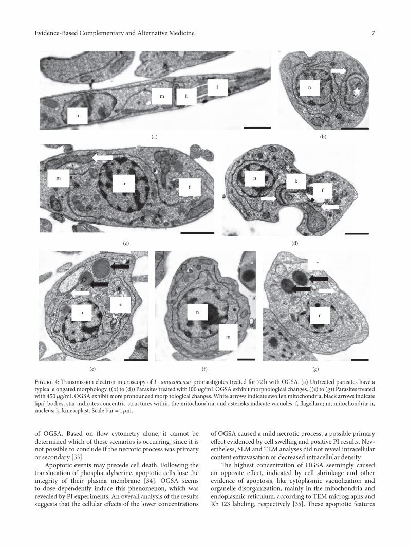
Transmission electron microscopy (TEM) of OGSA-treated promastigotes also indicated the presence of ultra-structural alterations (Figure 4). OGSA induced damagein parasite mitochondria, which presented disorganizedfeatures and the presence of inner concentric membranestructures. Extensive cytoplasmic vacuolization (Figure 4(e))and general disorganization of the organelles (Figure 4(f)),mainly related to the endoplasmic reticulum (Figures 4(b),4(e), and 4(g)), were important changes caused by OGSAtreatment. Alterations in the nucleus, number of flagella, orcytoplasmic membrane were not found by TEM analysis.
3.4. Alteration of Mitochondrial Transmembrane Potential(ΔΨm). TEM demonstrated that OGSA treatment alteredparasite mitochondria, and the ΔΨm was evaluated by flowcytometry using Rh 123, which accumulates inside healthymitochondria. The histograms revealed a decrease in totalRh 123 fluorescence intensity uponOGSA treatment, demon-strating that Rh 123 was not inside the mitochondria, proba-bly due to mitochondrial membrane depolarization. OGSAat concentrations of 100, 450, and 900 𝜇g/mL caused 46.23,60.04, and 75.63% decreases in total Rh 123 fluorescenceintensity, respectively, compared with the gray area, repre-senting the untreated control cells (Figure 5).
3.5. Induction of Phosphatidylserine Exposure. Phosphati-dylserine (PS) is a phospholipid that, under normal bio-chemical conditions, remains on the inner face of theplasma membrane or cytosolic face. During the apoptoticprocess, these molecules are translocated to the cell surface,working as a signal to be phagocytosed from defense cells.Annexin-V has an affinity for phosphatidylserine and thefluorescence emitted from FITC conjugated to annexin-Vsuggests apoptotic or late apoptotic processes if propidiumiodide fluorescence is negative or positive, respectively. Thetypically labeled parasites appeared at the upper and lower-right quadrants on the dot-plot graphic. The intensity ofannexin-V and PI fluorescence was increased up to 78.40%compared to intensity of untreated parasites, suggesting a lateapoptotic process (Figure 6). Annexin-V-FITC was able tolink to the phosphatidylserine on the inner face of the cellmembrane, indicating the loss of membrane integrity, whichcan be observed by the PI fluorescence.
4. Discussion
Current treatments for leishmaniasis are unsatisfactorybecause the parasite is also a eukaryotic organism and sharesmany features with its mammalian cell hosts [25]. Moreover,there has been an increase in the number of cases resistantto the available drugs recommended for treating cutaneousand visceral leishmaniasis. It is imperative to search for new,effective, safe, low-cost agents to replace the current ones [26].

6 Evidence-Based Complementary and Alternative Medicine
104103102101100
FSC-height
100
80
60
40
20
0
Cou
nts
(a)
104103102101100
FSC-height
100
80
60
40
20
0
Cou
nts
(b)
104103102101100
FSC-height
100
80
60
40
20
0
Cou
nts
(c)
104103102101100
FSC-height
100
80
60
40
20
0
Cou
nts
(d)
Figure 3: Cell volume of L. amazonensis promastigotes treated with OGSA fraction at concentrations of 100 𝜇g/mL (a), 450 𝜇g/mL (b), and900 𝜇g/mL (c) and with Actinomycin D (positive control) (d). The gray area corresponds to the untreated control cells.
The use of plants as medicine involves the investigationof active compounds, starting with the isolation of morphinefrom opium in the 19th century [27, 28]. Methods such asfractionation, enrichment of certain substances, isolation,and characterization of pharmacologically relevant com-pounds frommedicinal plants are currently being applied. Inaddition, drug research techniques have been used to stan-dardize herbal formulations and elucidate their compoundsconsidered as analytical markers [29].
In the present study, EHA, SAP, and OGSA exhibitedantiproliferative effects on promastigotes and intracellularamastigotes, along with reduced survival of intracellular par-asites in macrophages. However, OGSAwas the only fractionmore selective for the protozoan than for mammalian cells,and at 100 𝜇g/mL, close to its IC
50for promastigotes, did
not show any degree of hemolysis. In contrast, the hemolyticactivity of SAP was present even at low concentrations, asexpected. Saponins are known to disrupt erythrocytes byinteracting with sterols on erythrocyte membranes, increas-ing cell permeability and causing hemoglobin loss [30].
For these reasons, OGSA was selected and subjected toadditional investigations to study the effects of this com-pound on promastigote forms of L. amazonensis.
OGSA acted like an antileishmanial substance by affect-ing the parasite’s mitochondrial function even at lowerconcentrations, according to the variations in mitochondrialtransmembrane potential (ΔΨm), shown from flow cytom-etry experiments using Rh 123. This potential (ΔΨm) refersto the electrochemical gradient generated by the passage ofelectrons through complexes I to IV of the respiratory chain,resulting in the accumulation of protons between the externaland internal mitochondrial membranes. The return of theseprotons to the mitochondrial matrix through complex Vallows the phosphorylation of ADP to ATP, the main cellularenergy source [31]. Rhodamine 123 accumulates in an inverseproportion toΔΨm, and itmay be an indicator of cell damage.Maintaining transmembrane potential is essential for parasitesurvival [32].
Flow cytometry analysis indicated a late apoptotic processor necrosis after treatment with the higher concentration
Evidence-Based Complementary and Alternative Medicine 7
n
m k
f
(a)
n
(b)
mn
f
(c)
n k
f
(d)
n∗
(e)
n
m
(f)
n
∗
(g)
Figure 4: Transmission electron microscopy of L. amazonensis promastigotes treated for 72 h with OGSA. (a) Untreated parasites have atypical elongatedmorphology. ((b) to (d)) Parasites treatedwith 100 𝜇g/mLOGSA exhibitmorphological changes. ((e) to (g)) Parasites treatedwith 450𝜇g/mLOGSA exhibit more pronouncedmorphological changes.White arrows indicate swollenmitochondria, black arrows indicatelipid bodies, star indicates concentric structures within the mitochondria, and asterisks indicate vacuoles. f, flagellum; m, mitochondria; n,nucleus; k, kinetoplast. Scale bar = 1 𝜇m.
of OGSA. Based on flow cytometry alone, it cannot bedetermined which of these scenarios is occurring, since it isnot possible to conclude if the necrotic process was primaryor secondary [33].
Apoptotic events may precede cell death. Following thetranslocation of phosphatidylserine, apoptotic cells lose theintegrity of their plasma membrane [34]. OGSA seemsto dose-dependently induce this phenomenon, which wasrevealed by PI experiments. An overall analysis of the resultssuggests that the cellular effects of the lower concentrations
of OGSA caused a mild necrotic process, a possible primaryeffect evidenced by cell swelling and positive PI results. Nev-ertheless, SEM and TEM analyses did not reveal intracellularcontent extravasation or decreased intracellular density.
The highest concentration of OGSA seemingly causedan opposite effect, indicated by cell shrinkage and otherevidence of apoptosis, like cytoplasmic vacuolization andorganelle disorganization, mainly in the mitochondria andendoplasmic reticulum, according to TEM micrographs andRh 123 labeling, respectively [35]. These apoptotic features

8 Evidence-Based Complementary and Alternative Medicine
FL1-height104103102101100
120
100
80
60
40
20
0
Cou
nts
(a)
FL1-height104103102101100
120
100
80
60
40
20
0
Cou
nts
(b)
FL1-height104103102101100
120
100
80
60
40
20
0
Cou
nts
(c)
FL1-height104103102101100
120
100
80
60
40
20
0
Cou
nts
(d)
Figure 5: Rhodamine 123-labeledmitochondrialmembrane potential assay by flow cytometry.Thefigure showsL. amazonensispromastigotestreated with 100𝜇g/mL (a), 450 𝜇g/mL (b), and 900 𝜇g/mL (c) and with CCCP (positive control) (d). The gray area corresponds to theuntreated control cells.
appeared gradually and dose-dependently, eventually reach-ing late apoptosis and cell death by loss of cell membraneintegrity, causing changes in the osmotic balance between theparasites and the environment and jeopardizing the selectivepermeability of the plasma membrane. These interpretationsallow us to suggest that necrosis was a secondary effect fromthe highest concentration of OGSA treatment.
In addition, evidence of autophagy, like typical autopha-gosomes, was not found from TEM analysis, which is consid-ered the gold standard to study this process [36].However, theintense disorganization of the endoplasmic reticulum mightcreate these structures after a longer time of OGSA exposurepast 24 h.
Other glycosides have been reported as cell death induc-ers in some studies; however, they always present somecyclization in their chemical structure. The acyclic sesquiter-pene oligoglycosides studied in this paper do not possess anykind of cyclization but still induced cell death, which mayshow that their mechanism of action is distinct. Secoiridoid
glycosides from Swertia chirata cause protozoa cell death byinhibiting the catalytic activity of L. donovani topoisomeraseI [7]. A tetrasaccharide antigen found on the lipophospho-glycan membrane of the genus Leishmania may be exploredusing a synthetic carbohydrate-based vaccine for the treat-ment of leishmaniasis [37]. The quinovic acid glycosidesisolated from N. diderrichii show strong activity againstintracellular amastigotes of L. infantum, and the mechanismin this case was inhibition of parasite internalization in thepromastigote form [8]. In addition, cardiac glycosides areexceptional inducers of immunogenic cell death, an effectthat is associated with inhibition of the plasma membraneNa+/K+ ATPase [38].
In contrast, monodesmosidic saponins, such as thosepresent in S. saponaria L. [39], act specifically on the plasmamembrane of L. infantum promastigotes by nonspecificinteractions with the ergosterol present in these parasites.This interaction causes loss of integrity or changes the neg-ative charge of the carbohydrate portion of cell membranes.

Evidence-Based Complementary and Alternative Medicine 9
3.25%
2.70%
0.59%
FL2
-hei
ght
FL1-height
104
103
102
101
100
104103102101100
(a)
3.74%
1.01% 5.02%
FL2
-hei
ght
FL1-height
104
103
102
101
100
104103102101100
(b)
5.36% 36.10%
5.22%
FL2
-hei
ght
FL1-height
104
103
102
101
100
104103102101100
(c)
5.33% 78.40%
0.90%
FL2
-hei
ght
FL1-height
104
103
102
101
100
104103102101100
(d)
11.77% 48.21%
2,25%
FL2
-hei
ght
FL1-height
104
103
102
101
100
104103102101100
(e)
Figure 6: Phosphatidylserine exposure in untreated L. amazonensis promastigotes (a) and promastigotes treated with 100𝜇g/mL (b),450 𝜇g/mL (c), and 900𝜇g/mL (d) OGSA for 24 h using annexin-V FITC and PI. Miltefosine was used as positive control (e).

10 Evidence-Based Complementary and Alternative Medicine
This peculiar mechanism of action of saponins, which actexclusively on the cell membrane and their integrity tocause necrotic processes, may explain the difference betweenSAP and OGSA regarding their antiproliferative activity, theeffects on intracellular amastigotes, and toxicity in general,since the OGSA fraction greatly affected apoptotic processand the development of cell death.
When biological activities of vegetal species are investi-gated, it is important to determine which plant componentsare responsible for the observed activity. In addition, it isimportant to verify if the crude extract is more or lessactive than its fractions. Besides the investigation of theantiprotozoal and cytotoxic activities of a given sample, it isnecessary to consider other factors to elucidate the effectsof compounds tested on the parasites. Thus, the searchfor morphological, ultrastructural, and biochemical changesis important to help determine the probable mechanismresponsible for cell death.
5. Conclusion
Our results demonstrated inhibition of promastigote andintracellular amastigote forms of L. amazonensis from theextract and fractions of S. saponaria L. However, only theOGSA fraction was selective for different evolutionary formsof L. amazonensis and did not show hemolytic activity.OGSA inducedmorphological and ultrastructural alterationsin promastigote forms, and these results were confirmed bycytometry assays. Thus, our findings suggest that treatingwith high concentrations of the OGSA fraction can have anantileishmanial effect, inducing apoptotic processes followedby necrosis.
Conflicts of Interest
All the authors declare that there are no conflicts of interestin this study.
Acknowledgments
This researchwas supported byConselhoNacional deDesen-volvimento Cientıfico e Tecnologico (CNPq), Coordenacaode Aperfeicoamento de Pessoal de Nıvel Superior (Capes),Financiadora de Estudos e Projetos (FINEP), Complexode Centrais de Apoio a Pesquisa (COMCAP-UEM), andPrograma de Nucleos de Excelencia (PRONEX/FundacaoAraucaria).
References
[1] L. Lovato, B. L. Pelegrini, J. Rodrigues, A. J. Braz de Oliveira,and I. C. Piloto Ferreira, “Seed oil of Sapindus saponaria L.(Sapindaceae) as potential C16 to C22 fatty acids resource,”Biomass and Bioenergy, vol. 60, pp. 247–251, 2014.
[2] O. A. A. Guirado, “Potencial medicinal del genero SapindusL.(Sapindaceae) y de la especie Sapindus saponariaL,” RevistaCubana Planta Medicinal, vol. 10, pp. 3-4, 2005.
[3] A. Sharma, S. C. Sati, D. Sati, Maneesha, and S. K. Kothiyal,“Chemical constituents and bio activities of genus Sapindus,”
International Journal of Research inAyurveda andPharmacy, no.2, pp. 403–409, 2011.
[4] M. Murgu and E. Rodrigues Filho, “Dereplication of glyco-sides from Sapindus saponariausing liquid cromatography-mass spectrometry,” Journal of the Brazilian Chemical Society,vol. 17, no. 7, pp. 1291-1290, 2006.
[5] L. Maes, D. Vanden Berghe, N. Germonprez et al., “In Vitro andIn Vivo Activities of a Triterpenoid Saponin Extract (PX-6518)from the Plant Maesa balansae against Visceral LeishmaniaSpecies,” Antimicrobial Agents and Chemotherapy, vol. 48, no.1, pp. 130–136, 2004.
[6] R. A. I. da Luz, M. Vermeersch, M. Deschacht et al., “In vitroand in vivo prophylactic and curative activity of the triterpenesaponin PX-6518 against cutaneous Leishmania species,” Jour-nal of Antimicrobial Chemotherapy, vol. 66, no. 2, Article IDdkq444, pp. 350–353, 2011.
[7] S. Ray, H. K. Majumder, A. K. Chakravarty, S. Mukhopadhyay,R. R. Gil, and G. A. Cordell, “Amarogentin, a naturally occur-ring secoiridoid glycoside and a newly recognized inhibitor oftopoisomerase I from Leishmania donovani,” Journal of NaturalProducts, vol. 59, no. 1, pp. 27–29, 1996.
[8] C. Di Giorgio, M. Lamidi, F. Delmas, G. Balansard, and E.Ollivier, “Antileishmanial activity of quinovic acid glycosidesand cadambine acid isolated from Nauclea diderrichii,” PlantaMedica, vol. 72, no. 15, pp. 1396–1402, 2006.
[9] W. M. Abdel-Mageed, E. Y. Backheet, A. A. Khalifa, Z. Z.Ibraheim, and S. A. Ross, “Antiparasitic antioxidant phenyl-propanoids and iridoid glycosides fromTecomamollis,” Fitoter-apia, vol. 83, no. 3, pp. 500–507, 2012.
[10] S. A. Grevelink and E. A. Lerner, “Leishmaniasis,” Journal of theAmerican Academy of Dermatology, vol. 34, no. 2 I, pp. 257–272,1996.
[11] O. Genaro, “Leishmaniose Tegumentar Americana,” in Para-sitologia Humana, D. P. Neves, Ed., pp. 41–60, Atheneu, SaoPaulo, Brazil, 9th edition, 1998.
[12] World Health Organization, Parasitic Disease, World HealthOrganization, 2015, http://www.who.int/leishmaniasis/en/index.html.
[13] F. Chappuis, E. Alirol, D. T.Worku, Y.Mueller, and K. Ritmeijer,“High mortality among older patients treated with pentava-lent antimonials for visceral leishmaniasis in east africa andrationale for switch to liposomal amphotericin B,”AntimicrobialAgents and Chemotherapy, vol. 55, no. 1, pp. 455-456, 2011.
[14] R. L. M. Neto, L. M. A. Sousa, C. S. Dias, J. M. B. Filho, M.R. Oliveira, and R. C. B. Q. Figueiredo, “Morphological andphysiological changes in Leishmania promastigotes induced byyangambin, a lignan obtained fromOcotea duckei,” Experimen-tal Parasitology, vol. 127, no. 1, pp. 215–221, 2011.
[15] K. Seifert and S. L. Croft, “In vitroand in vivointeractionsbetween miltefosine and other antileishmanial drugs,” Antimi-crobial Agents and Chemotherapy, vol. 50, no. 1, pp. 73–79, 2006.
[16] V. Kaplum, J. Cogo, D. P. Sangi, T. Ueda-Nakamura, A. G.Correa, and C. V. Nakamura, “In vitro and in vivo activities of2,3-diarylsubstituted quinoxaline derivatives against Leishma-nia amazonensis,” Antimicrobial Agents and Chemotherapy, vol.60, no. 6, pp. 3433–3444, 2016.
[17] T. Mosmann, “Rapid colorimetric assay for cellular growth andsurvival: application to proliferation and cytotoxicity assays,”Journal of Immunological Methods, vol. 65, no. 1-2, pp. 55–63,1983.
[18] E. A. Britta, D. B. Scariot, H. Falzirolli et al., “Cell death andultrastructural alterations in Leishmania amazonensis caused

Evidence-Based Complementary and Alternative Medicine 11
by new compound 4-Nitrobenzaldehyde thiosemicarbazonederived from S-limonene,” BMC Microbiology, vol. 14, no. 1,article no. 236, 2014.
[19] D. B. Scariot, E. A. Britta, A. L. Moreira et al., “Inductionof Early Autophagic Process on Leishmania amazonensis bySynergistic Effect of Miltefosine and Innovative Semi-syntheticThiosemicarbazone,” Front. Microbiol, no. 8, p. 255, 2017.
[20] J. Kleeff, M. Kornmann, H. Sawhney, and M. Korc, “Actino-mycin D induces apoptosis and inhibits growth of pancreaticcancer cells,” International Journal of Cancer, vol. 89, no. 2, pp.399–407, 2000.
[21] M. L. R. Lim, T.Minamikawa, and P.Nagley, “TheprotonophoreCCCP induces mitochondrial permeability transition withoutcytochrome c release in human osteosarcoma cells,” FEBSLetters, vol. 503, no. 1, pp. 69–74, 2001.
[22] F. P. Garcia, J. Henrique da Silva Rodrigues, Z. U. Din et al.,“A3K2A3-induced apoptotic cell death of Leishmania amazo-nensis occurs through caspase- and ATP-dependent mitochon-drial dysfunction,” Apoptosis, vol. 22, no. 1, pp. 57–71, 2017.
[23] C. Paris, P. M. Loiseau, C. Bories, and J. Breard, “Milte-fosine induces apoptosis-like death in Leishmania donovanipromastigotes,” Antimicrobial Agents and Chemotherapy, vol.48, no. 3, pp. 852–859, 2004.
[24] V. Jimenez, R. Paredes, M. A. Sosa, and N. Galanti, “Naturalprogrammed cell death in T. cruzi epimastigotes maintained inaxenic cultures,” Journal of Cellular Biochemistry, vol. 105, no. 3,pp. 688–698, 2008.
[25] D. Lazarin-Bidoia, V. C. Desoti, T. Ueda-Nakamura, B. P. DiasFilho, C. V. Nakamura, and S. O. Silva, “Further evidenceof the trypanocidal action of eupomatenoid-5: Confirmationof involvement of reactive oxygen species and mitochondriaowing to a reduction in trypanothione reductase activity,” FreeRadical Biology and Medicine, vol. 60, pp. 17–28, 2013.
[26] P. R.Murray, K. S. Rosenthal, G. S. Kobayashi, andM. A. Pfaller,Microbiologia Medica, Guanabara Koogan, Rio de Janeiro, 6thedition, 2000.
[27] B. P. da Silva, D. A. Cortez, T. Y. Violin et al., “Antileishmanialactivity of a guaianolide from Tanacetum parthenium (L.)schultz bip,” Parasitology International, vol. 59, no. 4, pp. 643–646, 2010.
[28] A. D. Kinghorn, “Pharmacognosy in the 21st century,” Journal ofPharmacy and Pharmacology, vol. 53, no. 2, pp. 135–148, 2001.
[29] G. Samuelsson, Drugs of Natural Origin: a Textbook of Pharma-cognosy, Swedish Pharmaceutical Press, Stockholm, 5th edition,2004.
[30] M. J. Balunas and A. D. Kinghorn, “Drug discovery frommedicinal plants,” Life Sciences, vol. 78, no. 5, pp. 431–441, 2005.
[31] E. Baumann, G. Stoya, A. Volkner,W. Richter, C. Lemke, andW.Linss, “Hemolysis of human erythrocytes with saponin affectsthe membrane structure,” Acta Histochemica, vol. 102, no. 1, pp.21–35, 2000.
[32] S. W. Perry, J. P. Norman, J. Barbieri, E. B. Brown, and H. A.Gelbard, “Mitochondrial membrane potential probes and theproton gradient: a practical usage guide,” BioTechniques, vol. 50,no. 2, pp. 98–115, 2011.
[33] E. Brauchle, S. Thude, S. Y. Brucker, and K. Schenke-Layland,“Cell death stages in single apoptotic and necrotic cells mon-itored by Raman microspectroscopy,” Scientific Reports, vol. 4,article no. 4698, 2014.
[34] A. J. Kowaltowski and A. E. Vercesi, “Mitochondrial damageinduced by conditions of oxidative stress,” Free Radical Biologyand Medicine, no. 26, pp. 463–471, 1999.
[35] S. Elmore, “Apoptosis: a review of programmed cell death,”Toxicologic Pathology, vol. 35, no. 4, pp. 495–516, 2007.
[36] A. K. Au, H. Bayir, P. M. Kochanek, and R. S. B. Clark,“Evaluation of autophagy using mouse models of brain injury,”Biochimica et Biophysica Acta - Molecular Basis of Disease, vol.1802, no. 10, pp. 918–923, 2010.
[37] M. C. Hewitt and P. H. Seeberger, “Solution and solid-supportsynthesis of a potential leishmaniasis carbohydrate vaccine,”Journal of Organic Chemistry, vol. 66, no. 12, pp. 4233–4243,2001.
[38] L. Menger, E. Vacchelli, S. Adjemian et al., “Cardiac glycosidesexert anticancer effects by inducing immunogenic cell death,”Science Translational Medicine, vol. 4, no. 143, Article ID143ra99, 2012.
[39] F. Delmas, C. Di Giorgio, R. Elias et al., “Antileishmanialactivity of three saponins isolated from ivy, 𝛼- hederin, 𝛽-hederin and hederacolchiside A1, as compared to their actionon mammalian cells cultured in vitro,” Planta Medica, vol. 66,no. 4, pp. 343–347, 2000.