Acute Soft Skull Syndrome in an Adult Male with Sickle...
4
Exploratory Research and Hypothesis in Medicine 2019 vol. 4 | 90–93 Copyright: © 2019 Authors. This is an Open Access article distributed under the terms of the Creative Commons Attribution-Noncommercial 4.0 International License (CC BY-NC 4.0), permitting all non-commercial use, distribution, and reproduction in any medium, provided the original work is properly cited. Case Report Introduction Sickle cell disease (SCD) 1 includes a group of related genetic dis- eases affecting hemoglobin structure, 2 and is considered the most common genetic blood disorder among children. 3 SCD frequently occurs in individuals whose ethnic origin is Africa, 4 Middle East, 5 Asia, 6 Southern Europe, 7 Central or South America, 8 or the Carib- bean. 9 Sudan includes mosaic ethnic diversity that ranges from Af- ro-Arab and Arab to African tribal. 4 This ethnic diversity includes groups with Negroid genetic features, with a recognized history in areas such as Nilotes and Nuba. 10 Acute skull soft syndrome is an uncommon complication of sickle cell anemia. 11 The pathogenesis and mechanism underlying the complication of acute skull soft syndrome is not fully under- stood. In addition, no information has been gained from the pre- vious studies of acute skull soft syndrome for suffers in Sudan. The case of such presented herein is thus novel and of particular importance to Sudanese clinicians, 12 and we hope it will serve to increase knowledge and kindle awareness of this complication in Sudan. Case Report A 20-year old male with a known history of sickle cell anemia, di- agnosed at the age of 2 years-old, came to our clinic at Almawada (a private hospital) on May 18, 2019. The patient complained of headache and generalized joint pain that had begun 1 day prior to clinic presentation. The patient described the headache as constant, with mild frontal pain and not preceded by an aura nor associated or aggravated by anything, and partially relieved by paracetamol tablets. He denied any visual changes, vomiting, altered level of consciousness, neck pain, or variation of headache with positional changes or during the hours of the day. He also reported isolated joint pain involving wrists, knees and hips. The pain had severity score of 6/10 (according to self-judgment) and was dull in qual- ity but not interfering with movement. There was moderate grade fever and the pain had intermittent association with nausea, but no Acute Soft Skull Syndrome in an Adult Male with Sickle Cell Anemia in Sudan: A Case Report Ziryab Imad Taha 1,2* , Sulafa Eisa Mohammed 3 , Mohammed Elmujtba Adam Essa 1,4* , Walaa Mohamed Elsid 2 , Mustafa Mohamed Ali Hussein 1,4 , Sherihan Mohammed Elkundi Osman 4,5 , Hussein Osman Ahmed 4 , Mutwaly Defealla Yousif 1,4 and Abdelkareem A. Ahmed 1,6* 1 Department of Clinical Medicine, Medical and Cancer Research Institute (MCRI), Nyala, Sudan; 2 Department of Internal Medicine, Faculty of Medicine, University of Bahri, Khartoum, Sudan; 3 Department of Internal Medicine, National Ribat University, Khartoum, Sudan; 4 Department of Internal Medicine, Faculty of Medicine, AlFashir University, AlFashir, Sudan; 5 Department of Molecular Medi- cine, Institute of Endemic Diseases, University of Khartoum, Khartoum, Sudan; 6 Department of Physiology and Biochemistry, Faculty of Veterinary Science, University of Nyala, Nyala, Sudan Abstract Acute soſt skull syndrome is an uncommon complicaon of paents with sickle cell anemia. Here, we report a case of an adult paent in Sudan with the acute soſt syndrome, with our aim of providing more knowledge on this type of complicaon. The 20-year old paent, with a known history of sickle cell anemia, presented with a 1-day history of headache and joint pain. The complaint connued aſter admission, with increasing headache severity and development of rapid skull swelling, which indicated the rare sickle cell disease complicaon known as an acute soſt head syndrome. Conservave management resulted in good response and rapid recovery of this case of acute soſt skull syndrome with sickle cell anemia mainly related to skull infracon. Keywords: Adult; Acute soft skull syndrome; Sickle cell anemia; Conservative man- agement. Abbreviations: CT, computed tomography; MRI, magnetic resonance imaging; SCD, sickle cell disease. Received: September 21, 2019; Revised: October 17, 2019; Accepted: November 29, 2019 * Correspondence to: Mohammed Elmujtba Adam Essa Adam, Medical and Cancer Research Institute (MCRI), Department of Clinical Medicine, Faculty of Medicine, AlFashir University, AlFashir, Sudan. Tel: +249907009378, E-mail: Awadali818@ya- hoo.com; Ziryab Imad Taha, Department of Internal Medicine, Faculty of Medicine, University of Bahri, Khartoum, Sudan, Tel: +249912129921, E-mail: Ziryab2008@ yahoo.com; Abdelkareem Abdallah Ahmed, Department of Physiology and Biochem- istry, Faculty of Veterinary Science, University of Nyala, Nyala, P.O. Box: 155 Nyala, Sudan. Fax: +249711833123, E-mail: [email protected] How to cite this article: Taha ZI, Mohammed SE, Essa MEA, Elsid WM, Hus- sein MMA, Osman SME, Ahmed HO, Yousif MD, Ahmed AA. Acute Soft Skull Syndrome in an Adult Male with Sickle Cell Anemia in Sudan: A Case Report. Ex- ploratory Research and Hypothesis in Medicine 2019;4(4):90–93. doi: 10.14218/ ERHM.2019.00024.
Transcript of Acute Soft Skull Syndrome in an Adult Male with Sickle...
Exploratory Research and Hypothesis in Medicine 2019 vol. 4 | 90–93
Copyright: © 2019 Authors. This is an Open Access article distributed under the terms of the Creative Commons Attribution-Noncommercial 4.0 International License (CC BY-NC 4.0), permitting all non-commercial use, distribution, and reproduction in any medium, provided the original work is properly cited.
Case Report
Introduction
Sickle cell disease (SCD)1 includes a group of related genetic dis-eases affecting hemoglobin structure,2 and is considered the most common genetic blood disorder among children.3 SCD frequently occurs in individuals whose ethnic origin is Africa,4 Middle East,5 Asia,6 Southern Europe,7 Central or South America,8 or the Carib-bean.9 Sudan includes mosaic ethnic diversity that ranges from Af-ro-Arab and Arab to African tribal.4 This ethnic diversity includes groups with Negroid genetic features, with a recognized history in
areas such as Nilotes and Nuba.10
Acute skull soft syndrome is an uncommon complication of sickle cell anemia.11 The pathogenesis and mechanism underlying the complication of acute skull soft syndrome is not fully under-stood. In addition, no information has been gained from the pre-vious studies of acute skull soft syndrome for suffers in Sudan. The case of such presented herein is thus novel and of particular importance to Sudanese clinicians,12 and we hope it will serve to increase knowledge and kindle awareness of this complication in Sudan.
Case Report
A 20-year old male with a known history of sickle cell anemia, di-agnosed at the age of 2 years-old, came to our clinic at Almawada (a private hospital) on May 18, 2019. The patient complained of headache and generalized joint pain that had begun 1 day prior to clinic presentation. The patient described the headache as constant, with mild frontal pain and not preceded by an aura nor associated or aggravated by anything, and partially relieved by paracetamol tablets. He denied any visual changes, vomiting, altered level of consciousness, neck pain, or variation of headache with positional changes or during the hours of the day. He also reported isolated joint pain involving wrists, knees and hips. The pain had severity score of 6/10 (according to self-judgment) and was dull in qual-ity but not interfering with movement. There was moderate grade fever and the pain had intermittent association with nausea, but no
Acute Soft Skull Syndrome in an Adult Male with Sickle Cell Anemia in Sudan: A Case Report
Ziryab Imad Taha1,2*, Sulafa Eisa Mohammed3, Mohammed Elmujtba Adam Essa1,4*, Walaa Mohamed Elsid2, Mustafa Mohamed Ali Hussein1,4,
Sherihan Mohammed Elkundi Osman4,5, Hussein Osman Ahmed4, Mutwaly Defealla Yousif1,4 and Abdelkareem A. Ahmed1,6*
1Department of Clinical Medicine, Medical and Cancer Research Institute (MCRI), Nyala, Sudan; 2Department of Internal Medicine, Faculty of Medicine, University of Bahri, Khartoum, Sudan; 3Department of Internal Medicine, National Ribat University, Khartoum,
Sudan; 4Department of Internal Medicine, Faculty of Medicine, AlFashir University, AlFashir, Sudan; 5Department of Molecular Medi-cine, Institute of Endemic Diseases, University of Khartoum, Khartoum, Sudan; 6Department of Physiology and Biochemistry, Faculty of
Veterinary Science, University of Nyala, Nyala, Sudan
Abstract
Acute soft skull syndrome is an uncommon complication of patients with sickle cell anemia. Here, we report a case of an adult patient in Sudan with the acute soft syndrome, with our aim of providing more knowledge on this type of complication. The 20-year old patient, with a known history of sickle cell anemia, presented with a 1-day history of headache and joint pain. The complaint continued after admission, with increasing headache severity and development of rapid skull swelling, which indicated the rare sickle cell disease complication known as an acute soft head syndrome. Conservative management resulted in good response and rapid recovery of this case of acute soft skull syndrome with sickle cell anemia mainly related to skull infraction.
Keywords: Adult; Acute soft skull syndrome; Sickle cell anemia; Conservative man-agement.Abbreviations: CT, computed tomography; MRI, magnetic resonance imaging; SCD, sickle cell disease.Received: September 21, 2019; Revised: October 17, 2019; Accepted: November 29, 2019*Correspondence to: Mohammed Elmujtba Adam Essa Adam, Medical and Cancer Research Institute (MCRI), Department of Clinical Medicine, Faculty of Medicine, AlFashir University, AlFashir, Sudan. Tel: +249907009378, E-mail: [email protected]; Ziryab Imad Taha, Department of Internal Medicine, Faculty of Medicine, University of Bahri, Khartoum, Sudan, Tel: +249912129921, E-mail: [email protected]; Abdelkareem Abdallah Ahmed, Department of Physiology and Biochem-istry, Faculty of Veterinary Science, University of Nyala, Nyala, P.O. Box: 155 Nyala, Sudan. Fax: +249711833123, E-mail: [email protected] to cite this article: Taha ZI, Mohammed SE, Essa MEA, Elsid WM, Hus-sein MMA, Osman SME, Ahmed HO, Yousif MD, Ahmed AA. Acute Soft Skull Syndrome in an Adult Male with Sickle Cell Anemia in Sudan: A Case Report. Ex-ploratory Research and Hypothesis in Medicine 2019;4(4):90–93. doi: 10.14218/ERHM.2019.00024.
DOI: 10.14218/ERHM.2019.00024 | Volume 4 Issue 4, December 2019 91
Taha ZI. et al: Adult Sudanese male with soft skull syndrome Explor Res Hypothesis Med
shivering or rigger. The patient also reported a burning sensation during micturition. There were no complaints involving any other organ systems.
The patient had a history of multiple previous hospital ad-missions, due to sickle cell crises. He had no history of diabetes mellitus, hypertension or any similar conditions, but he had been diagnosed 1 month previous with gallstone (“not operated”). The patient was on regular folic acid tablets, 5 mg daily, and not known to be allergic to any type of medication. On examination, the pa-tient looked ill and in pain, and was pale and jaundiced. Vital signs were all within normal parameters, except for the body tempera-ture at 38 °C. Blood pressure was 120/80 and heart rate was 70 beats/m. The patient was fully conscious and oriented when the central nervous system tests were performed. The Mini-Mental State Examination (commonly referred to as the ‘MMSE’) score was 30/30. All cranial nerves were intact, and Kernig’s and Brudz-inski’s signs were negative.
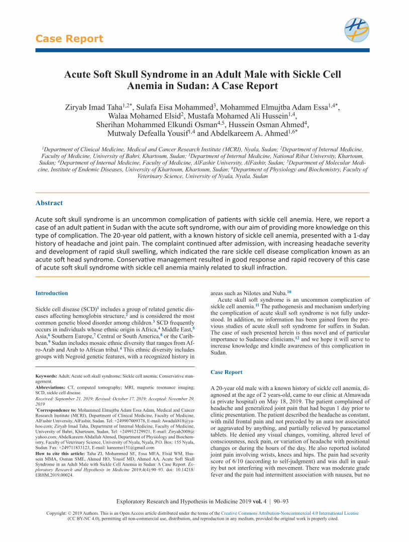
Upper limbs showed no muscle wasting or fasciculations, and there was intact normal tone sensation; power was grade 5, and deep tendon reflex was +2 bilaterally. The lower limbs showed no muscle wasting or fasciculations, normal tone and intact sensa-tion; power grade was 5, deep tendon reflex was +2 bilaterally, and gait was normal. Musculoskeletal examination of hands showed no swelling, tenderness, deformities, muscle wasting, skin rash or signs of dactylitis, and normal temperature and intact radial pulses. Phalen’s test was negative, with both knees and hips showing not swelling nor hotness but tenderness was present; the range of move-ment was normal. The cardiovascular examination also showed normal first and second heart sounds, with no murmurs or signs of heart failure. Clear breath sounds were heard bilaterally in chest examination. Results from blood sample testing for complete blood count, renal function and blood glucose are summarized in Table 1.Upon admission, the patient was immediately begun on intrave-nous normal saline (1 L/8 hours) with ciprofloxacin infusion (200 mg intravenously every 12 hours), tramadol tablets (once per day), paracetamol infusion (1 g/ 6 hours), and 4 U of packed red blood cells. On the third day of admission, the headache reached its maxi-mum, being associated with scalp swelling and tenderness (Fig. 1). There was no loss of consciousness, visual change, projectile vom-iting or neck stiffness. The patient denied history of head trauma, and stated he had never experienced such symptoms before. Again, the patient was negative for Kernig’s and Brudzinski’s signs. Brain
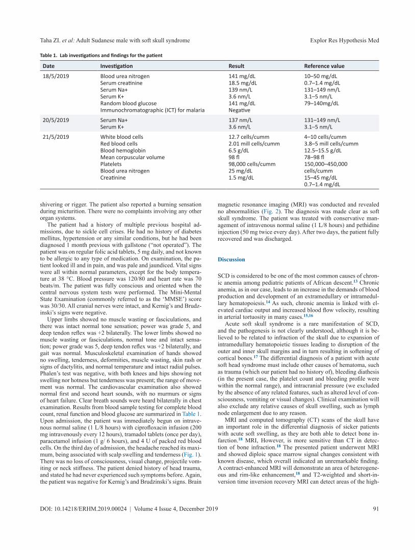
magnetic resonance imaging (MRI) was conducted and revealed no abnormalities (Fig. 2). The diagnosis was made clear as soft skull syndrome. The patient was treated with conservative man-agement of intravenous normal saline (1 L/8 hours) and pethidine injection (50 mg twice every day). After two days, the patient fully recovered and was discharged.
Discussion
SCD is considered to be one of the most common causes of chron-ic anemia among pediatric patients of African descent.13 Chronic anemia, as in our case, leads to an increase in the demands of blood production and development of an extramedullary or intramedul-lary hematopoiesis.14 As such, chronic anemia is linked with el-evated cardiac output and increased blood flow velocity, resulting in arterial tortuosity in many cases.15,16
Acute soft skull syndrome is a rare manifestation of SCD, and the pathogenesis is not clearly understood, although it is be-lieved to be related to infraction of the skull due to expansion of intramedullary hematopoietic tissues leading to disruption of the outer and inner skull margins and in turn resulting in softening of cortical bones.17 The differential diagnosis of a patient with acute soft head syndrome must include other causes of hematoma, such as trauma (which our patient had no history of), bleeding diathesis (in the present case, the platelet count and bleeding profile were within the normal range), and intracranial pressure (we excluded by the absence of any related features, such as altered level of con-sciousness, vomiting or visual changes). Clinical examination will also exclude any relative causes of skull swelling, such as lymph node enlargement due to any reason.
MRI and computed tomography (CT) scans of the skull have an important role in the differential diagnosis of sicker patients with acute soft swelling, as they are both able to detect bone in-farction.18 MRI, However, is more sensitive than CT in detec-tion of bone infraction.18 The presented patient underwent MRI and showed diploic space marrow signal changes consistent with known disease, which overall indicated an unremarkable finding. A contract-enhanced MRI will demonstrate an area of heterogene-ous and rim-like enhancement,18 and T2-weighted and short-in-version time inversion recovery MRI can detect areas of the high-
Table 1. Lab investigations and findings for the patient
Date Investigation Result Reference value
18/5/2019 Blood urea nitrogenSerum creatinineSerum Na+Serum K+Random blood glucoseImmunochromatographic (ICT) for malaria
141 mg/dL18.5 mg/dL139 nm/L3.6 nm/L141 mg/dLNegative
10–50 mg/dL0.7–1.4 mg/dL131–149 nm/L3.1–5 nm/L79–140mg/dL
20/5/2019 Serum Na+Serum K+
137 nm/L3.6 nm/L
131–149 nm/L3.1–5 nm/L
21/5/2019 White blood cellsRed blood cellsBlood hemoglobinMean corpuscular volumePlateletsBlood urea nitrogenCreatinine
12.7 cells/cumm2.01 mill cells/cumm6.5 g/dL98 fl98,000 cells/cumm25 mg/dL1.5 mg/dL
4–10 cells/cumm3.8–5 mill cells/cumm12.5–15.5 g/dL78–98 fl150,000–450,000 cells/cumm15–45 mg/dL0.7–1.4 mg/dL
DOI: 10.14218/ERHM.2019.00024 | Volume 4 Issue 4, December 201992
Taha ZI. et al: Adult Sudanese male with soft skull syndromeExplor Res Hypothesis Med
Fig. 1. During and after recovery from the acute attack of soft skull syndrome. (a) and (b) shows the patient with acute soft skull syndrome. (c) Shows the patient after recovery.
Fig. 2. Brain magnetic resonance imaging scans of the patient, indicating left maxillary polypoid sinusitis and diploic space marrow signal changes consist-ent with known disease. In conclusion: unremarkable study.

DOI: 10.14218/ERHM.2019.00024 | Volume 4 Issue 4, December 2019 93
Taha ZI. et al: Adult Sudanese male with soft skull syndrome Explor Res Hypothesis Med
intensity bone marrow infarction and edema.18 Although, neither one of those investigations could afforded by our patient.
Acute soft head syndrome due to sickle cell anemia is well known to be resolved by the same treatment of other sickle cell crises, that being conservative management (intravenous fluids and analgesics).18 Our patient did very well after receiving the in-travenous fluids and tramadol.
In conclusion, this complication of SCD is uncommon but other reported cases along with ours serve to emphasize the need and desire for recognizing skull infarction, perhaps resulting in sub-periosteal hematomas as causes of skull swellings and headache in sickle cell patients.
Patient consent
Consent was obtained from the patient.
Ethical approval
Ethical approval was obtained from Sudan Federal Ministry of health.
Financial disclosure
No funds or financial aid were received for this study.
Conflict of interest
All authors declare there are no conflicts of interest.
Author contributions
Case diagnosed and treatment supervision (ZIT), follow up (SEA, WME, SMEO), drafting of the manuscript (MEAE), analysis and interpretation (MMAH, HOA), critical revision of the manu-script for important intellectual content (MDY), study supervision (AAA).
References
[1] Alvarez OA, Hustace T, Voltaire M, Mantero A, Liberus U, Saint Fleur R. Newborn screening for sickle cell disease using point-of-care testing in low-income setting. Pediatrics 2019;144(4):e20184105. doi:10.1542/peds.2018-4105.
[2] Du S, Lin C, Tao YX. Updated mechanisms underlying sickle cell dis-ease-associated pain. Neurosci Lett 2019;712:134471. doi:10.1016/j.
neulet.2019.134471.[3] Pace BS, Ofori-Acquah SF, Peterson KR. Sickle cell disease: genet-
ics, cellular and molecular mechanisms, and therapies. Anemia 2012;2012:143594. doi:10.1155/2012/143594.
[4] Sabahelzain MM, Hamamy H. The ethnic distribution of sickle cell disease in Sudan. Pan Afr Med J 2014;18:13. doi:10.11604/pamj.2014.18.13.3280.
[5] El-Hazmi MAF, Al-Hazmi AM, Warsy AS. Sickle cell disease in Mid-dle East Arab countries. Indian J Med Res 2011;134(5):597–610. doi:10.4103/0971-5916.90984.
[6] Piel FB, Patil AP, Howes RE, Nyangiri OA, Gething PW, Williams TN, et al. Global distribution of the sickle cell gene and geographical confirmation of the malaria hypothesis. Nat Commun 2010;1:104. doi:10.1038/ncomms1104.
[7] Aguilar Martinez P, Angastiniotis M, Eleftheriou A, Gulbis B, Mañú Pereira MDM, Petrova-Benedict R, et al. Haemoglobinopathies in Europe: health & migration policy perspectives. Orphanet J Rare Dis 2014;9:97. doi:10.1186/1750-1172-9-97.
[8] Serjeant GR. The natural history of sickle cell disease. Cold Spring Harb Perspect Med 2013;3(10):a011783. doi:10.1101/cshperspect.a011783.
[9] Solovieff N, Hartley SW, Baldwin CT, Klings ES, Gladwin MT, Tay-lor JG6th, et al. Ancestry of African Americans with sickle cell disease. Blood Cells Mol Dis 2011;47(1):41–45. doi:10.1016/j.bcmd.2011.04.002.
[10] Babiker HM, Schlebusch CM, Hassan HY, Jakobsson M. Genetic vari-ation and population structure of Sudanese populations as indicated by 15 Identifiler sequence-tagged repeat (STR) loci. Investig Genet 2011;2(1):12. doi:10.1186/2041-2223-2-12.
[11] Akodu SO, Njokanma OF, Diaku-Akinwumi IN, Ubuane PO, Adediji UO. Acute soft head syndrome in children with sickle cell anaemia in lagos, Nigeria. Indian J Hematol Blood Transfus 2014;30(Suppl 1):67–69. doi:10.1007/s12288-013-0251-6.
[12] Elagib EM, Eltahir NIA, Essa Adam MEA, Yousif Haron MD, Mahmoud ZIT, Mohamed Yousif HH, et al. Catastrophic antiphospholipid syn-drome in combination with SLE treated by Rituximab: a case report and literature review. Lupus: Open Access 2019;4(1):137.
[13] Bello-Manga H, DeBaun MR, Kassim AA. Epidemiology and treat-ment of relative anemia in children with sickle cell disease in sub-Saharan Africa. Expert Rev Hematol 2016;9(11):1031–1042. doi:10.1080/17474086.2016.1240612.
[14] Alli NA, Wainwright RD, Mackinnon D, Poyiadjis S, Naidu G. Skull bone infarctive crisis and deep vein thrombosis in homozygous sickle cell disease- case report and review of the literature. Hematology 2007;12(2):169–174. doi:10.1080/10245330601111912.
[15] Phillips JK, Boyd R, Krockenberger MB, Burgio G. Progression of ane-mia and its relationship with renal function, blood pressure, and erythropoietin in rats with chronic kidney disease. Vet Clin Pathol 2015;44(3):342–354. doi:10.1111/vcp.12276.
[16] Moghe S, Pillai A, Guru KN, Nair PP. Idiopathic facial swelling second-ary to sickle cell anaemia. BMJ Case Rep 2012;2012:bcr2012007132. doi:10.1136/bcr-2012-007132.
[17] Neves FS, Oliveira LS, Torres MG, Toralles MB, da Silva MC, Cam-pos MI, et al. Evaluation of panoramic radiomorphometric indices related to low bone density in sickle cell disease. Osteoporos Int 2012;23(7):2037–2042. doi:10.1007/s00198-011-1810-z.
[18] Saito N, Nadgir RN, Flower EN, Sakai O. Clinical and radiologic mani-festations of sickle cell disease in the head and neck. Radiographics 2010;30(4):1021–1034. doi:10.1148/rg.304095171.


![[Mohamed (Mohamed El-Sharkawi) El-Sharkawi] Fundam](https://static.fdocuments.net/doc/165x107/577c781b1a28abe0548ec3ac/mohamed-mohamed-el-sharkawi-el-sharkawi-fundam.jpg)


![y± ; ~ d G y ± G · 2018-11-09 · -erhm -iptivw 'pyi 1mri x (mx] [mhi zer wivzmgi jsv mrhmzmhyepw erh kvsytw x 2ywx fi fssoih mr ehzergi x ,vsyt vexiw ezempefpi -erhm 8verwmx x](https://static.fdocuments.net/doc/165x107/5e6a6cd56429da078c646810/y-d-g-y-g-2018-11-09-erhm-iptivw-pyi-1mri-x-mx-mhi-zer-wivzmgi.jpg)