Acute Sinusitis Mimicking Antrochoanal Polyp - AJNR · Acute Sinusitis Mimicking Antrochoanal Polyp...
4
Matilde Nino-Murcia' Vijay M. Rao' Diran O. Mikaelian 2 Peter 80m 3 Received April 4, 1985; accepted after revision August 23, 1985 1 Department of Radiology, Thomas Jefferson University Hospital, Philadelphia, PA 19107. Ad- dress reprint requests to Vijay M. Rao. 2 Department of Otolaryngology, Thomas Jeffer- son University Hospital, Philadelphia, PA 19107. 3 Department of Radiology, Mt. Si nai School of Medicine, New York , NY 10029. AJNR 7:513-516, May/June 1986 0195-6108/86/0703-0513 © American Society of Neuroradiology Acute Sinusitis Mimicking Antrochoanal Polyp 513 Three cases of antral sinusitis that presented in an unusual fashion are reported. In each case, the inflamed, swollen sinus mucosa became redundant and prolapsed into the nasal cavity through a widened sinus ostium, mimicking the findings of an antro- choana I polyp. The cases represent the first report in the radiographic literature of this unusual manifestation of maxillary sinusitis. Antral sinusitis is a common condition whose radiographic and computed tomog- raphy (CT) manifestations have been well described [1-4]. These include: muco- periosteal thickening, air-fluid levels, sinus opacification, and bony changes. An antrochoanal polyp is a relatively unusual condition in which an antral polyp enlarges, widens the maxillary ostium, prolapses into the nasal cavity, and presents clinically as a nasal polyp. The radiographic and CT findings in this condition have also been well described [1, 2, 3, 5]. This paper presents three cases of acute sinusitis that had the radiographic and CT findings of an antrochoanal polyp, but were found at surgery to be caused by prolapse of hypertrophic sinus mucosa into the nose. Until now, these particular radiographic manifestations of sinusitis have not been described in the literature. Representative Case Reports Case 1 A 50-year-old black woman with a 2-month history of clear rhinorrhea and with chronic renal failure was admitted. There was no history of trauma or of exposure to dust or fumes . At physical examination, the right nasal cavity showed edematous mucosa, congestion, and deformity of the turbinates. Plain films and tomograms of the paranasal sinuses revealed a right antral soft-tissue mass that extended into the nasal cavity via a widened sinus ostium. A soft-tissue density was also seen in the inferior portion of the right ethmoid sinus (Fig. 1 A) . CT confirmed the above findings (Figs. 1 Band 1C), and the preoperative diagnosis was sinusitis with a probable antrochoanal polyp. The patient underwent a right Caldwell-Luc procedure with nasal antrostomy. Abundant purulent material and inflammatory mucosa were present; however, no gross polyp or cyst was found. Cultures were positive for light growth of a mixed flora. Pathologic examination revealed inflamed mucosa without evidence of polyp formation . Case 2 A 45-year-old black woman with a 10-year history of chronic sinusitis presented with left frontal pain and left nasal purulent discharge. Routine sinus films revealed opacification of the left maxillary antrum. Tomography and CT of the paranasal sinuses revealed opacification of the left maxillary antrum with extension of a soft-tissue density from the antrum into the nasal fossa through a widened sinus ostium. There was diffuse sclerosis of the remaining sinus walls (Figs. 2A and 2B). The preoperative diagnosis was chronic sinusitis with a probable antrochoanal polyp. At surgery, abundant purulent material was suctioned from the left
Transcript of Acute Sinusitis Mimicking Antrochoanal Polyp - AJNR · Acute Sinusitis Mimicking Antrochoanal Polyp...
Matilde Nino-Murcia' Vijay M. Rao'
Diran O. Mikaelian2
Peter 80m3
Received April 4, 1985; accepted after revision August 23, 1985
1 Department of Radiology, Thomas Jefferson University Hospital, Philadelphia, PA 19107. Address reprint requests to Vijay M. Rao.
2 Department of Otolaryngology, Thomas Jefferson University Hospital, Philadelphia, PA 19107.
3 Department of Radiology, Mt. Sinai School of Medicine, New York, NY 10029.
AJNR 7:513-516, May/June 1986 0195-6108/86/0703-0513 © American Society of Neuroradiology
Acute Sinusitis Mimicking Antrochoanal Polyp
513
Three cases of antral sinusitis that presented in an unusual fashion are reported. In each case, the inflamed, swollen sinus mucosa became redundant and prolapsed into the nasal cavity through a widened sinus ostium, mimicking the findings of an antrochoana I polyp. The cases represent the first report in the radiographic literature of this unusual manifestation of maxillary sinusitis.
Antral sinusitis is a common condition whose radiographic and computed tomography (CT) manifestations have been well described [1-4]. These include: mucoperiosteal thickening, air-fluid levels, sinus opacification, and bony changes. An antrochoanal polyp is a relatively unusual condition in which an antral polyp enlarges, widens the maxillary ostium, prolapses into the nasal cavity, and presents clinically as a nasal polyp. The radiographic and CT findings in this condition have also been well described [1, 2, 3, 5]. This paper presents three cases of acute sinusitis that had the radiographic and CT findings of an antrochoanal polyp, but were found at surgery to be caused by prolapse of hypertrophic sinus mucosa into the nose. Until now, these particular radiographic manifestations of sinusitis have not been described in the literature.
Representative Case Reports
Case 1
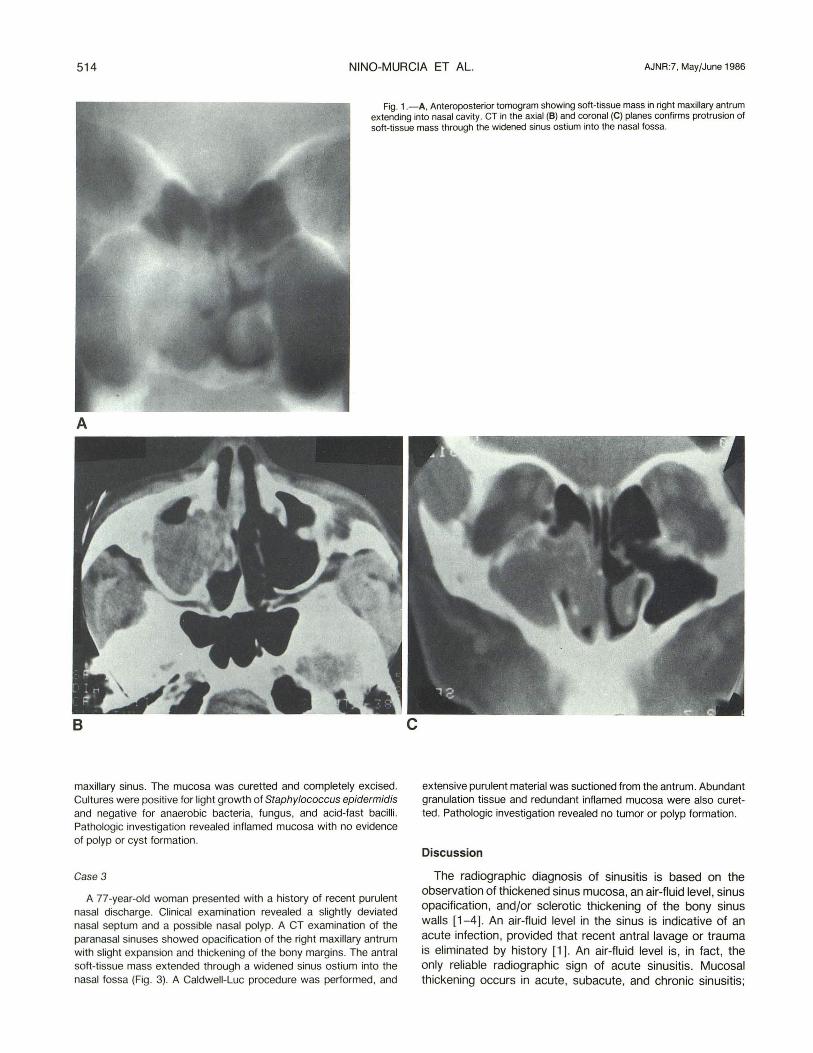
A 50-year-old black woman with a 2-month history of clear rhinorrhea and with chronic renal failure was admitted. There was no history of trauma or of exposure to dust or fumes . At physical examination , the right nasal cavity showed edematous mucosa, congestion , and deformity of the turbinates. Plain films and tomograms of the paranasal sinuses revealed a right antral soft-tissue mass that extended into the nasal cavity via a widened sinus ostium. A soft-tissue density was also seen in the inferior portion of the right ethmoid sinus (Fig . 1 A). CT confirmed the above findings (Figs . 1 Band 1 C), and the preoperative diagnosis was sinusitis with a probable antrochoanal polyp. The patient underwent a right Caldwell-Luc procedure with nasal antrostomy. Abundant purulent material and inflammatory mucosa were present; however, no gross polyp or cyst was found. Cultures were positive for light growth of a mixed flora. Pathologic examination revealed inflamed mucosa without evidence of polyp formation .
Case 2
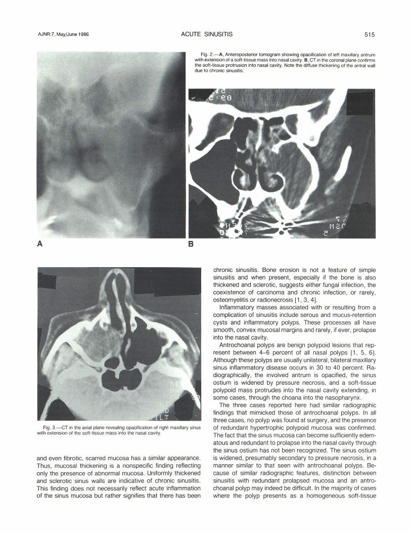
A 45-year-old black woman with a 10-year history of chronic sinusitis presented with left frontal pain and left nasal purulent discharge. Routine sinus films revealed opacification of the left maxillary antrum. Tomography and CT of the paranasal sinuses revealed opacification of the left maxillary antrum with extension of a soft-tissue density from the antrum into the nasal fossa through a widened sinus ostium. There was diffuse sclerosis of the remaining sinus walls (Figs. 2A and 2B). The preoperative diagnosis was chronic sinusitis with a probable antrochoanal polyp. At surgery , abundant purulent material was suctioned from the left
514 NINO-MURCIA ET AL. AJNR:7, May/June 1986
Fig. 1.-A, Anteroposterior tomogram showing soft-tissue mass in right maxillary antrum extending into nasal cavity. CT in the axial (8) and coronal (C) planes confirms protrusion of soft-tissue mass through the widened sinus ostium into the nasal fossa.
A
maxillary sinus. The mucosa was curetted and completely excised. Cultures were positive for light growth of Staphylococcus epidermidis and negative for anaerobic bacteria, fungus, and acid-fast bacilli. Pathologic investigation revealed inflamed mucosa with no evidence of polyp or cyst formation .
Case 3
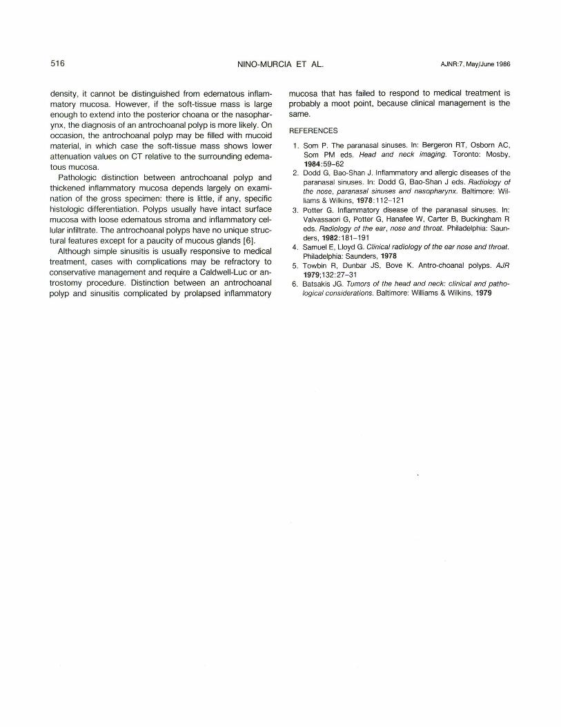
A 77-year-old woman presented with a history of recent purulent nasal discharge. Clinical examination revealed a slightly deviated nasal septum and a possible nasal polyp. A CT examination of the paranasal sinuses showed opacification of the right maxillary antrum with slight expansion and thickening of the bony margins. The antral soft-tissue mass extended through a widened sinus ostium into the nasal fossa (Fig . 3). A Caldwell-Luc procedure was performed, and
extensive purulent material was suctioned from the antrum. Abundant granulation tissue and redundant inflamed mucosa were also curetted . Pathologic investigation revealed no tumor or polyp formation .
Discussion
The radiographic diagnosis of sinusitis is based on the observation of thickened sinus mucosa, an air-fluid level, sinus opacification, and/or sclerotic thickening of the bony sinus walls [1-4]. An air-fluid level in the sinus is indicative of an acute infection, provided that recent antral lavage or trauma is eliminated by history [1]. An air-fluid level is, in fact, the only reliable radiographic sign of acute sinusitis. Mucosal thickening occurs in acute, subacute, and chronic sinusitis;
AJNR:7, May/June 1986 ACUTE SINUSITIS 515
Fig . 2.-A, Anteroposterior tomogram showing opacification of left maxillary antrum with extension of a soft-tissue mass into nasal cavity. B, CT in the coronal plane confirms the soft-tissue protrusion into nasal cavity . Note the diffuse thickening of the antral wall due to chronic sinusitis.
A B
Fig. 3.-CT in the axial plane revealing opacification of right maxillary sinus with extension of the soft-tissue mass into the nasal cavity.
and even fibrotic , scarred mucosa has a similar appearance. Thus, mucosal thickening is a nonspecific finding reflecting only the presence of abnormal mucosa. Uniformly thickened and sclerotic sinus walls are indicative of chronic sinusitis. This finding does not necessarily reflect acute inflammation of the sinus mucosa but rather signifies that there has been
chronic sinusitis. Bone erosion is not a feature of simple sinusitis and when present, especially if the bone is also thickened and sclerotic, suggests either fungal infection, the coexistence of carcinoma and chronic infection, or rarely , osteomyelitis or radionecrosis [1, 3, 4].
Inflammatory masses associated with or resulting from a complication of sinusitis include serous and mucus-retention cysts and inflammatory polyps . These processes all have smooth, convex mucosal margins and rarely, if ever, prolapse into the nasal cavity.
Antrochoanal polyps are benign polypoid lesions that represent between 4-6 percent of all nasal polyps [1 , 5, 6]. Although these polyps are usually unilateral , bilateral maxillary sinus inflammatory disease occurs in 30 to 40 percent. Radiographically, the involved antrum is opacified , the sinus ostium is widened by pressure necrosis, and a soft-tissue polypoid mass protrudes into the nasal cavity extending, in some cases, through the choana into the nasopharynx.
The three cases reported here had similar radiographic findings that mimicked those of antrochoanal polyps. In all three cases, no polyp was found at surgery, and the presence of redundant hypertrophic polypoid mucosa was confirmed . The fact that the sinus mucosa can become sufficiently edematous and redundant to prolapse into the nasal cavity through the sinus ostium has not been recognized. The sinus ostium is widened , presumably secondary to pressure necrosis, in a manner similar to that seen with antrochoanal polyps. Because of similar radiographic features , distinction between sinusitis with redundant prolapsed mucosa and an antrochoanal polyp may indeed be difficult. In the majority of cases where the polyp presents as a homogeneous soft-tissue
516 NINO-MURCIA ET AL. AJNR:7, May/June 1986
density, it cannot be distinguished from edematous inflammatory mucosa. However, if the soft-tissue mass is large enough to extend into the posterior choana or the nasopharynx , the diagnosis of an antrochoanal polyp is more likely. On occasion, the antrochoanal polyp may be filled with mucoid material , in which case the soft-tissue mass shows lower attenuation values on CT relative to the surrounding edematous mucosa.
Pathologic distinction between antrochoanal polyp and thickened inflammatory mucosa depends largely on examination of the gross specimen: there is little, if any, specific histologic differentiation. Polyps usually have intact surface mucosa with loose edematous stroma and inflammatory cellular infiltrate. The antrochoanal polyps have no unique structural features except for a paucity of mucous glands [6].
Although simple sinusitis is usually responsive to medical treatment, cases with complications may be refractory to conservative management and require a Caldwell-Luc or antrostomy procedure. Distinction between an antrochoanal polyp and sinusitis complicated by prolapsed inflammatory
mucosa that has failed to respond to medical treatment is probably a moot pOint, because clinical management is the same.
REFERENCES
1. Som P. The paranasal sinuses. In: Bergeron RT, Osborn AC, Som PM eds. Head and neck imaging. Toronto: Mosby, 1984:59-62
2. Dodd G, Bao-Shan J. Inflammatory and allergic diseases of the paranasal sinuses. In: Dodd G, Bao-Shan J eds. Radiology of the nose, paranasal sinuses and nasopharynx. Baltimore: Williams & Wilkins , 1978 :112-121
3. Potter G. Inflammatory disease of the paranasal sinuses. In: Valvassaori G, Potter G, Hanafee W, Carter B, Buckingham R eds. Radiology of the ear, nose and throat. Philadelphia: Saunders, 1982:181-191
4. Samuel E, Lloyd G. Clinical radiology of the ear nose and throat. Philadelphia: Saunders, 1978
5. Towbin R, Dunbar JS, Bove K. Antro-choanal polyps. AJR 1979;132:27-31
6. Batsakis JG. Tumors of the head and neck: clinical and pathological considerations. Baltimore: Williams & Wilkins , 1979