Acute Respiratory Failure - atimures.ro · Anesthesia & Intensive Care 2014 2 Respiratory failure...
86
Anesthesia & Intensive Care 2014 1 Acute Respiratory Failure Sanda-Maria Copotoiu
Transcript of Acute Respiratory Failure - atimures.ro · Anesthesia & Intensive Care 2014 2 Respiratory failure...
Anesthesia & Intensive Care 2014 1
Acute Respiratory Failure
Sanda-Maria Copotoiu
Anesthesia & Intensive Care 2014 2
Respiratory failure
• Definition
• Classification
• Physiology recall
• Diagnosis
• Respiratory monitoring
• Management
Anesthesia & Intensive Care 2014 3
Aims
1. Diagnose – rapid & accurate
2. Manage in due time the syndrome
3. Understand

Anesthesia & Intensive Care 2014 4
Respiratory failure
Definition
Acute or chronic impairment of respiratory
system function to maintain normal O2
and CO2 values when breathing room air.
Oxygenation failure
paO2 < normal predicted values for age & altitude
Inspired O2 concentration
V/P mismatch
Anesthesia & Intensive Care 2014 5
Respiratory failure
Ventilatory failure
CO2 eliminationpaCO2 >45mmHg
Most common causes:
Exacerbation of COPD
Asthma
Neuromuscular fatigue
Dyspnoea, tachypnoea, tachycardia,
accessory muscles of ventilation, altered
counsciousness
Anesthesia & Intensive Care 2014 6
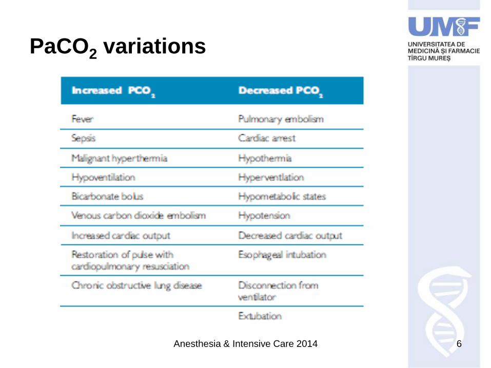
PaCO2 variations
Anesthesia & Intensive Care 2014 7
Classification criteria
Pathophysiology
Time
Etiology
Anesthesia & Intensive Care 2014 8
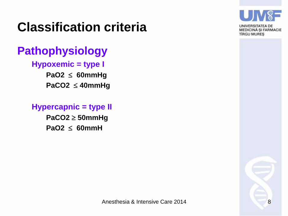
Classification criteria
PathophysiologyHypoxemic = type I
PaO2 60mmHg
PaCO2 40mmHg
Hypercapnic = type II
PaCO2 50mmHg
PaO2 60mmH
Anesthesia & Intensive Care 2014 9
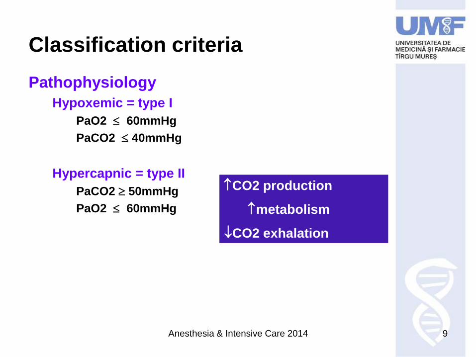
Classification criteria
Pathophysiology
Hypoxemic = type I
PaO2 60mmHg
PaCO2 40mmHg
Hypercapnic = type II
PaCO2 50mmHg
PaO2 60mmHg
CO2 production
metabolism
CO2 exhalation
Anesthesia & Intensive Care 2014 10
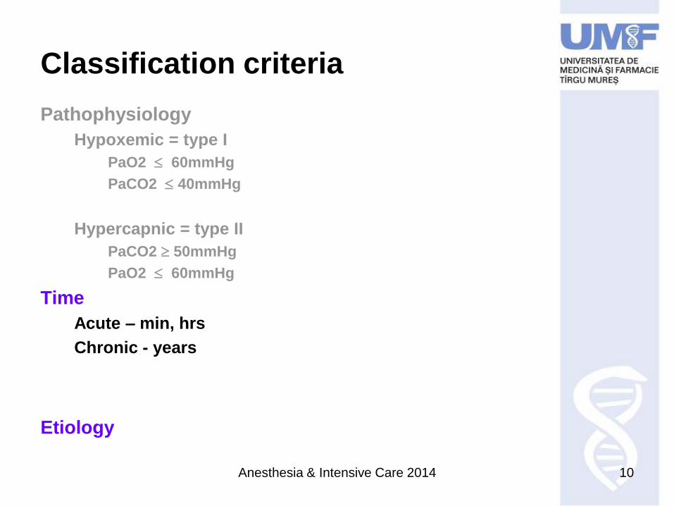
Classification criteria
Pathophysiology
Hypoxemic = type I
PaO2 60mmHg
PaCO2 40mmHg
Hypercapnic = type II
PaCO2 50mmHg
PaO2 60mmHg
Time
Acute – min, hrs
Chronic - years
Etiology
Anesthesia & Intensive Care 2014 11
Classification criteria
Etiology
• CNS
• Spinal cord
• Neuromuscular system
• Chest wall
• Airways – upper, lower
• Lung parenchyma
• CV system
Anesthesia & Intensive Care 2014 12
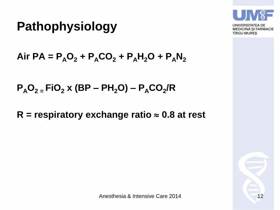
Pathophysiology
Air PA = PAO2 + PACO2 + PAH2O + PAN2
PAO2 = FiO2 x (BP – PH2O) – PACO2/R
R = respiratory exchange ratio 0.8 at rest
Anesthesia & Intensive Care 2014 13
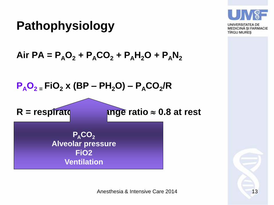
Pathophysiology
Air PA = PAO2 + PACO2 + PAH2O + PAN2
PAO2 = FiO2 x (BP – PH2O) – PACO2/R
R = respiratory exchange ratio 0.8 at rest
PACO2
Alveolar pressure
FiO2
Ventilation
Anesthesia & Intensive Care 2014 14
Oxygen cascade
Anesthesia & Intensive Care 2014 15
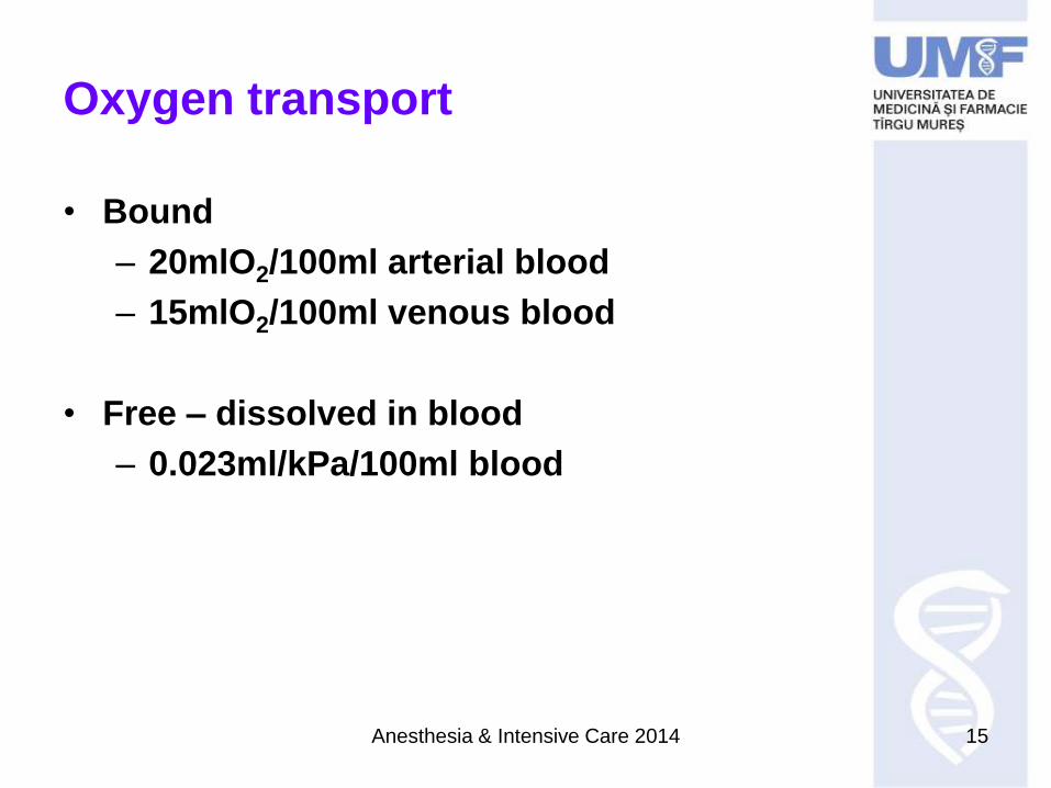
Oxygen transport
• Bound
– 20mlO2/100ml arterial blood
– 15mlO2/100ml venous blood
• Free – dissolved in blood
– 0.023ml/kPa/100ml blood
Anesthesia & Intensive Care 2014 16
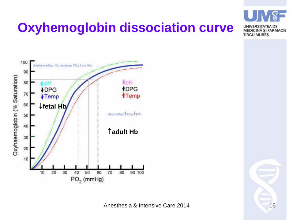
Oxyhemoglobin dissociation curve
adult Hb
fetal Hb
Anesthesia & Intensive Care 2014 17
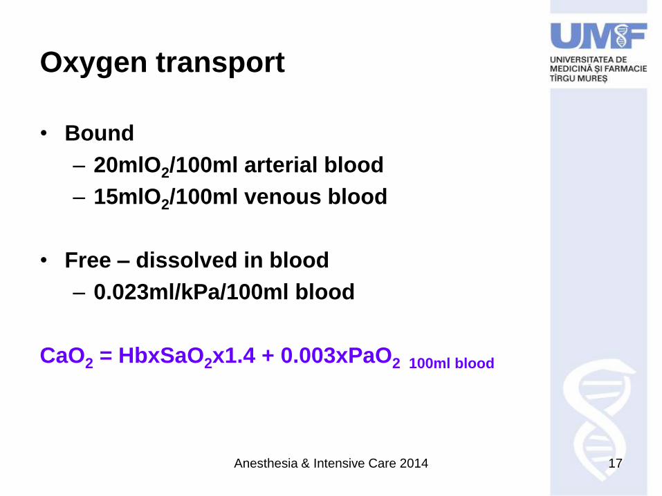
Oxygen transport
• Bound
– 20mlO2/100ml arterial blood
– 15mlO2/100ml venous blood
• Free – dissolved in blood
– 0.023ml/kPa/100ml blood
CaO2 = HbxSaO2x1.4 + 0.003xPaO2 100ml blood
Anesthesia & Intensive Care 2014 18
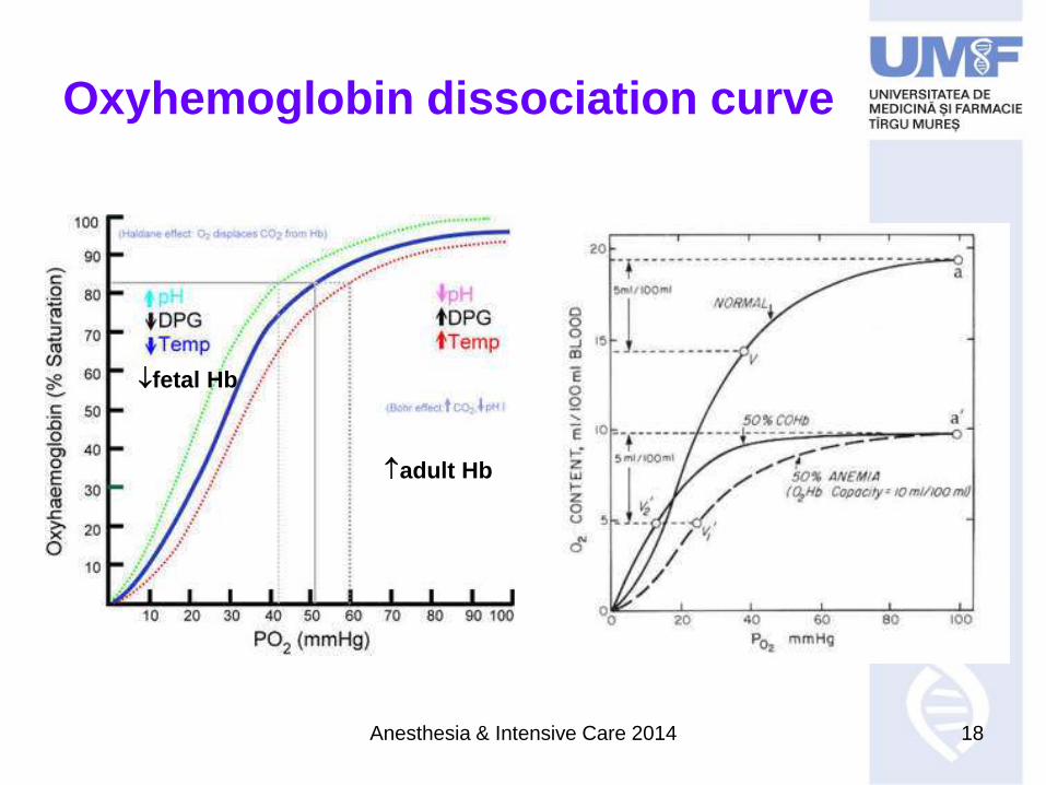
Oxyhemoglobin dissociation curve
adult Hb
fetal Hb
Anesthesia & Intensive Care 2014 19
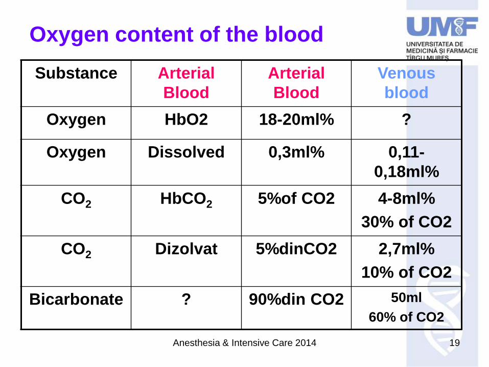
Oxygen content of the blood
Substance Arterial
Blood
Arterial
Blood
Venous
blood
Oxygen HbO2 18-20ml% ?
Oxygen Dissolved 0,3ml% 0,11-
0,18ml%
CO2 HbCO2 5%of CO2 4-8ml%
30% of CO2
CO2 Dizolvat 5%dinCO2 2,7ml%
10% of CO2
Bicarbonate ? 90%din CO2 50ml
60% of CO2
Anesthesia & Intensive Care 2014 20
Gas exchange
Lung units: alveoli + capillaries
Diffusion abnormalities
Anesthesia & Intensive Care 2014 21
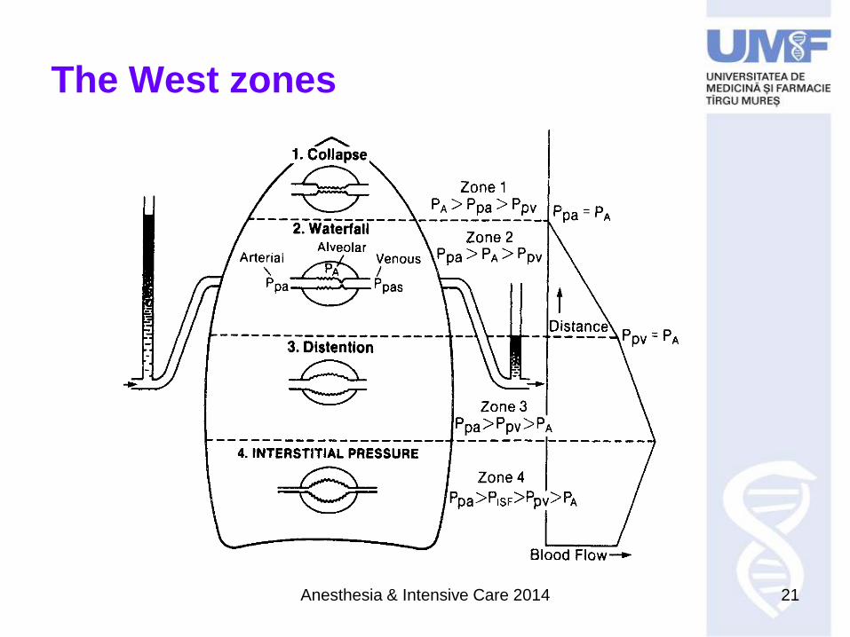
The West zones
Anesthesia & Intensive Care 2014 22
V/P
V/P = 1 ideal unit
V/P < 1 underventilated, normally perfused
V/P > 1 overventilation, underperfusion
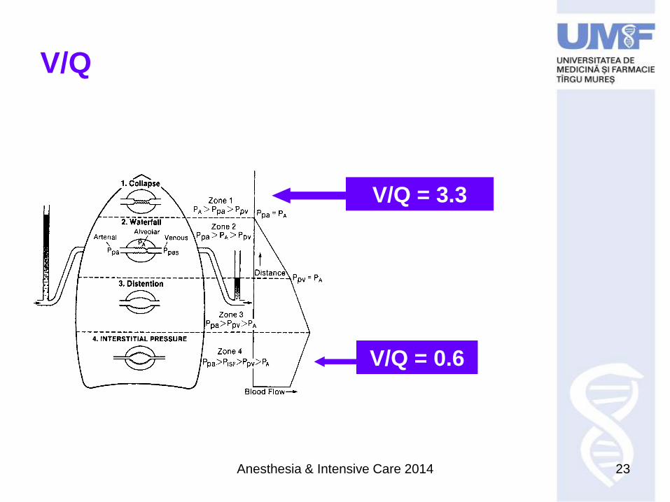
Anesthesia & Intensive Care 2014 23
V/Q
V/Q = 3.3
V/Q = 0.6
Anesthesia & Intensive Care 2014 24
Intrapulmonary shunt
• True V/Q = 0
• Shunt fraction; Qs 1 > V/Q > 0
Pulmonary vasoconstriction
Anesthesia & Intensive Care 2014 25
Intrapulmonary shunt
• True V/Q = 0
• Shunt fraction; Qs 1 > V/Q > 0
Pulmonary vasoconstriction
Causes
• Pneumonia
• Lung edema
• Atelectasis
• Collapse
• Pulmonary hemorrhage
• Lung contusion
Anesthesia & Intensive Care 2014 26
A-a gradient = difference
diagnosis
• a shunt
• a diffusion abnormality
Alveolar gas equation
PA = FiO2 x (BP – H2O) – PACO2/R
5 mmHg (0.5-1kPa) – 15mmHg
>15-20mmHg = lung disease
Anesthesia & Intensive Care 2014 27
Dead space ventilation
a. Air reaching only the conducting airways
= anatomic dead space
b. Air to the alveoli inert as to gas
exchange with the capillaries
A + b = physiologic dead space
dead space ventilation = 20-30% of VT
VD/VT = 0.2-0.3
Anesthesia & Intensive Care 2014 28
Dead space ventilation
• CO
• intra-alveolar pressure
stretching the alveolar capillaries
Anesthesia & Intensive Care 2014 29
Alveolar hypoventilation
• paCO2 O2
• Brainstem
– Trauma, haemorrhage, infarction, hypoxia,
infection.
– Metabolic encephalopathy
– Depressant drugs
• Spinal cord
– Trauma, tumor, transverse myelitis
• Nerve root injury
Anesthesia & Intensive Care 2014 30
Alveolar hypoventilation cont
• Nerve
– Trauma
– Neuropathy
– Motor neuron disease
• Nm junction
– Myastenia gravis
– Nm blocade
• Respiratory muscle fatigue
– Disuse atrophy
– Myopathy
– Malnutrition
• Respiratory system
– Airway obstruction (upper or
lower)
– Decreased lung, pleural or chest
wall compliance
Anesthesia & Intensive Care 2014 31
Lung compliance
• Volume change/unit pressure =ΔV/ ΔP
• Lung compliance 200ml/cmH2O
• Chest compliance 100ml/H2O
Anesthesia & Intensive Care 2014 32
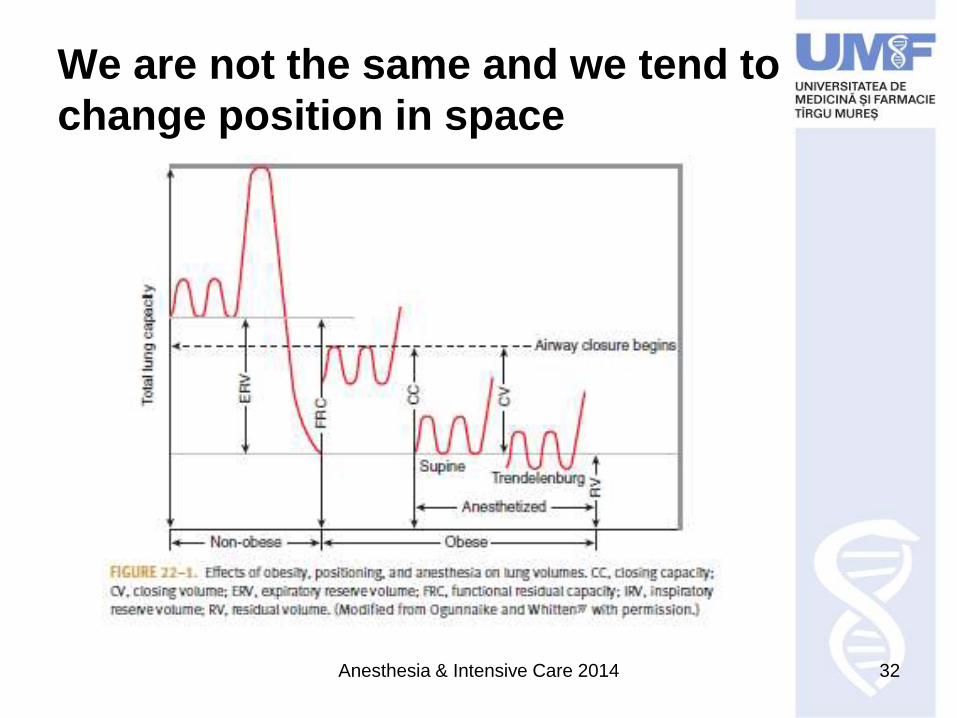
We are not the same and we tend to
change position in space
Anesthesia & Intensive Care 2014 33
Cost of breathing
2-3% of oxygen delivery
Anesthesia & Intensive Care 2014 34
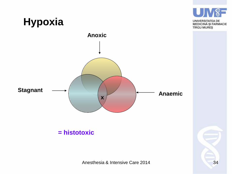
HypoxiaAnoxic
StagnantAnaemic
x
= histotoxic
Anesthesia & Intensive Care 2014 35
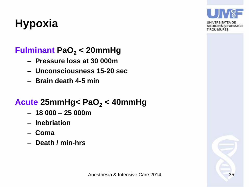
Hypoxia
Fulminant PaO2 < 20mmHg
– Pressure loss at 30 000m
– Unconsciousness 15-20 sec
– Brain death 4-5 min
Acute 25mmHg< PaO2 < 40mmHg
– 18 000 – 25 000m
– Inebriation
– Coma
– Death / min-hrs
Anesthesia & Intensive Care 2014 36
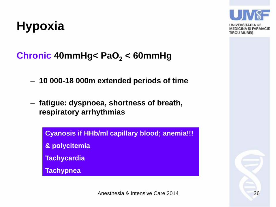
Hypoxia
Chronic 40mmHg< PaO2 < 60mmHg
– 10 000-18 000m extended periods of time
– fatigue: dyspnoea, shortness of breath,
respiratory arrhythmias
Cyanosis if HHb/ml capillary blood; anemia!!!
& polycitemia
Tachycardia
Tachypnea
Anesthesia & Intensive Care 2014 37
Hyperoxia
• FiO2 > 0.6
• Acute
• Chronic oxygen toxicity
Anesthesia & Intensive Care 2014 38
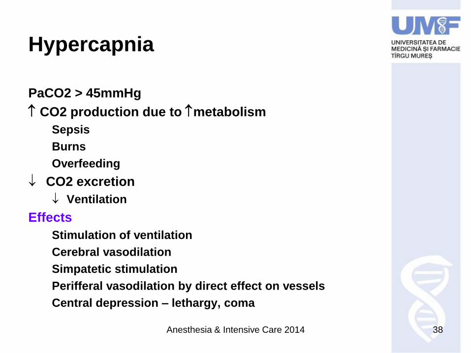
Hypercapnia
PaCO2 > 45mmHg
CO2 production due to metabolism
Sepsis
Burns
Overfeeding
CO2 excretion
Ventilation
Effects
Stimulation of ventilation
Cerebral vasodilation
Simpatetic stimulation
Perifferal vasodilation by direct effect on vessels
Central depression – lethargy, coma
Anesthesia & Intensive Care 2014 39
Hypercapnia
PaCO2 > 45mmHg CO2 production due to metabolism
Sepsis
Burns
Overfeeding
CO2 excretion
Ventilation
EffectsStimulation of ventilation
Cerebral vasodilation
Simpatetic stimulation
Perifferal vasodilation by direct effect on vessels
Central depression – lethargy, coma
Anesthesia & Intensive Care 2014 40
Hypercapnia
PaCO2 > 45mmHg CO2 production due to metabolism
Sepsis
Burns
Overfeeding
CO2 excretion
Ventilation
EffectsStimulation of ventilation
Cerebral vasodilation
Simpatetic stimulation
Perifferal vasodilation by direct effect on vessels
Central depression – lethargy, coma
Permissive hypercapnia
CO due to sympathetic
activity
splanchnic & renal blood flow
Anesthesia & Intensive Care 2014 41
Hypocapnia
PaCO2 35mmHg
– Cerebral vasoconstriction
• Ca pl muscle excitability
– Alcalosis
Anesthesia & Intensive Care 2014 42
Hypocapnia
PaCO2 35mmHg
– Cerebral vasoconstriction
• Ca pl muscle excitability
– Alcalosis
Address the cause!
Anesthesia & Intensive Care 2014 43
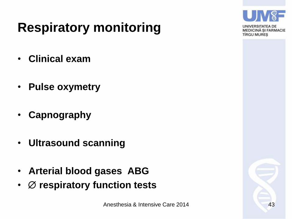
Respiratory monitoring
• Clinical exam
• Pulse oxymetry
• Capnography
• Ultrasound scanning
• Arterial blood gases ABG
• respiratory function tests
Anesthesia & Intensive Care 2014 44
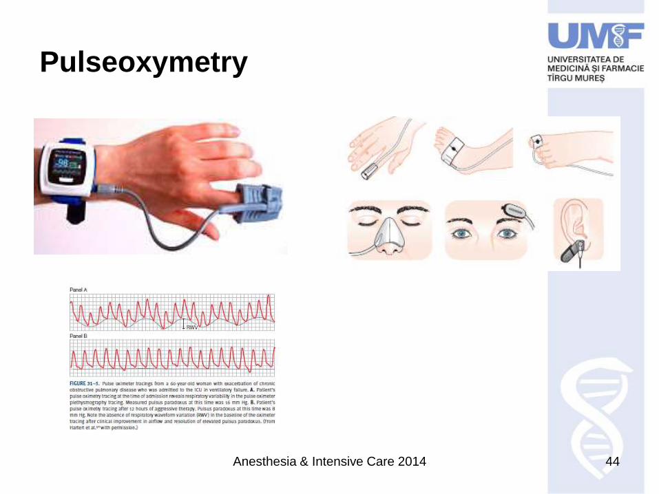
Pulseoxymetry
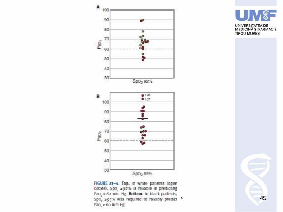
Anesthesia & Intensive Care 2014 45
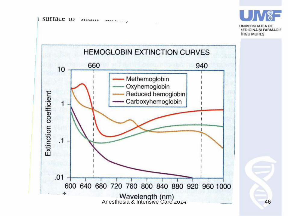
Anesthesia & Intensive Care 2014 46
Anesthesia & Intensive Care 2014 47
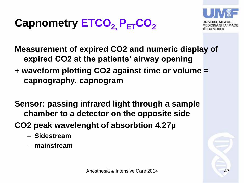
Capnometry ETCO2, PETCO2
Measurement of expired CO2 and numeric display of
expired CO2 at the patients’ airway opening
+ waveform plotting CO2 against time or volume =
capnography, capnogram
Sensor: passing infrared light through a sample
chamber to a detector on the opposite side
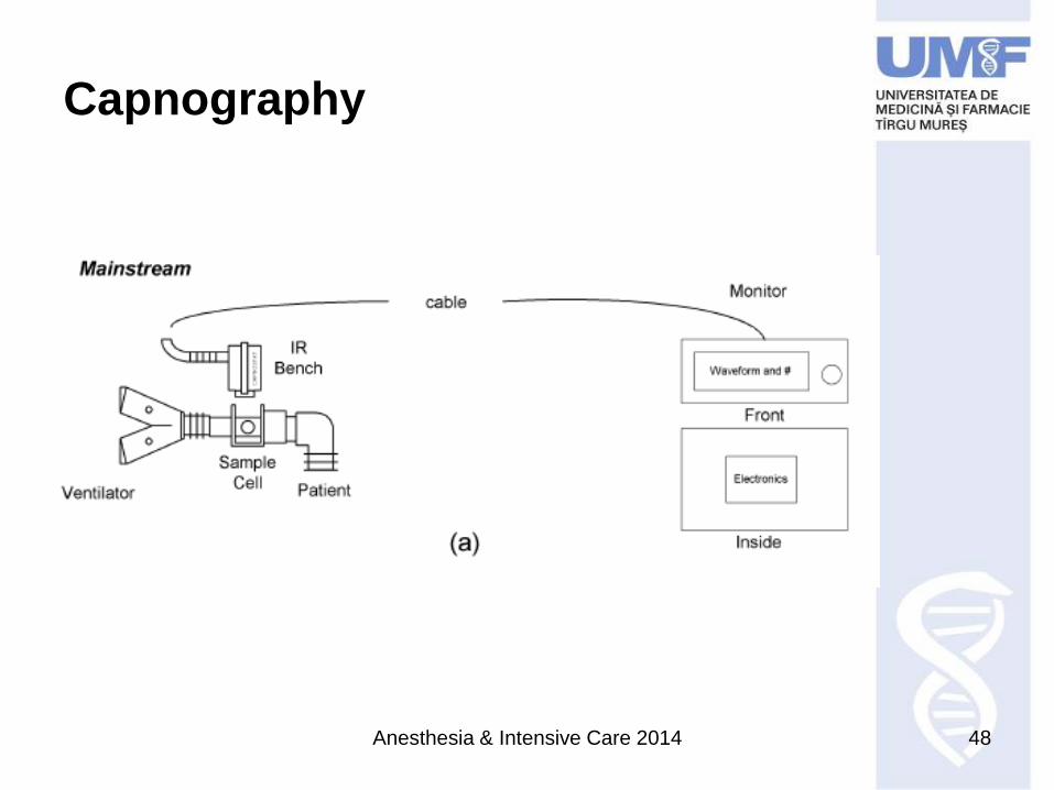
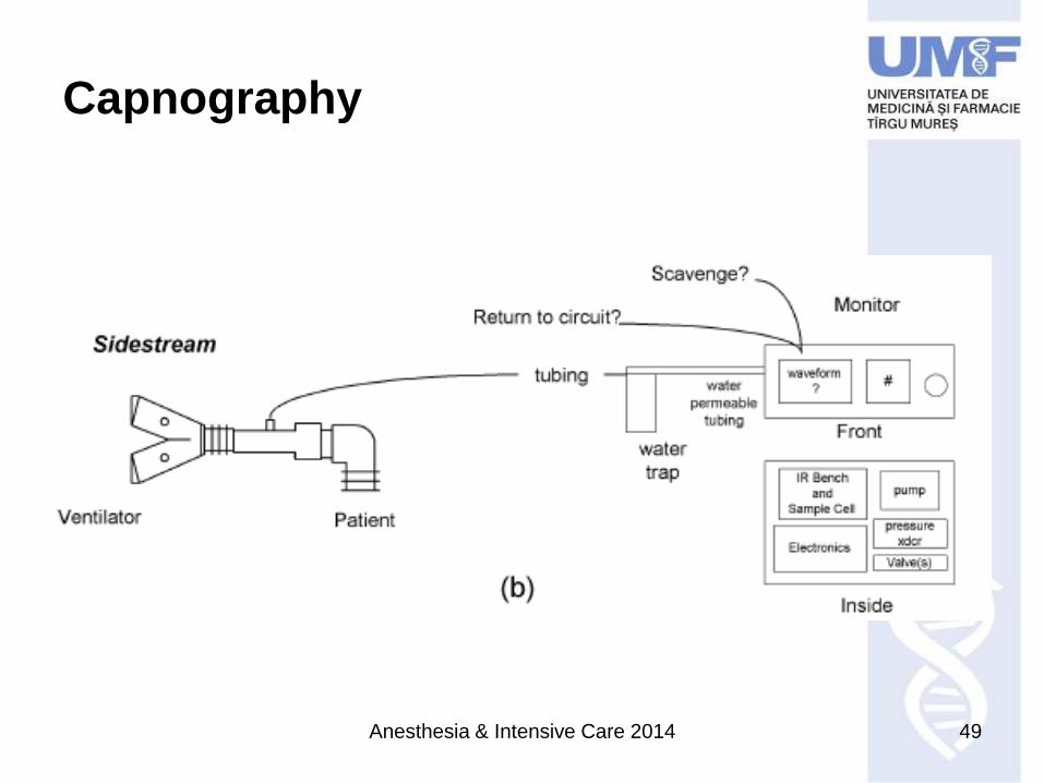
CO2 peak wavelenght of absorbtion 4.27μ
– Sidestream
– mainstream
Anesthesia & Intensive Care 2014 48
Capnography
Anesthesia & Intensive Care 2014 49
Capnography
Anesthesia & Intensive Care 2014 50
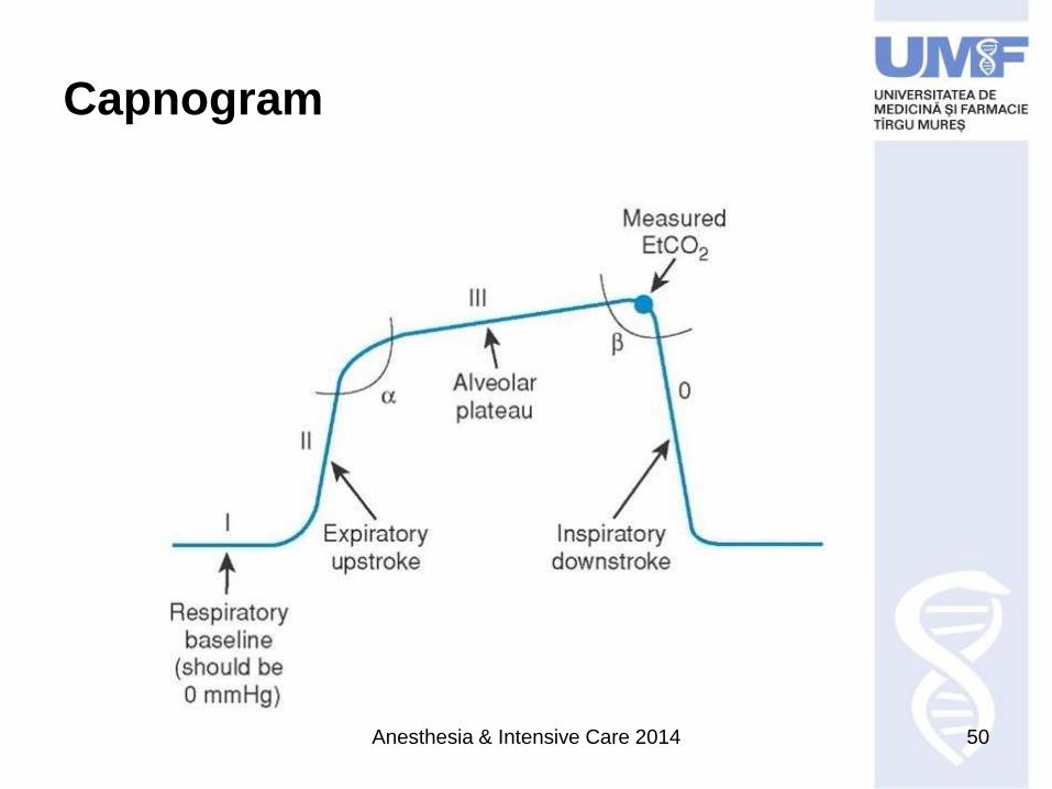
Capnogram
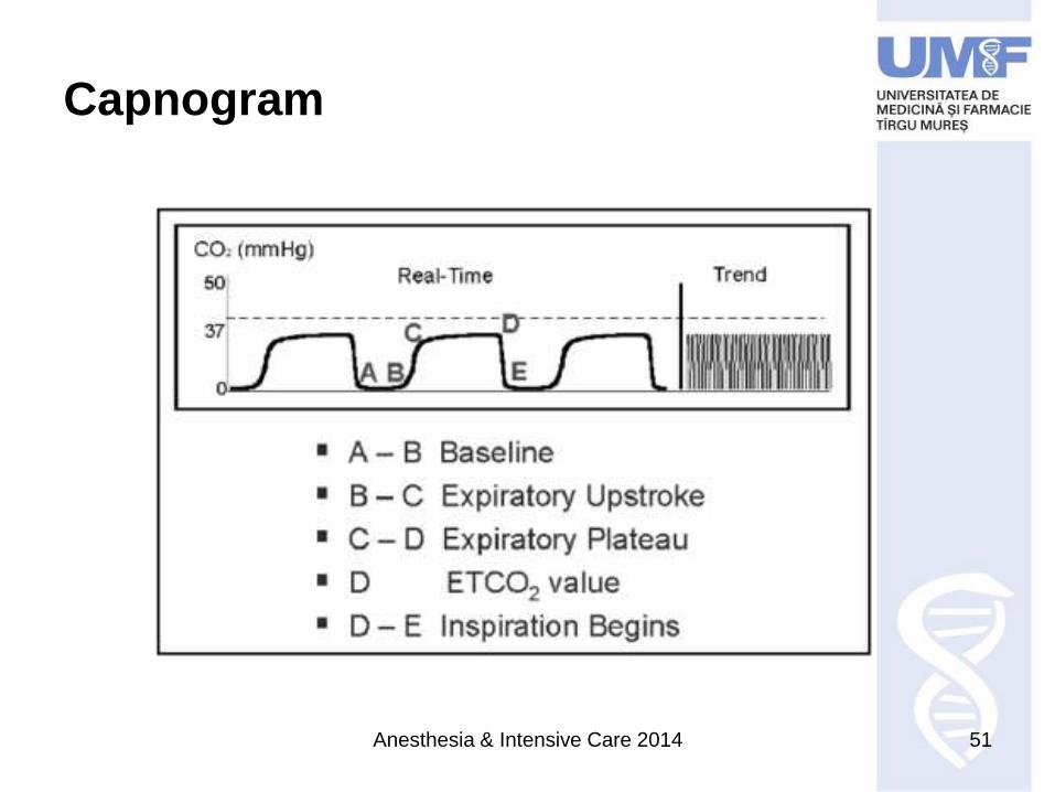
Anesthesia & Intensive Care 2014 51
Capnogram
Anesthesia & Intensive Care 2014 52
Capnography indications
• Diagnosis of pulmonary embolism
• Assessing lung recruitment response to PEEP
• Detection of intrinsic PEEP
• Evaluation of weaning
• Indirect marker of elevated dead space ventilation
• Assessment of CP resuscitation
• Indirect CO measuring by CO2 rebreathing
• Verification of endotracheal cannulation
• Detection of airway accidents
• Determination of feeding tube placement
Anesthesia & Intensive Care 2014 53
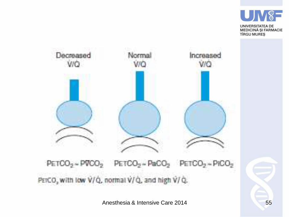
PaCO2 – PETCO2 gradient
• normal 4-5mmHg
• Critically ill pts
• Eg: COPD 7-16mmHg
• ALI, cardiogenic PE: 4-12mmHg
Caused by:
paCO2 reflects mean PCO2 in alveolar gas
PETCO2 approximates peak PaCO2
Anesthesia & Intensive Care 2014 54
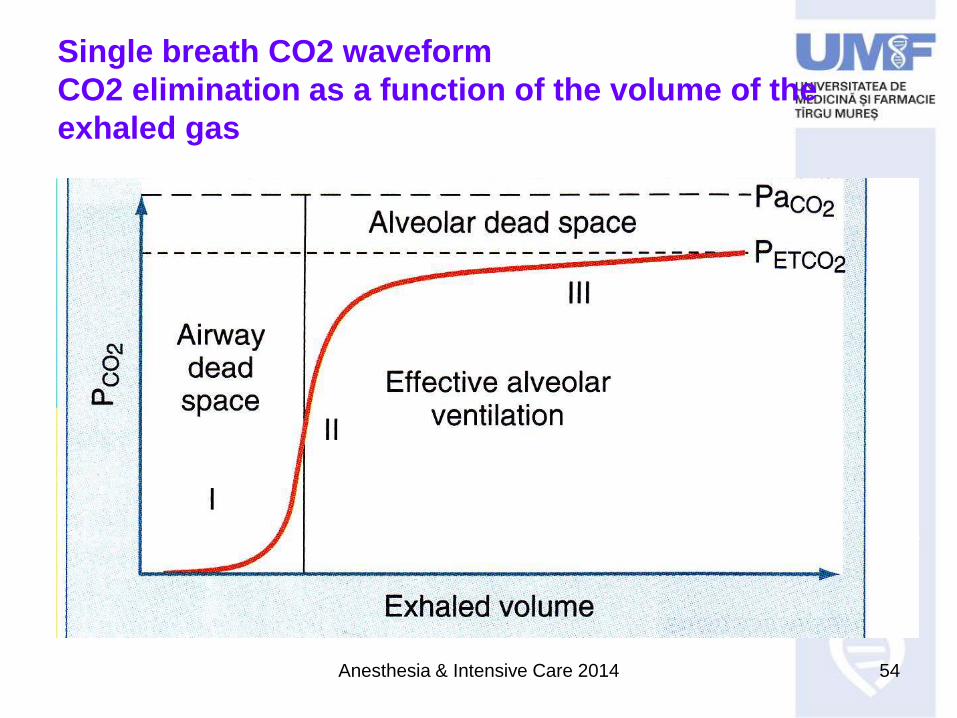
Single breath CO2 waveform
CO2 elimination as a function of the volume of the
exhaled gas
Anesthesia & Intensive Care 2014 55
Anesthesia & Intensive Care 2014 56
Management of ARF
a. Diagnosis & treatment of the underlying condition
b. Respiratory support adequate oxygenation to
the tissues since hypoxaemia is deleterious and
rapid reversal critical.
Anesthesia & Intensive Care 2014 57
Respiratory support
• Oxygen supplementation
• Mechanical ventilation
• Physical therapy
Anesthesia & Intensive Care 2014 58
Why do we need MV?
a. ARF or imminent despita maximal
treatment
Anesthesia & Intensive Care 2014 59
Why do we need MV?
a. ARF or imminent despita maximal
treatment
b. Following major surgery in GA
Anesthesia & Intensive Care 2014 60
Why do we need MV?
a. ARF or imminent despita maximal
treatment
b. Following major surgery in GA
c. Cardiogenic shock – to reduce the
oxygen cost of ventilation when CO
Anesthesia & Intensive Care 2014 61
Aspirations vs reality
Ideally:
MV would replicate the mechanics and physiology of
spontaneous respiration reaching adequate
oxygenation and ventilation.
Anesthesia & Intensive Care 2014 62
Aspirations vs reality
Ideally:
MV would replicate the mechanics and physiology of
spontaneous respiration reaching adequate
oxygenation and ventilation.
Reality
MV works with positive pressure during inspiration,
exhaling to athmospheric pressure or to a preset
PEEP.
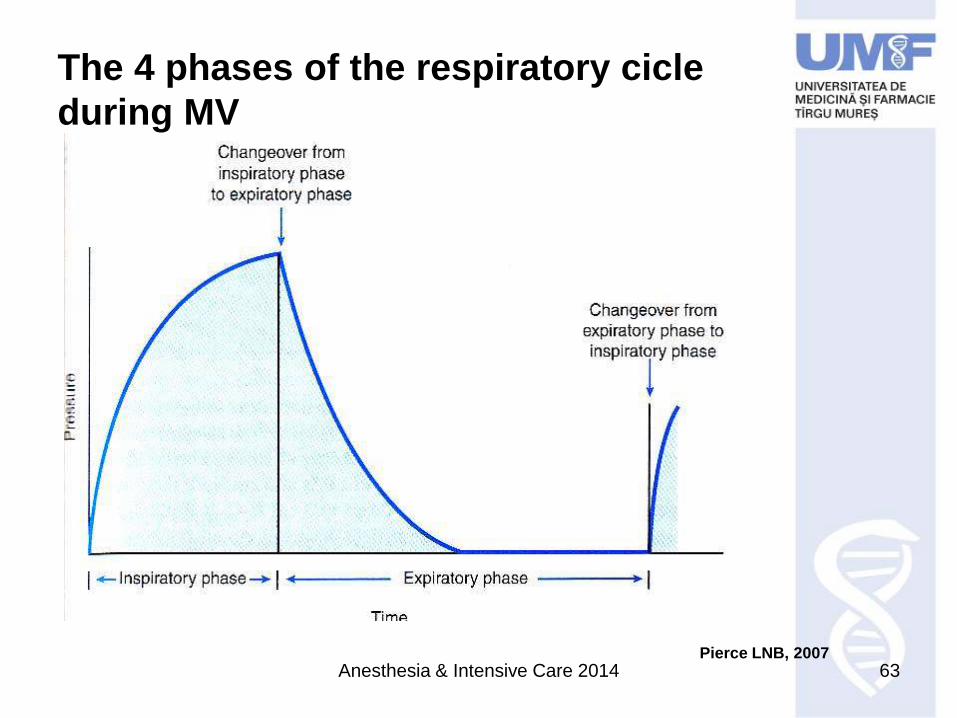
Anesthesia & Intensive Care 2014 63
The 4 phases of the respiratory cicle
during MV
Pierce LNB, 2007
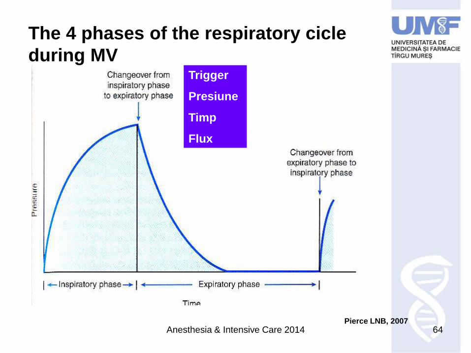
Anesthesia & Intensive Care 2014 64
The 4 phases of the respiratory cicle
during MVTrigger
Presiune
Timp
Flux
Pierce LNB, 2007
Anesthesia & Intensive Care 2014 65
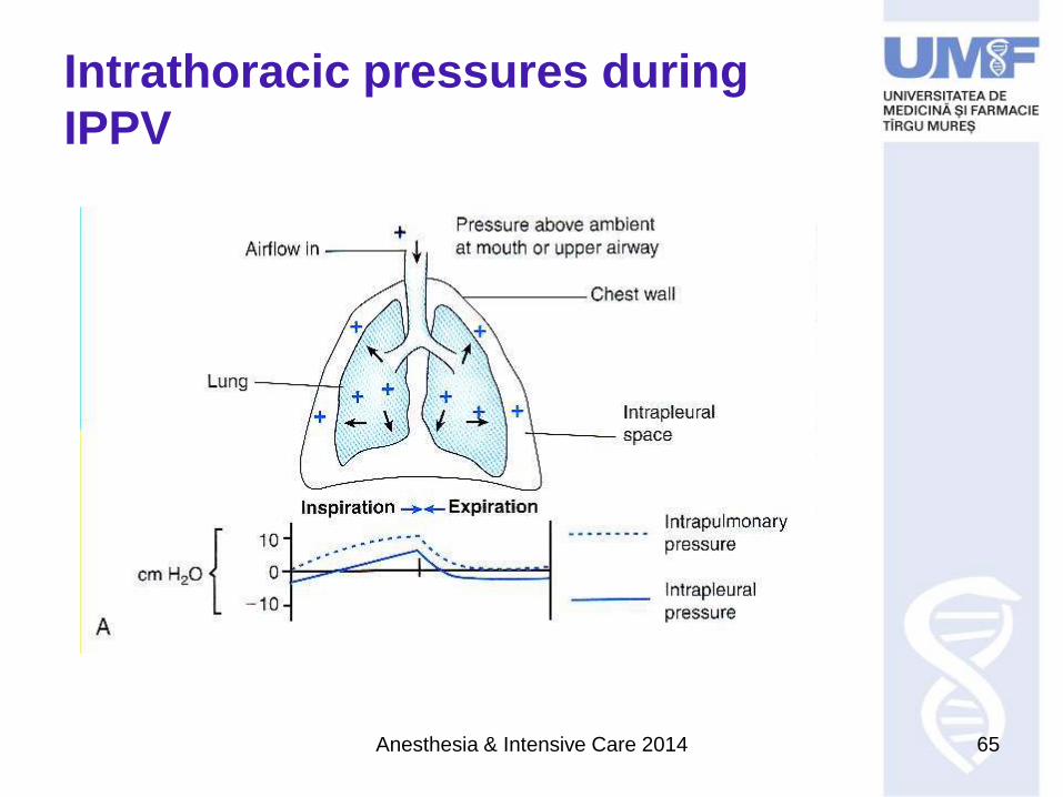
Intrathoracic pressures during
IPPV
Anesthesia & Intensive Care 2014 66
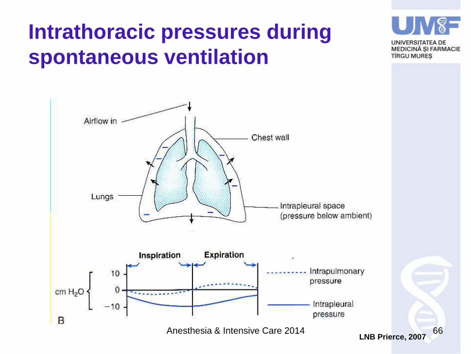
Intrathoracic pressures during
spontaneous ventilation
LNB Prierce, 2007
Anesthesia & Intensive Care 2014 67
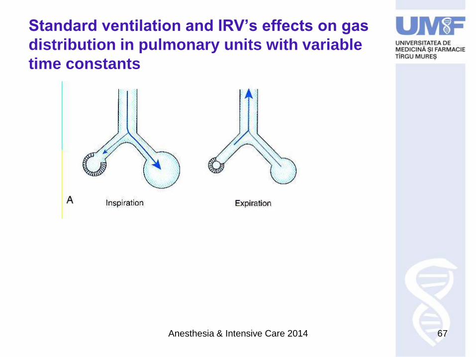
Standard ventilation and IRV’s effects on gas
distribution in pulmonary units with variable
time constants
Anesthesia & Intensive Care 2014 68
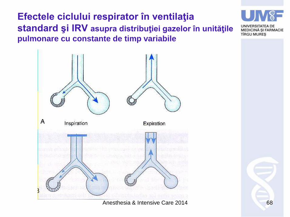
Efectele ciclului respirator în ventilaţia
standard şi IRV asupra distribuţiei gazelor în unităţile
pulmonare cu constante de timp variabile
Anesthesia & Intensive Care 2014 69
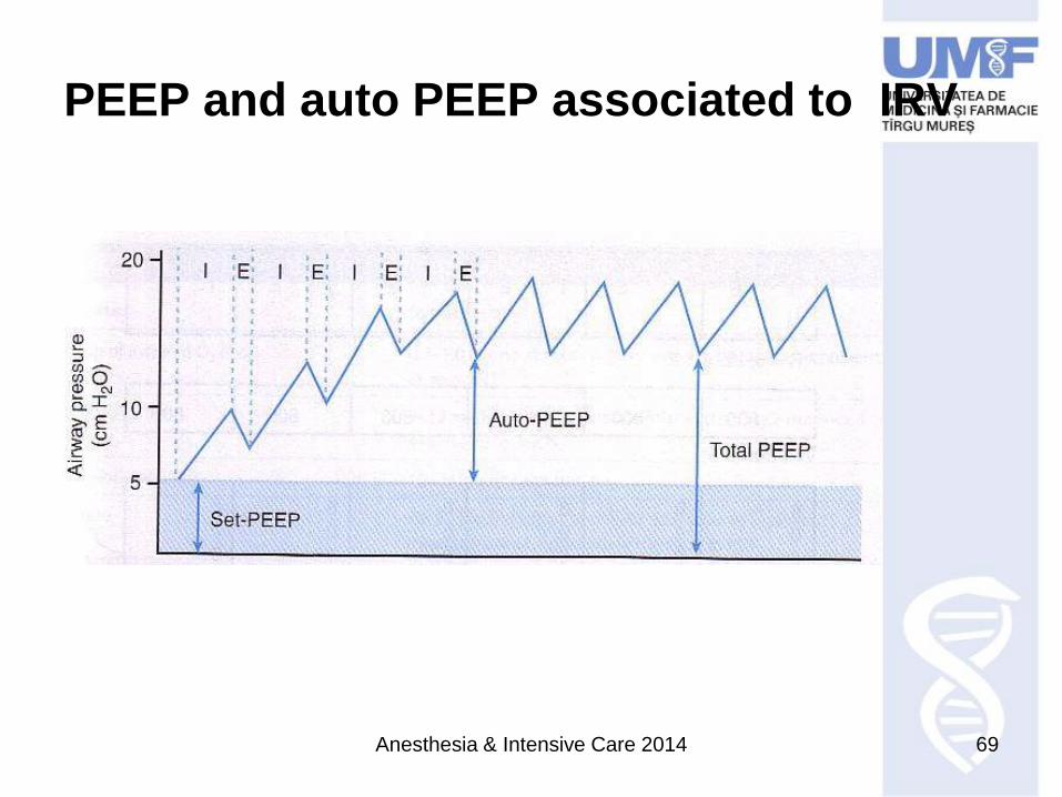
PEEP and auto PEEP associated to IRV
Anesthesia & Intensive Care 2014 70
Adverse effects associated to MV
PEEP
Hemodynamics
CO rapidlyDO2 due to P intrathoracic
Anesthesia & Intensive Care 2014 71
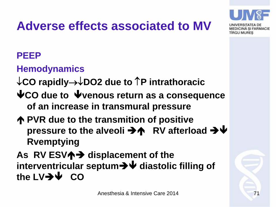
Adverse effects associated to MV
PEEP
Hemodynamics
CO rapidlyDO2 due to P intrathoracic
CO due to venous return as a consequence
of an increase in transmural pressure
PVR due to the transmition of positive
pressure to the alveoli RV afterload
Rvemptying
As RV ESV displacement of the
interventricular septum diastolic filling of
the LV CO
Anesthesia & Intensive Care 2014 72
Adverse effects associated to MV cont
Treatment
Intravascular volume replenishment +
inotropics and vasoactive drugs
!!! For a PEEP 10cmH2O use a Swan Ganz
catheter
Anesthesia & Intensive Care 2014 73
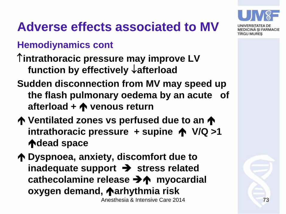
Adverse effects associated to MV
Hemodiynamics cont
intrathoracic pressure may improve LV
function by effectively afterload
Sudden disconnection from MV may speed up
the flash pulmonary oedema by an acute of
afterload + venous return
Ventilated zones vs perfused due to an
intrathoracic pressure + supine V/Q >1
dead space
Dyspnoea, anxiety, discomfort due to
inadequate support stress related
cathecolamine release myocardial
oxygen demand, arhythmia risk
Anesthesia & Intensive Care 2014 74
Adverse effects associated to MVcont
Physical effectsh– mecanical
Barotrauma – overdistention, peak inspiratory
pressure. Incid 7-25%
Incid of pneumothorax identical for HFJV and
standard MVCarbon GC et al, Chest 1983; 84;551
Gluch HE et al , Chest 1993; 103: 1413
Anesthesia & Intensive Care 2014 75
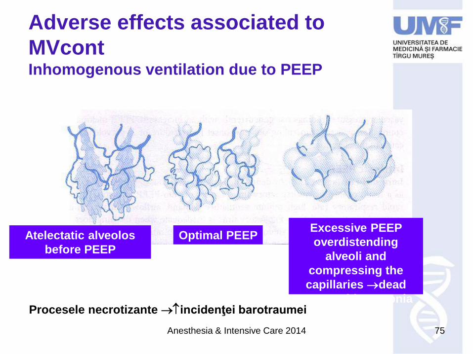
Adverse effects associated to
MVcontInhomogenous ventilation due to PEEP
Atelectatic alveolos
before PEEP
Optimal PEEP Excessive PEEP
overdistending
alveoli and
compressing the
capillaries dead
space + hipercapniaProcesele necrotizante incidenţei barotraumei
Anesthesia & Intensive Care 2014 76
Adverse effects associated to MVcont
VILI ventilation induced lung injury
Cause: excessive distention
• Alveoli rupture
– Pneumpmediastinum
– Pneumopericardium
– Subcutaneous emphysema
– Pneumothorax
– Gazeous emboli
Anesthesia & Intensive Care 2014 77
Adverse effects associated to MV cont
• parenchimal injury for transpulmonary distension
pressures >30-35cmH2O = diffuse alveolar injuries ,
cytokines, bacterial translocation
• Friction forces – repetitive opening/closure of the
alveoli (collapse)
• Acceleration of the initial rapid flows in the lungs
• The concept of “protective ventilation” with values <
normal
Anesthesia & Intensive Care 2014 78
cont
Desincronizarea pacient/ventilator
3 faze ale ciclului respirator asistat: trigger, target,
cycle
Oricare în contratimpoboseală musculară
Lupta cu ventilatorul
Sedare excesivă
suportului ventilator
Anesthesia & Intensive Care 2014 79
Efectele adverse asociate VM cont
Desincronizarea pacient/ventilator
3 faze ale ciclului respirator asistat: trigger, target,
cycle
Oricare în contratimpoboseală musculară
Anesthesia & Intensive Care 2014 80
Efectele adverse asociate VM cont
Desincronizarea pacient/ventilator
3 faze ale ciclului respirator asistat: trigger, target,
cycle
Oricare în contratimpoboseală musculară
Lupta cu ventilatorul
Anesthesia & Intensive Care 2014 81
Adverse effects associated to MV cont
Patient/ventilator dysynchrony
3 phases of the assited respiratory cycle : trigger,
target, cycle
Any couter timemuscular fatigue
Fighting the ventilator
Excessive sedation
ventilatory support
Anesthesia & Intensive Care 2014 82
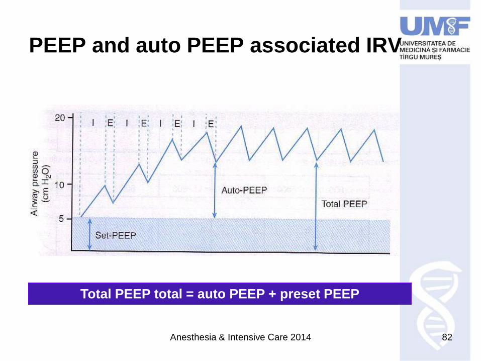
PEEP and auto PEEP associated IRV
Total PEEP total = auto PEEP + preset PEEP
Anesthesia & Intensive Care 2014 83
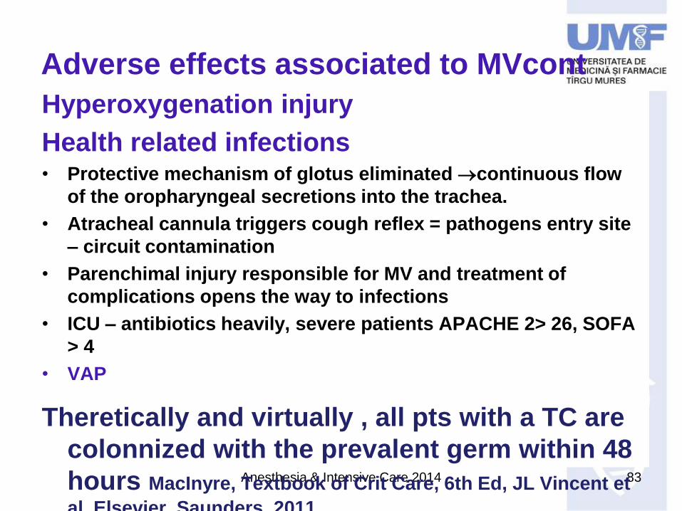
Adverse effects associated to MVcont
Hyperoxygenation injury
Health related infections• Protective mechanism of glotus eliminated continuous flow
of the oropharyngeal secretions into the trachea.
• Atracheal cannula triggers cough reflex = pathogens entry site
– circuit contamination
• Parenchimal injury responsible for MV and treatment of
complications opens the way to infections
• ICU – antibiotics heavily, severe patients APACHE 2> 26, SOFA
> 4
• VAP
Theretically and virtually , all pts with a TC are
colonnized with the prevalent germ within 48
hours MacInyre, Textbook of Crit Care, 6th Ed, JL Vincent et
al, Elsevier, Saunders, 2011,
Anesthesia & Intensive Care 2014 84
VAP prevention
Antibiotic strategies
Manipulating the circuits – change only if visible
contamination
Subglottic continuous drainage
BAL – inaccurate, confounding
Gastrointestinal hemorrhage
Anesthesia & Intensive Care 2014 85
Why VM?
Anesthesia & Intensive Care 2014 86
Why VM?