Acute Non-‐Traumatic Weakness Emergency Neurological Life ...
32
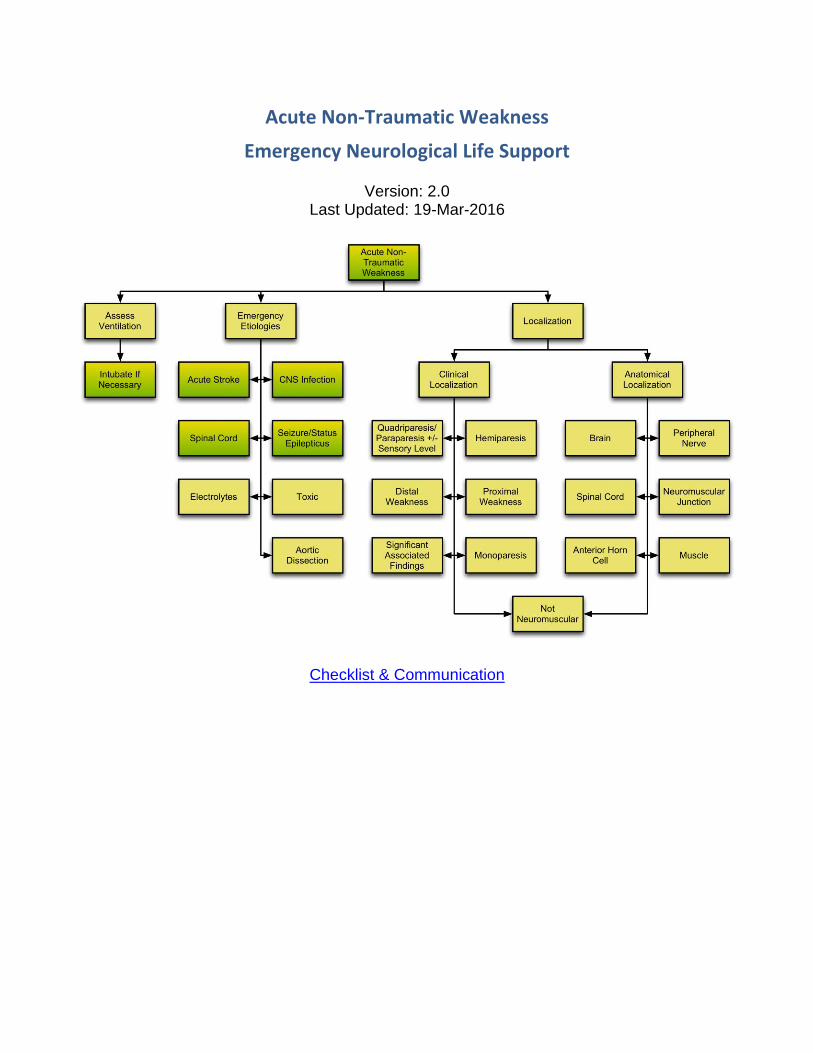
Acute Non-Traumatic Weakness Emergency Neurological Life Support Version: 2.0 Last Updated: 19-Mar-2016 Checklist & Communication
-
Upload
truongdang -
Category
Documents
-
view
222 -
download
2
Transcript of Acute Non-‐Traumatic Weakness Emergency Neurological Life ...
Acute Non-Traumatic Weakness
Emergency Neurological Life Support
Version: 2.0 Last Updated: 19-Mar-2016
Checklist & Communication
Acute Non-Traumatic Weakness: Table of Contents
Acute Non-traumatic Weakness ................................................................................................ 1 Emergency Neurological Life Support ....................................................................................... 1 Checklist .................................................................................................................................... 4 Communication .......................................................................................................................... 4 Acute Stroke .............................................................................................................................. 5
Within the time window? ........................................................................................................ 5 Acute Weakness ........................................................................................................................ 6
Patients presenting with any form of new weakness .............................................................. 6 Anatomical Localization ............................................................................................................. 7
Based on the location of nervous system pathology .............................................................. 7 Anterior Horn Cell ...................................................................................................................... 8
Alpha motoneuron .................................................................................................................. 8 Brain .......................................................................................................................................... 9
Cerebral cortex, white matter or brainstem ............................................................................ 9 Clinical Localization ..................................................................................................................10
Based on the presenting clinical symptoms and neurological examination ...........................10 CNS Infection ...........................................................................................................................11 Distal Weakness .......................................................................................................................12 Electrolyte Disturbance .............................................................................................................13
Glucose, potassium, phosphate, sodium ..............................................................................13 Emergency Etiologies ...............................................................................................................14
Exclude these time-sensitive emergency causes first ...........................................................14 Hemiparesis ..............................................................................................................................15
Unilateral body weakness .....................................................................................................15 Localizing the Cause of Acute Weakness.................................................................................16
This will help determine the cause ........................................................................................16 Monoparesis .............................................................................................................................17
Weakness of a single limb ....................................................................................................17 Muscle ......................................................................................................................................18 Need for Assisted Ventilation ....................................................................................................19
Do you need to intubate this patient? ....................................................................................19 Neuromuscular Junction ...........................................................................................................20 Not Neuromuscular Weakness .................................................................................................21
Consider psychiatric cause ...................................................................................................21 Other Urgent Causes ................................................................................................................22 Paraplegia from Aortic Dissection .............................................................................................23
Spinal cord infarct .................................................................................................................23 Peripheral Nerve .......................................................................................................................24 Proximal Weakness ..................................................................................................................25 Quadriparesis or Paraparesis with or without Sensory Level ....................................................26
Suggests spinal cord lesion ..................................................................................................26 Significant Associated Findings ................................................................................................27
Acute Non-Traumatic Weakness: Table of Contents
Other finding that may make the diagnosis clear ..................................................................27 Special Considerations for Intubation .......................................................................................29
Consider ................................................................................................................................29 Spinal Cord ...............................................................................................................................30
Non-traumatic cause .............................................................................................................30 Status Epilepticus or Seizure ....................................................................................................31
Postictal or non-convulsive status or Todd’s paresis ............................................................31 Toxic Cause ..............................................................................................................................32
Any toxin exposure?..............................................................................................................32
Acute Non-Traumatic Weakness: 4
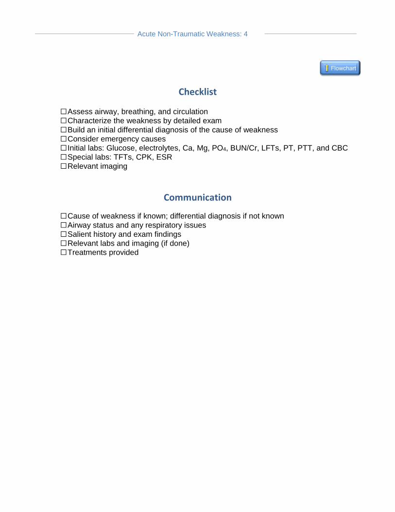
Checklist
☐ Assess airway, breathing, and circulation ☐ Characterize the weakness by detailed exam
☐ Build an initial differential diagnosis of the cause of weakness ☐ Consider emergency causes ☐ Initial labs: Glucose, electrolytes, Ca, Mg, PO4, BUN/Cr, LFTs, PT, PTT, and CBC ☐ Special labs: TFTs, CPK, ESR
☐ Relevant imaging
Communication
☐ Cause of weakness if known; differential diagnosis if not known
☐ Airway status and any respiratory issues ☐ Salient history and exam findings ☐ Relevant labs and imaging (if done) ☐ Treatments provided
Acute Non-Traumatic Weakness: 5
Acute Stroke
Within the time window?
If the patient has signs and symptoms consistent with acute stroke, especially if the patient is within the time window for thrombolysis or endovascular therapy, see the emergency evaluation of Acute Stroke.
Acute Non-Traumatic Weakness: 6
Acute Weakness
Patients presenting with any form of new weakness This topic provides an organized approach to the patient with new weakness not associated with or caused by trauma. If the patient has experienced trauma follow the links to the ENLS protocols Traumatic Brain Injury and Traumatic Spine Injury as appropriate. Based on the pattern of weakness, one can decide the degree of urgency for airway and ventilatory support and administration of time-sensitive treatments. Authors: Oliver Flower, MD Anna Finley Caulfield, MD Mark S. Wainwright, MD
Acute Non-Traumatic Weakness: 7
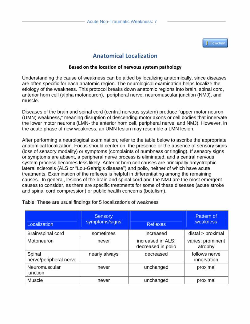
Anatomical Localization
Based on the location of nervous system pathology
Understanding the cause of weakness can be aided by localizing anatomically, since diseases are often specific for each anatomic region. The neurological examination helps localize the etiology of the weakness. This protocol breaks down anatomic regions into brain, spinal cord, anterior horn cell (alpha motoneuron), peripheral nerve, neuromuscular junction (NMJ), and muscle. Diseases of the brain and spinal cord (central nervous system) produce "upper motor neuron (UMN) weakness," meaning disruption of descending motor axons or cell bodies that innervate the lower motor neurons (LMN- the anterior horn cell, peripheral nerve, and NMJ). However, in the acute phase of new weakness, an UMN lesion may resemble a LMN lesion. After performing a neurological examination, refer to the table below to ascribe the appropriate anatomical localization. Focus should center on the presence or the absence of sensory signs (loss of sensory modality) or symptoms (complaints of numbness or tingling). If sensory signs or symptoms are absent, a peripheral nerve process is eliminated, and a central nervous system process becomes less likely. Anterior horn cell causes are principally amyotrophic lateral sclerosis (ALS or “Lou-Gehrig's disease”) and polio, neither of which have acute treatments. Examination of the reflexes is helpful in differentiating among the remaining causes. In general, lesions of the brain and spinal cord and the NMJ are the most emergent causes to consider, as there are specific treatments for some of these diseases (acute stroke and spinal cord compression) or public health concerns (botulism). Table: These are usual findings for 5 localizations of weakness
Localization
Sensory symptoms/signs
Reflexes
Pattern of weakness
Brain/spinal cord sometimes increased distal > proximal
Motoneuron never increased in ALS; decreased in polio
varies; prominent atrophy
Spinal nerve/peripheral nerve
nearly always decreased follows nerve innervation
Neuromuscular junction
never unchanged proximal
Muscle never unchanged proximal
Acute Non-Traumatic Weakness: 8
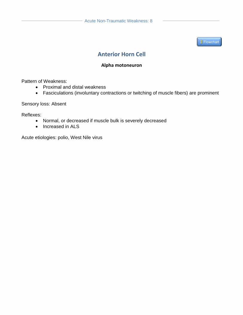
Anterior Horn Cell
Alpha motoneuron
Pattern of Weakness:
Proximal and distal weakness
Fasciculations (involuntary contractions or twitching of muscle fibers) are prominent
Sensory loss: Absent
Reflexes:
Normal, or decreased if muscle bulk is severely decreased
Increased in ALS
Acute etiologies: polio, West Nile virus
Acute Non-Traumatic Weakness: 9
Brain
Cerebral cortex, white matter or brainstem
Pattern of Weakness (UMN weakness):
Distal weakness more than proximal
Extensors weakness more than flexors (in upper extremity)
Hemiparesis or single limb
Sensory Loss:
May be present depending on whether cortex or sensory tracts are involved
Reflexes:
Increased; however, may be decreased initially but later increase
Acute Etiologies:
See ENLS protocols Acute Stroke, Subarachnoid Hemorrhage, and Status Epilepticus
Hypertensive encephalopathy
Acute Non-Traumatic Weakness: 10
Clinical Localization
Based on the presenting clinical symptoms and neurological examination
After performing the neurological examination, consider the pattern of weakness. Specifically, note whether all four limbs are weak (quadriparesis) and if there is a sensory level , or whether there is unilateral body weakness (hemiparesis), one limb weakness (monoparesis), distal extremity (hands, wrists, feet) weakness (distal weakness), or proximal muscle (axial muscles, deltoids, and hip flexors) weakness (proximal weakness)? Are there any significant associated findings? Accurately defining the presenting complaint helps generate a focused differential diagnosis. A good clinical history is essential, as the examination may be difficult or unreliable in the obtunded or confused patient. However, it should be possible to elicit whether the deficit is unilateral or bilateral, which anatomical region is affected, and whether there is a sensory deficit. With a cooperative patient, it should also be possible to establish whether the weakness is symmetrical or asymmetrical, and whether it is affecting the proximal or distal extremities. It is important to differentiate between an upper motor neuron (UMN) and a lower motor neuron (LMN) lesion in the acute setting, although this may be difficult in some situations. In well-established UMN lesions (brain and spinal cord), hyperreflexia, increased extremity tone, and a positive Babinski sign (great toe extension with lateral plantar stimulation) are seen on exam. In comparison, LMN lesions (from the anterior horn cells to the muscles) cause a flaccid, areflexic weakness, and with time, atrophy and fasciculations. However, in the acute phase, UMN lesions may mimic a LMN lesion (flaccid paralysis, normal or reduced extremity tone), and with decreased reflexes. In acute UMN lesions there is not enough time for atrophy to be evident.
Acute Non-Traumatic Weakness: 11
CNS Infection
Consider meningitis or encephalitis as a cause. See ENLS protocol Meningitis and Encephalitis.
Acute Non-Traumatic Weakness: 12
Distal Weakness
Vasculitic neuropathy
Toxin-induced peripheral neuropathy
Nerve compression syndromes Distal weakness is weakness mainly affecting the hands, wrists, and feet. It is typically caused by peripheral neuropathies that often present along with sensory symptoms. Patients have decreased grip strength and drop objects or develop gait disturbances due to foot drop. The pattern of weakness and history are of great significance. Of the many types of peripheral neuropathy, vasculitic and toxin-induced neuropathies are the most likely to produce acute weakness. It may also be produced by local nerve compression syndromes (e.g. carpal tunnel syndrome) that predominantly affects the distal extremities, causing both sensory and motor symptoms.
Acute Non-Traumatic Weakness: 13
Electrolyte Disturbance
Glucose, potassium, phosphate, sodium
Acute hypoglycemia, hypokalemia, hypophosphatemia or other electrolyte disturbances suggesting other organ dysfunction should be considered next.
Acute Non-Traumatic Weakness: 14
Emergency Etiologies
Exclude these time-sensitive emergency causes first
There are several time-sensitive causes of acute weakness that should be excluded quickly before moving on to a more comprehensive localization of the cause of weakness. Consider each of the causes before proceeding to localization.
Acute Non-Traumatic Weakness: 15
Hemiparesis
Unilateral body weakness
See ENLS protocols Acute Stroke, Intracerebral Hemorrhage, or Subarachnoid Hemorrhage
Intracranial mass
See ENLS protocol Meningitis and Encephalitis
Hypoglycemia/hyperglycemia
Postictal Todd's paresis
Hemiplegic migraine
Brown-Sequard syndrome Hemiparesis is partial paralysis affecting only one side of the body. Acute hemiplegia is complete paralysis of one side of the body. While acute hemiplegia is most commonly due to an ischemic stroke, other differential diagnoses must be considered because management varies. The history and demographic of the patient will narrow the diagnosis, and examination findings provide further clues. A blood glucose level and a non-contrast head computed tomography (CT) scan are part of the initial workup.
Acute Non-Traumatic Weakness: 16
Localizing the Cause of Acute Weakness
This will help determine the cause
Perform a neurological examination on the patient that includes:
Deep tendon reflexes
Strength testing of proximal and distal extremity muscles, and compare flexor versus extensor muscle strength, noting symmetry between sides
Judge diaphragmatic and chest wall muscle strength to determine if there is any respiratory insufficiency (single breath count from 1 to 20 [external intercostal muscles] and maximal inspiratory pressure or negative inspiratory force [diaphragm].)
After performing this focused neurological examination, determine the pattern of weakness.
Acute Non-Traumatic Weakness: 17
Monoparesis
Weakness of a single limb
See ENLS protocol Acute Stroke
Intracranial mass
Postictal Todd's paresis
Nerve compression syndromes
Diabetic lumbosacral radiculoplexus neuropathy
Acute poliomyelitis Monoparesis refers to paralysis of a single muscle, muscle group, or limb. Acute paralysis involving a single limb may be caused by a central or a peripheral lesion. Historical and examination factors may help to localize the lesion. For example, sudden onset right arm weakness with an associated dysphasia is most likely to result from a central lesion, whereas wrist drop in the right hand, with hypoesthesia on the back of the hand following falling asleep with the arm over the back of a chair, results from a peripheral nerve compression syndrome. Poliomyelitis is rare, but can occur in the unvaccinated.
Acute Non-Traumatic Weakness: 18
Muscle
Pattern of Weakness:
Proximal
Sensory Loss:
Absent Reflexes:
Normal unless muscles severely weak Acute etiologies:
Rhabdomyolysis

Acute Non-Traumatic Weakness: 19
Need for Assisted Ventilation
Do you need to intubate this patient?
Assess the patient's airway and potential need for assisted ventilation. If any of the following general, subjective or objective findings are present, consider intubation. General:
Increasing generalized muscle weakness?
Dysphagia?
Dysphonia?
Dyspnea on exertion and at rest? Subjective:
Rapid shallow breathing
Tachycardia
Weak cough
Interrupted or staccato speech (gasping for air)
Use of accessory muscles
Abdominal paradoxical breathing
Orthopnea (difficult or painful breathing except when erect)
Weakness of trapezius and neck muscles: inability to lift head
Inability to perform a single-breath count: count from 1 to 20 in single exhalation (Forced vital capacity 1.0 L is roughly equal to counting from 1 to 10)
Cough after swallowing Objective:
Decreased level of consciousness (have a lower threshold to control the airway if the patient requires transfer or movement to unmonitored areas)
Hypoxemia
Vital capacity (VC) < 1 L or 20 ml/kg, or 50% drop in VC in a day
Maximum inspiratory pressure > -30 cm H2O
Maximum expiratory pressure < 40 cm H2O
Nocturnal desaturation
Hypercarbia (a late finding)
Acute Non-Traumatic Weakness: 20
Neuromuscular Junction
Pattern of Weakness:
First in eye muscles, neck extensors, pharynx, and diaphragm
Followed by more generalized weakness
Sensory Loss: Absent
Reflexes:
Normal
Decreased if muscle is paralyzed
Acute Etiologies:
Botulism
Tick bite
Organophosphate toxicity
Myasthenic crisis
Acute Non-Traumatic Weakness: 21
Not Neuromuscular Weakness
Consider psychiatric cause
Some disease states may produce symptoms of generalized weakness or fatigue that do not have a neuromuscular basis. These may be medical emergencies in their own right, meriting urgent specific treatment. Consider:
Any severe medical illness can have weakness as a symptom, but generally these will become clinically obvious during the patient's evaluation
Diagnoses of exclusion:
Malingering
Conversion disorder
Anxiety disorders
Fibromyalgia
Chronic fatigue syndrome
Acute Non-Traumatic Weakness: 22
Other Urgent Causes
Consider:
Shock
Myocardial infarct
Addisonian crisis
Acute Non-Traumatic Weakness: 23
Paraplegia from Aortic Dissection
Spinal cord infarct
Acute aortic dissection can close the artery of Adamkiewicz that supplies the anterior spinal artery to the mid thoracic and lumbar spinal cord. The patient will have an anterior spinal artery syndrome (paraplegia with loss of pain and temperature sensation below the lesion but preservation of proprioception and light touch). Assess distal lower extremity pulses and consider CT Angiogram (CTA), ultrasound or other techniques to rule out aortic dissection if the patient has an acute anterior spinal artery syndrome.
Acute Non-Traumatic Weakness: 24
Peripheral Nerve
Pattern of Weakness:
In the distribution of the nerve, or diffusely present as stocking/glove weakness
Sensory Loss:
Present
Reflexes:
Decreased or absent
Acute Etiologies:
Guillain-Barré syndrome
Vasculitis
Acute Non-Traumatic Weakness: 25
Proximal Weakness
Acute myopathy
Guillain-Barré syndrome
Acute diabetic lumbosacral radiculoplexus neuropathy (DLRN)
Myasthenia gravis
Acute West Nile virus-associated paralysis
Lambert-Eaton myasthenic syndrome (LEMS) Proximal weakness is weakness predominantly affecting the axial muscles, deltoids, and hip flexors. Acute proximal weakness classically presents with difficulty rising from a chair or brushing hair. Patients may have difficulties flexing and extending their neck. The most common cause is myopathy. Less common causes include LEMS and myasthenia gravis. DLRN may be the presenting feature of diabetes mellitus. While poliomyelitis is very rare in western countries, it remains endemic elsewhere. West Nile virus, with similar semiology as acute poliomyelitis, is more common in the United States and Europe.
Acute Non-Traumatic Weakness: 26
Quadriparesis or Paraparesis with or without Sensory Level
Suggests spinal cord lesion
Quadriparesis/paraparesis ± sensory level
See ENLS protocol Spinal Cord Compression
Spinal cord infarction
Transverse myelitis
Generalized weakness: electrolyte and glucose abnormalities Quadriparesis/paraparesis is symmetrical weakness of either all four limbs (quadriparesis) or legs (paraparesis), characteristically with a sensory level. Often it is related to spinal cord injury. Non-traumatic spinal cord injury may occur from compression (e.g. epidural abscess, hematoma, expanding tumor, prolapsed intervertebral disc), ischemia (spinal cord infarction), or inflammation (transverse myelitis). In the acute phase, a flaccid paralysis below the level of cord injury and areflexia is typically seen, with an accompanying corresponding sensory level, although there is considerable variation. Neurological examination should localize the lesion in patients with acute paraparesis/plegia to the thoracic or lumbar region and quadriparesis/plegia to the cervical region. Sensory abnormalities localize in the vertical plane (cervical (C), thoracic (T), lumbar (L), or sacral). Key sensory levels to remember are T4 and T10, which correspond to the nipple and naval, respectively.
Acute Non-Traumatic Weakness: 27
Significant Associated Findings
Other finding that may make the diagnosis clear
Certain constellations of symptoms and signs can make specific, often unusual diagnoses more likely. Below is a list of diagnoses and significant associated findings. Stroke syndromes also have characteristic patterns which are too numerous and varied to discuss here. However, findings such as aphasia, agnosia, apraxia, and neglect with acute weakness or sensory signs should prompt consideration of acute stroke. Locked-in syndrome (usually due to basilar artery thrombosis; also consider residual neuromuscular blockade)
Acute tetraplegia
Facial muscles paralyzed except eyes
Clear sensorium Myasthenia gravis
Fatigable weakness in eyelids and extra-ocular muscles
Variable weakness elsewhere
No sensory symptoms Envenomation
History of animal bite
Descending paralysis
Possible coagulopathy
Rhabdomyolysis
Shock Hypertensive encephalopathy
Severe, refractory hypertension
Headache
Transient, migratory neurological non-focal deficits Guillain- Barré syndrome
Ascending paralysis following upper respiratory viral illness/infection Botulism
Descending symmetrical paralysis
No fever
Clear sensorium
Acute Non-Traumatic Weakness: 28
Organophosphate toxicity
Weakness with prominent cholinergic signs and symptoms Heavy metal toxicity
Heavy metal exposure
Prominent gastrointestinal symptoms
Multi-organ failure Periodic paralysis
Episodic proximal weakness
Family history of the same disorder Dermatomyositis
Heliotrope rash
Proximal weakness Acute intermittent porphyria
Abdominal pain
Proximal weakness
Psychiatric symptoms
Red urine Tick paralysis
Tick bite
Followed by ascending paralysis
Acute Non-Traumatic Weakness: 29
Special Considerations for Intubation
Consider
Special consideration for intubation:
Rapid sequence induction/intubation is advised.
Avoid use of succinylcholine if there is evidence of underlying progressive neuromuscular disease (precipitates acute hyperkalemia) such as Guillain-Barré, chronic neuromuscular weakness, or prolonged immobilization. Consider rocuronium as an alternative. Succinylcholine will be relatively ineffective to achieve muscle relaxation in myasthenia gravis, unless a higher dose is used (~2.5 times the standard dose). Conversely it is recommended to use half-dose of a nondepolarizing agent (rocuronium 0.5-0.6 mg/kg) in such patients because they may be more sensitive to nondepolarizing neuromuscular junction blockers.
Consider non-invasive assisted ventilation as a temporizing measure in a neurologically stable patient expected to have a rapid resolution (e.g., myasthenia gravis).
Prepare atropine/glycopyrolate, fluids, and vasopressors if there is evidence of autonomic instability.
See ENLS protocols Airway, Ventilation and Sedation, and Pharmacotherapy.
Acute Non-Traumatic Weakness: 30
Spinal Cord
Non-traumatic cause
Pattern of Weakness:
Acute flaccid quadriparesis/paraparesis (rarely hemiparesis) below the level of spinal cord compression
Distal weakness more than proximal
Extensors weaker than flexors Sensory Loss:
May be present depending on whether sensory tracts are involved; bilateral loss of sensation below a certain spinal level is diagnostic
Reflexes:
Elevated during acute brain insult; however, reflexes may be decreased initially and later increase
Acute etiologies:
Epidural abscess, tumor Acute spinal cord compression of non-traumatic cause should be suspected if the patient has weakness of both legs or both arms and legs with intact mental status and cranial nerves, especially if they have a history of cancer or complaint of back or neck pain. See ENLS protocol Spinal Cord Compression. If there is any sign of trauma, see ENLS protocol Traumatic Spine Injury.
Acute Non-Traumatic Weakness: 31
Status Epilepticus or Seizure
Postictal or non-convulsive status or Todd’s paresis
Patients who are comatose, or encephalopathic, may be postictal. A patient with focal neurological findings (typically hemiparesis) may have a Todd's paralysis caused by a generalized seizure in a brain with a prior injury. Also, a patient may be having non-convulsive status epilepticus. See the ENLS protocol Status Epilepticus.
Acute Non-Traumatic Weakness: 32
Toxic Cause
Any toxin exposure?
Consider organophosphate or carbon monoxide exposure among others.