
Acute moderate exercise elicits increased dorsolateral ...jtoomim.org/brain-training/Acute moderate...
9
Acute moderate exercise elicits increased dorsolateral prefrontal activation and improves cognitive performance with Stroop test Hiroki Yanagisawa a , Ippeita Dan b , Daisuke Tsuzuki b , Morimasa Kato a , Masako Okamoto b , Yasushi Kyutoku b , Hideaki Soya a, ⁎ a Laboratory of Exercise Biochemistry, University of Tsukuba Graduate School of Comprehensive Human Sciences, 1-1-1 Tennodai, Tsukuba 305-8574, Japan b National Food Research Institute, 2-1-12 Kannondai, Tsukuba 305-8642, Japan abstract article info Article history: Received 10 July 2009 Revised 1 December 2009 Accepted 3 December 2009 Available online 16 December 2009 Keywords: Optical topography Cognitive performance Acute moderate intensity exercise Color-word matching Stroop task Dorsolateral prefrontal cortex A growing number of human studies have reported the beneficial influences of acute as well as chronic exercise on cognitive functions. However, neuroimaging investigations into the neural substrates of the effects of acute exercise have yet to be performed. Using multichannel functional near-infrared spectroscopy (fNIRS), we sought cortical activation related to changes in the Stroop interference test, elicited by an acute bout of moderate exercise, in healthy volunteers (N = 20). The compactness and portability of fNIRS allowed on-site cortical examination in a laboratory with a cycle ergometer, enabling strict control of the exercise intensity of each subject by assessing their peak oxygen intake (V · o 2peak ). We defined moderate exercise intensity as 50% of a subject's peak oxygen uptake (50%V · o 2peak ). An acute bout of moderate exercise caused significant improvement of cognitive performance reflecting Stroop interference as measured by reaction time. Consistent with previous functional neuroimaging studies, we detected brain activation due to Stroop interference (incongruent minus neutral) in the lateral prefrontal cortices in both hemispheres. This Stroop- interference-related activation was significantly enhanced in the left dorsolateral prefrontal cortex due to the acute bout of moderate exercise. The enhanced activation significantly coincided with the improved cognitive performance. This suggests that the left dorsolateral prefrontal cortex is likely the neural substrate for the improved Stroop performance elicited by an acute bout of moderate exercise. fNIRS, which allows physiological monitoring and functional neuroimaging to be combined, proved to be an effective tool for examining the cognitive effects of exercise. © 2009 Elsevier Inc. All rights reserved. Introduction A body of human and animal studies have reported the beneficial influence of exercise on cognitive and brain functions. Accordingly, exercise is drawing increasing research attention as a possible lifestyle factor for improving neurocognitive functions, and preventing or delaying dementia (Cotman et al., 2007; Hillman et al., 2008). So far, the majority of studies have focused on the chronic effects of exercise, while studies on acute exercise effects on cognition have only started to draw growing attention (Tomporowski, 2003). Recent studies provide evidence that an acute bout of moderate aerobic exercise improves cognitive performance in a choice reaction task (Chmura et al., 1998), a simple reaction time task (Collardeau et al., 2001), as well as confliction tasks such as Eriksen flanker and Stroop tasks (Hogervorst et al., 1996; Kamijo et al., 2004, 2007). Keeping pace with the examination of the cognitive effects of acute exercise, the search for their neural substrates has been accelerated mainly in the field of event-related potential (ERP) research. P300 (or P3) is a component believed to indicate the brain activity required to maintain working memory when the mental model of the stimulus environment is updated (Donchin & Coles, 1988), and is thus regarded as an appropriate neural substrate for improved cognitive perfor- mance. Several studies have generally demonstrated increased amplitude and shortened latency of P300 components in relation to the performance improvements caused by an acute bout of exercise (Hillman et al., 2003, 2008; Kamijo et al., 2004, 2007; Magnie et al., 2000; Nakamura et al., 1999; Polich and Lardon, 1997). ERP provides high temporal information about brain activities, but it provides only rough information regarding where in the brain the effect was originated. In order to examine which brain regions change activation in response to exercise, the application of different neuroimaging techniques would be beneficial. As a promising neuroimaging technique for investigating the acute effects of exercise on cognition, we introduced functional near-infrared spectroscopy (fNIRS): an optical method that non-invasively monitors cerebral hemodynamics by measuring changes in the attenuation of near- infrared light passing through tissue (Koizumi et al., 2003; Obrig and Villringer, 2003; Villringer and Chance, 1997). In many studies, fNIRS has proven to be effective in assessing oxygenation changes in NeuroImage 50 (2010) 1702–1710 ⁎ Corresponding author. E-mail address: [email protected] (H. Soya). 1053-8119/$ – see front matter © 2009 Elsevier Inc. All rights reserved. doi:10.1016/j.neuroimage.2009.12.023 Contents lists available at ScienceDirect NeuroImage journal homepage: www.elsevier.com/locate/ynimg
Transcript of Acute moderate exercise elicits increased dorsolateral ...jtoomim.org/brain-training/Acute moderate...
NeuroImage 50 (2010) 1702–1710
Contents lists available at ScienceDirect
NeuroImage
j ourna l homepage: www.e lsev ie r.com/ locate /yn img
Acute moderate exercise elicits increased dorsolateral prefrontal activation andimproves cognitive performance with Stroop test
Hiroki Yanagisawa a, Ippeita Dan b, Daisuke Tsuzuki b, Morimasa Kato a, Masako Okamoto b,Yasushi Kyutoku b, Hideaki Soya a,⁎a Laboratory of Exercise Biochemistry, University of Tsukuba Graduate School of Comprehensive Human Sciences, 1-1-1 Tennodai, Tsukuba 305-8574, Japanb National Food Research Institute, 2-1-12 Kannondai, Tsukuba 305-8642, Japan
⁎ Corresponding author.E-mail address: [email protected] (H. Soya
1053-8119/$ – see front matter © 2009 Elsevier Inc. Adoi:10.1016/j.neuroimage.2009.12.023
a b s t r a c t
a r t i c l e i n f oArticle history:Received 10 July 2009Revised 1 December 2009Accepted 3 December 2009Available online 16 December 2009
Keywords:Optical topographyCognitive performanceAcute moderate intensity exerciseColor-word matching Stroop taskDorsolateral prefrontal cortex
A growing number of human studies have reported the beneficial influences of acute as well as chronicexercise on cognitive functions. However, neuroimaging investigations into the neural substrates of theeffects of acute exercise have yet to be performed. Using multichannel functional near-infrared spectroscopy(fNIRS), we sought cortical activation related to changes in the Stroop interference test, elicited by an acutebout of moderate exercise, in healthy volunteers (N=20). The compactness and portability of fNIRS allowedon-site cortical examination in a laboratory with a cycle ergometer, enabling strict control of the exerciseintensity of each subject by assessing their peak oxygen intake (V
·o2peak). We defined moderate exercise
intensity as 50% of a subject's peak oxygen uptake (50%V·o2peak). An acute bout of moderate exercise caused
significant improvement of cognitive performance reflecting Stroop interference as measured by reactiontime. Consistent with previous functional neuroimaging studies, we detected brain activation due to Stroopinterference (incongruent minus neutral) in the lateral prefrontal cortices in both hemispheres. This Stroop-interference-related activation was significantly enhanced in the left dorsolateral prefrontal cortex due to theacute bout of moderate exercise. The enhanced activation significantly coincided with the improvedcognitive performance. This suggests that the left dorsolateral prefrontal cortex is likely the neural substratefor the improved Stroop performance elicited by an acute bout of moderate exercise. fNIRS, which allowsphysiological monitoring and functional neuroimaging to be combined, proved to be an effective tool forexamining the cognitive effects of exercise.
© 2009 Elsevier Inc. All rights reserved.
Introduction
A body of human and animal studies have reported the beneficialinfluence of exercise on cognitive and brain functions. Accordingly,exercise is drawing increasing research attention as a possible lifestylefactor for improving neurocognitive functions, and preventing ordelaying dementia (Cotman et al., 2007; Hillman et al., 2008).
So far, themajority of studies have focused on the chronic effects ofexercise, while studies on acute exercise effects on cognition haveonly started to draw growing attention (Tomporowski, 2003). Recentstudies provide evidence that an acute bout of moderate aerobicexercise improves cognitive performance in a choice reaction task(Chmura et al., 1998), a simple reaction time task (Collardeau et al.,2001), as well as confliction tasks such as Eriksen flanker and Strooptasks (Hogervorst et al., 1996; Kamijo et al., 2004, 2007).
Keeping pace with the examination of the cognitive effects of acuteexercise, the search for their neural substrates has been acceleratedmainly in the field of event-related potential (ERP) research. P300 (or
).
ll rights reserved.
P3) is a component believed to indicate the brain activity required tomaintain working memory when the mental model of the stimulusenvironment is updated (Donchin & Coles, 1988), and is thus regardedas an appropriate neural substrate for improved cognitive perfor-mance. Several studies have generally demonstrated increasedamplitude and shortened latency of P300 components in relation tothe performance improvements caused by an acute bout of exercise(Hillman et al., 2003, 2008; Kamijo et al., 2004, 2007; Magnie et al.,2000; Nakamura et al., 1999; Polich and Lardon, 1997).
ERP provides high temporal information about brain activities, butit provides only rough information regarding where in the brain theeffect was originated. In order to examine which brain regions changeactivation in response to exercise, the application of differentneuroimaging techniques would be beneficial. As a promisingneuroimaging technique for investigating the acute effects of exerciseon cognition, we introduced functional near-infrared spectroscopy(fNIRS): an optical method that non-invasively monitors cerebralhemodynamics by measuring changes in the attenuation of near-infrared light passing through tissue (Koizumi et al., 2003; Obrig andVillringer, 2003; Villringer and Chance, 1997). In many studies, fNIRShas proven to be effective in assessing oxygenation changes in
1703H. Yanagisawa et al. / NeuroImage 50 (2010) 1702–1710
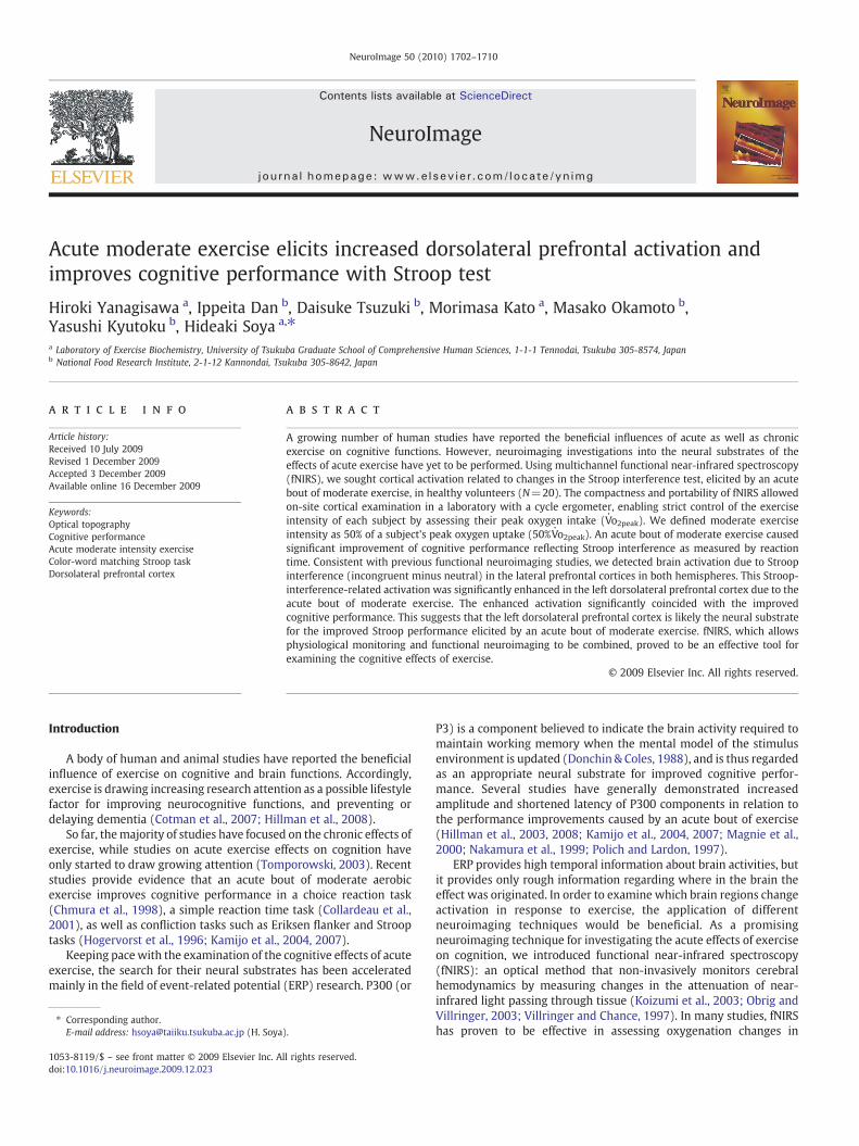
response to cortical activities, utilizing the tight coupling betweenneuronal activity and regional cerebral blood flow. In contrast to otherneuroimaging methods, fNIRS requires only compact experimentalsystems, is portable, and can be easily installed in a gym (Timinkul etal., 2008) (Fig. 1). This is advantageous for our study as exerciseintensity can be strictly controlled using gym facilities, and on-siteneuroimaging allows precise control of the interval between exerciseand brain measurement. Moreover, since fNIRS allows for the leastrestrictive measuring environment among neuroimaging modalities,possible influences on cognitive tasks can be kept minimal.
An important factor that needed to be controlled for this study wasthe exercise intensity for each subject. Behavioral studies and recentERP studies have shown that the effects of acute exercise on cognitiveperformance and brain response differ depending on the exerciseintensity: The best improvements are generally achieved with amoderate intensity (Kamijo et al., 2004, 2007). Nevertheless, the samedegree of difficulty of a physical task will have different impacts oneach subject depending on an individual's fitness level. Therefore, weassessed the peak oxygen intake (V
·o2peak) for each subject and
defined a moderate exercise intensity as 50% of a given subject'sV·o2peak (50%V
·o2peak).
For the cognitive task, we chose the color–word matching Strooptask, a classical measure of prefrontal cortex (PFC) function (MacLeod,1991), because it has been studied extensively using many neuroima-
Fig. 1. Experimental settings for fNIRS experiments. (A) Pilot studies 1 and 2.Physiological parameters were measured while subjects performed exercise using arecumbent type cycle ergometer. (B) fNIRS measurements. Brain activity was measuredwhile subjects performed the color-word matching Stroop task.
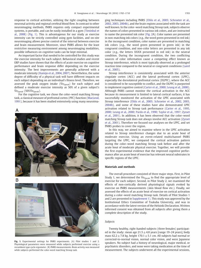
ging techniques including fNIRS (Ehlis et al., 2005; Schroeter et al.,2002, 2003, 2004b), and the brain regions associated with the task arewell known. In the color–wordmatching Stroop task, subjects observethe names of colors presented in various ink colors, and are instructedto name the presented ink color (Fig. 2A). Color names are presentedin non-matching ink colors (e.g., the word green presented in red ink)in the incongruent condition, color names are presented in matchingink colors (e.g., the word green presented in green ink) in thecongruent condition, and non-color letters are presented in any inkcolor (e.g., the letters XXXX presented in red ink) in the neutralcondition. During the incongruent condition, the two conflictingsources of color information cause a competing effect known asStroop interference, which is most typically observed as a prolongedreaction time compared to the neutral or congruent conditions (Lairdet al., 2005).
Stroop interference is consistently associated with the anteriorcingulate cortex (ACC) and the lateral prefrontal cortex (LPFC),especially the dorsolateral prefrontal cortex (DLPFC), where the ACCis considered to be susceptible to conflict, and the DLPFC is purportedto implement cognitive control (Carter et al., 2000; Leung et al., 2000).Although fNIRS cannot monitor the cortical activation in the ACCbecause its measurement is limited to lateral cortical surfaces, it hassuccessfully monitored the activation of the LPFC associated withStroop interference (Ehlis et al., 2005; Schroeter et al., 2002, 2003,2004b), and some of these studies have also demonstrated LPFCactivation related to Stroop task performance (Carter et al., 1995,2000; Leung et al., 2000; Pardo et al., 1990; Taylor et al., 1997; Zyssetet al., 2001). In addition, it has been observed that the color–wordmatching Stroop task does not always involve ACC activation (Zyssetet al., 2001). Therefore we focused our analyses on the LPFC, and setfNIRS probes to cover the region.
In this way, we aimed to examine where in the LPFC activationrelated to Stroop interference changes due to an acute bout ofmoderate exercise. Using an event-related multichannel fNIRStargeting the LPFC, we compared the cortical activation patternduring the color–word matching Stroop task before and after theacute bout of moderate physical exercise. Together, we will providethe first experimental evidence that the improved cognitive perfor-mance after an acute bout of exercise has relevant neural substrates inspecific regions of the LPFC.
Materials and methods
The overall procedure consisted of three major steps. First, in PilotStudy 1, we determined the V
·o2peak to find the appropriate level of
exercise for each subject. Second, in Pilot Study 2, we examined theeffects of non-cortically derived physiological signals evoked byexercise on fNIRS measurements (skin blood flow etc.). Finally, weassessed the effects of an acute bout of exercise on cortical activationduring a color–word matching Stroop task. Details of Pilot Studies 1and 2 are presented in Supplement 1). This study was approved by theInstitutional Ethics Committee of Tsukuba University, and was inaccordancewith the latest version of the Helsinki Declaration.Writteninformed consent was obtained from all subjects after giving them acomplete description of the study.
Subjects
Twenty healthy, right-handed subjects (three females) participat-ed in the study: mean age 21.5±4.8 years [range 19–24 years], bodymass 62.2±7.8 kg, height 170.1±5.1 cm. All subjects had normal orcorrected-to-normal vision, normal color vision, and were Japanesespeakers. No subject had a history of neurological, major medical, orpsychiatric disorders, and none were taking medication at the time ofmeasurement. The subjects underwent all the experimental sessions,
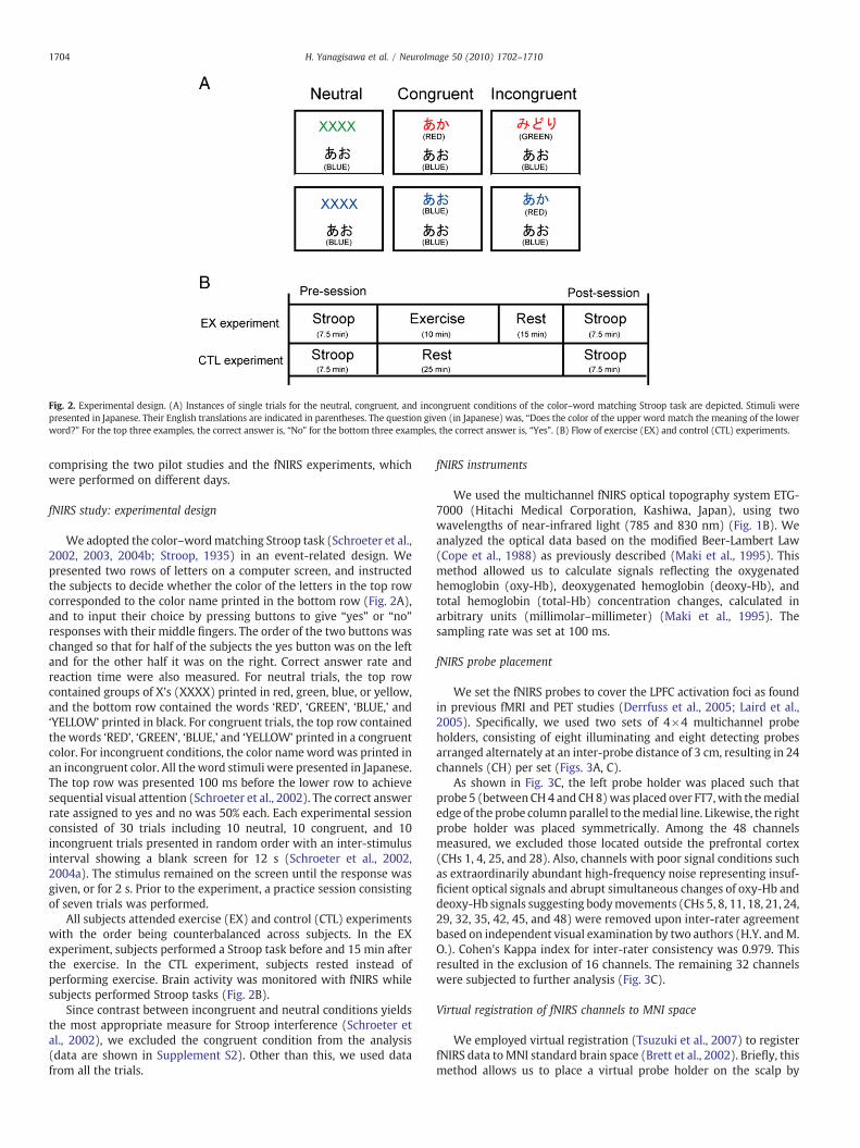
Fig. 2. Experimental design. (A) Instances of single trials for the neutral, congruent, and incongruent conditions of the color–word matching Stroop task are depicted. Stimuli werepresented in Japanese. Their English translations are indicated in parentheses. The question given (in Japanese) was, “Does the color of the upper word match the meaning of the lowerword?” For the top three examples, the correct answer is, “No” for the bottom three examples, the correct answer is, “Yes”. (B) Flow of exercise (EX) and control (CTL) experiments.
1704 H. Yanagisawa et al. / NeuroImage 50 (2010) 1702–1710
comprising the two pilot studies and the fNIRS experiments, whichwere performed on different days.
fNIRS study: experimental design
We adopted the color–wordmatching Stroop task (Schroeter et al.,2002, 2003, 2004b; Stroop, 1935) in an event-related design. Wepresented two rows of letters on a computer screen, and instructedthe subjects to decide whether the color of the letters in the top rowcorresponded to the color name printed in the bottom row (Fig. 2A),and to input their choice by pressing buttons to give “yes” or “no”responses with their middle fingers. The order of the two buttons waschanged so that for half of the subjects the yes button was on the leftand for the other half it was on the right. Correct answer rate andreaction time were also measured. For neutral trials, the top rowcontained groups of X's (XXXX) printed in red, green, blue, or yellow,and the bottom row contained the words ‘RED’, ‘GREEN’, ‘BLUE,’ and‘YELLOW’ printed in black. For congruent trials, the top row containedthe words ‘RED’, ‘GREEN’, ‘BLUE,’ and ‘YELLOW’ printed in a congruentcolor. For incongruent conditions, the color nameword was printed inan incongruent color. All the word stimuli were presented in Japanese.The top row was presented 100 ms before the lower row to achievesequential visual attention (Schroeter et al., 2002). The correct answerrate assigned to yes and no was 50% each. Each experimental sessionconsisted of 30 trials including 10 neutral, 10 congruent, and 10incongruent trials presented in random order with an inter-stimulusinterval showing a blank screen for 12 s (Schroeter et al., 2002,2004a). The stimulus remained on the screen until the response wasgiven, or for 2 s. Prior to the experiment, a practice session consistingof seven trials was performed.
All subjects attended exercise (EX) and control (CTL) experimentswith the order being counterbalanced across subjects. In the EXexperiment, subjects performed a Stroop task before and 15 min afterthe exercise. In the CTL experiment, subjects rested instead ofperforming exercise. Brain activity was monitored with fNIRS whilesubjects performed Stroop tasks (Fig. 2B).
Since contrast between incongruent and neutral conditions yieldsthe most appropriate measure for Stroop interference (Schroeter etal., 2002), we excluded the congruent condition from the analysis(data are shown in Supplement S2). Other than this, we used datafrom all the trials.
fNIRS instruments
We used the multichannel fNIRS optical topography system ETG-7000 (Hitachi Medical Corporation, Kashiwa, Japan), using twowavelengths of near-infrared light (785 and 830 nm) (Fig. 1B). Weanalyzed the optical data based on the modified Beer-Lambert Law(Cope et al., 1988) as previously described (Maki et al., 1995). Thismethod allowed us to calculate signals reflecting the oxygenatedhemoglobin (oxy-Hb), deoxygenated hemoglobin (deoxy-Hb), andtotal hemoglobin (total-Hb) concentration changes, calculated inarbitrary units (millimolar–millimeter) (Maki et al., 1995). Thesampling rate was set at 100 ms.
fNIRS probe placement
We set the fNIRS probes to cover the LPFC activation foci as foundin previous fMRI and PET studies (Derrfuss et al., 2005; Laird et al.,2005). Specifically, we used two sets of 4×4 multichannel probeholders, consisting of eight illuminating and eight detecting probesarranged alternately at an inter-probe distance of 3 cm, resulting in 24channels (CH) per set (Figs. 3A, C).
As shown in Fig. 3C, the left probe holder was placed such thatprobe 5 (betweenCH4 and CH8)was placed over FT7,with themedialedge of the probe columnparallel to themedial line. Likewise, the rightprobe holder was placed symmetrically. Among the 48 channelsmeasured, we excluded those located outside the prefrontal cortex(CHs 1, 4, 25, and 28). Also, channels with poor signal conditions suchas extraordinarily abundant high-frequency noise representing insuf-ficient optical signals and abrupt simultaneous changes of oxy-Hb anddeoxy-Hb signals suggesting bodymovements (CHs 5, 8, 11, 18, 21, 24,29, 32, 35, 42, 45, and 48) were removed upon inter-rater agreementbased on independent visual examination by two authors (H.Y. andM.O.). Cohen's Kappa index for inter-rater consistency was 0.979. Thisresulted in the exclusion of 16 channels. The remaining 32 channelswere subjected to further analysis (Fig. 3C).
Virtual registration of fNIRS channels to MNI space
We employed virtual registration (Tsuzuki et al., 2007) to registerfNIRS data toMNI standard brain space (Brett et al., 2002). Briefly, thismethod allows us to place a virtual probe holder on the scalp by
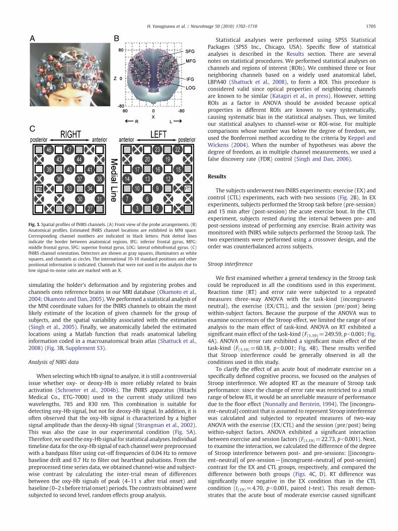
Fig. 3. Spatial profiles of fNIRS channels. (A) Front view of the probe arrangements. (B)Anatomical profiles. Estimated fNIRS channel locations are exhibited in MNI space.Corresponding channel numbers are indicated in black letters. Pink dotted linesindicate the border between anatomical regions. IFG: inferior frontal gyrus, MFG:middle frontal gyrus, SFG: superior frontal gyrus, LOG: lateral orbitofrontal gyrus. (C)fNIRS channel orientation. Detectors are shown as gray squares, illuminators as whitesquares, and channels as circles. The international 10-10 standard positions and otherpositional information is indicated. Channels that were not used in the analysis due tolow signal-to-noise ratio are marked with an X.
1705H. Yanagisawa et al. / NeuroImage 50 (2010) 1702–1710
simulating the holder's deformation and by registering probes andchannels onto reference brains in our MRI database (Okamoto et al.,2004; Okamoto and Dan, 2005). We performed a statistical analysis ofthe MNI coordinate values for the fNIRS channels to obtain the mostlikely estimate of the location of given channels for the group ofsubjects, and the spatial variability associated with the estimation(Singh et al., 2005). Finally, we anatomically labeled the estimatedlocations using a Matlab function that reads anatomical labelinginformation coded in a macroanatomical brain atlas (Shattuck et al.,2008) (Fig. 3B, Supplement S3).
Analysis of NIRS data
When selectingwhich Hb signal to analyze, it is still a controversialissue whether oxy- or deoxy-Hb is more reliably related to brainactivation (Schroeter et al., 2004b). The fNIRS apparatus (HitachiMedical Co., ETG-7000) used in the current study utilized twowavelengths, 785 and 830 nm. This combination is suitable fordetecting oxy-Hb signal, but not for deoxy-Hb signal. In addition, it isoften observed that the oxy-Hb signal is characterized by a highersignal amplitude than the deoxy-Hb signal (Strangman et al., 2002).This was also the case in our experimental condition (Fig. 5A).Therefore,weused the oxy-Hb signal for statistical analyses. Individualtimeline data for the oxy-Hb signal of each channelwere preprocessedwith a bandpass filter using cut-off frequencies of 0.04 Hz to removebaseline drift and 0.7 Hz to filter out heartbeat pulsations. From thepreprocessed time series data, we obtained channel-wise and subject-wise contrast by calculating the inter-trial mean of differencesbetween the oxy-Hb signals of peak (4–11 s after trial onset) andbaseline (0–2 s before trial onset) periods. The contrasts obtainedweresubjected to second level, random effects group analysis.
Statistical analyses were performed using SPSS StatisticalPackages (SPSS Inc., Chicago, USA). Specific flow of statisticalanalyses is described in the Results section. There are severalnotes on statistical procedures. We performed statistical analyses onchannels and regions of interest (ROIs). We combined three or fourneighboring channels based on a widely used anatomical label,LBPA40 (Shattuck et al., 2008), to form a ROI. This procedure isconsidered valid since optical properties of neighboring channelsare known to be similar (Katagiri et al., in press). However, settingROIs as a factor in ANOVA should be avoided because opticalproperties in different ROIs are known to vary systematically,causing systematic bias in the statistical analyses. Thus, we limitedour statistical analyses to channel-wise or ROI-wise. For multiplecomparisons whose number was below the degree of freedom, weused the Bonferroni method according to the criteria by Keppel andWickens (2004). When the number of hypotheses was above thedegree of freedom, as in multiple channel measurements, we used afalse discovery rate (FDR) control (Singh and Dan, 2006).
Results
The subjects underwent two fNIRS experiments: exercise (EX) andcontrol (CTL) experiments, each with two sessions (Fig. 2B). In EXexperiments, subjects performed the Stroop task before (pre-session)and 15 min after (post-session) the acute exercise bout. In the CTLexperiment, subjects rested during the interval between pre- andpost-sessions instead of performing any exercise. Brain activity wasmonitored with fNIRS while subjects performed the Stroop task. Thetwo experiments were performed using a crossover design, and theorder was counterbalanced across subjects.
Stroop interference
We first examined whether a general tendency in the Stroop taskcould be reproduced in all the conditions used in this experiment.Reaction time (RT) and error rate were subjected to a repeatedmeasures three-way ANOVA with the task-kind (incongruent-neutral), the exercise (EX/CTL), and the session (pre/post) beingwithin-subject factors. Because the purpose of the ANOVA was toexamine occurrences of the Stroop effect, we limited the range of ouranalysis to the main effect of task-kind. ANOVA on RT exhibited asignificant main effect of the task-kind (F(1,19)=249.59, pb0.001; Fig.4A). ANOVA on error rate exhibited a significant main effect of thetask-kind (F(1,19)=60.18, pb0.001; Fig. 4B). These results verifiedthat Stroop interference could be generally observed in all theconditions used in this study.
To clarify the effect of an acute bout of moderate exercise on aspecifically defined cognitive process, we focused on the analyses ofStroop interference. We adopted RT as the measure of Stroop taskperformance: since the change of error rate was restricted to a smallrange of below 8%, it would be an unreliable measure of performancedue to the floor effect (Nunnally and Berstein, 1994). The [incongru-ent–neutral] contrast that is assumed to represent Stroop interferencewas calculated and subjected to repeated measures of two-wayANOVA with the exercise (EX/CTL) and the session (pre/post) beingwithin-subject factors. ANOVA exhibited a significant interactionbetween exercise and session factors (F(1,19)=22.73, pb0.001). Next,to examine the interaction, we calculated the difference of the degreeof Stroop interference between post- and pre-sessions: [[incongru-ent–neutral] of pre-session− [incongruent–neutral] of post-session]contrast for the EX and CTL groups, respectively, and compared thedifference between both groups (Figs. 4C, D). RT difference wassignificantly more negative in the EX condition than in the CTLcondition (t(19)=4.70, pb0.001, paired t-test). This result demon-strates that the acute bout of moderate exercise caused significant
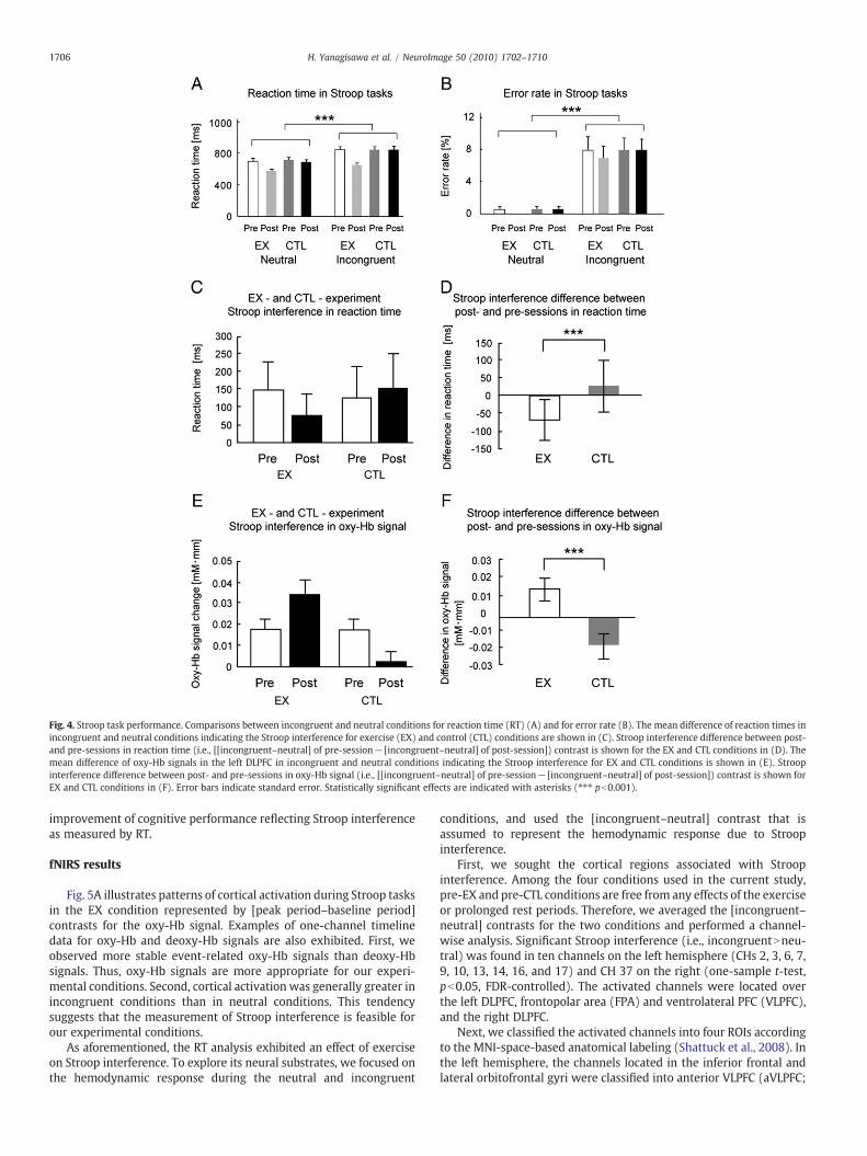
Fig. 4. Stroop task performance. Comparisons between incongruent and neutral conditions for reaction time (RT) (A) and for error rate (B). The mean difference of reaction times inincongruent and neutral conditions indicating the Stroop interference for exercise (EX) and control (CTL) conditions are shown in (C). Stroop interference difference between post-and pre-sessions in reaction time (i.e., [[incongruent–neutral] of pre-session− [incongruent–neutral] of post-session]) contrast is shown for the EX and CTL conditions in (D). Themean difference of oxy-Hb signals in the left DLPFC in incongruent and neutral conditions indicating the Stroop interference for EX and CTL conditions is shown in (E). Stroopinterference difference between post- and pre-sessions in oxy-Hb signal (i.e., [[incongruent–neutral] of pre-session− [incongruent–neutral] of post-session]) contrast is shown forEX and CTL conditions in (F). Error bars indicate standard error. Statistically significant effects are indicated with asterisks (⁎⁎⁎ pb0.001).
1706 H. Yanagisawa et al. / NeuroImage 50 (2010) 1702–1710
improvement of cognitive performance reflecting Stroop interferenceas measured by RT.
fNIRS results
Fig. 5A illustrates patterns of cortical activation during Stroop tasksin the EX condition represented by [peak period–baseline period]contrasts for the oxy-Hb signal. Examples of one-channel timelinedata for oxy-Hb and deoxy-Hb signals are also exhibited. First, weobserved more stable event-related oxy-Hb signals than deoxy-Hbsignals. Thus, oxy-Hb signals are more appropriate for our experi-mental conditions. Second, cortical activation was generally greater inincongruent conditions than in neutral conditions. This tendencysuggests that the measurement of Stroop interference is feasible forour experimental conditions.
As aforementioned, the RT analysis exhibited an effect of exerciseon Stroop interference. To explore its neural substrates, we focused onthe hemodynamic response during the neutral and incongruent
conditions, and used the [incongruent–neutral] contrast that isassumed to represent the hemodynamic response due to Stroopinterference.
First, we sought the cortical regions associated with Stroopinterference. Among the four conditions used in the current study,pre-EX and pre-CTL conditions are free from any effects of the exerciseor prolonged rest periods. Therefore, we averaged the [incongruent–neutral] contrasts for the two conditions and performed a channel-wise analysis. Significant Stroop interference (i.e., incongruentNneu-tral) was found in ten channels on the left hemisphere (CHs 2, 3, 6, 7,9, 10, 13, 14, 16, and 17) and CH 37 on the right (one-sample t-test,pb0.05, FDR-controlled). The activated channels were located overthe left DLPFC, frontopolar area (FPA) and ventrolateral PFC (VLPFC),and the right DLPFC.
Next, we classified the activated channels into four ROIs accordingto the MNI-space-based anatomical labeling (Shattuck et al., 2008). Inthe left hemisphere, the channels located in the inferior frontal andlateral orbitofrontal gyri were classified into anterior VLPFC (aVLPFC;
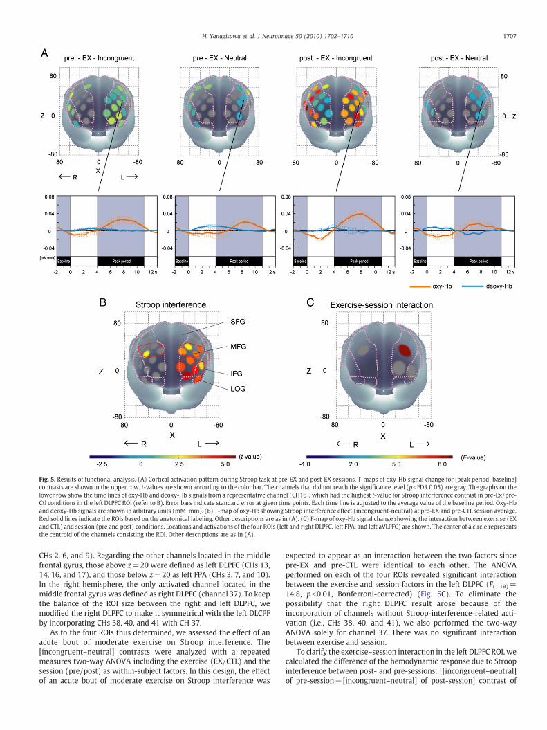
Fig. 5. Results of functional analysis. (A) Cortical activation pattern during Stroop task at pre-EX and post-EX sessions. T-maps of oxy-Hb signal change for [peak period–baseline]contrasts are shown in the upper row. t-values are shown according to the color bar. The channels that did not reach the significance level (pbFDR 0.05) are gray. The graphs on thelower row show the time lines of oxy-Hb and deoxy-Hb signals from a representative channel (CH16), which had the highest t-value for Stroop interference contrast in pre-Ex/pre-Ctl conditions in the left DLPFC ROI (refer to B). Error bars indicate standard error at given time points. Each time line is adjusted to the average value of the baseline period. Oxy-Hband deoxy-Hb signals are shown in arbitrary units (mM·mm). (B) T-map of oxy-Hb showing Stroop interference effect (incongruent-neutral) at pre-EX and pre-CTL session average.Red solid lines indicate the ROIs based on the anatomical labeling. Other descriptions are as in (A). (C) F-map of oxy-Hb signal change showing the interaction between exersise (EXand CTL) and session (pre and post) conditions. Locations and activations of the four ROIs (left and right DLPFC, left FPA, and left aVLPFC) are shown. The center of a circle representsthe centroid of the channels consisting the ROI. Other descriptions are as in (A).
1707H. Yanagisawa et al. / NeuroImage 50 (2010) 1702–1710
CHs 2, 6, and 9). Regarding the other channels located in the middlefrontal gyrus, those above z=20 were defined as left DLPFC (CHs 13,14, 16, and 17), and those below z=20 as left FPA (CHs 3, 7, and 10).In the right hemisphere, the only activated channel located in themiddle frontal gyrus was defined as right DLPFC (channel 37). To keepthe balance of the ROI size between the right and left DLPFC, wemodified the right DLPFC to make it symmetrical with the left DLCPFby incorporating CHs 38, 40, and 41 with CH 37.
As to the four ROIs thus determined, we assessed the effect of anacute bout of moderate exercise on Stroop interference. The[incongruent–neutral] contrasts were analyzed with a repeatedmeasures two-way ANOVA including the exercise (EX/CTL) and thesession (pre/post) as within-subject factors. In this design, the effectof an acute bout of moderate exercise on Stroop interference was
expected to appear as an interaction between the two factors sincepre-EX and pre-CTL were identical to each other. The ANOVAperformed on each of the four ROIs revealed significant interactionbetween the exercise and session factors in the left DLPFC (F(1,19)=14.8, pb0.01, Bonferroni-corrected) (Fig. 5C). To eliminate thepossibility that the right DLPFC result arose because of theincorporation of channels without Stroop-interference-related acti-vation (i.e., CHs 38, 40, and 41), we also performed the two-wayANOVA solely for channel 37. There was no significant interactionbetween exercise and session.
To clarify the exercise–session interaction in the left DLPFC ROI, wecalculated the difference of the hemodynamic response due to Stroopinterference between post- and pre-sessions: [[incongruent–neutral]of pre-session− [incongruent–neutral] of post-session] contrast of

1708 H. Yanagisawa et al. / NeuroImage 50 (2010) 1702–1710
oxy-Hb signal for the EX and CTL groups, respectively, and comparedthe difference between both groups (Figs. 4E, F). Oxy-Hb signaldifference was significantly greater in the EX condition than in the CTLcondition (t(19)=3.54, pb0.001, paired t-test). This result demon-strates that the acute bout of moderate exercise led to an increasedStroop interference-related cortical activation in the left DLPFC.
Association between behavioral and fNIRS results
We examined the association between the Stroop-interference-related reaction time shortening and left DLPFC activation induced byexercise. Conventional correlation analyses were not suitable for thecurrent data: both reaction time and oxy-Hb signals entail substantialindividual differences, and a subtraction procedure to contrast outStroop interference further jeopardizes the quantification of eachparameter and narrows the range of parameters leading to a flooreffect (Nunnally and Bernstein, 1994). Therefore, we performed aMcNemar test: a robust nonparametric procedure, applicable to assesscorrespondence between two incidences (Siegel and Castellan, 1988).We examined whether the exercise-induced enhancement of Stroopinterference-reflected in reaction time coincided with the exercise-induced Stroop-interference-related increase of oxy-Hb in a binom-inal manner. Specifically, the following contrast, [[[incongruent–neutral] of pre-session− [incongruent–neutral] of post-session] in EXcondition− [[incongruent–neutral] of pre-session− [incongruent–neutral] of post-session] in CTL condition], was calculated for RTand oxy-Hb signal data, respectively, and they were subjected to theMcNemar test. This revealed that the frequency of the coincidence asindicated in Table 1 was significant (χmc(1,20)
2 =11.53, pb0.001).Thus, we concluded that the improved cognitive performancedemonstrated in RT reflecting Stroop interference and left DLPFCactivation elicited by exercise significantly coincided.
Discussion
In this study,wehave for thefirst time applied fNIRSmeasurementsto assess the neural substrates underlying the cognitive effect of anacute bout of moderate exercise. fNIRS measurements in a laboratoryenabled strict control of the exercise intensity across subjects to be setat 50%V
·o2peak. Since most of the ERP studies performed thus far have
not applied strict criteria related to physicalfitness, the current study isvaluable in that it couples physiological monitoring with functionalneuroimaging to examine the cognitive effects of an acute exercisebout. The on-site measurements further allowed the monitoring ofphysiological parameters, including MCA V mean (middle cerebralarterymean blood velocity), SBF (skin blood flow), CO2, and HR (heartrate), and thus fNIRS measurements were devoid of possiblecontamination by exercise-induced physiological noise. These techni-cal merits of fNIRS have led to the revelation of a neural substrate forimproved cognitive performance after an acute bout of moderateexercise increased activation of the DLPFC (dorsolateral prefrontalcortex) in the left hemisphere to cope with Stroop interference.
From the behavioral measurements showing a shorter RT in theneutral compared to that in the incongruent condition, we verified
Table 1Contingency table for McNemar Test on association between reaction time and oxy-Hbsignal.
oxy-Hb signal Totals
− +
RT − 2 16 18+ 1 1 2Totals 3 17 20
Frequency of exercise-induced Stroop interference-related shortening of reaction timeand Stroop interference-related oxy-Hb increase in the left DLPFC is summarized. RTstands for reaction time.
that the Stroop effect could be stably observed in the conditions usedin this experiment even after an acute bout of moderate exercise.Based on this observation, we assessed the effect of an acute bout ofmoderate exercise on Stroop interference, and confirmed thatperformance was improved. This is consistent with a former studythat reports an acute-exercise-induced enhancement of Stroopinterference (Hogervorst et al., 1996).
Given that the acute bout of moderate exercise caused improvedcognitive performance to cope with the Stroop interference, the nextstep is to search for its neural substrates. In the current study, weobserved significant oxy-Hb signal increases associated with theStroop interference (incongruent–neutral contrast) in the LPFCcovering the left FPA, aVLPFC, and bilateral DLPFC (Fig. 5B). Thisspatially specific activation pattern roughly matches the results ofprevious fNIRS studies on Stroop interference, reporting bilateral LPFCactivation (Schroeter et al., 2002, 2003, 2004b), and left DLPFCactivation around FC3 when the ROI was only the left hemisphere(Ehlis et al., 2005) (Fig. 6).
Former fMRI and PET studies have also consistently reported LPFCactivation reflecting Stroop interference along with activation of ACC(Derrfuss et al., 2005; Laird et al., 2005) (Fig. 6). Since fNIRS cannotcapture frontomedian activation due to the limited depth penetrationof NIR light (Villringer and Chance, 1997), we limit our functionalarguments to the LPFC.
Reviewing the Stroop-task-related LPFC activation consistentlyobserved in former studies, Banich et al. (2000, 2001) postulated thatthe LPFC activation may reflect interference processing/responseinhibition. Considering these previous studies, the LPFC activationmay be regarded as a tight neural substrate for improved performanceon Stroop interference. In the current study, we observed that theimproved cognitive performance after an acute bout of moderateexercise coincided with increased activation of the left DLPFC due toStroop interference, providing the first experimental evidence forneural substrate of the improved cognitive performance after an acutebout of moderate exercise.
Although this would be a phenomenologically interestingfinding, the causal relation between an acute bout of exerciseand improved cognitive performance may not at first appearcoherent. However, the current study provides a crucial functionalclue to revealing the effects of an acute exercise bout on cognitiveperformance. Interestingly, in the CTL experiment, where subjectsperformed the Stroop tasks after just sitting still for 25 minwithout performing any exercise, we observed slightly decreasedcortical activation related to Stroop interference in the left DLPFC(Fig. 4F). Our first intention was to provide a control conditiondevoid of any exercise. However, this brought about decreasedarousal levels among the subjects: most of them expressedsleepiness during the rest period before the session. This led usto postulate that the left DLPFC activation coinciding with theStroop interference may be due to the increased level ofwakefulness caused by an acute bout of moderate exercise.Intriguingly, recent fNIRS studies have reported decreased LPFCactivation due to fatigue (Suda et al., 2009) and sleepiness (Suda etal., 2008) during a word-generation task, although such effects onStroop interference have yet to be explored. Indeed, afterreviewing P300 ERP experiments performed on natural andenvironmentally induced states, including exercise, Polich andKok (1995) hypothesized that cognitive processing speed isinfluenced by changes in arousal state. The arousal hypothesishas provided an explanation for the inverted U-shaped function oftask performance against physical strength (Yerkes and Dodson,1908). As arousal states increase with physical exertion, cognitiveperformance improves to an optimal point, after which furtherincreases in physical exertion result in decreased arousal levels anddeteriorating performance (Tomporowski and Ellis, 1985). Thephysical exertion required of participants in the current study was
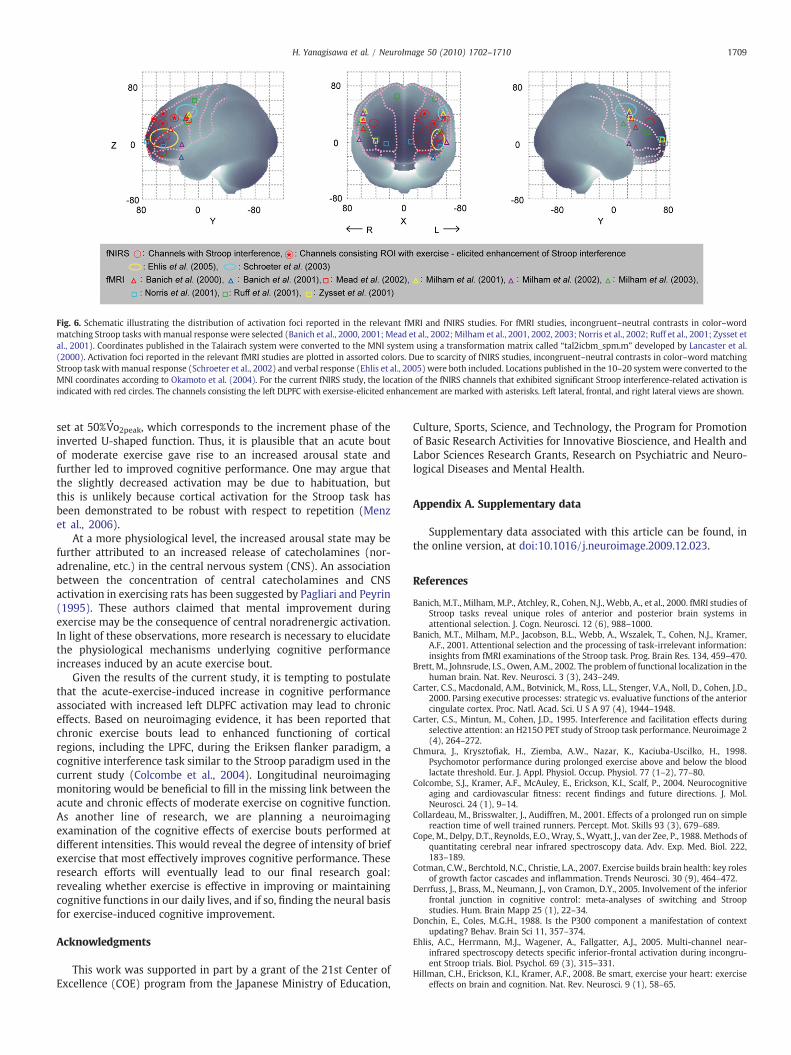
Fig. 6. Schematic illustrating the distribution of activation foci reported in the relevant fMRI and fNIRS studies. For fMRI studies, incongruent–neutral contrasts in color–wordmatching Stroop tasks withmanual response were selected (Banich et al., 2000, 2001;Mead et al., 2002; Milham et al., 2001, 2002, 2003; Norris et al., 2002; Ruff et al., 2001; Zysset etal., 2001). Coordinates published in the Talairach system were converted to the MNI system using a transformation matrix called “tal2icbm_spm.m” developed by Lancaster et al.(2000). Activation foci reported in the relevant fMRI studies are plotted in assorted colors. Due to scarcity of fNIRS studies, incongruent–neutral contrasts in color–word matchingStroop task withmanual response (Schroeter et al., 2002) and verbal response (Ehlis et al., 2005) were both included. Locations published in the 10–20 systemwere converted to theMNI coordinates according to Okamoto et al. (2004). For the current fNIRS study, the location of the fNIRS channels that exhibited significant Stroop interference-related activation isindicated with red circles. The channels consisting the left DLPFC with exersise-elicited enhancement are marked with asterisks. Left lateral, frontal, and right lateral views are shown.
1709H. Yanagisawa et al. / NeuroImage 50 (2010) 1702–1710
set at 50%V·o2peak, which corresponds to the increment phase of the
inverted U-shaped function. Thus, it is plausible that an acute boutof moderate exercise gave rise to an increased arousal state andfurther led to improved cognitive performance. One may argue thatthe slightly decreased activation may be due to habituation, butthis is unlikely because cortical activation for the Stroop task hasbeen demonstrated to be robust with respect to repetition (Menzet al., 2006).
At a more physiological level, the increased arousal state may befurther attributed to an increased release of catecholamines (nor-adrenaline, etc.) in the central nervous system (CNS). An associationbetween the concentration of central catecholamines and CNSactivation in exercising rats has been suggested by Pagliari and Peyrin(1995). These authors claimed that mental improvement duringexercise may be the consequence of central noradrenergic activation.In light of these observations, more research is necessary to elucidatethe physiological mechanisms underlying cognitive performanceincreases induced by an acute exercise bout.
Given the results of the current study, it is tempting to postulatethat the acute-exercise-induced increase in cognitive performanceassociated with increased left DLPFC activation may lead to chroniceffects. Based on neuroimaging evidence, it has been reported thatchronic exercise bouts lead to enhanced functioning of corticalregions, including the LPFC, during the Eriksen flanker paradigm, acognitive interference task similar to the Stroop paradigm used in thecurrent study (Colcombe et al., 2004). Longitudinal neuroimagingmonitoring would be beneficial to fill in the missing link between theacute and chronic effects of moderate exercise on cognitive function.As another line of research, we are planning a neuroimagingexamination of the cognitive effects of exercise bouts performed atdifferent intensities. This would reveal the degree of intensity of briefexercise that most effectively improves cognitive performance. Theseresearch efforts will eventually lead to our final research goal:revealing whether exercise is effective in improving or maintainingcognitive functions in our daily lives, and if so, finding the neural basisfor exercise-induced cognitive improvement.
Acknowledgments
This work was supported in part by a grant of the 21st Center ofExcellence (COE) program from the Japanese Ministry of Education,
Culture, Sports, Science, and Technology, the Program for Promotionof Basic Research Activities for Innovative Bioscience, and Health andLabor Sciences Research Grants, Research on Psychiatric and Neuro-logical Diseases and Mental Health.
Appendix A. Supplementary data
Supplementary data associated with this article can be found, inthe online version, at doi:10.1016/j.neuroimage.2009.12.023.
References
Banich, M.T., Milham, M.P., Atchley, R., Cohen, N.J., Webb, A., et al., 2000. fMRI studies ofStroop tasks reveal unique roles of anterior and posterior brain systems inattentional selection. J. Cogn. Neurosci. 12 (6), 988–1000.
Banich, M.T., Milham, M.P., Jacobson, B.L., Webb, A., Wszalek, T., Cohen, N.J., Kramer,A.F., 2001. Attentional selection and the processing of task-irrelevant information:insights from fMRI examinations of the Stroop task. Prog. Brain Res. 134, 459–470.
Brett, M., Johnsrude, I.S., Owen, A.M., 2002. The problem of functional localization in thehuman brain. Nat. Rev. Neurosci. 3 (3), 243–249.
Carter, C.S., Macdonald, A.M., Botvinick, M., Ross, L.L., Stenger, V.A., Noll, D., Cohen, J.D.,2000. Parsing executive processes: strategic vs. evaluative functions of the anteriorcingulate cortex. Proc. Natl. Acad. Sci. U S A 97 (4), 1944–1948.
Carter, C.S., Mintun, M., Cohen, J.D., 1995. Interference and facilitation effects duringselective attention: an H215O PET study of Stroop task performance. Neuroimage 2(4), 264–272.
Chmura, J., Krysztofiak, H., Ziemba, A.W., Nazar, K., Kaciuba-Uscilko, H., 1998.Psychomotor performance during prolonged exercise above and below the bloodlactate threshold. Eur. J. Appl. Physiol. Occup. Physiol. 77 (1–2), 77–80.
Colcombe, S.J., Kramer, A.F., McAuley, E., Erickson, K.I., Scalf, P., 2004. Neurocognitiveaging and cardiovascular fitness: recent findings and future directions. J. Mol.Neurosci. 24 (1), 9–14.
Collardeau, M., Brisswalter, J., Audiffren, M., 2001. Effects of a prolonged run on simplereaction time of well trained runners. Percept. Mot. Skills 93 (3), 679–689.
Cope, M., Delpy, D.T., Reynolds, E.O., Wray, S., Wyatt, J., van der Zee, P., 1988. Methods ofquantitating cerebral near infrared spectroscopy data. Adv. Exp. Med. Biol. 222,183–189.
Cotman, C.W., Berchtold, N.C., Christie, L.A., 2007. Exercise builds brain health: key rolesof growth factor cascades and inflammation. Trends Neurosci. 30 (9), 464–472.
Derrfuss, J., Brass, M., Neumann, J., von Cramon, D.Y., 2005. Involvement of the inferiorfrontal junction in cognitive control: meta-analyses of switching and Stroopstudies. Hum. Brain Mapp 25 (1), 22–34.
Donchin, E., Coles, M.G.H., 1988. Is the P300 component a manifestation of contextupdating? Behav. Brain Sci 11, 357–374.
Ehlis, A.C., Herrmann, M.J., Wagener, A., Fallgatter, A.J., 2005. Multi-channel near-infrared spectroscopy detects specific inferior-frontal activation during incongru-ent Stroop trials. Biol. Psychol. 69 (3), 315–331.
Hillman, C.H., Erickson, K.I., Kramer, A.F., 2008. Be smart, exercise your heart: exerciseeffects on brain and cognition. Nat. Rev. Neurosci. 9 (1), 58–65.
1710 H. Yanagisawa et al. / NeuroImage 50 (2010) 1702–1710
Hillman, C.H., Snook, E.M., Jerome, G.J., 2003. Acute cardiovascular exercise andexecutive control function. Int. J. Psychophysiol. 48 (3), 307–314.
Hogervorst, E., Riedel, W., Jeukendrup, A., Jolles, J., 1996. Cognitive performance afterstrenuous physical exercise. Percept. Mot. Skills 83 (2), 479–488.
Kamijo, K., Nishihira, Y., Hatta, A., Kaneda, T., Wasaka, T., Kida, T., Kuroiwa, K., 2004.Differential influences of exercise intensity on information processing in the centralnervous system. Eur. J. Appl. Physiol. 92 (3), 305–311.
Kamijo, K., Nishihira, Y., Higashiura, T., Kuroiwa, K., 2007. The interactive effect ofexercise intensity and task difficulty on human cognitive processing. Int. J.Psychophysiol. 65 (2), 114–121.
Katagiri, A., Dan, I., Tuzuki, D., Okamoto, M., Yokose, N., Igarashi, & K., H. T., Fujiwara, T.,Katayama, Y., Yamaguchi, Y., Sakatani, K., in press. Mapping of optical pathlength ofhuman adult head at multiwavelengths in near infrared. Advances in ExperimentalMedicine and Biology.
Keppel, G., Wickens, D.W., 2004. Design and Analysis: A Researcher’s Handbook, 4th ed.Prentice Hall, Upper Saddle River.
Koizumi, H., Yamamoto, T., Maki, A., Yamashita, Y., Sato, H., Kawaguchi, H., Ichikawa, N.,2003. Optical topography: practical problems and new applications. Appl. Opt. 42(16), 3054–3062.
Laird, A.R., McMillan, K.M., Lancaster, J.L., Kochunov, P., Turkeltaub, P.E., Pardo, J.V., Fox,P.T., 2005. A comparison of label-based review and ALE meta-analysis in the Strooptask. Hum Brain Mapp 25 (1), 6–21.
Lancaster, J.L., Woldorff, M.G., Parsons, L.M., Liotti, M., Freitas, C.S., et al., 2000.Automated Talairach atlas labels for functional brain mapping. Hum. Brain Mapp.10 (3), 120–131.
Leung, H.C., Skudlarski, P., Gatenby, J.C., Peterson, B.S., Gore, J.C., 2000. An event-relatedfunctional MRI study of the Stroop color word interference task. Cereb. Cortex 10(6), 552–560.
MacLeod, C.M., 1991. Half a century of research on the Stroop effect: an integrativereview. Psychol. Bull. 109 (2), 163–203.
Magnie, M.N., Bermon, S., Martin, F., Madany-Lounis, M., Suisse, G., Muhammad, W.,Dolisi, C., 2000. P300, N400, aerobic fitness, and maximal aerobic exercise.Psychophysiology 37 (3), 369–377.
Maki, A., Yamashita, Y., Ito, Y., Watanabe, E., Mayanagi, Y., Koizumi, H., 1995. Spatial andtemporal analysis of human motor activity using noninvasive NIR topography.Med. Phys. 22 (12), 1997–2005.
Mead, L.A., Mayer, A.R., Bobholz, J.A., Woodley, S.J., Cunningham, J.M., Hammeke, T.A.,Rao, S.M., 2002. Neural basis of the Stroop interference task: response competitionor selective attention? J. Int. Neuropsychol. Soc. 8 (6), 735–742.
Menz, M.M., Neumann, J., Muller, K., Zysset, S., 2006. Variability of the BOLD responseover time: an examination of within-session differences. Neuroimage 32 (3),1185–1194.
Milham, M.P., Banich, M.T., Barad, V., 2003. Competition for priority in processingincreases prefrontal cortex's involvement in top-down control: an event-relatedfMRI study of the Stroop task. Brain Res. Cogn. Brain Res. 17 (2), 212–222.
Milham, M.P., Banich, M.T., Webb, A., Barad, V., Cohen, N.J., Wszalek, T., Kramer, A.F.,2001. The relative involvement of anterior cingulate and prefrontal cortex inattentional control depends on nature of conflict. Brain Res. Cogn. Brain Res. 12 (3),467–473.
Milham, M.P., Erickson, K.I., Banich, M.T., Kramer, A.F., Webb, A., Wszalek, T., Cohen, N.J.,2002. Attentional control in the aging brain: insights from an fMRI study of theStroop task. Brain Cogn. 49 (3), 277–296.
Nakamura, Y., Nishimoto, K., Akamatu, M., Takahashi, M., Maruyama, A., 1999. Theeffect of jogging on P300 event related potentials. Electromyogr. Clin.Neurophysiol. 39 (2), 71–74.
Norris, D.G., Zysset, S., Mildner, T., Wiggins, C.J., 2002. An investigation of the value ofspin-echo-based fMRI using a Stroop color–word matching task and EPI at 3 T.Neuroimage 15 (3), 719–726.
Nunnally, J., Berstein, I., 1994. Psychometric theory (3rd ed.). Applied PsychologicalMeasurement 19, 303–305, McGraw-Hill, New York.
Obrig, H., Villringer, A., 2003. Beyond the visible—imaging the human brain withlight. J. Cereb. Blood Flow Metab. 23 (1), 1–18.
Okamoto, M., Dan, H., Sakamoto, K., Takeo, K., Shimizu, K., et al., 2004. Three-dimensional probabilistic anatomical cranio-cerebral correlation via the inter-national 10–20 system oriented for transcranial functional brain mapping.Neuroimage 21 (1), 99–111.
Okamoto, M., Dan, I., 2005. Automated cortical projection of head-surface locations fortranscranial functional brain mapping. Neuroimage 26 (1), 18–28.
Pardo, J.V., Pardo, P.J., Janer, K.W., Raichle, M.E., 1990. The anterior cingulate cortexmediates processing selection in the Stroop attentional conflict paradigm. Proc.Natl. Acad. Sci. U S A 87 (1), 256–259.
Pagliari, R., Peyrin, L., 1995. Norepinephrine release in the rat frontal cortex under treadmillexercise: a study with microdialysis. J Appl Physiol 78 (6), 2121–2130.
Polich, J., Kok, A., 1995. Cognitive and biological determinants of P300: an integrativereview. Biol. Psychol. 41 (2), 103–146.
Polich, J., Lardon, M.T., 1997. P300 and long-term physical exercise. Electroencephalogr.Clin. Neurophysiol. 103 (4), 493–498.
Ruff, C.C., Woodward, T.S., Laurens, K.R., Liddle, P.F., 2001. The role of the anteriorcingulate cortex in conflict processing: evidence from reverse Stroop interference.Neuroimage 14 (5), 1150–1158.
Schroeter, M.L., Zysset, S., Kruggel, F., von Cramon, D.Y., 2003. Age dependency of thehemodynamic response as measured by functional near-infrared spectroscopy.Neuroimage 19 (3), 555–564.
Schroeter, M.L., Zysset, S., Kupka, T., Kruggel, F., Yves von Cramon, D., 2002. Near-infrared spectroscopy can detect brain activity during a color–word matchingStroop task in an event-related design. Hum. Brain Mapp. 17 (1), 61–71.
Schroeter, M.L., Zysset, S., von Cramon, D.Y., 2004a. Shortening intertrial intervals inevent-related cognitive studies with near-infrared spectroscopy. Neuroimage 22(1), 341–346.
Schroeter, M.L., Zysset, S., Wahl, M., von Cramon, D.Y., 2004b. Prefrontal activation dueto Stroop interference increases during development—an event-related fNIRSstudy. Neuroimage 23 (4), 1317–1325.
Shattuck, D.W., Mirza, M., Adisetiyo, V., Hojatkashani, C., Salamon, G., Narr, K.L.,Poldrack, R.A., Bilder, R.M., Toga, A.W., 2008. Construction of a 3D probabilistic atlasof human cortical structures. Neuroimage 39 (3), 1064–1080.
Siegel, S., Castellan, N., 1988. Nonparametric Statistics for the Behavioral Sciences,Second edition. McGraw-Hill, New York.
Singh, A.K., Dan, I., 2006. Exploring the false discovery rate in multichannel NIRS.Neuroimage 33 (2), 542–549.
Singh, A.K., Okamoto, M., Dan, H., Jurcak, V., Dan, I., 2005. Spatial registration ofmultichannel multi-subject fNIRS data to MNI space without MRI. Neuroimage 27(4), 842–851.
Strangman, G., Boas, D.A., Sutton, J.P., 2002. Non-invasive neuroimaging using near-infrared light. Biol. Psychiatry 52 (7), 679–693.
Stroop, J., 1935. Studies of interference in serial verbal reactions. J. Exp. Psychol. 18,643–662.
Suda, M., Fukuda, M., Sato, T., Iwata, S., Song, M., Kameyama, M., Mikuni, M., 2009.Subjective feeling of psychological fatigue is related to decreased reactivity inventrolateral prefrontal cortex. Brain Res. 1252, 152–160.
Suda, M., Sato, T., Kameyama, M., Ito, M., Suto, T., Yamagishi, Y., Uehara, T., Fukuda, M.,Mikuni, M., 2008. Decreased cortical reactivity underlies subjective daytime lightsleepiness in healthy subjects: a multichannel near-infrared spectroscopy study.Neurosci. Res. 60 (3), 319–326.
Taylor, S.F., Kornblum, S., Lauber, E.J., Minoshima, S., Koeppe, R.A., 1997. Isolation ofspecific interference processing in the Stroop task: PET activation studies.Neuroimage 6 (2), 81–92.
Timinkul, A., Kato, M., Omori, T., Deocaris, C.C., Ito, A., Kizuka, T., Asada, T., Soya, H.,2008. Enhancing effect of cerebral blood volume by mild exercise in healthy youngmen: a near-infrared spectroscopy study. Neurosci. Res. 61 (3), 242–248.
Tomporowski, P.D., 2003. Effects of acute bouts of exercise on cognition. Acta Psychol.(Amst) 112 (3), 297–324.
Tomporowski, P.D., Ellis, N.R., 1985. The effects of exercise on the health, intelligence,and adaptive behavior of institutionalized severely and profoundly mentallyretarded adults: a systematic replication. Appl. Res. Ment. Retard. 6 (4), 465–473.
Tsuzuki, D., Jurcak, V., Singh, A.K., Okamoto, M., Watanabe, E., Dan, I., 2007. Virtual spatialregistration of stand-alone fNIRS data to MNI space. Neuroimage 34 (4), 1506–1518.
Villringer, A., Chance, B., 1997. Non-invasive optical spectroscopy and imaging ofhuman brain function. Trends Neurosci. 20 (10), 435–442.
Yerkes, R.M., Dodson, J.D., 1908. The relation of strength of stimulus to rapidity of habit-formation. J. Comp. Neurol. Psychol. 18, 459–482.
Zysset, S., Muller, K., Lohmann, G., von Cramon, D.Y., 2001. Color–wordmatching Strooptask: separating interference and response conflict. Neuroimage 13 (1), 29–36.


