Acute leukemia I&II - KSUMSCksumsc.com/download_center/2nd/2) GNT Block/Teams work/Haemato… ·...
12
Important. Extra. Doctor’s Notes Objectives: • To understand the definition of acute leukemia and recognize the general features of leukemia • To understand the general concepts of leukemia pathogenesis. • To understand the clinical presentation and recognize the importance of early diagnosis of acute leukemia • To understand the general themes of classification and the basic tool of diagnosis • To recognize the most common presenting features of acute myeloid leukemia and their significance in therapeutic approaches • To know the most important indicators implicated in prognosis of acute myeloid leukemia. • To emphasize on the general aspects of leukemia including definition, common feature and general classification and the basic diagnostic tool for acute leukemia • To understand the clinical features of acute lymphoblastic leukemia • To understand the difference between T-ALL and B-ALL in term of clinical and pathological features • To recognize the most important prognostic factor for ALL. References: 436 girls & boys’ slides 435 teamwork slides Editing file Do you have any suggestions? Please contact us! @haematology436 E-mail: [email protected] or simply use this form Acute leukemia I&II
Transcript of Acute leukemia I&II - KSUMSCksumsc.com/download_center/2nd/2) GNT Block/Teams work/Haemato… ·...

Important. Extra. Doctor’s Notes
Objectives:• To understand the definition of acute leukemia and recognize the general features of leukemia• To understand the general concepts of leukemia pathogenesis.• To understand the clinical presentation and recognize the importance of early diagnosis of acute
leukemia• To understand the general themes of classification and the basic tool of diagnosis• To recognize the most common presenting features of acute myeloid leukemia and their
significance in therapeutic approaches• To know the most important indicators implicated in prognosis of acute myeloid leukemia.
• To emphasize on the general aspects of leukemia including definition, common feature and general classification and the basic diagnostic tool for acute leukemia
• To understand the clinical features of acute lymphoblastic leukemia• To understand the difference between T-ALL and B-ALL in term of clinical and pathological
features• To recognize the most important prognostic factor for ALL.
References:436 girls & boys’ slides435 teamwork slides
Editing file
Do you have any suggestions? Please contact us!
@haematology436 E-mail: [email protected]
or simply use this form
Acute leukemia I&II

we recommend you to watch the video first
● Leukemia is named by pathologist Virchow in 1845 which means white cells because PCV shows high white cells and low Red cells which is abnormal .
● Aggressive malignant hematopoietic disorders.
● Accumulation of abnormal blasts (Immature precursors of WBCs) in bone marrow and blood leading to:
1- Bone marrow failure (anemia , neutropenia , thrombocytopenia)
2- Organ infiltration ( hepatosplenomegy , lymphadenopathy)
Pathogenesis :
Genetic
predispositionEnvironmental
factorsInfection
Previous
therapy
Other
hematological
disorders
Unknown
Mechanism
Block of differentiation ,Enhanced proliferation & Decreased
apoptosis
Genetic alteration in the immature precursors
Radio or Chemo
Note: one single factor can NOT lead to cancer
Example: mutation in
P53
E.g. HIV, and malaria
The stem cell will divide to daughter cell and blast cell which this blast will NOT differentiate to another cell
They will give them mutation that lead to proliferation
This will increase the life of cells which will lead to accumulation of blast
Acute leukemia
10:18
HEMATOLOGY TEAM 436
2

Epidemiology :
AL represent about 8% of neoplastic disease & cause about 4% of malignancy related deaths ! (cure rate is very high)
● Acute myeloid leukemia is more common in adults > 15 per 100.000/year.
● Acute lymphoid leukemia is usually affecting children 76% of childhood leukemia.
Acute Leukemia Classification:
1- Acute Myeloid Leukemia (AML). In Adults
2- Acute Lymphoid Leukemia (ALL). In children
3- Acute Leukemia of Ambiguous Lineage.
Basis of Classification
1- Clinical history. (previous therapy) 2- Morphology.
3- Flow cytometry. (BM or Periphery) 4- Chromosomal typing. 5-Molecular study.
1- Light microscopy: (Microscopy in malignancies is important)
(blood smear to see blasts, bone marrow aspirate & biopsy )
• Blast count : it should be >20% (more than or equal 20) out of the total cells
• Blast morphology :
●Myeloblast:
❑ Size: medium-Large
❑ Nucleous: round, oval or irregular
❑ Nucleolus: prominent
❑ Cytoplasm: abundant, granular
Auer rods is an important characteristic!
● Lymphoblast: ❑ Size: small- medium ❑ Nucleous: round ❑ Nucleolus: not prominent ❑ Cytoplasm: scanty ,agranular and may be vacuolated
Auer rods
In this stage is acute and undifferentiation
In this stage is chronic
HEMATOLOGY TEAM 436
3

•2-Flow cytometry: a very important technique
• Laser based technology allows for cells counting & detection of their surface &cytoplasmic markers by suspending them in a stream of fluid followed by analysis through electronic system. (CD; cluster of differentiation)
•3-Chromosomal Karyotype
•Set of the chromosomes from one cell during metaphase to study the numerical(deletion &trisomy) and structural ( translation &inversion ) abnormality.
Stem Cell Markers: (CD34& TDT)
Myeloid B-Lymphoid T-Lymphoid
MPOCD13
CD33
CD14
CD64
CD41
CD235a
CD10
CD19CD22
CD79aTo diagnose B-ALL We
need at least to markers,usually one is CD19 and
the other could be anyone from the list.
CD3CD4
CD5
CD7
CD8
When CD34 or/and TDT positive means acute leukemia
enough to diagnose AML
enough to diagnose
T-ALL
HEMATOLOGY TEAM 436
4

4- Molecular studies:
Several techniques used to detect and localize the presence or absence of specific DNA sequences on chromosomes .
Recurrent genetic abnormalities:
Fluorescent In-Situ Hybridization
(FISH)
Polymerase Chain Reaction
(PCR)
Karyotype Molecular
t (8;21) AML1-ETO
t (16;16) or inv(16)
CBFB-MYH11
t (15;17) PML-RARA
t (9;11) MLLT1-MLL
Karyotype Molecular
t (9;22) BCR-ABL1
t (4;11) AF4-MLL
t (12;21) ETV6-RUNX1
t (5;14) IL3-IGH
AML ALL
HEMATOLOGY TEAM 436
5

Acute Myeloid Leukemia (AML):• Group of hematopoietic neoplasms caused by proliferation of malignant myeloid
blasts in bone marrow and blood.
• The blast ≥20% or t(8;21) t (16;16) or t(15;17).
• More in Adults (do occur in infants!).
• Worse than ALL.
*This classification is based on morphology and flow cytometry.*Important to know the notes about M3, M4 and M5 .- M6 is very rare .
FBA Classification:-
Type
Block
differentiation of
(lead to
accumulation of)
Genes Notes
M2 (Acute myeloblastic leukemia with
maturation)Myeloblast t(8;21) -
M3 (Acute promyelocytic leukemia) promyelocyte t(15;17)Assosiated
with DIC
M4 (Acute myelomonocytic leukemia)Both Myeloblast
& Monoblastst or inv(16;16) Assosiated
with Gum
HypertrophyM5 (Acute monocytic leukemia) Monocytes t(9;11)
الجدول األخضر هو المهم
HEMATOLOGY TEAM 436
6

Clinical features of AML:
1. Pancytopenia : (deficiency in the mature cells of al blood components) acute onset:
• ↓WBC→ infection (fever ,septic shock)
• ↓Hb (because the BM is shut down, BM does not produce mature cells)→ anemia (fatigue ,headache , pallor ,SOB….)
• ↓platelets →bleeding (bruises , epistaxis ,menorrhagia…)
2. Organ infiltration:
• Hepatosplenomegaly Why ? because these two are extramedulary hemaptosis (neonatal life)
• Lymphadenopathy (rare)
• Myeloid sarcoma (solid tumor composed of immature RBCs)
• gum hypertrophy
•CNS disease
3. Leucostasis: (increased blood viscosity)
4. Disseminated intravascular coagulation (DIC): Widespread activation of coagulation system leading to intravascular fibrin deposition & consumption of platelet and coagulation factors which can be manifested as bleeding (85%) or thrombosis (15%) → more with acute promyelocytic leukemia (M3) (Why ? Abnormal promyelocytes contain numerous primary granules that increase the risk for DIC so M3 within 24h either cure (95%) or kill )
- WHO Classification:
• 1-t(8;21)• 2- t(16;16)• 3- t(15;17), • Prognosis: good
AML with recurrent genetic abnormalities:
• Blasts ≥ 20%• significant dysplasia, • Prognosis: Poor
Myelodisplasia related AML:
• Blasts ≥ 20%• Previous chemotherapy • Prognosis: Poor
Therapy related AML:
• Blasts ≥ 20%• Genetic: N• No dysplasia • Prognosis: Standard
AML not otherwise specified (FAB):
More with Acute
Monoblastic
Leukemia (M4&M5)
HEMATOLOGY TEAM 436
7

Prognosis Better Prognosis:-• Genetics: t(8;21) , inv(16;16) or t(15;17) (if you discover and treat them early)
• Age: < 60 years• Primary better than secondary (as chemotherapy either transplantation or death )
Treatment• Chemotherapy:- AML: M0-M8 but not M3 ( same protocol)- AML: M3 with target therapy (ATRA or arsenic) (Treatment is withall-trans-retinoic acid (ATRA, a vitamin A derivative), which binds the alteredreceptor and causes the blasts to mature)
• Stem cell transplantation.
Case Study:-• 65 years old male presented to ER with fatigue ,fever and nose bleeding for 2
weeks.• On Examination : moderate hepatosplenomegaly & multiple bruises. • CBC : WBC :40 x109/L HB: 7g/dL PLT: 51 x109/L
• Flow cytometry :- The blast are positive for CD34 (indicate Acute Leukemia),CD13,CD33,CD117 and MPO - They are negative for CD3 (-ve for B-ALL), CD10,CD19&CD79a (-ve for T-ALL).
• Karyotype: t(8;21).
- Diagnosis: • AML with maturation (M2) (FAB classification) • AML with t(8;21) (WHO classification )
Indicate AML
HEMATOLOGY TEAM 436
8
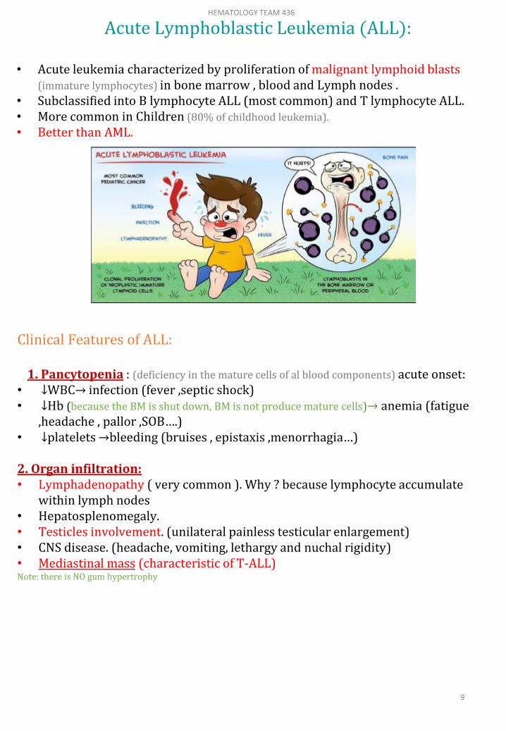
Clinical Features of ALL:
1. Pancytopenia : (deficiency in the mature cells of al blood components) acute onset:• ↓WBC→ infection (fever ,septic shock)• ↓Hb (because the BM is shut down, BM is not produce mature cells)→ anemia (fatigue
,headache , pallor ,SOB….)• ↓platelets →bleeding (bruises , epistaxis ,menorrhagia…)
2. Organ infiltration:• Lymphadenopathy ( very common ). Why ? because lymphocyte accumulate
within lymph nodes • Hepatosplenomegaly.• Testicles involvement. (unilateral painless testicular enlargement) • CNS disease. (headache, vomiting, lethargy and nuchal rigidity)• Mediastinal mass (characteristic of T-ALL)Note: there is NO gum hypertrophy
Acute Lymphoblastic Leukemia (ALL):
• Acute leukemia characterized by proliferation of malignant lymphoid blasts(immature lymphocytes) in bone marrow , blood and Lymph nodes .
• Subclassified into B lymphocyte ALL (most common) and T lymphocyte ALL.• More common in Children (80% of childhood leukemia).
• Better than AML.
HEMATOLOGY TEAM 436
9

Morphological subtypes (FAB)
Treatment:
• Chemotherapy (high cure rate)
• Stem cell transplantation
L3 (Burkitt’s) represents mature lymphoid neoplasmso it is a type of lymphoma not Acute lymphoblastic leukaemia
L1 L2 L3
L1 L2 L3 Burkitt’s
Morphology Homogenous Heterogeneous Homogenous
Size Small Variable Small
Cytoplasm Little More Vaculated
Nucleoli Not prominent Prominent Prominent
Genetics Variable Variable t(8;14) c-Myc
B cell T cell
MarkersCD19, CD10,
CD79aCD3
Percentage 80% 20%
Age Younger Older
Clinical -Mediastinal
mass CNS Relapse
WBC count Less Higher
Prognosis Better Worse
Geneticst(9;22), t(4;11),
t(12;21)-
Immunophenotypic Subtypes (WHO):
WorseBetter
<2 - >10 yrs2 - 10 yrsAge
MaleFemaleGender
HighLowWBC count
T cellB cellCell type
OthersCommon
They mean positive CD10
B-ALL phenotyp
e
Hypodiploidyt(9;22)
Hyperdiploidyt(12;21)
B-ALL genetics
YesNoCNS
involvement
Prognosis:
CD34 and TDT negative
HEMATOLOGY TEAM 436
10

Remember :
- Acute leukaemia is a fatal neoplastic condition.
- 20% or more blasts = Acute leukaemia.
- Diagnosis requires special investigations.
- Auer rods = AML.
- AML M3 = DIC & target therapy.
- Gum hypertrophy = mostly M4 or M5.
- Mediastinal = T-ALL.
- Subtypes of AML (M0-M8) + cytogenetic abnormalities.
- Subtypes of ALL (T or B cell).
- Main lineages markers are MPO , CD19 and CD3.
- Stem cell markers are CD34,TDT.
- FAB classification based mainly on morphology.
- WHO classification focused more on genetics.
THIS SLIDE IS: IMPORTANT X (109)
HEMATOLOGY TEAM 436
11

1-Talal is 25 years old from Hafer Albaten, his blast count were 8% and he has a mutation in t(8;21) So, he doesn’t have acute lymphoblastic leukemia:
A-true B-false
2-which one is true about acute lymphoblastic leukemia:
A-Burkitt’s is a type of ALL B-present with gum hypertrophy C-present with lymphadenopathy
3-A 16 female patient came to the hospital with fever, fatigue and multiple bruises. A blood sample was taken and under the microscope the cells appeared homogeneous with small sizes and not prominent nuclei, then she diagnosed with acute lymphoblastic leukemia, according to the morphological features and (FAB) classification what is the subtype of the disease:
A- L2 B-L3 Burkitt’s C-L1
4- which one is NOT clinical feature of acute myeloid leukemia:
A-lymphadenopathy B-myeloid sarcomaC-CNS disease D-mediastinal mass
Team Members:
Mosaed Al-Nowaiser
Mohammad Al-Mania
Mohammad Al-Kahil
Essam Al-Shahrani
Yara Aligi
1-A
2-C
3-C
4-D
5-B
6-B
Good Luck!
Team Leaders:
Abdulaziz Al-Hussainy
Safa Al-Osaimi
5-A patient came to the hospital, he noticed that his testis is enlarge , and hesometimes have epistaxisalso he is short of breath. The doctor diagnosed him with acute lymphoblasticleukemia on further examination the doctor did flow cytometry, the results were:CD19-negative CD10-negative, CD3-positive, what is the diagnosis?
A- B cell ALL B- T cell ALL C-acute myeloid leukemia
6-A 65 years old male patient with splenomegaly and Disseminated intravascularcoagulation, genetic test revealed a translocation between chromosome 15 and 17 t(15;17) what is the diagnosis?
A-Acute monoblastic leukemiaB-Acute promyelocytic leukemiaC-Acute myeloid leukemia with maturationD-Acute myeloid leukemia without maturation
MCQs:HEMATOLOGY TEAM 436
12