ACUTE KIDNEY INJURY (AKI) - جامعة المنصورة€¦ · creatinine (IVU is not feasible)....
54
ACUTE KIDNEY INJURY (AKI) Nephrology unit Mansoura Faculty of Medicine
Transcript of ACUTE KIDNEY INJURY (AKI) - جامعة المنصورة€¦ · creatinine (IVU is not feasible)....

ACUTE KIDNEY INJURY
(AKI)
Nephrology unitMansoura Faculty of Medicine
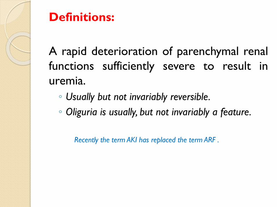
Definitions:
A rapid deterioration of parenchymal renal
functions sufficiently severe to result in
uremia.
◦ Usually but not invariably reversible.
◦ Oliguria is usually, but not invariably a feature.
Recently the term AKI has replaced the term ARF .

3


Stage Increase in Serum
Creatinine
Urine Output
1 1.5-2 times baseline
OR
0.3 mg/dl increase from
baseline
<0.5 ml/kg/h for >6 h
2 2-3 times baseline <0.5 ml/kg/h for >12 h
3 3 times baseline OR
0.5 mg/dl increase if
baseline>4mg/dl
OR
Any RRT given
<0.3 ml/kg/h for >24 h
OR
Anuria for >12 h
Definition of Acute Kidney Injury (AKI) based on
“Acute Kidney Injury Network”
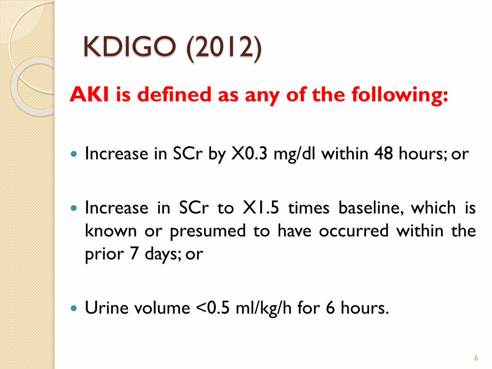
KDIGO (2012)
AKI is defined as any of the following:
Increase in SCr by X0.3 mg/dl within 48 hours; or
Increase in SCr to X1.5 times baseline, which is
known or presumed to have occurred within the
prior 7 days; or
Urine volume <0.5 ml/kg/h for 6 hours.
6
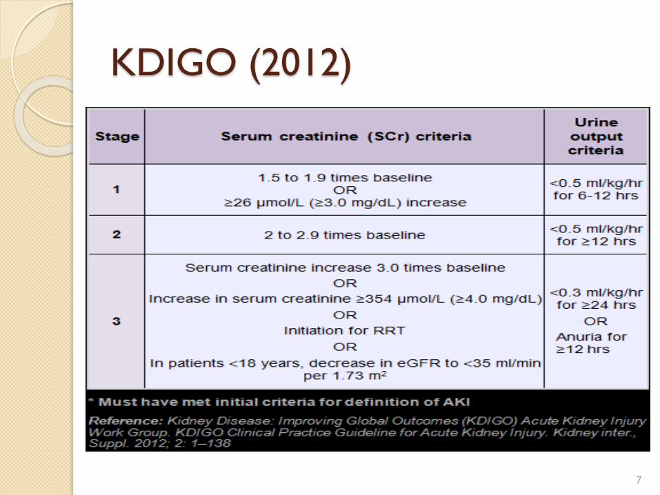
KDIGO (2012)
7

Classification of
AKI
8

9

10

In pre-renal◦ Renal tissue is intact
◦ Kidney biopsy shows normal renal histology.
◦ Oliguria and high serum creatinine are due to functional
impairment.
Since there is no structural renal damage, early diagnosis and
correction of renal hypoperfusion results in immediate
diuresis and rapid drop in serum creatinine and blood urea
levels.
If hypoperfusion is severe or neglected, renal compensatory
mechanisms will fail and acute tubular necrosis occurs

In post-renal
◦ The obstruction of the urinary tract results in
increasing the pressure above the level of the
obstruction.
◦ When this back pressure exceeds that of the
filtration pressure in the renal glomeruli, the
process of urine formation will stop with
progressive accumulation of wastes and
increase of serum creatinine and blood urea.
12

Intrinsic Renal
This includes:
◦ Acute tubular necrosis (ATN)
◦ Acute interstitial nephritis
◦ Acute glomerulonephritis

14

15

Acute tubular necrosis
(ATN)
16

Acute Tubular Necrosis
ATN can be induced by:
◦ Renal hypoperfusion (ischemia)
◦ Exposure to nephrotoxins (exogenous or
endogenous toxins)
◦ A combination of both.

Causes of Ischaemic ATN:
A-Blood Loss
• Haemorrhage (post partum, surgical or gastrointestinal).
• Major trauma
B-Fluid Loss
• Gastrointestinal (vomiting or diarrhoea)
• Renal (aggressive diuresis or polyuria)
C-Third Space
• Haematoma
• Illius
• Peritonitis
D-Severe vasodilatation as in septicemia, rapid edema formation, liver cell
failure.
E-Renovascular disease
• Renal artery occlusion by stenosis, embolism or compression.
• Renal vein thrombosis or compression.

Causes of Toxic ATN(A)Exogenous nephrotoxins include:
Antibiotics: Aminoglycosides Amphotericin
Cephalosporin Acyclovir
Sulfonamide
Tetracyclines Bacitracin
Anaesthetic agents: Methoxy fluorane
Contrast Media:
Analgesics: Phenacetin
Metals: as Mercury, lead, arsenic, bismuth, cadmium, antimony
organic solvents: Glycols
Poisons: snake bite, stings, bacterial toxins.
(B)Endogenous nephrotoxins include
Pigments: Crystals:
Myoglobin Uric acid
Hemoglobin Calcium
Methemoglobin Oxalate

20


Acute cortical necrosis:
Is a subset of ATN in which there is a
massive necrosis of the tubules and
glomeruli of the renal cortex.
The condition may be focal or diffuse with
irreversible damage of the kidneys.
It is suspected when ATN fails to recover
after 4-6 weeks.
Acute cortical necrosis usually occurs with
complicated pregnancy as postpartum
hemorrhage and abruptio placenta

23
Clinical features of AKI

Usually, the patient gives history of the
etiologic cause such as:
Trauma
Shock
Hemolysis
Drug intake
Infection
Stone disease
Procedure requiring contrast media
ICU admission

25

Patient may notice a change in
1. Urine volume and character
2. Oliguria is common, but in 10-50% of
cases urine volume will be normal or
even higher (Non-oliguric).
3. Absolute anuria is highly suggestive of
obstructive AKI (post-renal) or very
severe form of ATN (cortical necrosis).
26

Manifestation of salt and water
retention
Edema
Puffiness
Hypertension
Heart failure
27

Manifestations of uremia
By time, manifestations of uremia appear as:
acidotic breathing, dyspnea, nausea, vomiting,
headache, muscle twitches and even frank
encephalopathy and coma.
Patient may present as well with complications
28

Complications of AKI
29

Cardiovascular
• pulmonary odema • arrhythmias
• hypertension • pericardial effusion
• myocardial infarction • pulmonary embolism
Metabolic
• hyponatremia • hyperkalemia
• acidosis • hypocalcemia
• hyperphosphatemia
Neurologic
• coma • seizures
Gastrointestinal
• gastritis • gastroduodenal ulcers
Haematologic
• anaemia • hemorrhagic diathesis
Infections
• pneumonia • septicemia
• UTI


Symptoms Possible Diagnosis
Example Of Clinical Cases

33
Investigations of
AKI
A- Urinary indices:-
May be helpful in the differentiation
between pre-renal failure and acute
tubular necrosis.
Diuretics should not be given during the
preceding 48 hours for these parameters
to be valid.

35

B- Urinary sediment:
Centrifugation of fresh urine sample andexamination of the urinary sediment may behelpful in diagnosing different causes of AKI.
In pre-renal failure and in ischaemic ATNurinary sediment is usually free.
Urine sediment in Acute Nephrotic Syndromeand in RPGN is characteristic.

Urine Examination

C- Renal Imaging:
1. Plain film of the abdomen:
This will show kidney parity, size, shape, calcification and stones.
2. Renal Ultrasonography and Echo-Doppler of renal vs:◦ US safely assesses kidney size, shape and echogenicity.
◦ Cortical thinning or oedema can sometimes be seen clearly.
◦ Also, it can exclude obstructive uropathy (back pressure changes).
Echo-Doppler of renal vessels can exclude occlusion of the renal arteries and
veins.
3. Retrograde and antegrade pyelography:
Provide the most reliable information on the patency of the ureter.

Role of Ultrasound
39

4. Angiography:
Is useful mainly when an acute reversible renovascular event is
suspected such as embolization, thrombosis or
involvement in a dissecting aortic aneurysm. It carries
the risk of exposure to contrast media which could be nephrotoxic.
5. C.T. studies:
Provide reliable information on kidney parity, size, shape and
presence of hydronephrosis.
6. Magnetic Resonance urography:
*Recently MRI urography (MRU) without use of contrast
media can provide films similar to IVP.
*It is thus of great value to exclude U.T. obstruction without
the risk of contrast media nephropathy.
*It is important to know that Gadolinium an MRI contrast is
nephrotoxic,.

MR urography shows bilateral hydroureteronephrosis in a patient with 4.8 mg/dl serum
creatinine (IVU is not feasible). Note the hypointense ureteric stone bilaterally (arrows).

D. Renal biopsy:
Indications
Urine: RC cast, WC cast, Proteinuria
Unexplained
Systemic disease
Prolonged course (more than 3 weeks).

TREATMENT OF AKI
43

A-Treatment of the causee.g. any condition causing renal hypoperfusion, exposure to toxic
drug or chemical or systemic disease.

B- Conservative measures:
1- fluid balance:
Careful monitoring of intake/output and body weight is very
important to avoid overload and hypovolemia.
The 1st may lead to pulmonary edema while the 2nd may aggravate renal ischemia.
Patient should receive fluids =
daily urine output + other sensible losses e.g. vomitus or diarrhea + amount
equals the insensible loss which is around 600 c.c. for 60kg body weight
patient.
Fluids could be given orally or (if not possible), it could be given
intravenously.

2- Electrolytes and acid-base balance:• Prevent and treat hyperkalemia.
• Avoid hyponatremia.
• Keep serum bicarbonate above 16 mmol/L.
• Minimize hyperphosphatemia by giving phosphate binders (e.g. Ca Co3 & AL
hydroxide) with meals.
• Treat hypocalcaemia.
3-Treatment of hyperkalemia: Calcium gluconate I.V.
Glucose 50% + Insulin
Na Hco3 I.V.
K-exchange resins (e.g. resonium)
Avoid diets and drugs causing hyperkalaemia
Dialysis

4- Nutritional support:
With rare exceptions, Na & K restriction is appropriate.
The place of dietary ptn restriction is controversial:◦ Hope to avoid dialysis → 40gm/day
◦ Pt treated with HD → 70gm/day
◦ Hypercatabolic pt will need ↑ nitrogen intake
5- Drugs:
• Review all medications.
• Adjust dosage for renal failure.

C-Dialysis
The indications of dialysis in AKI are:
a.Clinical:
• Poor clinical state, nausea, confusion.
• Fluid overload, pulmonary oedema.
• Preoperatively.
b.Biochemical:
• Plasma K+ > 7 mmol/L.
• Plasma bicarbonate < 12 mmol/L
• Arterial pH < 7.15.
48

Prevention of AKI
49
The timing of intervention to prevent ATN is important.
Protective agents must be administered at the time of, or
immediately following potential renal insult. This intervention
may prevent or at least blunt the severity of ATN.
The intervention could be through the following approaches.
In different combinations according to the clinical situation:
• Volume expansion by isotonic saline loading.
• Diuretic as furosemide (to change ATN to polyuric-easy
manageable type)
• Non dihydropyridine Calcium channel blockers asVerapamil .
• Vasodilating agents as dopamine in renal dose 1-2 ug/kg/min.
(are not effective) .

In case of contrast media, the following additional
points should be adopted, these are:-
Avoid unnecessary contrast procedures.
Avoid multiple contrast exposure within a few days.
Avoid contrast exposure in high risk patient.
Use the smallest dose possible.
Use non-ionic , low viscosity contrast (good evidence).
Hydration by isotonic saline in a dose of 3ml/kg/h. for 12h
before and after contrast exposure (has the best evidence).
Acetyl cysteine sachets 600mg/kg tds , 2days before and after
exposure (good evidence).
Washing the contrast out immediately after the technique (e.g.
coronary angiography) by hemodialysis, is of no value.

Prognosis of AKI
52

:
The mortality of AKI remains high, ranging between 50-80% in
surgical and post-traumatic cases.
It is generally lower in AKI due to drugs and toxins.
About 75% of deaths occur in the first week of AKI, and 25-
50% of these deaths are due to the underlying disease.
The overall prognosis is better in non-oliguric than in oliguric
renal failure.
The factors influencing patient survival in AKI include the
following:
◦ Etiology of AKI.
◦ Severity of AKI.
◦ Number and severity of coexisting illness.
◦ Patient's age.
◦ Presence of complications