Acute Ischemic Stroke By
33
Acute Ischemic Stroke By: Pat Hall A manuscript submitted in partial fulfillment of the requirement for the degree of MASTER OF NURSING WASHINGTON STATE UNIVERSITY College of Nursing Intercollegiate Center for Nursing Education May, 1999
Transcript of Acute Ischemic Stroke By
Acute Ischemic Stroke
By:
Pat Hall
A manuscript submitted in partial fulfillment of� the requirement for the degree of�
MASTER OF NURSING�
WASHINGTON STATE UNIVERSITY� College ofNursing�
Intercollegiate Center for Nursing Education�
May, 1999�
To the Faculty ofWashington State University:
The members of the committee appointed to examine the ICNE Research requirements and manuscript ofPat M. Hall find it satisfactory and recommend that it be accepted.
Dr. Lorna Schumann, chair
Dr. Renee Hoeksel
ACKNOWLEDGMENTS
I wish to express my sincere gratitude to all of those who have made a "distant dream a
true reality" in obtaining my Masters in Nursing degree and becoming an Adult Nurse
Practitioner.
I thank God for "selectively" answering my deluge ofprayers and for giving me the
strength to persevere. Without his gentle guidance I would not have accomplished this dream.
A special thank you to my committee:
Dr. Lorna Schumann, my committee chair:
Your constant support, positive strokes and clinical expertise made the uphill climb
a little less steep. I always felt that you believed in my ability to succeed even
though there were times that I had my doubts! Thank you for all your help in
getting my manuscript published.
Dr. Renee Hoeksel and Dr. Mary Anne Reynolds:
Thank you for all your support, not only serving on my committee, but through
out graduate school. I appreciate all you have done.
To Margaret Ruby, for answering all my questions in a calm manner, keeping me
organized and on track. I have appreciated all your assistance.
To the Acute Care Nurse Practitioner gang: Janice Linehan, Annie Stone, Mary Hoerner
and Kim Tucker. We have shared many miles and laughs together.
To my husband, Gary, and children, Luke, Matt, Brett and Lauren. I appreciate all your
love and support. This accomplishment would not have happened without you.
ill

ACUTE ISCHEMIC STROKE�
By:�
Pat Hall� Washington State University�
May, 1999�
ABSTRACT�
Stroke is the third leading cause ofdeath in adults in the United States, behind ischemic
heart disease and all forms ofcancer. Stroke is also the leading cause of serious, long-term adult
disability and the financial burden in lost productivity and health care expenses are estimated at
$30 billion annually. Despite the magnitude of the problem, prevention strategies and treatment
methods are incompletely used. The American Heart Association developed guidelines for the
management ofacute ischemic stroke stressing the need for rapid response from the public,
emergency medical services and clinicians. Thrombolytics, more specifically, tissue plasminogen
activator {TPA(Activase)}has increased interest in treating acute ischemic stroke and
demonstrated improved outcomes when given within three hours of onset of symptoms in
selected individuals. Whether treated with thrombolytics or not, stroke patients can experience a
high incidence ofcomplications due to neurological involvement and are in need of supportive
care.
IV
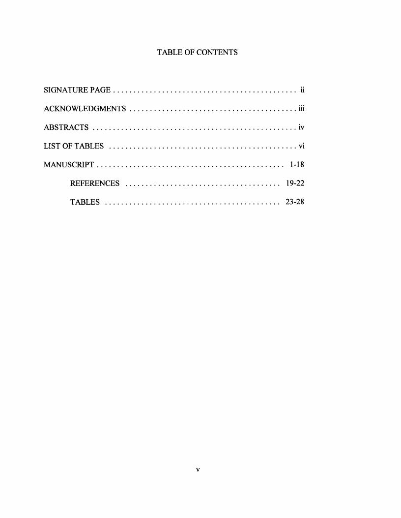
TABLE OF CONTENTS
SIGNATURE PAGE ii�
ACKNOWLEDGMENTS iii�
ABSTRACTS iv�
LIST OF TABLES vi�
MAN'USCRIPT . . . . . . . . . . . . . . . . . . . . . . . . . . . . . . . . . . . . . . . . . . . . .. 1-18�
REFERENCES 19-22�
TABLES 23-28�
v
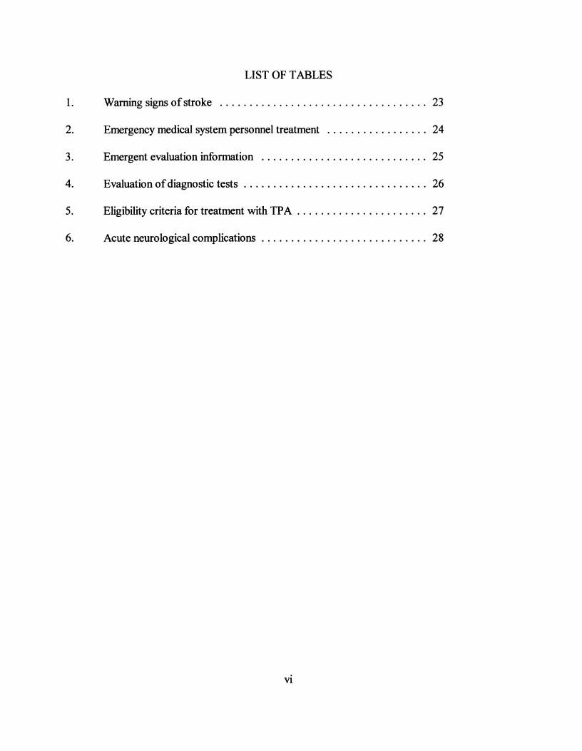
LIST OF TABLES�
1. Warning signs of stroke 23�
2. Emergency medical system personnel treatment 24�
3. Emergent evaluation information 25�
4. Evaluation ofdiagnostic tests . . . . . . . . . . . . . . . . . . . . . . . . . . . . . .. 26�
5. Eligibility criteria for treatment with TPA . . . . . . . . . . . . . . . . . . . . .. 27�
6. Acute neurological complications 28�
VI
1
One ofthe most common yet serious illnesses is ischemic stroke. It is the third leading
cause ofdeath in adults in the United States, behind ischemic heart disease and all forms ofcancer
(Selman, Tarr, & Landis, 1997). Stroke, also referred to as "brain attack", occurs 500,000 or
more times a year with approximately 158,000 Americans dying in 1995 (Stroke Statistics, 1998).
Data collected by the American Heart Association (AHA) indicates that in the United States
there is a stroke every minute and a person dies of stroke about every 3 ~ minutes (Stroke
Statistics, 1998).
The National Center for Health Statistics (Spotlight on Stroke, 1996) reports that nearly 3
million Americans have suffered a stroke, or 1 per 100 population. The incidence of stroke
doubles with every decade after 55 years ofage. Five percent ofmales aged 65 and over and 6%
of females in the same age group have suffered a stroke. Stroke rates are 50% higher in African
American men than in white men, and 130% higher in African-American women than in white
women. According to information from the United States Department ofHealth and Human
Services (USDllliS,1995), about one-third of the increased risk in African-Americans is
attributed to cardiovascular risk factors, another third to factors related to family income, and
one-third is unexplained. First-ever strokes account for about 75% of acute events and recurrent
strokes for about 25% with a recurrence rate of7-10% per year which is highest in the first year
after a first stroke (USDllliS,1995).
In the Framingham study, recurrence was found to be particularly common for thrombotic
stroke and more frequent in men (USDllliS, 1995), The Southeast has the greatest prevalence of
cerebrovascular disease and is referred to as the "stroke belt"; the lowest prevalence is in the
Northwest. Stroke affects more people every year than any other neurological illness.
2
Alzheimer's disease comes the closest with 400,000 new cases each year compared to 500,000
new cases of stroke each year (Lyden, 1997).
Stroke is also the leading cause of serious, long-term adult disability in the United States
with more than 3,000,000 people in 1991 having survived a stroke (USDIlliS, 1995).
Furthermore, stroke accounts for more than half ofall patients hospitalized for acute neurological
disease. Statistics indicate that 31 % of stroke survivors needed help caring for themselves, 20%
required help walking, 71 % had impaired ability to work and 16% had to be institutionalized
(USDIlliS, 1995). Approximately, 10% of stroke survivors are without disability and are able to
function independently.
The financial burden with lost productivity and health care expenses are estimated to be
nearly 30 billion annually (USDIlliS, 1995, Matcher, 1998). This includes $17 billion in direct
health care costs and anothe~ $13 billion in indirect costs. In 1996, for people under the age of
65, the average cost ofa stroke from hospital admission to discharge was $18,244 with the
average length of stay being 6.7 days. Seventy-five percent of stroke patients are able to return
home after completing a comprehensive rehabilitation program and their mean duration of survival
after the stroke is 7.5 years (Schnell, 1997; Reddy and Reddy, 1997).
Despite the magnitude of the problem both financially and emotionally, prevention
strategies and treatment methods are incompletely used. A study by the AHA (Adams, 1994)
revealed that nearly two-thirds ofpersons surveyed could not identify one warning sign ofa
stroke and there is a perception by the public that stroke is not a medical emergency. Less than
one-third ofpatients with an acute stroke are admitted to the hospital within 24 hours ofonset of
symptoms and less than 50% are brought to the hospital by emergency transport (Selman, et a!.,
3
1997). Starkman (1996), feels that the public is in denial when it comes to acknowledging that a
loved one has experienced a stroke. The public recognizes that the stroke has taken place, but do
not want to acknowledge that an acute neurological deficit has occurred. Stroke is many people's
greatest fear.
Despite this, there has been an increase in public awareness of stroke risk factors and the
need for changes in lifestyle, such as cessation ofcigarette smoking, decreasing alcohol and
caloric intake, controlling blood lipids and diabetes mellitus. The reduction of stroke mortality
and morbidity over the past 25 years is largely due to hypertension control, atherosclerosis
prevention, therapy for cardiac disease to eliminate embolic sources, and surgical therapy for
stroke prevention (AminotI: 1998; National Stroke Association, 1998).
Risk factors are divided into those that are potentially modifiable and those that are not.
Modifiable risk factors are: transient ischemic attack (TIA), hypertension, diabetes mellitus, atrial
fibrillation, left ventricular hypertrophy, and cigarette smoking. The paramount modifiable risk
factor for stroke is hypertension and is especially strong for levels above 160/95 mm Hg
(USDHHS, 1995). Cigarette smoking increased the risk of stroke by about 50% in both sexes
and all age groups, and the risk was directly related to the number ofcigarettes smoked per day.
Heavy consumption ofalcoho~ cocaine use, and obesity have also been linked to an increase of
stroke. Prior stroke, age, sex(male), race, and family history are nonmodifiable factors.
Increasing age is the strongest fixed factor with 72% of strokes occurring in people 65 years or
older (USDHHS, 1995).
PathophysioloK)'
The pathogenesis of stroke can be divided into two broad categories: ischemic and
4
hemorrhagic. Approximately 80 to 85% of strokes are ischemic, with the remainder (17%) being
hemorrhagic (Selman, et aI., 1997; National Stroke Association, 1997). About two-thirds of
hemorrhagic strokes are intracerebral hemorrhages commonly due to poorly controlled
hypertension and the remaining third are subarachnoid hemorrhages, caused most often by
aneurysmal rupture (Frankel & Kothari, 1997).
Approximately, two-thirds of ischemic strokes are caused by in situ thrombotic occlusions
in either large or small vessels most often caused by atherosclerosis (Frankel & Kothari, 1997).
Thrombosis may occur anywhere along a carotid artery or its branches, with a frequent site being
at the bifurcation ofthe common carotid into the internal and external carotid arteries.
Thrombotic stroke is a common stroke in diabetics patients.
About one-third of ischemic strokes are embolic, with the clot traveling through the
arterial circulation system until it lodges in a vessel too small to allow passage, which in turn,
blocks blood flow. Embolic events usually stem from cardiac sources such as atrial fibrillation,
endocarditis, and artificial cardiac valve replacement. Less commorl1y, the clot forms at the
carotid bifurcation and either blocks blood flow or gives rise to emboli that lodge in cerebral
vessels, most often being the middle cerebral artery (Rordort: Koroshetz, Copen & Cramer,
1998). The incidence ofcerebral embolism increases after age 40. (Schnell, 1997).
Under stroke conditions the brain is perfused with blood at the expense ofother less vital
organs. When blood flow is blocked to a part ofthe brain, there is a limited amount of time
before irreversible tissue injury occurs. Hypoxia (inadequate oxygenation) can cause cerebral
ischemia. The longer the duration of ischemia, the greater the likelihood of irreversible injury.
The extent of ischemia is not uniform throughout all portions ofthe brain. Severe reduction of
5
blood flow can kill brain cells within minutes, whereas, a relatively mild decrease in blood flow
can be tolerated for hours without permanent injury.
Normal cerebral blood flow (CBF) is 55 mI./I00 g./minute (AminotI: 1998). Lyden,
Rapp, Babcock, & Rothrock (1994) state that after a vascular occlusion, a portion of the ischemic
brain will survive for hours around the core ofthe ischemic zone where CBF is near zero. The
neurons in the penumbra, an area surrounding the ischemic core that is receiving blood from
collateral circulation, may not function when the CBF is between 12 and 18 mI./I00g/minute, but
may survive up to a few hours and be potentially recoverable. The ischemic penumbra may
survive ifearly reperfusion is restored. Within the area of the ischemia is a core that contains
cells that are highly dependent on the occluded artery and this area probably cannot be salvaged
(Steiner & Hacke, 1998).
The severity ofsymptoms experienced after the stroke depends on the size and number
ofcollateral vessels. The earlier perfusion is restored to the ischemic penumbra, the greater the
likelihood that the injury will be reversible. According to Heiss, Grond and Thiel (1997), much of
the tissue initially below the conventional viability threshold can survive if sufficient reperfusion is
achieved. However, most tissue that remains hypoperfused will become necrotic and some tissue
may become necrotic, despite good initial flow and reperfusion. The amount ofedema that takes
place may also lead to neurological changes that sometimes resolves within hours to days.
Early Recoenition and Treatment
Prior to 1995, acute stroke was largely considered an unfortunate medical problem
requiring supportive care and monitoring. The National Institute ofNeurological Disorders and
Stroke (NINDS) study (1995), reported on the use oftissue plasminogen activator ( TPA
6
{Activase}) for acute stroke management. With this report, there has been a change in the
philosophy ofmost of the medical community toward the urgency of swift treatment.
Pepe (1997), addressed the need for a Chain ofRecovery at the National Symposium on
Rapid Identification and Treatment ofAcute Stroke. The key links include: 1) immediate
identification of stroke symptoms and appropriate reaction by bystanders (or the patients
themselves), 2.) early access to emergency medical services (EMS), 3.) rapid EMS response,
treatment, and evacuation to designated centers capable of immediately providing definitive
diagnosis and treatment ofstroke, 4.) early communication to alert the specialty receiving center,
ensuring preparation and immediate mobilization ofresources for the stroke patient, 5.) rapid
diagnosis and intervention at those designated receiving centers, 6.) specialized treatment and
evaluation ofcomplications, precipitating factors, and accompanying conditions and 7.)
appropriate rehabilitation, when applicable.
The AHA under the guidelines for the management ofpatients with acute ischemic stroke
(1996), also stress the need for rapid response that includes the entire community. The
recognition of stroke occurs at three levels: the public, nonphysician EMS, and clinician. The
successful outcome begins with the signs ofstroke recognized by the patient or bystanders. This
requires a wide public awareness ofthe signs and symptoms ofstroke and the immediate contact
ofthe EMS. The EMS personnel should be instructed in rapid recognition, evaluation, treatment,
and transport ofpatients with stroke. A baseline assessment and quick notification ofthe hospital
can save valuable time. The clinician should suspect a stroke whenever there is a sudden onset of
focal neurological signs. Initial evaluation should assess the patient's airway, ventilation, and
circulation with a neurological exam that should be completed in 5 to 10 minutes. Emergent
8
evaluation is needed to confirm the cause of the stroke, provide information regarding the
possible reversibility, give clues about possible etiology, predict likelihood of immediate
complications and begin appropriate treatment.
Guideline for Clinical Decision Makioe
Established guidelines are important tools for minimizing delays and enhance appropriate
treatment. The AHA stroke council and American Academy ofNeurology jointly develoPed
guidelines with the emergence ofthrombolytics in the treatment ofacute ischemic stroke.
Although details of treatment protocols vary across the nation, many centers are devising
strategies for rapid diagnosis and institution ofappropriate therapy. Just as every hospital has a
plan for heart attack, every hospital needs a plan for stroke. These guidelines are meant to
expedite and improve the care given but cannot be considered effective if those in the health care
profession are not open to implement the care.
The earlier the treatment is started after the onset of stroke symptoms the better.
Therefore, when an individual develops symptoms, the "911 " emergency telephone system should
be called immediately for transport and evaluation at a medical facility. There should not be delay
to see ifthe symptoms resolve on their own or to notify relatives or private physician for advice.
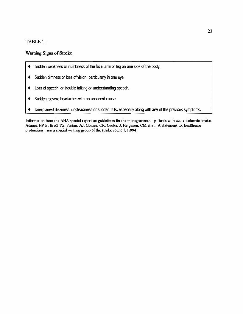
The AHA has stepped up it's campaign to make the public aware ofthe need to know the
warning signs of stroke (Table 1) and to act quickly.
Part ofthe problem in the delayed treatment ofa stroke patient is the subtle clinical
presentation. Most stroke patients do not evoke the same level ofanxiety or action as one
experiencing an acute myocardial infarction or trauma. Lack of response and anxiety applies not
only to the lay person witnessing the symptoms, but also those in the medical field who are not

9
educated to current medical practices. Prehospital providers can have a profound influence on the
patient's outcome by reducing the time required to deliver the patient to a medical facility
(Adams, 1996).
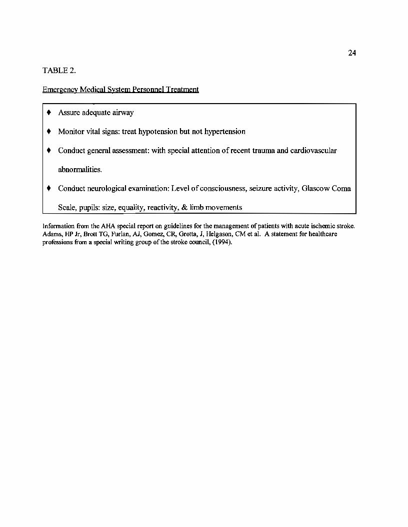
Emergency medical system (EMS) personnel should be knowledgeable ofthe rapid
recognition ofthe signs and symptoms ofan evolving stroke (Table 2), treatment and transport to
a medical facility. The baseline assessment should only take a few minutes.
According to Adams (1996), intravenous access, supplemental oxygen and cardiac
monitoring should begin during transport by the EMS. Notification of the medical facility should
take place with information regarding patient assessment, time of stroke occurrence, and
estimated time ofarrival. This enables the staffat the medical facility to assemble a "stroke team"
and meet the patient on arrival. This "stroke team" should consist, at a minimum, ofthe
emergency room physician or nurse practitioner who initially assesses the patient, an emergency
nurse who will initially care for the patient, a consulting or admitting physician (usually a
neurologist) who will provide long-term care, radiology personnel (technician and radiologist) for
cranial computed tomography (CT) and possible magnetic resonance imaging (MRI), and
laboratory personnel. Some facilities encourage a family member to ride with the EMS personnel
to facilitate quick access to the patient's medical history and events that occurred prior to the
stroke.
The AHA recommends that once the patient has arrived, an eyewitness account (if
possible) ofthe stroke should be obtained to establish the time and mode ofsymptom onset,
which is crucial to treatment. Further patient information should also include history ofprior
seizures, evidence oftrauma, infection, diabetes, illicit drug use, and other significant medical
10
history. Initial management ofpatients with suspected stroke should include a thorough physical
and neurological examination. Respiratory and cardiac function also need to be assessed and
supported ifappropriate. A complete metabolic panel (CMP), complete blood count (CBC),
prothrombin time (PT), partial prothrombin time (PTT), and bedside glucose need to be obtained
along with a CT scan.
Bock, at the National Symposium on Rapid Identification and Treatment ofAcute Stroke
(1997) discussed the paradoxial situation that exists where the victim who might gain the most
from aggressive diagnostic and therapeutic interventions is left alone to silently extend their
damage while others, who have poorer prognoses and less to gain, are given the benefit of
expedited care. All the more reason to recognize subtle changes mentioned by the patient or
family member and act quickly.
It is important to recognize various stroke "mimics" during the initial emergency
evaluation. These can include: recent seizures, suicide gestures/efforts, conversion disorders,
cocaine or amphetamine use, SAH despite a normal CT scan, transient global amnesia, systemic
infection, and toxic/metabolic encephalopathy (Selman, et al. 1997). Other neuromedical
problems that can be mistaken for stroke include migraine complicated by hemiparesis, brain
tumor, carpal tunnel syndrome associated with hand numbness, and brachial plexopathy with arm
weakness (Starkman, & Dobkin, 1995). Table 3 lists the information necessary for emergent
evaluation. Table 4 provides information on evaluation of diagnostic tests.
DiaeDostic Testio&
An emergency CT scan is absolutely necessary for patients suspected ofhaving a stroke
because ofthe subsequent therapeutic decisions depend on the results. CT is the modality of
11
choice and is usually requested to exclude SAH. It is widely available, allows for close
monitoring, and is tolerated by gravely ill patients. National Stroke Association (1998) reports
that a CT scan does not show definitive changes ofcerebral infarction for 24 to 48 hours after
onset, but subtle signs of ischemia may appear within 3 hours. It can define almost all
intracerebral hematomas>1 cm. in diameter and more than 95% of subarachnoid hemorrhages.
The findings require a high-quality CT scanner and must be carefully reviewed by a radiologist.
The MRI has some advantages over a CT scan, however, it is not widely available. There
is better contrast resolution ofall parenchymal structures, significantly better sensitivity to detect
abnormal tissues and show evidence of ischemic stroke sooner than a CT scan. Demaerel (1996),
states the advantage ofMRI is better visualization of small white matter, brainstem and cortical
infarcts. It also plays an important role in excluding more unusual causes of stroke, such as
vasculitis, arterial dissection, and multiple sclerosis.
A study in Radiology (Sorensen, 1996) reports that the thirty minute long MRI may be the
fastest way to fully evaluate patients with neurological change by providing details of flow
patterns in the Circle ofWillis, extent and location ofany acute ischemic tissue changes or
perfusion abnormalities. Disadvantages include difficulty monitoring seriously ill patients, time
needed to perform the procedure and motion artifacts in patients unable to follow commands to
remain still, claustrophobia, and those with any implanted metal (pacemakers).
Two new MRI techniques that use diffusion-weighted imaging and perfusion imaging
appear to be more sensitive in depicting infarcted tissue by changes in water mobility and
providing information regarding poorly perfused vascular territories with indication on how much
brain tissue is at risk (Demaerel, 1996; Caplan, 1998). Focal cerebral ischemia can be detected
12
earlier with the diffusion-weighted and perfusion imaging than with a CT scan or with the
traditional MRI (Sorensen, et aI, 1996; Everdingen, Grond, Kappelle, Ramos, & Malie, 1998)).
If the clinical presentation, laboratory data and results ofboth CT scan and MRI are
consistent with the diagnosis ofacute ischemic stroke, the indications, contraindications and
relative contraindications for thrombolytic therapy need to be considered. In general,
thrombolytics expedite clot lysis and restore circulation which may limit the extent ofbrain injury
and improve the outcome ofthe stroke. In the late 1960's and early 1970's intracranial bleeding
was a frequent complication and the therapy was abandoned. At that time, there was no CT
diagnostics, consequently many patients were treated that may have had SAH and the 3 hour time
frame was not instituted (Adams, et aI, 1996).
The NINDS study (Marler, 1995), demonstrated neurological improvement after a stroke
when given within a three hour onset of stroke symptoms in selected individuals. There was also
a significant improvement at 24 hours and at three months. Following the NINDS study, the
recommended dose ofTPA is 0.9mg/kg (maximum 90 mg). The initial 10% is given as an
intravenous bolus over one minute and the remaining infused over 60 minutes. No anticoagulants
or aspirin should be given within the first 24 hours after TPA. The AHA, working in conjunction
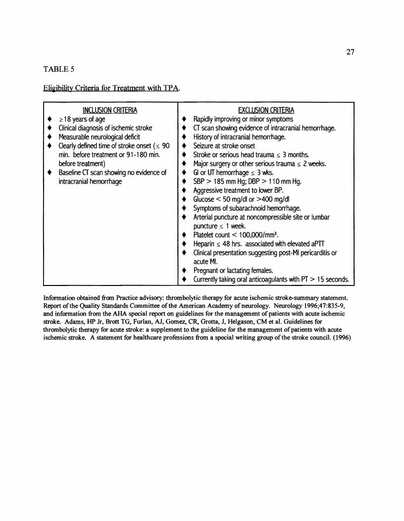
with the American Academy ofNeurology, have provided eligibility criteria guidelines listed in
Table 5.
Thrombolytic therapy is not without risk. Symptomatic intracranial hemorrhage (ICH)
occurred in 6.4% ofpatients treated within the NINDS-sponsored study (Adams, et aI., 1996).
However, the overall rate was lower than reported in other studies and despite the hemorrhage,
the rate ofdeath or severe disability was less in the actively treated group.
Other thrombolytic agents such as streptokinase and urokinase wiIllyse clots and have

13
been used for the treatment of stroke. Streptokinase has been documented to have serious
adverse effects and questionable efficacy in treating stroke. Urokinase has been used but
generally as a thrombolytic agent delivered via a catheter directly to the clot obstructed artery or
vein (National Stroke Association, 1998; Albers, 1997).
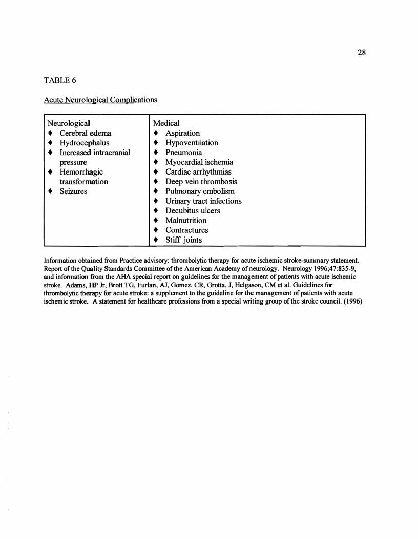
Complications
The majority of stroke patients should be admitted to the hospital because ofthe high
incidence ofcomplications due to neurological involvement. Emergent supportive care and
treatment ofacute complications is initiated whether the stroke is ischemic or hemorrhagic in
nature. Most stroke patients with the exception ofhemorrhagic patients, die from complications
related to the stroke, rather than the stroke itself (Starkman & Dobkin, 1995). There are many
acute neurological complications that can occur (Table 6) after a stroke. Cerebral edema,
seizures and hemorrhagic conversion are three ofthe most acute complications.
Fifteen percent of ischemic stroke victims develop severe brain edema which peaks at 3 to
5 days and is usually not a problem for the first 24 hours except for those with large cerebral
infarctions. Brain edema and elevated intracranial pressure caused by occlusions ofmajor
intracranial arteries and large multilobar infarctions commonly result in death within the first
week. Brain edema can lead to further extension ofthe original infarction or additional injury to
other areas ofthe brain with additional neurological damage. Treatment may include the use of
osmotic diuretics, hyperventilation, drainage ofcerebral spinal fluid, and surgery. Controversial
management ofcerebral edema is the use ofcorticosteroids due to the increase of infection,
furosemide, mannitol and barbiturates (Adams, et aI., 1994; Albers, 1997)
Seizures are most likely to occur within 24 hours of stroke and are usually partial, with or
without secondary generalization. Overall prognosis of stroke is not altered with intermittent
14
seizures, which may reoccur in 20% to 80% ofcases. Prophylactic administration of
anticonvulsants is not recommended for stroke patients not experiencing seizures. Treatment
depends on the type and frequency of seizures with phenytoin, carbamazepine, lorazepam and
diazepam most commonly used. (Adams, et aI., 1994; USDHSS, 1995; Albers, 1997)
According to Adams, et. aI., (1996), the treatment ofthrombolysis-related bleeding
depends on the location and size of the hematoma, ability to control the bleeding mechanically,
risk ofworsening the neurological condition or death, interval between drug administration and
hemorrhage, and the type of thrombolytic drug given. The location, size and etiology of stroke
may influence the bleeding complication. In the NINDS stroke trial (Marler, 1995), 6.4% of
those in the TPA group experienced symptomatic brain hemorrhage and approximately half of
those patients died. Many persons presenting with a stroke are taking aspirin, ticlopidine, or
warfarin which may influence the eligibility for, or success of thrombolytic therapy in the setting
ofan acute ischemic stroke. There was a higher incidence ofhemorrhage in those patients
presenting to the emergency room with very severe deficits or early evidence ofcerebral edema
on their pretreatment CT scan.
Treatment for those suspected ofhaving intracranial hemorrhage include STAT blood
draw ofprothrombin time (PT), partial thromboplastin time (PTT), hematocrit, hemoglobin,
platelet count and fibrinogen. Blood should be typed and crossed. IfTPA is being infused, it
should be discontinued immediately. If intracranial bleeding is suspect, an emergent CT scan is
needed and a neurosurgeon should be consulted. Some active bleeding from intravenous or
arterial sites can be controlled mechanically with direct compression.
Hypoxia caused from partial airway obstruction, hypoventilation, aspiration pneumonia
and atelectesis can result in anaerobic metabolism and decrease in energy stores which in turn can
15
increase the extent ofbrain injury. Maintaining adequate oxygenation is a critical component in
the emergent care ofa stroke patient. Intubation may be necessary when the level of
consciousness deteriorates. Hypoxia should be monitored by arterial blood gases and pulse
oximetry.
Optimum management ofhypertension following a stroke remains controversial. There
are many reasons for an elevated blood pressure: stress from the stroke, a full bladder, pain,
underlying hypertension, physiological response to brain hypoxia, or increased intracranial
pressure. Once any or all of these problems are taken care of: the blood pressure may move to
within normal limits. Lowering of blood pressure should, therefore, be done cautiously because
the patient's neurological status may be compromised with the use ofantihypertensive drugs.
Most guidelines recommend minimal to no initial treatment ofmild to moderately elevated
blood pressure during the first several hours with an ischemic stroke, but more aggressive
treatment in patients with intracerebral or subarachnoid hemorrhage (Broderick, 1997). Agents
used to treat elevated blood pressure should be those that are easily titrated with a quick onset of
action, such as labetalol or low dose enalapril. In the NINDS stroke study, exclusion was made if
the systolic blood pressure was >185 mm Hg or diastolic 110 mm Hg or ifaggressive treatment
was needed to reach those limits.
Hypotension is rarely a problem in stroke patients and may signify dehydration, diminished
cardiac output or arrhythmia. Correction of the above problems during the first few hours ofa
stroke are optimal. The use of intravenous fluids, vasopressors, colloid solutions, and cardiac
medications may be needed (Adams, et aI., 1994; Albers, 1997; Broderick, 1997).
According to the AHA guidelines for the management ofpatients with acute ischemic
stroke (1994), hyperglycemia can be a response to a serious brain injury and it is not certain ifthe
16
elevated glucose would make the stroke worse. Some studies have correlated poor outcome after
a stroke with an elevated blood glucose. However, hypoglycemia can produce focal symptoms
that may mimic stroke (Albers, 1997). Both hyperglycemia and hypoglycemia should be treated.
NINDS exclusion criteria for receiving TPA is a glucose <50 mg/dl or > 400 mg/dl.
Acute myocardial infarction occurs concomitantly with stroke onset in up to 3% of
patients and atrial fibrillation in 18%. Fewer than 2% ofpatients with acute myocardial infarction
develop ischemic strokes and 85% of such strokes occur within the first month (Starkman &
Dobkin,. 1995).
Supportive Care
All patients admitted for stroke should be considered at risk for neurological worsening
with approximately 25% ofpatients deteriorating during the first 24 to 48 hours after admission
(Adams, et aI1994). Changes in neurological function are often hard to predict, therefore, stroke
patients should be admitted to a unit that has nursing personnel trained to promptly recognize
subtle changes in neurological status. Supportive care should include facilitating measures both
medical and surgical, observe changes that may prompt interventions, prevention of
complications, preventative measure offuture strokes, and rehabilitation to restore neurological
function (Aminoft: 1998).
Assessment ofvital signs and neurological status should take place frequently during the
first 24 hours. The patient is usually placed on bed rest, however, early mobilization will decrease
the chance ofpneumonia, deep vein thrombosis, pulmonary embolism, contractures, and decubitis
ulcers. Frequent turning and passive range ofmotion can be started during the first 24 hours.
These patients are at high risk ofaspiration, choking, excessive coughing and vomiting.
Oral intake should be held for at least 24 to 48 hours (Schnell, 1997). Assessment ofthe ability to
17
swallow should take place to decrease the possibility ofaspiration. Pneumonia is the leading
cause ofdeath due to aspiration (Starkman & Dobkin, 1995). Patients with infarctions in the
brain stem, multiple strokes, large hemispheric lesions, or depressed consciousness are at greatest
risk ofaspiration (Adams, et aI., 1994).
Malnutrition is ofconcern if the patient is unable to sustain nutrition. Ifnecessary, a
feeding tube or gastrostomy tube may be inserted to implement caloric intake and administration
ofmedication. Further patient care includes keeping the head ofthe bed raised 30 degrees,
pulmonary toileting, encouragement ofcoughing and postural drainage. Vital signs should be
followed with special attention given to the temperature which may signify pneumonia and
appropriate antibiotic therapy instituted, along with serial chest Xrays.
Due to immobility and inability to complain ofpain, stroke patients are at high risk for
deep vein thrombosis (DVT). Starkman & Dobkins (1995), rate the incidence ofDVT, as high as
75% IIp to 2 weeks after a stroke. Pulmonary embolism is the fourth commonest cause ofdeath
in patients who have had a stroke, which usually occurs when the DVT involves the vessels
proximal to the popliteal vein. Early mobilization, subcutaneous heparin, aspirin, alternating
pressure stockings and elastic support stockings are suggested modes oftreatment.
Sixty percent ofpatients that have had a stroke, experience urinary incontinence but only
15% remain incontinent at six months (Ayers, et aI., 1995). Secondary septicemia occurs in 5%
of stroke patients (Adams et aI., 1994). The use of indwelling catheters should be avoided due to
the increased incidence of infection.
Rehabilitation
Twenty to thirty percent of stroke survivors are likely to require inpatient rehabilitation
(Starkman & Dobkins, 1995). Neurological and functional recovery occurs most rapidly in the
18
first 1 to 3 months after stroke (USDllliS,1995). In the Framingham study, improvement in
motor strength and performance of self-care functions slowed 3 months after stroke, but continue
at a reduced rate throughout the first 12 months, especially in the patients with cerebral
infarctions. Stroke rehabilitation begins during the acute hospitalization, as soon as the diagnosis
ofstroke is established and life-threatening complications are under control. Rehabilitation has
been described as "the planned withdrawal of support" in which services are provided when
needed and removed when no longer needed (USDllliS, 1995). Rehabilitation should include
prevention of further strokes and complications, proper management ofgeneral health functions,
mobilizing the patient, resumption of self-care activities, emotional support and education for
patient and family.
Summary
To optimize the recovery outcome ofthose with acute ischemic stroke, several steps need
to be taken and strengthened by the public and medical personnel. These include: immediate
identification of stroke symptoms and appropriate actions, quick access to EMS, rapid EMS
response, treatment and evacuation, early communication to the medical facility, rapid diagnosis
and interventions, specialized treatment, evaluation ofcomplications, precipitating and
accompanying factors, and appropriate rehabilitation when applicable.
19
References
Adams, H. P., Brott T.G., Crowell, R. M., Furlan, A.J., Gomez, C.R., Grotta, J., Helgason,
C.M., Marler, J. R., Woolson, R. F., Zivin, J. A., Feinberg, W., Mayberg, M. (1994).
Guidelines for the manaaement ofpatients with acute ischemic stroke. A statement for healthcare
professionals from a special writing group of the stroke council, American Heart Association.
Adams, H. P., Brott T.G., Furlan, A.J., Gomez, C.R., Grotta, J., Helgason, C.M.,
Kwiatkowski, T., Lyden, P. D., Marler, J. R., Tomer, J., Feinberg, W., Mayberg, M., Thies, W.
(1996). Guidelines for thrombolytic therapy for acute stroke: A supplement to the auidelines for
the manaaement ofpatients with acute ischemic stroke. A statement for healthcare professionals
from a special writing group of the stroke council, American Heart Association.
Albers, G. W., (1997). Medical treatment for acute ischemic stroke. The American Journal of
Medicine Sacco, R. L. (ed.) Continuing education series; New approaches to the treatment of
ischemic stroke. Part II: Established treatments for ischemic stroke.
AminotI: M. J. (1998). Nervous system. Tierney, L. M., McPhee, S. J., Papadadis, M. A.,
(Eds.), Current medical diaanosis & treatment. (37th ed.), (pp 929-936). Stanford, CT: appleton &
Lange.
Babcock, T., Lyden, P.D., Rapp, K., Rothrock, J. (1994). Ultra-rapid identification, triage,
and enrollment of stroke patients into clinical trials. Journal of Stroke Cerebrovascular Disease,
~(2), 106-113.
Bock, B.F. (1997). Response system for patients presenting with acute stroke. Marler, J. R.,
Jones, P. W., & Emr, M. (Eds.), Proceedinas ofa National Symposium on Rapid Identification
and Treatment ofAcute Stroke (NIH Publication No. 97-4239, pp.55-56) . Bethesda, MD.
20
Broderick, J. P. (1997). Response system for patients presenting with acute stroke. Marler, J.
R., Jones, P. W., & Emr, M. (Eds.), Proceedings ofa National Symposium on Rapid
Identification and Treatment ofAcute Stroke (NIH Publication No. 97-4239, pp.63-68 ).
Bethesda, MD.
Caplan, L. R. (1998). Cerebrovascular disease (stroke). Stein, J. H. (Ed.). Internal Medicine
(5 th ed. ). (pp. 997-1017). St. Louis, MO: Mosby, Inc.
Demaere~ P. (1996). The role of imaging in acute stroke patients. Acute stroke: Current
Approaches to Management, 1, 3-5.
Everdingen, K. J., Grond, J., Kappelle, L. J., Ramos, L. M.P., & Malie, W.P. T. M., (1998).
Diffusion weighted magnetic resonance imaging in acute stroke. Stroke,29, 1783-1790.
Franke~ M. R., & Kot~ R. U. (eds.) (1997) Acute ischemic stroke: A new paradigm
(monograph produced by Gardiner-Caldwell SynerMed). Emory University School ofMedicine,
The Robert W. Woodruff Health Sciences Center.
Furlan, A. J. (1997). Hospital care ofacute stroke. Marler, J. R., Jones, P. W., & Emr, M.
(Eds.), Proceedings ofa National Symposium on Rapid Identification and Treatment ofAcute
Stroke (NIH Publication No. 97-4239, pp.75-82). Bethesda, MD.
Heiss, W.D., Grond, M., Thiel, A (1997), Ischemic brain tissue salvaged from infarction with
alteplase. Lancet, 349 1599-1600.
Lyden, P.D., (1997). Magnitude of the problem of stroke and the significance ofacute
intervention. (Keynote address), Marler, J. R., Jones, P. W., & Emr, M. (Eds.), Proceedings ofa
National Symposium on Rapid Identification and Treatment ofAcute Stroke (NIH Publication
No. 97-4239, ppl-4). Bethesda, MD.�
Lyden, P. D., Rapp, K., Babcock,T., & Roghrock, J. (1994). Ultra-rapid identification, triage,�

21
and enrollment ofstroke patients into clinical trials. Journal of Stroke and Cerbrovascular Disease
~ (2). 106-113.
Marler, J. (1995, December 14). Tissue plasminogen activator for acute ischemic stroke.
The national institute ofneurological disorders and stroke rt-PA stroke study group. The New
England Journal ofMedicine, 333(24), 581-1587.
Matcher, D. B. (1998). The value of stroke prevention and treatment. Neurology, 51 (supp 3),
S31-S35. 603-606.
National Stroke Association (1997). StrokelBrain Attack Reporter's Handbook. Englewood.
Pagana, K.D., Pagana, T.J. (1998) Mosby's Manual of Diagnostic and LaboratoQ' Test. St.
Louis, MO: Mosby.
Pepe, P. E. (1997). The initial links in the chain of recovery for brain attack-access,
prehospital care, notification, and transport. Marler, J. R., Jones, P. W., & Emr, M. (Eds.),
Proceedings ofa National Symposium on Rapid Identification and Treatment ofAcute Stroke
(NIH Publication No. 97-4239, pp. 17-28). Bethesda, MD.
Reddy, M. P. & Reddy, V. (1997, April) Stroke rehabilitation. American Family Physician
~(5), 1742-1752.
Rordort: G., Koroshetz, W.J., Copen, W. A., & Cramer, S. C. (1998). Regional ischemia and
ischemia injury in patients with acute middle cerebral artery stroke as defined by early diffusion
weighted and perfusion-weighted MRI. Stroke,(29), 939-943.
Schnell, S. S., (1997). Nursing care ofclients with cerebrovascular disorders Black, J. M.,
Matassarin-Jacobs, E. (Eds.), Medical-surgical nursing: Clinical management for continuity of
care. (5 th ed.), (pp 771-833).Philadelphia, PA: W. B. Saunders.
Selman, W. R., Tarr, R., & Landis, D.M.D. (1997, June). Brain attack: emergency treatment
22
of ischemic stroke. American Family Physician.. 55(8), 2655-2663.
Sorensen, A. G., Buonanno, F. S., Gonzalez, R. G., Schwamm, L. H., Lev, M.G., Huang
Hellinger, F. R., Reese, T. G., WeisskotI: R. M., Davis, T. L., Suwanwela, N., Finklestein, S. P.,
Rosen, B. R. Koroshetz, W. J. (1996). Hyperacute stroke: evaluation with combined multisection
diffusion-weighted and hemodynamically weighted echo-planar MR imaging. Radiology,199391
401.
Spotlight on: Stroke. (1996, September). National Center for Health Statistics
<http://www.cdc.gov/nchs...acts/96sheets/mhc996.htm> (1998, March 10).
Starkman, S., (1996). Acute stroke and emergency medicine: the physician's perspective.
(Interview). Acute Stroke: Current Approaches to Management, 1, 6-8.
Starkman, S. & Dobkin, B. (1995).Cerebral vascular emergencies. Ayers, S. M., Grenvik, A.,
Holbrook, P. R., Shoemaker, W. C. (Eds.), Textbook of Critical Care (3rd ed.), (pp. 1539-1545).
Philadelphia, PA: W. B. Saunders.
Steiner, T. & Hacke, W. (1998). Combination therapy with neuroprotectants and
thrombolytics in acute ischemic stroke. European Neurology, 40, 1-8.
Stroke (brain attacks) statistics. (1998) American Heart Association (1998).
<http://www.amhrt.org/scientific/hsstats98/index.html> (1998, March 6).
co.
Stroke: The first hours. (1998). National Stroke Association.
<http://www.stroke.org/ns800_strokeinfolib.html> . (1998, March 5).
U.S. Department ofHealth and Human Services. (1995). Post-stroke rehabilitation (USDHHS
Publication No. 95-0662). Rockville, MD: Author.
23
TABLE 1.�
Warning Signs of Stroke�
+ Sudden v.eakness or numbness of the face, arm or leg on one side of the body.
+ Sudden dimness or loss of vision, particularly in one eye.
+ Loss of speech, or trouble talking or understanding speech.
+ Sudden, severe headaches with no apparent cause.
+ Unexplained dizziness, unsteadiness or sudden falls, especially along with any of the previous symptoms.
Information from the AHA special report on guidelines for the management of patients with acute ischemic stroke. Adams, lIP Jr, Brott TG, Furlan, AJ, Gomez, CR, Grotta, J, Helgason, CM et al. A statement for healthcare professions from a special writing group of the stroke council, (1994).
24
TABLE 2.
EmerGency Medical System Personnel Treatment
+� Assure adequate airway
+� Monitor vital signs: treat hypotension but not hypertension
+� Conduct general assessment: with special attention of recent trauma and cardiovascular�
abnormalities.�
+� Conduct neurological examination: Level ofconsciousness, seizure activity, Glascow Coma
Scale, pupils: size, equality, reactivity, & limb movements
Information from the AHA special report on guidelines for the managenlent of patients with acute ischemic stroke. Adams, HP Jr, Brott TG, Furlan, AJ, Gomez, CR, Grotta, J, Helgason, CM et at. A statement for healthcare professions from a special writing group of the stroke council, (1994).
25
TABLE 3
Emer~ent Evaluation Information
+� Time ofonset of symptoms
+� Blood tests to include:�
Complete Blood Count (CBC)�
Complete Metabolic Panel (CMP)
Prothrombin Time (PT)
Partial Prothrombin Time (PTT)
Glucose (can be done by finger stick)
+� Physical and neurological examination
+� 12-lead electrocardiagram (EKG)
+� Noncontrast brain CT scan
+� *MRI (ifhemorrhage excluded by CT
scan)
*An MRI is not standard practice at many medical institutions, but is included due to new
research that is available.
26
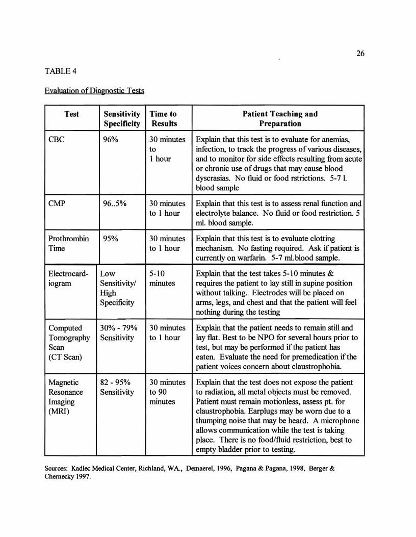
TABLE 4
Evaluation ofDiaanostic Tests
Test Sensitivity Time to Patient Teaching and Specificity Results Preparation
CBC 96% 30 minutes Explain that this test is to evaluate for anemias, to infection, to track the progress ofvarious diseases, 1 hour and to monitor for side effects resulting from acute
or chronic use ofdrugs that may cause blood dyscrasias. No fluid or food rstrictions. 5-7 1. blood sample
CMP 96..5% 30 minutes Explain that this test is to assess renal function and to 1 hour electrolyte balance. No fluid or food restriction. 5
mI. blood sample.
Prothrombin 95% 30 minutes Explain that this test is to evaluate clotting Time to 1 hour mechanism. No fasting required. Ask ifpatient is
currently on warfarin. 5-7 mI.blood sample.
Electrocard- Low 5-10 Explain that the test takes 5-10 minutes & 10gram Sensitivity/ minutes requires the patient to lay still in supine position
High without talking. Electrodes will be placed on Specificity arms, legs, and chest and that the patient will feel
nothing during the testing
Computed 30% -79% 30 minutes Explain that the patient needs to remain still and Tomography Sensitivity to 1 hour lay flat. Best to be NPO for several hours prior to Scan test, but may be performed if the patient has (CT Scan) eaten. Evaluate the need for premedication if the
patient voices concern about claustrophobia.
Magnetic 82 - 95% 30 minutes Explain that the test does not expose the patient Resonance Sensitivity to 90 to radiation, all metal objects must be removed. Imaging minutes Patient must remain motionless, assess pt. for (MRI) claustrophobia. Earplugs may be worn due to a
thumping noise that may be heard. A microphone allows communication while the test is taking place. There is no food/fluid restriction, best to empty bladder prior to testing.
Sources: Kadlec Medical Center, Richland, WA., Demaerel,1996, Pagana & Pagana, 1998, Berger & Chemecky 1997.
27
TABLE 5
EliGibility Criteria for Treatment with TPA.
INCLUSION CRITERIA +� ~ 18 years of age +� Clinical diagnosis of ischemic stroke +� Measurable neurological deficit +� Clearly defined time of stroke onset (~ 90�
min. before treatment or 91-180 min.� before treatment)�
+� Baseline cr scan showing no evidence of� intracranial hemorrhage�
EXCLUSION CRITERIA +� Rapidly improving or minor symptoms +� a scan showing evidence of intracranial hemorrhage. +� History of intracranial hemorrhage. +� Seizure at stroke onset +� Stroke or serious head trauma ~ 3 months. +� Major surgery or other serious trauma ~ 2weeks. +� GI or UT hemorrhage ~ 3wks. +� SBP> 185 mm Hg; DBP > 110 mm Hg. +� Aggressive treatment to lower BP. +� Glucose < 50 mg/dl or >400 mg/dl +� Symptoms of subarachnoid hemorrhage. +� Arterial puncture at noncompressible site or lumbar
puncture ~ 1week. +� Platelet count < 100,OOO/mm3•
+� Heparin ~ 48 hrs. associated with elevated aPTT +� Clinical presentation suggesting post-MI pericarditis or
acute MI. +� Pregnant or lactating females. +� Currently taking oral anticoagulants with PT > 15seconds.
Information obtained from Practice advisory: thrombolytic therapy for acute ischemic stroke-summary statement. Report of the Quality Standards Committee of the American Academy of neurology. Neurology 1996;47:835-9, and information from the AHA special report on guidelines for the management ofpatients with acute ischemic stroke. Adams, HP Jr, Brott TG, Furlan, AJ, Gomez, CR, Grotta, J, Helgason, CM et al. Guidelines for thrombolytic therapy for acute stroke: a supplement to the guideline for the management ofpatients with acute ischemic stroke. A statement for healthcare professions from a special writing group of the stroke council. (1996)
28
TABLE 6
Acute Neurological Complications
Neurological� Medical +� Cerebral edema + Aspiration +� Hydrocephalus + Hypoventilation +� Increased intracranial + Pneumonia�
pressure + Myocardial ischemia� + Hemorrhagic + Cardiac arrhythmias�
transformation + Deep vein thrombosis� +� Seizures + Pulmonary embolism
+� Urinary tract infections +� Decubitus ulcers +� Malnutrition +� Contractures +� Stiff joints
Information obtained from Practice advisory: thrombolytic therapy for acute ischemic stroke-summary statement. Report of the Quality Standards Committee of the American Academy of neurology. Neurology 1996;47:835-9, and information from the AHA special report on guidelines for the management of patients with acute ischemic stroke. Adams, HP Jr, Brott TG, Furlan, AJ, Gomez, CR, Grotta, J, Helgason, CM et al. Guidelines for thrombolytic therapy for acute stroke: a supplement to the guideline for the management ofpatients with acute ischemic stroke. A statement for healthcare professions from a special writing group of the stroke council. (1996)