Acute encephalopathy in children with tuberous sclerosis ...
9
Numoto et al. Orphanet J Rare Dis (2021) 16:5 https://doi.org/10.1186/s13023-020-01646-8 RESEARCH Acute encephalopathy in children with tuberous sclerosis complex Shingo Numoto 1* , Hirokazu Kurahashi 1 , Atsushi Sato 2 , Masaya Kubota 3 , Takashi Shiihara 4 , Tohru Okanishi 5 , Ryuta Tanaka 6 , Ichiro Kuki 7 , Tetsuhiro Fukuyama 8 , Mitsuru Kashiwagi 9 , Mitsuru Ikeno 10 , Kazuo Kubota 11 , Manami Akasaka 12 , Masakazu Mimaki 13 and Akihisa Okumura 1 Abstract Objective: We examined the clinical manifestations of acute encephalopathy (AE) and identify risk factors for AE in children with tuberous sclerosis complex (TSC). Methods: The clinical data of 11 children with clinically diagnosed TSC associated with AE and 109 children with clinically diagnosed TSC alone aged 4 years or older were collected from 13 hospitals. Results: Of the 11 children with AE, 5 had histories of febrile seizures (FS), and all had histories of febrile status epilep‑ ticus (FSE). AE developed within 24 h after fever onset in all children with seizures lasting 30 min or longer. All children developed coma after seizure cessation. Head magnetic resonance imaging (MRI) revealed widespread abnormalities in the cerebral cortex, subcortical white matter, corpus callosum, basal ganglia, and thalamus. One child died; seven had severe neurological sequelae; and the other three, mild sequelae. Logistic regression analysis revealed that a his‑ tory of FSE was correlated with the development of AE. Significance: AE in children with TSC was characterized by sudden onset after fever, followed by coma, widespread brain edema evident on MRI, and poor outcomes. A history of FSE was a risk factor for the development of AE. Keywords: Clinical neurology history, Prognosis, Status epilepticus, Infantile spasms, MRI © The Author(s) 2021. Open Access This article is licensed under a Creative Commons Attribution 4.0 International License, which permits use, sharing, adaptation, distribution and reproduction in any medium or format, as long as you give appropriate credit to the original author(s) and the source, provide a link to the Creative Commons licence, and indicate if changes were made. The images or other third party material in this article are included in the article’s Creative Commons licence, unless indicated otherwise in a credit line to the material. If material is not included in the article’s Creative Commons licence and your intended use is not permitted by statutory regulation or exceeds the permitted use, you will need to obtain permission directly from the copyright holder. To view a copy of this licence, visit http://creativecommons.org/licenses/by/4.0/. The Creative Commons Public Domain Dedication waiver (http://creativeco mmons.org/publicdomain/zero/1.0/) applies to the data made available in this article, unless otherwise stated in a credit line to the data. Introduction Tuberous sclerosis complex (TSC) is an autosomal-dom- inant genetic disorder caused by mutation of the TSC1 or TSC2 gene [1], characterized by multiple hamarto- mas in the skin, brain, heart, kidney, and lungs [2, 3]. A hyperactive mammalian target of rapamycin (mTOR) pathway plays a key role in the pathophysiology of TSC and seizure development in patients with the condition [4, 5]. TSC is one of the major genetic causes of epilepsy; about 85% of TSC patients present with seizures [6–8], especially in infancy [9]. Patients with TSC may exhibit multiple types of seizures refractory to antiepileptic drugs (AEDs) [10, 11]. Many authors have evaluated epilepsy in patients with TSC, but little is known about acute encephalopathy (AE) [12–14], which is character- ized by impaired consciousness with or without other neurologic findings such as seizures lasting for > 24 h. We encountered a boy with TSC complicated by AE. He had a history of epileptic spasms followed by focal sei- zures, but the seizures were well controlled by vigabatrin and carbamazepine. His psychomotor development was slightly delayed, and he exhibited mild autistic features. At 16 months of age, he developed febrile status epilep- ticus (FSE). e seizure lasted for 35 min, whereas com- plete recovery of consciousness was seen within 8 h and no neurological sequelae was recognized. At 23 months of age, he developed AE. Although he received intensive care, marked brain edema developed, followed by brain Open Access *Correspondence: [email protected]‑med‑u.ac.jp 1 Department of Pediatrics, Aichi Medical University, 1‑1 Yazako Karimata, Nagakute, Aichi 480‑1195, Japan Full list of author information is available at the end of the article
Transcript of Acute encephalopathy in children with tuberous sclerosis ...
Numoto et al. Orphanet J Rare Dis (2021) 16:5 https://doi.org/10.1186/s13023-020-01646-8
RESEARCH
Acute encephalopathy in children with tuberous sclerosis complexShingo Numoto1* , Hirokazu Kurahashi1, Atsushi Sato2, Masaya Kubota3, Takashi Shiihara4, Tohru Okanishi5, Ryuta Tanaka6, Ichiro Kuki7, Tetsuhiro Fukuyama8, Mitsuru Kashiwagi9, Mitsuru Ikeno10, Kazuo Kubota11, Manami Akasaka12, Masakazu Mimaki13 and Akihisa Okumura1
Abstract
Objective: We examined the clinical manifestations of acute encephalopathy (AE) and identify risk factors for AE in children with tuberous sclerosis complex (TSC).
Methods: The clinical data of 11 children with clinically diagnosed TSC associated with AE and 109 children with clinically diagnosed TSC alone aged 4 years or older were collected from 13 hospitals.
Results: Of the 11 children with AE, 5 had histories of febrile seizures (FS), and all had histories of febrile status epilep‑ticus (FSE). AE developed within 24 h after fever onset in all children with seizures lasting 30 min or longer. All children developed coma after seizure cessation. Head magnetic resonance imaging (MRI) revealed widespread abnormalities in the cerebral cortex, subcortical white matter, corpus callosum, basal ganglia, and thalamus. One child died; seven had severe neurological sequelae; and the other three, mild sequelae. Logistic regression analysis revealed that a his‑tory of FSE was correlated with the development of AE.
Significance: AE in children with TSC was characterized by sudden onset after fever, followed by coma, widespread brain edema evident on MRI, and poor outcomes. A history of FSE was a risk factor for the development of AE.
Keywords: Clinical neurology history, Prognosis, Status epilepticus, Infantile spasms, MRI
© The Author(s) 2021. Open Access This article is licensed under a Creative Commons Attribution 4.0 International License, which permits use, sharing, adaptation, distribution and reproduction in any medium or format, as long as you give appropriate credit to the original author(s) and the source, provide a link to the Creative Commons licence, and indicate if changes were made. The images or other third party material in this article are included in the article’s Creative Commons licence, unless indicated otherwise in a credit line to the material. If material is not included in the article’s Creative Commons licence and your intended use is not permitted by statutory regulation or exceeds the permitted use, you will need to obtain permission directly from the copyright holder. To view a copy of this licence, visit http://creat iveco mmons .org/licen ses/by/4.0/. The Creative Commons Public Domain Dedication waiver (http://creat iveco mmons .org/publi cdoma in/zero/1.0/) applies to the data made available in this article, unless otherwise stated in a credit line to the data.
IntroductionTuberous sclerosis complex (TSC) is an autosomal-dom-inant genetic disorder caused by mutation of the TSC1 or TSC2 gene [1], characterized by multiple hamarto-mas in the skin, brain, heart, kidney, and lungs [2, 3]. A hyperactive mammalian target of rapamycin (mTOR) pathway plays a key role in the pathophysiology of TSC and seizure development in patients with the condition [4, 5]. TSC is one of the major genetic causes of epilepsy; about 85% of TSC patients present with seizures [6–8], especially in infancy [9]. Patients with TSC may exhibit multiple types of seizures refractory to antiepileptic
drugs (AEDs) [10, 11]. Many authors have evaluated epilepsy in patients with TSC, but little is known about acute encephalopathy (AE) [12–14], which is character-ized by impaired consciousness with or without other neurologic findings such as seizures lasting for > 24 h. We encountered a boy with TSC complicated by AE. He had a history of epileptic spasms followed by focal sei-zures, but the seizures were well controlled by vigabatrin and carbamazepine. His psychomotor development was slightly delayed, and he exhibited mild autistic features. At 16 months of age, he developed febrile status epilep-ticus (FSE). The seizure lasted for 35 min, whereas com-plete recovery of consciousness was seen within 8 h and no neurological sequelae was recognized. At 23 months of age, he developed AE. Although he received intensive care, marked brain edema developed, followed by brain
Open Access
*Correspondence: [email protected]‑med‑u.ac.jp1 Department of Pediatrics, Aichi Medical University, 1‑1 Yazako Karimata, Nagakute, Aichi 480‑1195, JapanFull list of author information is available at the end of the article
Page 2 of 9Numoto et al. Orphanet J Rare Dis (2021) 16:5
herniation. EEG revealed generalized slowing, followed by extremely low voltage EEG activity. He died 20 days after AE onset. We discussed this child with other pedi-atric neurologists and found that similar cases had been observed in other hospitals. This prompted us to investi-gate AE in children with TSC.
This study examined the clinical manifestations of AE to identify risk factors for AE in children with TSC. We presumed that excitotoxicity attributable to prolonged seizures is the principal cause of the irreversible brain lesions. A better understanding of the clinical manifes-tations of AE will aid clinicians in treating children with TSC, as early identification of the problem will facilitate appropriate treatment. Identification of children at high risk is essential to ensure that caregivers of children with TSC receive appropriate information. We thus performed a retrospective multicenter study.
Materials and methodsThis study was approved by the Ethics Committee of Aichi Medical University Hospital. TSC was clinically diagnosed using the criteria of the 2012 International TSC Consensus Conference [14]. In all patients, two or more measure features were recognized, fulfilling the cri-teria of the definite diagnosis of TSC, although genetic analysis was rarely performed. We formed a research group to clarify the clinical features of and risk factors for AE in children with TSC. We invited researchers to join the group using the mailing list of the Annual Zao Conference on Pediatric Neurology (http://sites .googl e.com/site/zaose minar /). The mailing list includes more than 1,000 pediatric neurologists from all over Japan. First, in November 2017, the senior author (AO) com-menced enrolment of patients with TSC who had expe-rienced AE. Here, febrile seizure (FS) was defined as a seizure accompanied by pyrexia of 38 °C or higher without central nervous system infection or acute meta-bolic derangement lasting for 30 min or less. FSE was defined as a seizure accompanied by pyrexia of 38 °C or higher lasting for 30 min or longer, with recovery of consciousness within 24 h and complete recovery with-out neurological sequelae. AE was defined as a condition characterized by impaired consciousness with or with-out other neurologic findings, such as seizures, involun-tary movement, and delirious behavior, lasting for > 24 h in children with infection symptoms, including fever, cough, and diarrhea, according to our previous study [15]. Coma was defined as marked loss of consciousness when a patient could not be awakened by painful stimuli, which could not be attributable to sedative effects of the drugs. Eight hospitals, including ours, wherein pediatric neurologists had encountered children with TSC com-plicated by AE joined the research group. A total of 11
children with clinically diagnosed TSC associated with AE were reported. One of them had been reported else-where as a case report [16]. Demographic information, disease manifestations and complications of TSC before the onset of AE, any family history of TSC, epilepsy, or FS, clinical manifestations, laboratory data obtained during AE, treatments, and outcomes of the 11 children were collected using a structured questionnaire. In addi-tion, MRI data on all children were obtained. Outcomes were divided into three categories: death, severe sequelae (no verbal communication and/or bedridden), and mild sequelae (capable of verbal communication and sitting unaided).
We also collected clinical data on children with clini-cally diagnosed TSC who had not experienced AE to assess the risk factors for AE in children with TSC. As mentioned below, the age at onset of AE was less than 4 years in all but one child, suggesting that children with TSC who are younger than 4 years of age may develop AE in the future, even if they had no history of AE. There-fore, we excluded children with TSC under 4 years of age from the control group. The 106 control children who were 4 years of age or older and who had never had AE were recruited from 13 hospitals: the aforementioned eight hospitals that had seen children with AE and 5 additional hospitals. We retrospectively collected the fol-lowing information: sex, any history of FS, FSE, and non-febrile SE, any family history of TSC, epilepsy and FS, and complications of TSC such as subependymal giant cell astrocytoma (SEGA), cognitive disorder, any epilepsy, epileptic spasms, and focal seizures observed by 4 years of age. The genetic data were not evaluated because genetic examinations were performed only in a few patients, so we could not discuss the difference between genetic diagnosis and clinical diagnosis.
All statistical analyses were performed using EZR ver. 1.37 (http://www.jichi .ac.jp/saita ma-sct/Saita maHP.files /statm ed.html) [17]. A p-value < 0.05 was considered to indicate statistical significance. To define risk factors for AE in children with TSC, we first compared clinical variables between children with and without AE using Fisher’s exact test. Then, we performed logistic regression analysis to identify contributors to the occurrence of AE. We used stepwise selection based on p-values to identify risk factors for the occurrence of AE.
ResultsClinical features of AE in children with TSCTable 1 lists the demographic features of children with AE. The median age at AE onset was 22 months (range, 16–52 months). All but one child developed AE before 4 years of age. Five children had histories of FS and all had histories of FSE. Ten children had epilepsy and
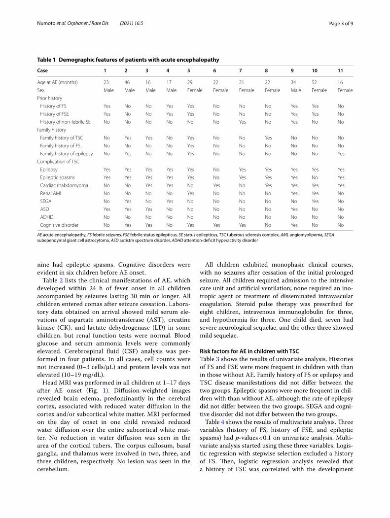
Page 3 of 9Numoto et al. Orphanet J Rare Dis (2021) 16:5
nine had epileptic spasms. Cognitive disorders were evident in six children before AE onset.
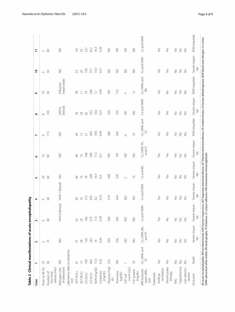
Table 2 lists the clinical manifestations of AE, which developed within 24 h of fever onset in all children accompanied by seizures lasting 30 min or longer. All children entered comas after seizure cessation. Labora-tory data obtained on arrival showed mild serum ele-vations of aspartate aminotransferase (AST), creatine kinase (CK), and lactate dehydrogenase (LD) in some children, but renal function tests were normal. Blood glucose and serum ammonia levels were commonly elevated. Cerebrospinal fluid (CSF) analysis was per-formed in four patients. In all cases, cell counts were not increased (0–3 cells/µL) and protein levels was not elevated (10–19 mg/dL).
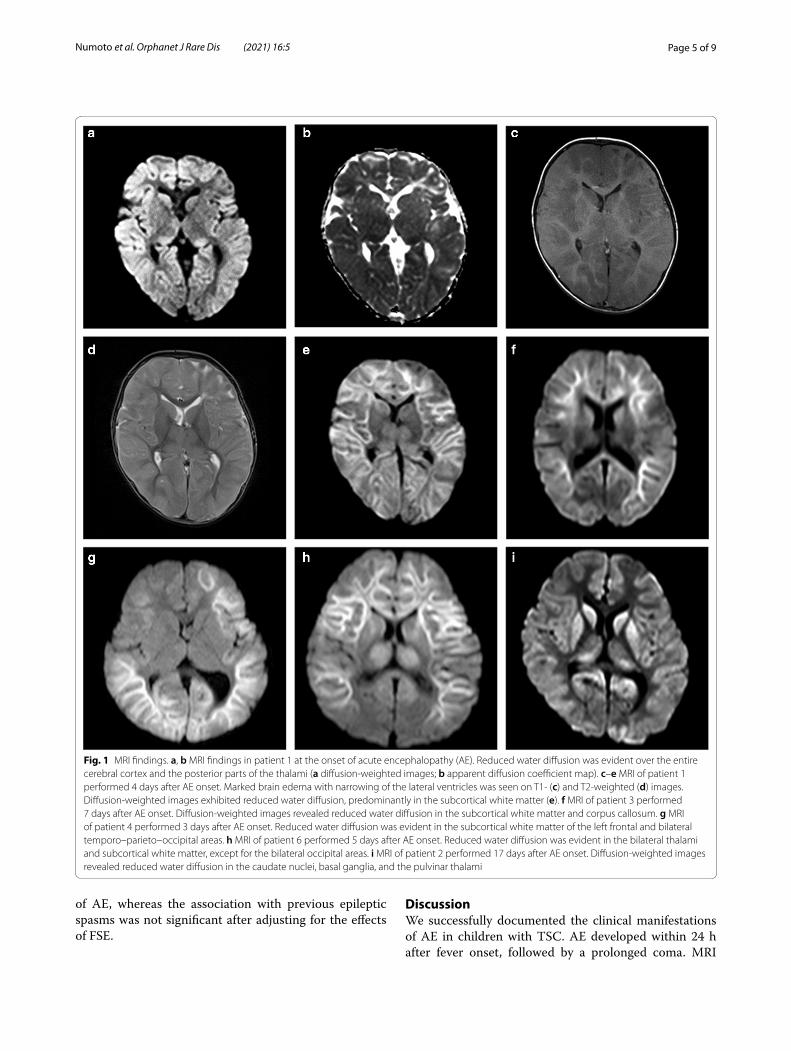
Head MRI was performed in all children at 1–17 days after AE onset (Fig. 1). Diffusion-weighted images revealed brain edema, predominantly in the cerebral cortex, associated with reduced water diffusion in the cortex and/or subcortical white matter. MRI performed on the day of onset in one child revealed reduced water diffusion over the entire subcortical white mat-ter. No reduction in water diffusion was seen in the area of the cortical tubers. The corpus callosum, basal ganglia, and thalamus were involved in two, three, and three children, respectively. No lesion was seen in the cerebellum.
All children exhibited monophasic clinical courses, with no seizures after cessation of the initial prolonged seizure. All children required admission to the intensive care unit and artificial ventilation; none required an ino-tropic agent or treatment of disseminated intravascular coagulation. Steroid pulse therapy was prescribed for eight children, intravenous immunoglobulin for three, and hypothermia for three. One child died, seven had severe neurological sequelae, and the other three showed mild sequelae.
Risk factors for AE in children with TSCTable 3 shows the results of univariate analysis. Histories of FS and FSE were more frequent in children with than in those without AE. Family history of FS or epilepsy and TSC disease manifestations did not differ between the two groups. Epileptic spasms were more frequent in chil-dren with than without AE, although the rate of epilepsy did not differ between the two groups. SEGA and cogni-tive disorder did not differ between the two groups.
Table 4 shows the results of multivariate analysis. Three variables (history of FS, history of FSE, and epileptic spasms) had p-values < 0.1 on univariate analysis. Multi-variate analysis started using these three variables. Logis-tic regression with stepwise selection excluded a history of FS. Then, logistic regression analysis revealed that a history of FSE was correlated with the development
Table 1 Demographic features of patients with acute encephalopathy
AE acute encephalopathy, FS febrile seizures, FSE febrile status epilepticus, SE status epilepticus, TSC tuberous sclerosis complex, AML angiomyolipoma, SEGA subependymal giant cell astrocytoma, ASD autistm spectrum disorder, ADHD attention-deficit hyperactivity disorder
Case 1 2 3 4 5 6 7 8 9 10 11
Age at AE (months) 23 46 16 17 29 22 21 22 34 52 16
Sex Male Male Male Male Female Female Female Female Male Female Female
Prior history
History of FS Yes No No Yes Yes No No No Yes Yes No
History of FSE Yes No No Yes Yes No No No Yes Yes No
History of non‑febrile SE No No No No No No Yes No Yes No No
Family history
Family history of TSC No Yes Yes No Yes No No Yes No No No
Family history of FS No No No No Yes No No No No No No
Family history of epilepsy No Yes No No Yes No No No No No Yes
Complication of TSC
Epilepsy Yes Yes Yes Yes Yes No Yes Yes Yes Yes Yes
Epileptic spasms Yes Yes Yes Yes Yes No Yes Yes Yes No Yes
Cardiac rhabdomyoma No No Yes Yes No Yes No Yes Yes Yes Yes
Renal AML No No No No Yes No No No Yes Yes No
SEGA No Yes No Yes No No No No No Yes No
ASD Yes Yes Yes No No No No No Yes No No
ADHD No No No No No No No No No No No
Cognitive disorder No Yes Yes No Yes Yes Yes No Yes No No
Page 4 of 9Numoto et al. Orphanet J Rare Dis (2021) 16:5
Tabl
e 2
Clin
ical
man
ifest
atio
ns o
f acu
te e
ncep
halo
path
y
AE a
cute
enc
epha
lopa
thy,
ND
not
det
erm
ined
, HH
V hu
man
her
pesv
irus,
AST
Asp
arta
te a
min
otra
nsfe
rase
, ALT
Ala
nine
am
inot
rans
fera
se, C
K cr
eatin
e ki
nase
, LD
lact
ate
dehy
drog
enas
e, B
UN
blo
od u
rea
nitr
ogen
, Cx
cort
ex,
SWM
sub
cort
ical
whi
te m
atte
r, BG
bas
al g
angl
ia, T
h th
alam
us, C
C co
rpus
cal
losu
m, I
VIG
intr
aven
ous
imm
unog
lobu
lin
Case
12
34
56
78
910
11
Feve
r to
AE
(h)
233
04
2410
68
104
3
Dur
atio
n of
th
e in
itial
se
izur
e (m
in)
3070
8060
6090
115
105
3050
80
Path
ogen
(site
of
det
ectio
n)N
DN
DH
HV‑
6 (b
lood
)H
HV‑
7 (b
lood
)N
DN
DN
DH
HV‑
6(b
lood
)In
fluen
za(n
asal
sw
ab)
ND
ND
Labo
rato
ry d
ata
on p
rese
nta‑
tion
AST
(IU
/L)
4747
6285
4648
4430
3853
161
ALT
(IU
/L)
1328
2025
1619
1228
1120
33
CK
(IU/L
)10
913
518
431
578
148
5785
7010
922
7
LD
(IU
/L)
463
262
319
435
309
299
409
352
296
377
812
BU
N (m
g/dL
)13
.314
11.5
8.2
14.4
17.2
18.9
13.6
1214
.316
.3
Cre
atin
ine
(mg/
dL)
0.32
0.44
0.39
0.33
0.4
0.3
0.44
0.21
0.45
0.51
0.28
Glu
cose
(mg/
dL)
232
303
358
219
248
ND
286
329
ND
ND
ND
Am
mon
ia
(μg/
dL)
ND
256
343
434
320
147
243
103
152
ND
ND
CSF
cel
l co
unt (
/µL)
1N
DN
DN
D2
ND
0N
D3
ND
ND
CSF
pro
tein
(m
g/dL
)18
ND
ND
ND
10N
D19
ND
12N
DN
D
MRI
‑red
uced
w
ater
diff
u‑si
on
Cx,
SW
M, a
nd
ThC
x, S
WM
, BG
, an
d Th
Cx
and
SWM
Cx
and
SWM
Cx
and
BGC
x, S
WM
, Th,
an
d CC
Cx,
SW
M, a
nd
CCC
x an
d SW
MC
x, S
WM
, and
BG
Cx
and
SWM
Cx
and
SWM
Trea
tmen
t
Art
ifici
al
vent
ilatio
nYe
sYe
sYe
sYe
sYe
sYe
sYe
sYe
sYe
sYe
sYe
s
Ste
roid
pul
se
ther
apy
Yes
No
No
Yes
Yes
Yes
Yes
Yes
No
Yes
Yes
IVIG
No
No
No
Yes
No
Yes
No
No
No
No
Yes
Hyp
othe
rmia
No
No
No
Yes
No
No
No
No
Yes
Yes
No
Use
cat
echo
‑la
min
eN
oN
oN
oN
oN
oN
oN
oN
oN
oN
oN
o
Out
com
eD
eath
Seve
re s
eque
‑la
eSe
vere
seq
ue‑
lae
Seve
re s
eque
‑la
eSe
vere
seq
ue‑
lae
Seve
re s
eque
‑la
eM
ild s
eque
lae
Seve
re s
eque
‑la
eM
ild s
eque
lae
Seve
re s
eque
‑la
eM
ild s
eque
lae
Page 5 of 9Numoto et al. Orphanet J Rare Dis (2021) 16:5
of AE, whereas the association with previous epileptic spasms was not significant after adjusting for the effects of FSE.
DiscussionWe successfully documented the clinical manifestations of AE in children with TSC. AE developed within 24 h after fever onset, followed by a prolonged coma. MRI
Fig. 1 MRI findings. a, b MRI findings in patient 1 at the onset of acute encephalopathy (AE). Reduced water diffusion was evident over the entire cerebral cortex and the posterior parts of the thalami (a diffusion‑weighted images; b apparent diffusion coefficient map). c–e MRI of patient 1 performed 4 days after AE onset. Marked brain edema with narrowing of the lateral ventricles was seen on T1‑ (c) and T2‑weighted (d) images. Diffusion‑weighted images exhibited reduced water diffusion, predominantly in the subcortical white matter (e). f MRI of patient 3 performed 7 days after AE onset. Diffusion‑weighted images revealed reduced water diffusion in the subcortical white matter and corpus callosum. g MRI of patient 4 performed 3 days after AE onset. Reduced water diffusion was evident in the subcortical white matter of the left frontal and bilateral temporo–parieto–occipital areas. h MRI of patient 6 performed 5 days after AE onset. Reduced water diffusion was evident in the bilateral thalami and subcortical white matter, except for the bilateral occipital areas. i MRI of patient 2 performed 17 days after AE onset. Diffusion‑weighted images revealed reduced water diffusion in the caudate nuclei, basal ganglia, and the pulvinar thalami
Page 6 of 9Numoto et al. Orphanet J Rare Dis (2021) 16:5
revealed brain edema with reduced water diffusion, pre-dominantly in the subcortical white matter. Outcomes were poor (severe neurological sequelae or death in most patients) despite various treatments. We also found that a history of FSE was associated with the development of AE.
It is remarkable that the clinical manifestations of AE were similar among the children. All experienced AE within 1 day of fever onset, seizures lasting for 30 min, a monophasic clinical course with coma, and widespread MRI abnormalities. We presume that excitotoxicity attributable to prolonged seizures is the principal cause of the irreversible brain lesions because all children had a prolonged seizure refractory to antiepileptic drugs. Hypercytokinemia may be also involved in AE patho-genesis, but the laboratory abnormalities of our patients were milder than those of patients with acute necrotizing encephalopathy [18, 19], which is considered to be caused by a “cytokine storm” associated with multiorgan failure and disseminated intravascular coagulation. Marked ele-vations of enzymes such as AST and LD were common in children with acute necrotizing encephalopathy immedi-ately after disease onset [20]. Hypercytokinemia may play
only a limited role in the development of AE in children with TSC. On the other hand, elevated blood glucose and serum ammonia levels were common in our patients, suggesting metabolic derangement; this may be a sequela of critical illness caused by AE.
MRI revealed widespread abnormalities in all children. Reduced water diffusion (indicating cytotoxic edema) was evident, predominantly in the subcortical white mat-ter, and conventional MRI suggested edema in the cer-ebral cortex. A similar MRI pattern is seen in children with Dravet syndrome complicated by AE [15, 21]. Oku-mura et al. reported 15 such children and showed that brain edema and reduced water diffusion in the cortical and/or subcortical white matter were characteristic of the condition [15]. Notably, a prolonged seizure is an initial symptom of AE in children with Dravet syndrome. Wide-spread MRI abnormalities with cytotoxic edema may be neuroimaging features of AE in children with TSC.
Initial laboratory abnormalities and CSF analysis abnormalities were mild (or absent) in our children. This implies that the brain disorders of children with AE exhibited sudden onset and rapid progression. All children presented with a seizure induced by fever; dis-tinguishing AE from less severe seizures is clinically dif-ficult on initial presentation. Laboratory data may not be helpful; no marked abnormalities are present. However, hyperglycemia was common in AE children on presenta-tion. Hyperglycemia is correlated with adverse outcomes of status epilepticus and AE [22–24] and may be a con-venient predictor of AE.
The outcomes of children with TSC complicated by AE were poor, although intensive treatment was performed. Treatments included supportive efforts to stabilize the general condition, seizure control, and neuroprotection. Although all patients required intensive care and artifi-cial ventilation, their general condition was appropriately maintained. No patient developed shock, serious mul-tiorgan failure, or disseminated intravascular coagula-tion. Seizure control was achieved in all patients after the aggressive use of antiepileptic drugs. A recent consensus treatment for status epilepticus refers to prompt recog-nition and the need for very early treatment to reduce morbidity and mortality, drug requirements, and seizure duration [25, 26]. Studies employing buccal or intrana-sal midazolam found that delivery via non-intravenous routes was a practical, rapid, reasonably safe, and effec-tive alternative to intravenous lorazepam or diazepam as a first-line treatment for early status epilepticus in out-of-hospital settings [27, 28]. No such rescue drugs (example: buccal midazolam) are yet available in Japan. Neuroprotective treatment will be a subject of a future study. Several pharmacological and non-pharmacolog-ical treatments including intravenous immunoglobulin,
Table 3 Univariate analysis
AE acute encephalopathy, FS febrile seizures, FSE febrile status epilepticus, SE status epilepticus, SEGA subependymal giant cell astrocytoma
Patients with AE(N = 11)
Patients with no AE(N = 106)
p-value
Sex (male: female) 5:6 63:43 0.52
History of FS 5 (45%) 17 (16%) 0.032
History of FSE 5 (45%) 14 (13%) 0.017
History of non‑febrile SE
2 (18%) 15 (14%) 0.66
Family history of TSC 4 (36%) 23 (22%) 0.28
Family history of FS 1 (9%) 3 (3%) 0.34
Family history of epilepsy
3 (27%) 10 (9%) 0.11
SEGA 3 (27%) 25 (24%) 0.72
Cognitive disorder 6 (55%) 51/103 (50%) > 0.99
Any epilepsy 10 (91%) 93 (88%) > 0.99
Epileptic spasms 9 (82%) 48 (45%) 0.027
Focal seizures 8 (73%) 85 (83%) 0.69
Table 4 Multivariate analysis
FSE febrile status epilepticus
Odds ratio (95% confidence interval)
p-value
History of FSE 4.70 (1.25–17.7) 0.022
Epileptic spasms 2.56 (0.62–10.5) 0.19
Page 7 of 9Numoto et al. Orphanet J Rare Dis (2021) 16:5
corticosteroids, neuroactive steroids, and hypothermia have been used to treat patients who presented with sta-tus epilepticus [29–32], but neither efficacy nor tolerabil-ity has been investigated.
Our index case died of AE. Shepherd et al. explored the causes of death of TSC patients and found that 9 of 40 TSC patients who died had status epilepticus [11]. The age at death ranged from infancy to adulthood. Shehata et al. reported that 2 of 21 patients with TSC complicated by status epilepticus died [12]. These reports did not give detailed clinical and genetic information, and it is uncer-tain whether the dead patients met our criteria for AE. Welin et al., who used national registry data to estimate the prevalence of epilepsy and mortality associated with TSC in Sweden [33]. The causes of death were directly related to TSC in 15 of 30 patients who died, including 3 who died of epilepsy. No additional information was provided. Amin et al. reported that renal disease was a major cause of mortality in TSC patients and for sudden unexpected death from epilepsy [34]. No information on status epilepticus was given. Although the frequencies of AE may be low, more attention should be paid to AE to improve the long-term outcomes of patients with TSC.
We found that a history of FSE was a risk factor for AE in children with TSC. Nearly half of children with AE had experienced FSE before AE onset. Little atten-tion has been paid to the relationship between TSC and FS. No study has adequately investigated the rate or clinical manifestations of FS in children with TSC. Notably, a history of FS in our study was more frequent (16%) in children with TSC but without AE than in the general population (3–8% in Japan). This suggests sev-eral different scenarios. One possible explanation is that children with TSC may be intrinsically susceptible to FS. However, no data support this hypothesis. Exper-imental and/or epidemiological studies are required. Another possibility is that mTOR pathway plays a role in FSE. The association between mutations in mTOR pathway genes and epileptic network has reported, and studies in rodent models of status epilepticus dem-onstrate that mTOR signaling is activated by status epilepticus [35]. However, this biological hypothesis is unclear because there have been no studies on the relation between mTOR pathway and fever. Another possibility is that genes other than TSC1/TSC2 may contribute to AE development. Mutations in SCN1A and PCDH19 are well known to cause several types of epilepsy that are associated with FS [36, 37]. Mutations in SCN1B, SCN2A, SCN9A, GABRG2, CACNA1H, and STX1B have been found in families exhibiting genetic epilepsy with FS [38]. It is possible that some genetic variants may modify the phenotypes of TSC, increasing susceptibility to FS. It is also possible that initial FSE
may precipitate FSE recurrence, increasing the risk of AE in children with TSC. Maytal et al. reported that development of FSE in an otherwise normal child did not increase the risk of subsequent FS during the first few years following the initial episode [39]. By contrast, the FEBSTAT study revealed that the risk of subsequent FSE was significantly increased in those with an initial FSE compared to a simple FS and that any MRI abnor-mality increased the risk 3.4 fold [40]. These results may support the hypothesis that FSE occurrence may increase the risk of later FSE /AE in children with TSC.
Our study has several limitations. The selection of con-trol children with TSC may have affected the results. A distinct feature of TSC is that disorders of various organs appear at different ages. The clinical manifestations of TSC develop with age, and the extent of each symptom or complication changes constantly. The severity of clini-cal manifestations varies widely, even in a single patient, according to age. A neonate with TSC may have no epi-leptic seizures but may have seizures in the future. Simi-larly, a young infant with no history of AE may develop AE in the future. Therefore, we believe that the clinical variables should be compared at specific ages. We found that the age at the onset of AE in most cases was 4 years of age or younger. Thus, we excluded children with TSC under 4 years of age from the control group and com-pared clinical variables that were recognized by 4 years of age; the appropriateness of such exclusion may be con-troversial. The time at which clinical information was collected may affect our results. We could not perform genetic analysis of all children. It is possible that the risk of AE may be correlated with the type of TSC1/TSC2 mutation. Genetic analysis would yield useful informa-tion on AE development in children with TSC. Finally, this was a retrospective study with a small number of patients. The results of this study should be validated by prospective studies with more sophisticated designs.
ConclusionWe clarified the clinical manifestations of AE and risk factors for the condition in children with TSC. AE in such children was characterized by sudden onset after fever and was followed by coma, widespread brain edema evident on MRI, and poor outcomes. A history of FSE was a risk factor for AE development. Our results will be useful when imparting information to caregivers and will aid clinicians who encounter children with TS with a his-tory of FSE.
AcknowledgementsThe authors are grateful to the patients and their relatives for their efforts. Statistical analysis conducted by Akihisa Okumura MD, PhD.
Page 8 of 9Numoto et al. Orphanet J Rare Dis (2021) 16:5
Authors’ contributionsSN, HK and AO have designed the study and drafted the manuscript. AS, MK, TS, TO, RT, IK, TF, MK, MI, KK, MA and MM participated in data acquisition. SN and HK analyzed the date. All the authors read and approved the final manuscript.
FundingThis work is supported by Japan Agency for Medical Research and Develop‑ment (AMED) (19ek0109311h0001) and The Ministry of Health, Labour and Welfare Japan (H30‑Nanji‑Ippan‑007 and H29‑Nanji‑Ippan‑010).
Availability of data and materialsAnonymized data and materials can be made available upon reasonable request to the corresponding author.
Ethics approval and consent to participateThis study is approved by the Ethics Committee of Aichi Medical University Hospital. We confirm that we have read the Journal’s position on issues involved in ethical publication and affirm that this manuscript is consistent with those guidelines.
Consent for publicationNot applicable.
Competing interestsAnonymized data and materials can be made available upon reasonable request to the corresponding author.
Author details1 Department of Pediatrics, Aichi Medical University, 1‑1 Yazako Karimata, Nagakute, Aichi 480‑1195, Japan. 2 Department of Pediatrics, The University of Tokyo Hospital, Tokyo, Japan. 3 Division of Neurology, National Center for Child Health and Development, Tokyo, Japan. 4 Department of Neurology, Gunma Children’s Medical Center, Shibukawa, Gunma, Japan. 5 Department of Child Neurology, Seirei Hamamatsu General Hospital, Hamamatsu, Japan. 6 Department of Child Health, Ibaraki Pediatric Education and Training Station, University of Tsukuba, Mito, Japan. 7 Department of Pediatric Neurology, Osaka City General Hospital, Osaka, Japan. 8 Division of Neurology, Nagano Children’s Hospital, Nagano, Japan. 9 Department of Pediatrics, Hirakata City Hospital, Osaka, Japan. 10 Department of Pediatrics, Faculty of Medicine, Juntendo University, Tokyo, Japan. 11 Department of Pediatrics, Gifu University Graduate School of Medicine, Gifu, Japan. 12 Department of Pediatrics, School of Medi‑cine, Iwate Medical University, Morioka, Japan. 13 Department of Pediatrics, Teikyo University School of Medicine, Tokyo, Japan.
Received: 16 June 2020 Accepted: 9 December 2020
References 1. European Chromosome 16 Tuberous Sclerosis Consortium Group.
Identification and characterization of the tuberous sclerosis gene on chromosome 16. Cell. 1993;75:1305–15.
2. Curatolo P, Bombardieri R, Jóźwiak S. Tuberous sclerosis. Lancet. 2008;23(372):657–68.
3. Schwartz RA, Fernández G, Kotulska K, Jóźwiak S. Tuberous sclerosis complex: advances in diagnosis, genetics, and management. J Am Acad Dematol. 2007;57:189–202.
4. Wong M. Mammalian target of rapamycin(mTOR) pathways in neurologi‑cal diseases. Biomed J. 2013;36:40–50.
5. Lasarge CL, Danzer SC. Mechanisms regulating neuronal excitability and seizure development following mTOR pathway hyperactivation. Front Mol Neurosci. 2014;7:18.
6. Kingswood JC, d’Augères GB, Belousova E, Ferreira JC, Carter T, Castel‑lana R, et al. TuberOus SClerosis registry to increase disease Awareness (TOSCA)—baseline data on 2093 patients. Orphanet J Rare Dis. 2017;12:2.
7. Crino PB, Nathanson KL, Henske EP. The tuberous sclerosis complex. N Engl J Med. 2006;355:1345–56.
8. Chu‑Shore CJ, Major P, Camposano S, Muzykewicz D, Thiele EA. The natural history of epilepsy in tuberous sclerosis complex. Epilepsia. 2010;51:1236–41.
9. Kossoff EH, Thiele EA, Pfeifer HH, Muzykewicz D, Thiele EA. Tuberous sclerosis complex and ketogenic diet. Epilepsia. 2005;46:1684–6.
10. Curatolo P, Jóźwiak S, Nabbout R. Management of epilepsy associated with tuberous sclerosis complex (TSC): clinical recommendations. Eur J Paediatr Neurol. 2012;16:582–6.
11. Shepherd CW, Gomez MR, Lie JT, Crowson CS. Causes of death in patients with tuberous sclerosis. Mayo Clin Proc. 1991;66:792–6.
12. Shehata HS, AbdelGhaffar HM, Nasreldin M, Elmazny A, Abdelalim A, Sabbah A, et al. Clinical patterns and outcomes of status epilepticus in patients with tuberous sclerosis complex. Ther Clin Risk Manag. 2017;13:779–85.
13. Liu X, Zhang Y, Hao Y, Chen Y, Chen C. Tuberous sclerosis complex pre‑senting as convulsive status epilepticus followed by hypoxic cerebropa‑thy: a case report. Medicine (Baltimore). 2019;98:e15545.
14. Northrup H, Krueger DA. International Tuberous Sclerosis Complex Consensus Group. Tuberous sclerosis complex diagnostic criteria update: recommendations of the. Iinternational tuberous sclerosis complex consensus conference. Pediatr Neurol. 2012;2013(49):243–54.
15. Okumura A, Uematsu M, Imataka G, Tanaka M, Okanishi T, Kubota T, et al. Acute encephalopathy in children with Dravet syndrome. Epilepsia. 2012;53:79–86.
16. Okanishi T, Fujimoto A, Motoi H, Kanai S, Nishimura M, Yamazoe T, et al. Total corpus callosotomy for epileptic spasms after acute encephalopa‑thy with biphasic seizures and late reduced diffusion (AESD) in a case with tuberous sclerosis complex. Brain Dev. 2017;39:431–4.
17. Kanda R. Investigation of the freely available easy‑to‑use software ‘EZR’ for medical statistics. Bone Marrow Transpl. 2013;48:452–8.
18. Ichiyama T, Isumi H, Ozawa H, Matsubara T, Morishima T, Furukawa S. Cer‑ebrospinal fluid and serum levels of cytokines and soluble tumor necrosis factor receptor in influenza virus‑associated encephalopathy. Scand J Infect Dis. 2003;35:59–61.
19. Kansagra SM, Gallentine WB. Cytokine storm of acute necrotizing encephalopathy. Pediatr Neurol. 2011;45:400–2.
20. Mizuguchi M, Yamanouchi H, Ichiyama T, Shiomi M. Acute encepha‑lopathy associated with influenza and other viral infections. Acta Neurol Scand Suppl. 2007;186:45–56.
21. Tian X, Ye J, Zeng Q, Zhang J, Yang X, Liu A, et al. The clinical outcome and neuroimaging of acute encephalopathy after status epilepticus in Dravet syndrome. Dev Med Child Neurol. 2018;60:566–73.
22. Chiewthanakul P, Noppaklao P, Sawanyawisuth K, Tiamkao S. Hyperglyce‑mia associated with seizure control in status epilepticus. Epilepsy Behav. 2015;49:155–7.
23. Rathakrishnan R, Sidik NP, Huak CY, Wilder‑Smith EP. Generalised con‑vulsive status epilepticus in Singapore: clinical outcomes and potential prognostic markers. Seizure. 2009;18:202–5.
24. Shima T, Okumura A, Kurahashi H, Numoto S, Abe S, Ikeno M, et al. A nationwide survey of norovirus‑associated encephalitis/encephalopathy in Japan. Brain Dev. 2019;41:263–70.
25. Silbergleit R, Durkalski V, Lowenstein D, Conwit R, Pancioli A, Palesch Y, et al. Intramuscular versus intravenous therapy for prehospital status epilepticus. N Engl J Med. 2012;366:591–600.
26. Betjemann JP, Lowenstein DH. Status epilepticus in adults. Lancet Neurol. 2015;14:615–24.
27. Brigo F, Nardone R, Tezzon F, Trinka E. A common reference‑based indirect comparison meta‑analysis of buccal versus intranasal midazolam for early status epilepticus. CNS Drugs. 2015;29:741–57.
28. Brigo F, Nardone R, Tezzon F, Trinka E. Nonintravenous midazolam versus intravenous or rectal diazepam for the treatment of early status epilepticus: a systematic review with meta‑analysis. Epilepsy Behav. 2015;49:325–36.
29. Lin JJ, Wang Y, Lan SY, Chan OW, Hsia SH, Chou ML, et al. Combina‑tion of intravenous immunoglobulin and steroid pulse therapy improves outcomes of febrile refractory status epilepticus. Epilepsy Res. 2018;142:100–5.
30. Zeiler FA, Matuszczak M, Teitelbaum J, Kazina CJ, Gillman LM. Intravenous immunoglobulins for refractory status epilepticus, part I: a scoping systematic review of the adult literature. Seizure. 2017;45:172–80.
Page 9 of 9Numoto et al. Orphanet J Rare Dis (2021) 16:5
• fast, convenient online submission
•
thorough peer review by experienced researchers in your field
• rapid publication on acceptance
• support for research data, including large and complex data types
•
gold Open Access which fosters wider collaboration and increased citations
maximum visibility for your research: over 100M website views per year •
At BMC, research is always in progress.
Learn more biomedcentral.com/submissions
Ready to submit your researchReady to submit your research ? Choose BMC and benefit from: ? Choose BMC and benefit from:
31. Legriel S, Lemiale V, Schenck M, Chelly J, Laurent V, Daviaud F, et al. Hypothermia for neuroprotection in convulsive status epilepticus. N Engl J Med. 2016;375:2457–67.
32. Rosenthal ES, Claassen J, Wainwright MS, Husain AM, Vaitkevicius H, Raines S, et al. Brexanolone as adjunctive therapy in super‑refractory status epilepticus. Ann Neurol. 2017;82:342–52.
33. Welin KO, Carlqvist P, Svensson A, et al. Epilepsy in tuberous sclerosis patients in Sweden—healthcare utilization, treatment, morbidity, and mortality using national register data. Seizure. 2017;53:4–9.
34. Amin S, Lux A, Calder N, Althin R, Eklund E, Rask O. Causes of mortality in individuals with tuberous sclerosis complex. Dev Med Child Neurol. 2017;59:612–7.
35. Peter BC. Mechanistic target of rapamycin (mTOR) signaling in status epilepticus. Epilepsy Behav. 2019;101:106550.
36. Escayg A, Goldin AL. Sodium channel SCN1A and epilepsy: mutations and mechanisms. Epilepsia. 2010;51:1650–8.
37. Marini C, Darra F, Specchio N, Mei D, Terracciano A, Parmeggiani L, et al. Focal seizures with affective symptoms are a major feature of PCDH19 gene‑related epilepsy. Epilepsia. 2012;53:2111–9.
38. Zhang YH, Burgess R, Malone JP, Glubb GC, Helbig KL, Vadlamudi L, et al. Genetic epilepsy with febrile seizures plus: refining the spectrum. Neurol‑ogy. 2017;89:1210–9.
39. Maytal J, Shinnar S. Febrile status epilepticus. Pediatrics. 1990;86:611–6. 40. Hesdorffer DC, Shinnar S, Lax DN, Wood R. Risk factors for subsequent
febrile seizures in the FEBSTAT study. Epilepsia. 2016;57:1042–7.
Publisher’s NoteSpringer Nature remains neutral with regard to jurisdictional claims in pub‑lished maps and institutional affiliations.