Acute Dermatologic Emergencies of the Inpatient · Acute Dermatologic Emergencies of the Inpatient...
77
Acute Dermatologic Emergencies of the Inpatient Sarah Taylor, MD, FAAD Eisenhower Army Medical Center
Transcript of Acute Dermatologic Emergencies of the Inpatient · Acute Dermatologic Emergencies of the Inpatient...

Acute Dermatologic Emergencies of the Inpatient Sarah Taylor, MD, FAAD
Eisenhower Army Medical Center

Objectives
Identify potentially life-threatening dermatologic conditions in the inpatient
Discuss the most common disease processes which should prompt a dermatology consult
Discuss management of inpatient dermatologic emergencies
Introduction
20% of outpatient primary care visits are for “skin issues”
In the inpatient setting, both common and potentially life-threatening dermatologic conditions occur
Quickly identifying manifestations of serious skin disease can significantly reduce morbidity and mortality

When to worry…
Erythroderma
Mucous membrane involvement
Blisters or desquamation
Rapid purpura
Skin pain

History is critical!
Known/underlying dermatologic conditions Atopic derm, psoriasis, etc which can flare
All prescribed and OTC meds Intermittent drugs very important
Exposures Travel, pets, other contacts, occupation
Where did rash start? How did it progress?
Pruritus vs skin pain Skin pain is ominous

Exam is critical!
**More is missed by not looking than by not knowing
Distribution
Don’t forget to check mouth, eyes and genitals
Morphology
Papules? pustules? blisters?
Blanching vs non-blanching
Distinguishes contained vascular inflammation (dilation) vs “leaky” vasculature (vasculitis)

Red flags
Erythroderma
Mucous membrane involvement
Blisters or desquamation
Rapid purpura
Skin pain

Erythroderma
Redness over > 90% of skin surface
Multiple causes
Drug Eruption
Toxic Shock syndrome
Psoriasis Contact Dermatitis
Atopic Dermatitis
Cutaneous T cell lymphoma
Seborrheic dermatitis
Pityriasis rubra pilaris

Erythroderma
Check for:
Fever
Systemic Symptoms
Rapid progression
Multi-organ dysfunction
Excess vasodilatation leading to:
Hypotension
Electrolyte imbalance
Congestive heart failure

Erythroderma
Case:
60 yr old male with recent COPD exacerbation, hx of HTN, HLD, and mild psoriasis
On lisinopril, simvastatin, advair, and given 1 week of oral prednisone for COPD flare
Develops low-grade fever, shaking chills, diffuse erythema


Q1. Which drug is most likely responsible for the patient’s presentation?
A. Simvastatin
B. Lisinopril
C. Prednisone
D. Advair
A. B. C. D.
17%
11%
50%
22%

Pustular/Erythrodermic Psoriasis Occurs when known psoriatics are given oral steroids
B-blockers, indomethacin, antimalarials can also cause flare
Flare with pustules when tapered
Can be life threatening High cardiac output state
Electrolyte issues, elevated ESR
Leukocytosis
Can affect multiple organ systems
Management: Monitor closely
Cyclosporine (2.5-5mg/kg/day) or Infliximab Quick acting, then transition to other agents long-term

Erythroderma
Case
40 yr old male recent rotator cuff repair
Develops fever, hypotension, and faint rash on the abdomen
After hospitalization, widespread erythroderma, decreased urine output, diarrhea, vomiting, and confusion ensue


Toxic Shock Syndrome
**Fever, hypotension, blanching, sunburn-like rash
Menstrual (<5% mortality) and non-menstrual cases ( 20% mortality): Post-surgical, sinusitis, postpartum, respiratory infection post
influenza, etc
S. aureus exotoxins act as superantigens
activate large numbers of T cells, triggering extensive cytokine production ( IL1, IL2, TNF, and IFN)

TSS Diagnostic Criteria

Toxic Shock Management
Supportive care
Check for any foreign bodies– tampons, contraceptive sponges, nasal packing
Clindamycin + oxacillin or nafcillin, or vancomycin ( if MRSA)

Erythroderma
Case:
52 yr old M with hx gout, hypertension, and hyperlidemia.
on enapril for years
simvastatin and allopurinol added 4 weeks ago.
Presents with pruritic eruption on his face, upper trunk, and upper and lower extremities
Fevers prior to rash and facial edema
Labs reveal leukocytosis with eosinophilia, elevated BUN/Crt and elevated transaminases


Q2. Which drug should we stop?
A. Enalapril
B. Allopurinol
C. Simvastatin
D. All of the above
A. B. C. D.
10% 10%
0%
81%

DRESS
Drug Reaction with Eosinophilia and Systemic Symptoms ( drug hypersensitivity rxn)
Clinical presentation: Erythematous morbilliform rash and facial edema Lymphadenopathy- limited or generalized Fever Leukocytosis with eosinophilia and/or atyp lymphs Transaminitis
Systemic abnormalities which can affect nearly every organ system
10% mortality Hepatic necrosis most common


Fig 2
Journal of the American Academy of Dermatology 2013 68, 693.e1-693.e14DOI: (10.1016/j.jaad.2013.01.033)
Copyright © 2013 American Academy of Dermatology, Inc. Terms and Conditions

Drug Eruptions
Simple cutaneous eruptions:
Without systemic involvement
Exanthematous in appearance, truncal
Remove offending drug, pt improves over 7-10 days
Complex drug eruptions:
DRESS
Drug Induced Hypersensitivity
SJS/TEN
Prominent systemic involvement
Widespread eruption +/- mucosal surfaces

Common drugs associated with drug reaction with eosinophilia and systemic symptoms syndrome
Drug category Drug name
Anticonvulsant
Carbamazepine, lamotrigine,
phenobarbital, phenytoin, valproic acid, and zonisamide
Antimicrobial
Ampicillin, cefotaxime, dapsone, ethambutol, isoniazid, linezolid,
metronidazole, minocycline, pyrazinamide, quinine, rifampin, sulfasalazine, streptomycin,
trimethoprim-sulfamethoxazole, and vancomycin
Antiviral Abacavir, nevirapine, and zalcitabine
Antidepressant Bupropion and fluoxetine
Antihypertensive Amlodipine and captopril
Biologic Efalizumab and imatinib
NSAID Celecoxib and ibuprofen
Miscellaneous Allopurinol, epoetin alfa, mexiletine, and ranitidine

DRESS Pathogenesis
Role for HHV-6 reactivation
Latent period of 2-6 weeks after drug started
Precise pathogenesis unclear
* Drug detoxification problem
Certain HLA types more pre-disposed to drug reactions with certain drugs
Ie, HLA-B5701 allele and abacavir-induced DRESS in Caucasian patients

DRESS management
Stop offending drug
Limit any new/unnecessary drugs while in-house
All organ systems at risk, but certain drugs preferentially target certain systems

Drugs associated with specific internal organ risk in drug reaction with eosinophilia and systemic symptoms syndrome
Medication Clinical abnormality
Allopurinol Renal
Ampicillin Cardiac
Carbamazepine Renal
Dapsone Hepatic and renal
Minocycline Hepatic, pulmonary, and cardiac
Phenytoin Hepatic

DRESS management
Sometimes tough to determine which drug is offender
Patch testing and lymphocyte transformation test?
Life-threatening DRESS
PO or IV steroids, 1-2mg/kg and taper over 3-6 months after clinical/lab stabilization
Add in topical steroids as well
Avoid empiric abx or unnecessary NSAIDS
DRESS with exfoliation
Steroids + care in burn or ICU setting

DRESS long-term
Most will recover Cutaneous manifestations can take weeks
+/-scarring, hyperpigmentation
Can have life-long systemic damage
Long-term endocrine effects: Thyroid fxn should be routinely screened for at
least 2 years
Pancreatic fxn should also be assessed Fulminant Type I DM can develop weeks-months
later

Red flags
Erythroderma
Mucous membrane involvement
Blisters or desquamation
Rapid purpura
Skin pain

Desquamation/Blisters/Pain
Need to rapidly evaluate and determine rapid course of action
Skin separation and/or skin pain is an ominous sign
Immediately consider:
SJS/TEN
Staph scalded skin
Severe acute graft vs host disease
Acute generalized exanthematous pustulosis

Desquamation/Blisters/Pain
Case:
35 yr old F with recent UTI
Started on Bactrim
Dysphagia, eye pain
Flu-like symptoms
Skin sloughing/pain

Q3. What is the next best step?
A. Consult dermatology
B. Stop all non-life-sustaining drugs
C. Request a tissue biopsy/frozen section to confirm diagnosis
D. All of the above
A. B. C. D.
0%
90%
0%
10%

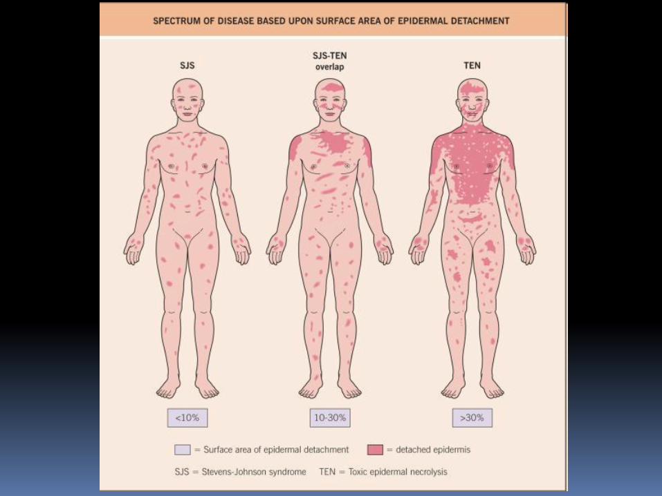
Stevens Johnson/Toxic Epidermal Necrolysis
Clinical Presentation: Fever, chills, anorexia prodrome- “flu-like” Morbilliform rash that rapidly evolves to widespread
sloughing of skin and mucosa Prominent dysphagia and dysuria Painful eyes and stinging/painful skin
1-2 weeks within initiation of offending drug **As opposed to DRESS (2-6 weeks)
Leukopenia **As opposed to leukocytosis in DRESS
“Acute Skin Failure” extensive sloughing of internal and external
mucocutaneous membranes


Fig 1
Journal of the American Academy of Dermatology 2013 69, 173.e1-173.e13DOI: (10.1016/j.jaad.2013.05.003)
Copyright © 2013 American Academy of Dermatology, Inc. Terms and Conditions

Fig 3
Journal of the American Academy of Dermatology 2013 69, 173.e1-173.e13DOI: (10.1016/j.jaad.2013.05.003)
Copyright © 2013 American Academy of Dermatology, Inc. Terms and Conditions

SJS/TEN

Fig 2
Journal of the American Academy of Dermatology 2013 69, 187.e1-187.e16DOI: (10.1016/j.jaad.2013.05.002)
Copyright © 2013 American Academy of Dermatology, Inc. Terms and Conditions

Common Drugs implicated in SJS/TEN
Sulfa drugs, sulfasalazine
Allopurinol
Tetracyclines ( minocycline)
Anticonvulsants
Carbamazepine, lamotrigine, phenytoin, phenobarbital
NSAIDS
Nevirapine

SJS/TEN Pathogenesis
Hypersensitivity rxn to drug Mycoplasma, dengue, CMV and contrast medium also
implicated
T-cell mediated disease CD8+ cells as mediate keratinocyte death Soluble Fas ligand, TNFalpha, granzymeB/perforin, &
granulysin mediate apoptosis
Certain HLA’s increase risk Patients of East Asian descent (HLAB1502)should
have testing prior to carbamazepine therapy All patients ( HLA-B5701) before abacavir therapy HLAB5801 (Han Chinest) and allopurinol

SJS/TEN Management
Early derm consult Frozen section can quickly identify TEN vs other Full thickness epidermal necrosis
Early ophtho Eyesight preservation paramount
Early urology Manage like burn victim
Severe dysfunction of ocular, pulmonary, CV, GI and renal systems
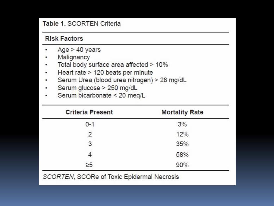
Up to 30% mortality Sepsis, GI bleeds, PE, MI, pulmonary edema
65% 5 yr survival rate

SJS/TEN management
Withdraw offending drug/stop unnecessary drugs Major predictor of survival
Supportive/Burn unit level care NO PROPHYLACTIC ABX!
Sterile handling of patient Optimized wound care
Culture-directed abx only Q48 hrs for skin, blood, catheters, urinary, gastric tubes
Steroids controversial IVIG– FasL not main mediator- granulysin is Enbrel? Cyclosporine? Success in small case series

Fig 1
Journal of the American Academy of Dermatology 2014 71, 278-283DOI: (10.1016/j.jaad.2014.04.044)
Copyright © 2014 American Academy of Dermatology, Inc. Terms and Conditions

SJS/TEN Sequelae
Cutaneous scarring
Ocular lesions
Dental complications
GU issues- stenosis, adhesions
Pulmonary disease

Desquamation/Blisters/Pain
Case: 45 yr old Asian female
Facial swelling, fevers, leukocytosis (PMN’s)
Takes ranitidine, multivitamin
Took naproxen 3 days ago for back pain

Acute Generalized Exanthematous Pustulosis
Rapid onset 2-5 days after drug initiated
Pinpoint sheets of nonfollicular, sterile pustules on erythematous background
Erythema on body folds, face before generalizing
Mucous membrane common, but usually only 1 surface, non-erosive
Fever, facial edema, leukocytosis :
PMN’s(90%), but occasionally eos
Self-limiting in 15 days once drug removed


AGEP
Antibiotics- sulfa, B-lactams, quinolones, TCN’s, Vanc
NSAIDS
Hydroxychloroquine
Terbinafine
Diltiazem, nifedipine, furosemide
Allopurinol
5% of cases- no trigger identified Likely viral, etc.

AGEP
Most important to differentiate from DRESS and/or pustular psoriasis flare
Biopsy helpful! Subcorneal pustules
Treatment includes stopping drug, topical /po steroids and antihistamines
* 5% mortality due to secondary infection
* If relapse/refractory, etanercept and cyclosporine have success

ICharacteristic findings of severe cutaneous drug reactions
DRESS SJS/TEN AGEP Erythroderma
Onset of eruption 2-6 weeks 1-3 weeks 48 hours 1-3 weeks
Duration of eruption (weeks) Several 1-3 <1 Several
Fever +++ +++ +++ +++
Mucocutaneous features
Facial edema, morbilliform
eruption, pustules,
exfoliative dermatitis, tense
bullae, and possible target
lesions
Bullae, atypical target
lesions, and
mucocutaneous erosions
Facial edema, pustules,
tense bullae, possible
target lesions, and possible
mucosal involvement
Erythematous plaques and
edema affecting >90% of
the total skin surface with
or without diffuse
exfoliation
Histological pattern of skin Perivascular lymphocytic
infiltrate Epidermal necrosis Subcorneal pustules
Nonspecific, unless
reflecting Sézary syndrome
or other lymphoma
Lymph node enlargement +++ – + +
Lymph node histology Lymphoid hyperplasia – –
No, unless reflecting
Sézary syndrome or other
malignancy
Hepatitis +++ ++ ++ –
Other organ involvement
Interstitial nephritis,
pneumonitis, myocarditis,
and thyroiditis
Tubular nephritis and
tracheobronchial necrosis Possible Possible
Neutrophils ↑ ↓ ↑↑↑ ↑
Eosinophils ↑↑↑ − ↑ ↑
Atypical lymphocytes + − − +
Mortality (%) 10 5-35 5 5-15

Red flags
Erythroderma
Mucous membrane involvement
Blisters or desquamation
Rapid purpura
Skin pain

Purpura
Purpura: Extravasation of red cells
“Palpable purpura” Leukocytoclastic vasculitis (LCV) Implies inflammation damaging vessel wall
Non-palpable Petechiae- pinpoint ( benign to severe) Macular- > 1-2 mm
Retiform: “Net-like” purpura
Antiphospholipid antibody syndrome Calciphylaxis

Petechiae
Non-Platelet related:
Trauma (valsalva, retching, compression)
Scurvy, amyloid,infection
Platelet-related:
ITP, TTP, DIC, HUS
NSAIDs

Purpura
If associated with fever, likely systemic inflammatory process or infection Rocky M0untain Spotted Fever, DIC,
Meningococcemia
Palpable purpura: Idiopathic 45-55%
Infection 15-20%
Inflammatory 15-20%
Medication 10-15%
Malignancy < 5%

Calciphylaxis

Rocky Mountain Spotted Fever

Antiphospholipid AB Syndrome

Coumadin Necrosis

Purpura
Case:
20 yr old asplenic male
Fever, chills, myalgias,stiff neck, hypotension
Petechiae which have progressed to purpura, and finally, frank bullous hemorrhagic lesions within hours
“The sickest he has ever felt”


Q4. What is the most important next step in management?
1. Call dermatology for biopsy
2. Initiate antibiotics
3. Start 1-2mg/kg oral steroids
4. Wait until lumbar puncture performed to initiate antibiotics
1. 2. 3. 4.
58%
5%
16%21%

Meningococcemia
Flu-like prodrome
Angular, gun-metal grey centered purpuric lesions
Purpura fulminans (in setting of DIC and infxn), shock, amputation, death
Require prompt care to save life and limb: 30min time to abx

We are more than Botox- but we love that too.

Evaluation
Please take < 90 seconds to evaluate this session.
Time permitting, speaker will take questions following evaluation.
Responses are not displayed and are important in maintaining high quality education.

The overall performance of the speaker:
1. Poor
2. Fair
3. Average
4. Good
5. Excellent
1. 2. 3. 4. 5.
0% 0%
92%
8%
0%

How well were the learning objectives met?
1. Poor
2. Fair
3. Average
4. Good
5. Excellent
PoorFa
ir
Avera
geGood
Excelle
nt
0% 0%
93%
7%0%

Did speaker present a balanced view of therapeutic options?
1. Yes
2. No
3. N/A
YesNo
N/A
100%
0%0%

How useful will this session be in your practice?
1. Poor
2. Fair
3. Average
4. Good
5. Excellent
PoorFa
ir
Avera
geGood
Excelle
nt
0% 0%
73%
23%
5%

As a result of this program, do you intend to change your patient care?
1. Yes
2. No
YesNo
5%
95%

Thank you!