Acute Coronary Syndromes
47
1 Acute Coronary Syndromes Acute Coronary Syndromes EMS Professions EMS Professions Temple College Temple College
-
Upload
leigh-simpson -
Category
Documents
-
view
35 -
download
4
description
Acute Coronary Syndromes. EMS Professions Temple College. The History of Paramedics Begins with Cardiac Care. The original Paramedic idea was based upon the need for rapid response to, identification of and emergency care for victims of: Sudden Cardiac Death (SCD) - PowerPoint PPT Presentation
Transcript of Acute Coronary Syndromes

1
Acute Coronary Acute Coronary SyndromesSyndromes
EMS ProfessionsEMS Professions
Temple CollegeTemple College

2
The History of Paramedics The History of Paramedics Begins with Cardiac CareBegins with Cardiac Care
The The originaloriginal Paramedic idea was Paramedic idea was based upon the need for rapid based upon the need for rapid response to, identification of and response to, identification of and emergency care for victims of:emergency care for victims of:
Sudden Cardiac Death (SCD)Sudden Cardiac Death (SCD)Acute Myocardial Infarction (AMI)Acute Myocardial Infarction (AMI)
3
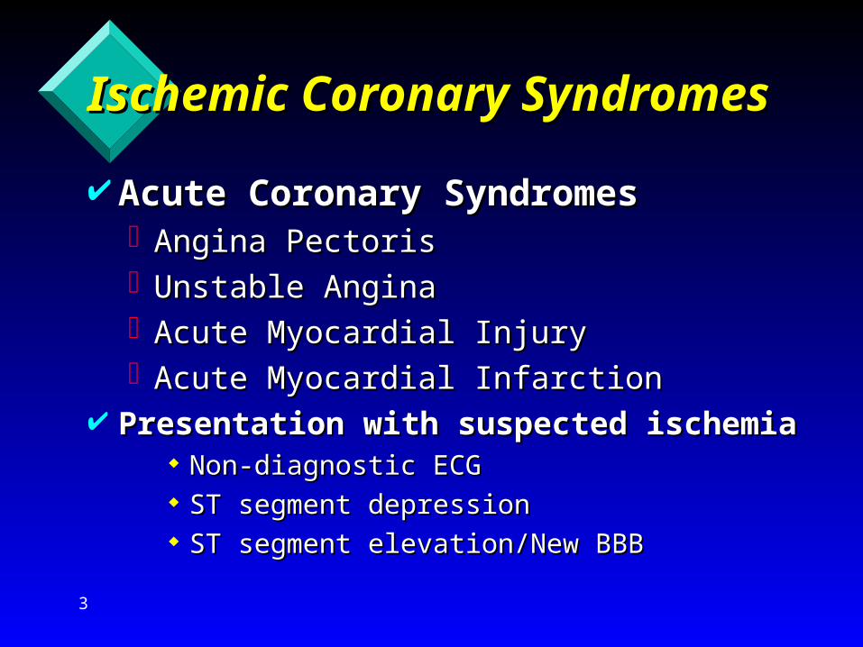
Ischemic Coronary Ischemic Coronary SyndromesSyndromes Acute Coronary SyndromesAcute Coronary Syndromes
Angina PectorisAngina PectorisUnstable AnginaUnstable AnginaAcute Myocardial InjuryAcute Myocardial InjuryAcute Myocardial InfarctionAcute Myocardial Infarction
Presentation with suspected ischemia Presentation with suspected ischemia Non-diagnostic ECGNon-diagnostic ECG ST segment depressionST segment depression ST segment elevation/New BBBST segment elevation/New BBB

4
Ischemic Coronary Ischemic Coronary SyndromesSyndromes Angina PectorisAngina Pectoris
Acute pain, usually in the chest, Acute pain, usually in the chest, resulting from an increased demand resulting from an increased demand for oxygen and a decreased ability to for oxygen and a decreased ability to provide itprovide it
Usually due to a partially occluded Usually due to a partially occluded coronary artery or vasospasmcoronary artery or vasospasm

5
Ischemic Coronary Ischemic Coronary SyndromesSyndromes
Angina PectorisAngina PectorisTypical PresentationTypical Presentation
Squeezing, Crushing, Heavy, TightSqueezing, Crushing, Heavy, Tight– Fist to chest = Levine’s signFist to chest = Levine’s sign
Pain/Discomfort may radiate to shoulders, Pain/Discomfort may radiate to shoulders, arms, neck, back, jaw or epigastriumarms, neck, back, jaw or epigastrium
Usually lasts 3-5 min and rarely exceeds Usually lasts 3-5 min and rarely exceeds 15 min15 min
Not changed by swallowing, coughing, Not changed by swallowing, coughing, deep breathing or positional changesdeep breathing or positional changes

6
Ischemic Coronary Ischemic Coronary SyndromesSyndromes
Angina PectorisAngina PectorisTypical PresentationTypical Presentation
AnxietyAnxiety Diaphoresis or clammy skinDiaphoresis or clammy skin Nausea, vomitingNausea, vomiting Shortness of breathShortness of breath WeaknessWeakness PalpitationsPalpitations SyncopeSyncope

7
Ischemic Coronary Ischemic Coronary SyndromesSyndromes
Angina PectorisAngina PectorisUsually Provoked by:Usually Provoked by:
ExerciseExercise EatingEating Emotion/StressEmotion/Stress
Usually Relieved by:Usually Relieved by: Rest; Removal of provoking factorRest; Removal of provoking factor NitroglycerinNitroglycerin

8
Ischemic Coronary Ischemic Coronary SyndromesSyndromes
Stable Angina PectorisStable Angina PectorisReasonably Predictable frequency, Reasonably Predictable frequency,
onset, durationonset, durationRelief predictable with rest, Relief predictable with rest,
nitroglycerinnitroglycerin

9
Ischemic Coronary Ischemic Coronary SyndromesSyndromes
Stable Angina PectorisStable Angina PectorisTreatment GoalsTreatment Goals
Reduce myocardial oxygen demandReduce myocardial oxygen demand Improve myocardial oxygen supplyImprove myocardial oxygen supply

10
Ischemic Coronary Ischemic Coronary SyndromesSyndromes Stable Angina PectorisStable Angina Pectoris
TreatmentTreatment Physical/Psychological restPhysical/Psychological rest Position of comfort, sitting or supinePosition of comfort, sitting or supine OxygenOxygen ECG MonitorECG Monitor
– Assess the underlying rhythmAssess the underlying rhythm Nitroglycerin, 0.4 mg SL q 5 min as long Nitroglycerin, 0.4 mg SL q 5 min as long
as BP > 90 mm Hgas BP > 90 mm Hg– Continue until pain relieved or contraindicatedContinue until pain relieved or contraindicated
11
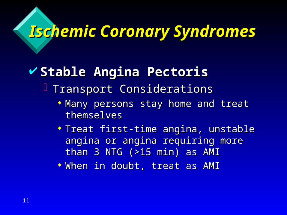
Ischemic Coronary Ischemic Coronary SyndromesSyndromes
Stable Angina PectorisStable Angina PectorisTransport ConsiderationsTransport Considerations
Many persons stay home and treat Many persons stay home and treat themselvesthemselves
Treat first-time angina, unstable angina Treat first-time angina, unstable angina or angina requiring more than 3 NTG or angina requiring more than 3 NTG (>15 min) as AMI(>15 min) as AMI
When in doubt, treat as AMIWhen in doubt, treat as AMI

12
Ischemic Coronary Ischemic Coronary SyndromesSyndromes Stable Angina PectorisStable Angina Pectoris
Variant Angina (Prinzmetal’s Angina)Variant Angina (Prinzmetal’s Angina) Occurs at restOccurs at rest Episodes at regular times of dayEpisodes at regular times of day Results from coronary vasospasmsResults from coronary vasospasms Treated long term with calcium channel Treated long term with calcium channel
blockersblockers May result in abnormal 12 lead ECG May result in abnormal 12 lead ECG
changes that resolve with minimal changes that resolve with minimal treatmenttreatment

13
Ischemic Coronary Ischemic Coronary SyndromesSyndromes Unstable AnginaUnstable Angina
Prolonged chest pain/ischemic Prolonged chest pain/ischemic symptoms or an symptoms or an atypicalatypical presentation of angina without ECG or presentation of angina without ECG or laboratory evidence of AMI (Injury)laboratory evidence of AMI (Injury)
Usually associated with significant or Usually associated with significant or progressing occlusion of a coronary progressing occlusion of a coronary artery or severe vasospasmartery or severe vasospasm
Considered “Pre-infarction Angina”Considered “Pre-infarction Angina”

14
Ischemic Coronary Ischemic Coronary SyndromesSyndromes Unstable AnginaUnstable Angina
May have Typical or Atypical Signs & May have Typical or Atypical Signs & SymptomsSymptoms Atypical PresentationAtypical Presentation
– Increased frequency or duration of Increased frequency or duration of episodesepisodes
– Onset with less exertion than normalOnset with less exertion than normal– Increased severity of symptomsIncreased severity of symptoms– Requires greater number of NTG tablets Requires greater number of NTG tablets
to relieve symptomsto relieve symptoms

15
Ischemic Coronary Ischemic Coronary SyndromesSyndromes Unstable AnginaUnstable Angina
Treatment same as Angina Treatment same as Angina PLUSPLUS:: IV, NS (no dextrose), TKO IV, NS (no dextrose), TKO
– Some exceptions to restricting fluidSome exceptions to restricting fluid 12 Lead ECG12 Lead ECG
– Assess for RVIAssess for RVI Morphine sulfate, 2 - 4 mg q 5-15 min slow Morphine sulfate, 2 - 4 mg q 5-15 min slow
IV titrated to pain relief and BP > 90IV titrated to pain relief and BP > 90 Aspirin, 160-325 mg POAspirin, 160-325 mg PO
– Chewed & swallowed if possibleChewed & swallowed if possible– Determine if hypersensitive to ASADetermine if hypersensitive to ASA

16
Ischemic Coronary Ischemic Coronary SyndromesSyndromes Unstable AnginaUnstable Angina
Treatment Treatment Metoprolol, 5 mg slow IV q 5 min to 15 mg Metoprolol, 5 mg slow IV q 5 min to 15 mg
total, prn for total, prn for HR/BP in absence of HR/BP in absence of contraindicationscontraindications
In longer or interfacility transports, consider:In longer or interfacility transports, consider:– Nitroglycerin IV infusion, 10-20 mcg/minNitroglycerin IV infusion, 10-20 mcg/min– HeparinHeparin– GP IIB/IIIA inhibitorsGP IIB/IIIA inhibitors
Thrombolytics Checklist (just in case)Thrombolytics Checklist (just in case) Transport, destination?Transport, destination?

17
Ischemic Coronary Ischemic Coronary SyndromesSyndromes Acute Myocardial InjuryAcute Myocardial Injury
Presentation of Unstable Angina or Presentation of Unstable Angina or Acute Ischemia with Acute Ischemia with potentialpotential for for myocardium salvage (penumbra)myocardium salvage (penumbra)
Diagnostic evidence of Injury (ECG or Diagnostic evidence of Injury (ECG or elevated Enzymes)elevated Enzymes)
Does not necessarily imply necrosis of Does not necessarily imply necrosis of the myocardiumthe myocardium
Presentation, Signs and Symptoms are Presentation, Signs and Symptoms are the same as Acute MIthe same as Acute MI

18
Ischemic Coronary Ischemic Coronary SyndromesSyndromes
Acute Myocardial Infarction (AMI)Acute Myocardial Infarction (AMI)Necrosis of myocardial tissue caused by Necrosis of myocardial tissue caused by
a lack of oxygenation and blood flow a lack of oxygenation and blood flow resulting from an occluded coronary resulting from an occluded coronary arteryartery
Often also used to describe acute injury Often also used to describe acute injury when extent of necrosis is unknown but when extent of necrosis is unknown but imminentimminent
Diagnostic evidence of injury is present Diagnostic evidence of injury is present (elevated enzymes and possibly ECG)(elevated enzymes and possibly ECG)
19
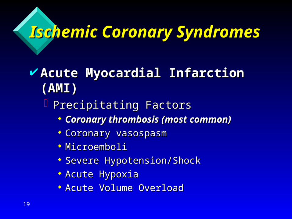
Ischemic Coronary Ischemic Coronary SyndromesSyndromes
Acute Myocardial Infarction Acute Myocardial Infarction (AMI)(AMI)Precipitating FactorsPrecipitating Factors
Coronary thrombosis (most common)Coronary thrombosis (most common) Coronary vasospasmCoronary vasospasm MicroemboliMicroemboli Severe Hypotension/ShockSevere Hypotension/Shock Acute HypoxiaAcute Hypoxia Acute Volume OverloadAcute Volume Overload

20
Ischemic Coronary Ischemic Coronary SyndromesSyndromes
Acute Myocardial Infarction Acute Myocardial Infarction (AMI)(AMI)Location, size of infarct and severity Location, size of infarct and severity
depends on site of vessel occlusiondepends on site of vessel occlusion majority involve left ventriclemajority involve left ventricle LCALCA
– anterior, septal, lateralanterior, septal, lateral RCARCA
– inferior, right ventricleinferior, right ventricle

21
Ischemic Coronary Ischemic Coronary SyndromesSyndromes
Acute Myocardial Infarction Acute Myocardial Infarction (AMI)(AMI)Often defined further asOften defined further as
subendocardial: involves only subendocardial: involves only subendocardial musclesubendocardial muscle
transmural: full thickness of ventricular transmural: full thickness of ventricular wall involvedwall involved

22
Anatomy of Plaque Disruption
• Thin, friable fibrous capseparating substantialthrombogenic lipid corefrom blood
• Lumen could be well preserved
Adapted from Libby P. Circulat ion. 1995;91:2844-2850, with permission.
• Thick fibrous capprotecting thrombogeniclipid core from blood
• More luminal narrowing
Lumen Lumen
Shoulder region
Lipid core
Media
“Vulnerable” Plaque “Stable” Plaque
Lipid coreFibrous cap
Evolution of AMIEvolution of AMI

23
Plaque Rupture, Stenosis, and Thrombosis
Adapted from Davies MJ. In: Schlant RC, Alexander RW, eds. The Heart, Arteriesand Veins. 8th ed. 1994:1009-1020, with permission.
Plaque rupture— intraplaque thrombus
Mural thrombus
Occlusivethrombosis
Total chronicocclusion
Healed plaque— increased stenosis
Healed plaque — decreased stenosis
Recanalizedlumen
Evolution of AMIEvolution of AMI

24
Plaque Rupture and Thrombus Progression
Adapted from Fuster V. N Engl J Med. 1992;326:242-250, with permission.
Lipid-rich
plaque
Plaquedisruptio
n
Thrombus
Lysis and residual thrombus
Disease progression
Completeocclusion
Reocclusion
Partial (labile) occlusion Recurrent pain
Unstableangina
AMI
Evolution of AMIEvolution of AMI

25
Coronary Artery Without Coronary Artery Without Evidence of PlaqueEvidence of Plaque
Source: University of Utah WebPath

26
Coronary Artery with Coronary Artery with Significant Plaque Formation Significant Plaque Formation
In addition to reduced Lumen size, there is also a calcified portion (right side of photo)
Source: University of Utah WebPath

27
Coronary Artery with Coronary Artery with Significant Plaque Formation Significant Plaque Formation
Source: University of Utah WebPath

28
Rupture of Atheromatous Rupture of Atheromatous Plaque Results in Thrombus Plaque Results in Thrombus Formation Formation Rupture of “Vulnerable” plaque’s soft Rupture of “Vulnerable” plaque’s soft
lipid core is the initiating event in most lipid core is the initiating event in most acute ischemic coronary eventsacute ischemic coronary events
Occlusion is dependent on clot formation Occlusion is dependent on clot formation and and accompanying fibrinolysis and and accompanying fibrinolysis
A thrombotic occlusion that is relatively A thrombotic occlusion that is relatively persistent (i.e., 2 to 4 hours or longer) persistent (i.e., 2 to 4 hours or longer) may result in acute myocardial infarctionmay result in acute myocardial infarction

29
Rupture of Atheromatous Rupture of Atheromatous Plaque Results in Thrombus Plaque Results in Thrombus FormationFormation Repeated thrombus formations may Repeated thrombus formations may
further decrease the lumen sizefurther decrease the lumen size
Intermittent non-occlusive thrombus Intermittent non-occlusive thrombus formation results in Unstable Anginaformation results in Unstable Angina
Incomplete occlusion may also result in Incomplete occlusion may also result in MI possibly due to coronary artery MI possibly due to coronary artery spasmspasm
30
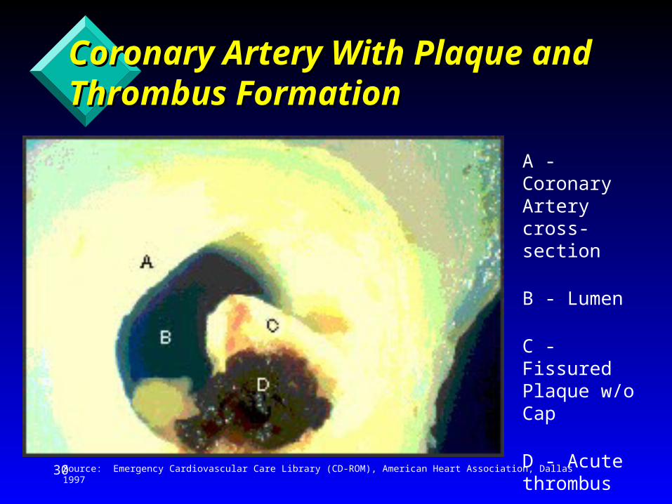
Coronary Artery With Plaque Coronary Artery With Plaque and Thrombus Formation and Thrombus Formation
A - Coronary Artery cross-section
B - Lumen
C - Fissured Plaque w/o Cap
D - Acute thrombus
Source: Emergency Cardiovascular Care Library (CD-ROM), American Heart Association, Dallas 1997

31
Plaque and Thrombus Plaque and Thrombus Formation Resulting in Formation Resulting in OcclusionOcclusion
Source: University of Utah WebPath

32
Coronary Artery ThrombusCoronary Artery Thrombus
Source: University of Utah WebPath
The external anterior view of the heart shows a dark clot formation in this artery

33
Coronary Artery Occlusion:The Evolution of Infarction
Progression of myocardial necrosis with time since occlusion
Adapted from Saltissi S, Mushahwar SS. Postgrad Med J. 1995;71:534-541, with permission.
4 h30 min 6 - 12 h
Normalmyocardium
“At risk”myocardium, ischemic but viable
Necrosis startingsubendocardially
Normalmyocardium
Completed infarctinvolving whole areaat risk
Normalmyocardium
“At risk”myocardium, ischemic but viable
Necrosis extendingtowardssubepicardium
Evolution of Evolution of Infarction/NecrosisInfarction/Necrosis

34
Ischemic Coronary Ischemic Coronary SyndromesSyndromes
Acute Myocardial Infarction (AMI)Acute Myocardial Infarction (AMI)PresentationPresentation
Similar to Angina but Similar to Angina but – Last longerLast longer– Not easily relieved with rest or NTGNot easily relieved with rest or NTG– Sx/Sx may be more severe (feeling of impending doom)Sx/Sx may be more severe (feeling of impending doom)– Pain often radiates to arms, neck, jaw, back, epigastriumPain often radiates to arms, neck, jaw, back, epigastrium
Some present atypically with complaints of only Some present atypically with complaints of only weakness or shortness of breathweakness or shortness of breath
DysrhythmiasDysrhythmias Sudden Cardiac DeathSudden Cardiac Death

35
Ischemic Coronary Ischemic Coronary SyndromesSyndromes Acute Myocardial Infarction (AMI)Acute Myocardial Infarction (AMI)
PresentationPresentation 10-20% have “silent” MI (no chest pain)10-20% have “silent” MI (no chest pain)
– common in elderly, older women, diabeticscommon in elderly, older women, diabetics If adding chest pain to the patient’s list of If adding chest pain to the patient’s list of
Sx/Sx completes a clear picture of AMI, then Sx/Sx completes a clear picture of AMI, then the patient is having an AMI!!the patient is having an AMI!!
Vital Signs and monitoring ECG leads DO NOT Vital Signs and monitoring ECG leads DO NOT provide DIAGNOSTIC evidence of AMI!!provide DIAGNOSTIC evidence of AMI!!
Clinical diagnosis in absence of 12 Lead ECG Clinical diagnosis in absence of 12 Lead ECG or Enzyme changesor Enzyme changes
36
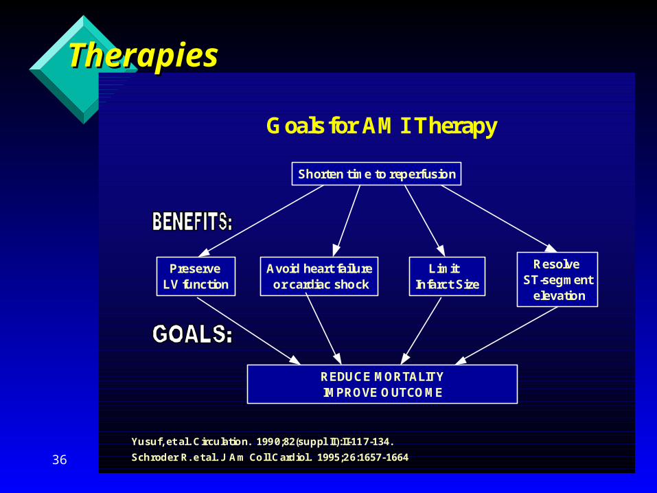
Goals for AMI Therapy
Shorten time to reperfusion
Preserve LV function
Avoid heart failure or cardiac shock
Limit Infarct Size
ResolveST-segment elevation
REDUCE MORTALITYIMPROVE OUTCOME
Yusuf, et al. Circulation. 1990;82(suppl II):II-117-134.
Schroder R. et al. J Am Coll Cardiol. 1995;26:1657-1664
TherapiesTherapies

37
Ischemic Coronary Ischemic Coronary SyndromesSyndromes Acute Myocardial Infarction Acute Myocardial Infarction
(AMI)(AMI)Treatment GoalsTreatment Goals
Decrease myocardial oxygen demandDecrease myocardial oxygen demand– Remove physical/psychological stressorsRemove physical/psychological stressors– Relieve painRelieve pain– Reduce workload of the heart (BP, HR)Reduce workload of the heart (BP, HR)
Inhibit further clot formationInhibit further clot formation Rapid identification/diagnosisRapid identification/diagnosis Transport for reperfusion therapyTransport for reperfusion therapy

38
Ischemic Coronary Ischemic Coronary SyndromesSyndromes Acute Myocardial Infarction (AMI)Acute Myocardial Infarction (AMI)
Treatment same as Angina Treatment same as Angina PLUSPLUS:: IV, NS, large boreIV, NS, large bore
– TKO with some exceptionsTKO with some exceptions– No dextrose containing solutionsNo dextrose containing solutions– Fluid boluses appropriate in some casesFluid boluses appropriate in some cases– 2nd line if time permits2nd line if time permits– Minimize number of attemptsMinimize number of attempts
12 Lead ECG12 Lead ECG– Diagnostic evidence of AMI presentDiagnostic evidence of AMI present– Assess for RVIAssess for RVI

39
Ischemic Coronary Ischemic Coronary SyndromesSyndromes Acute Myocardial Infarction (AMI)Acute Myocardial Infarction (AMI)
Treatment Treatment Morphine sulfate, 2 - 4 mg q 5-15 min slow IVMorphine sulfate, 2 - 4 mg q 5-15 min slow IV
– Maintain BP > ~ 90 mm HgMaintain BP > ~ 90 mm Hg– Titrated to Pain reliefTitrated to Pain relief– Reduce PVR and workload on the heartReduce PVR and workload on the heart
Aspirin, 160-325 mg POAspirin, 160-325 mg PO– Chewed & swallowed if possibleChewed & swallowed if possible– Determine if hypersensitive to ASADetermine if hypersensitive to ASA
““MONA greets all patients”MONA greets all patients”

40
Ischemic Coronary Ischemic Coronary SyndromesSyndromes
Acute Myocardial Infarction (AMI)Acute Myocardial Infarction (AMI)TreatmentTreatment
Metoprolol, 5 mg slow IV q 5 min to 15 mg total, Metoprolol, 5 mg slow IV q 5 min to 15 mg total, prn for prn for HR/BP in absence of contraindications HR/BP in absence of contraindications
In longer or interfacility transports, consider:In longer or interfacility transports, consider:– Nitroglycerin IV infusionNitroglycerin IV infusion– HeparinHeparin
Thrombolytics ChecklistThrombolytics Checklist– Exclusions for thrombolysisExclusions for thrombolysis

41
Ischemic Coronary Ischemic Coronary SyndromesSyndromes
Acute Myocardial Infarction Acute Myocardial Infarction (AMI)(AMI)Treatment Treatment
Transport for reperfusion therapy; Transport for reperfusion therapy; Destination?Destination?
– Thrombolysis vs Coronary Artery CatheterizationThrombolysis vs Coronary Artery Catheterization– For patients with associated pulmonary edema, For patients with associated pulmonary edema,
hypotension or cardiogenic shock, consider hypotension or cardiogenic shock, consider transport to facility with capability of transport to facility with capability of angiography & revascularizationangiography & revascularization

42
Acute Ischemic Syndromes:Diagnostic Considerations
Thrombolytics are not appropriate in all acute ischemicsyndromes
• Not all acute ischemic syndromes are AMIs
• ST-segment elevation suggests thrombic occlusionand need for immediate reperfusion
• No proven benefit of thrombolytic therapy in patientswithout ST-segment elevation
• Patients with ST-segment depression and/or T-waveinversion are currently not candidates for thrombolytictherapy
Considerations for Considerations for FibrinolyticsFibrinolytics

43
Contraindications for Contraindications for FibrinolyticsFibrinolytics Lack of diagnostic 12 Lack of diagnostic 12
Lead ECG changesLead ECG changes Chest pain < 20 min or Chest pain < 20 min or
> 12 hours> 12 hours Not oriented, can not Not oriented, can not
cooperatecooperate History of stroke or TIAHistory of stroke or TIA Known bleeding Known bleeding
disorderdisorder Active internal bleeding Active internal bleeding
in past 2-4 weeksin past 2-4 weeks
Surgery or trauma in Surgery or trauma in past 3 weekspast 3 weeks
Terminal illnessTerminal illness Jaundice, hepatitis, Jaundice, hepatitis,
kidney failurekidney failure Use of anticoagulantsUse of anticoagulants Systolic BP < 180 Systolic BP < 180
mm Hgmm Hg Diastolic BP < 110 Diastolic BP < 110
mm Hgmm Hg

44
Ischemic Coronary Ischemic Coronary SyndromesSyndromes
““Ischemic and injured tissue have reduced blood flow Ischemic and injured tissue have reduced blood flow but but maymay be salvaged. The area of the Penumbra may be salvaged. The area of the Penumbra may be viable for several hours after onset of occlusion.”be viable for several hours after onset of occlusion.”Source: Emergency Cardiovascular Care Library (CD-ROM), American Heart Association, Dallas, 1997Source: Emergency Cardiovascular Care Library (CD-ROM), American Heart Association, Dallas, 1997

45
Ischemic Coronary Ischemic Coronary SyndromesSyndromes
Sudden Cardiac Death (SCD or SCA)Sudden Cardiac Death (SCD or SCA)Sudden, unexpected biologic death presumably Sudden, unexpected biologic death presumably
resulting from cardiovascular diseaseresulting from cardiovascular diseaseMost common rhythm of SCA is Ventricular Most common rhythm of SCA is Ventricular
FibrillationFibrillationMay be primary or secondary VFMay be primary or secondary VFChain of Survival is the greatest determinant of Chain of Survival is the greatest determinant of
outcomeoutcomeTreatment based on ECG rhythm & arrest Treatment based on ECG rhythm & arrest
eventsevents

46
Time is Time is Muscle!!!Muscle!!!

47
References and ResourcesReferences and Resources
Advanced Cardiac Life SupportAdvanced Cardiac Life Support, Edited by R O Cummins, MD, American , Edited by R O Cummins, MD, American Heart Association, Dallas, 1997Heart Association, Dallas, 1997
““Emergency Cardiovascular Care Library” (CD-ROM), American Heart Emergency Cardiovascular Care Library” (CD-ROM), American Heart Association and ProEducation International, Dallas, 1997Association and ProEducation International, Dallas, 1997
Eisenberg, M S, Eisenberg, M S, Life in the Balance: Emergency Medicine and the Quest to Life in the Balance: Emergency Medicine and the Quest to Reverse Sudden DeathReverse Sudden Death, Oxford University Press, New York, 1997, Oxford University Press, New York, 1997
““A Definition of Advanced Types of Atherosclerotic Lesions and a A Definition of Advanced Types of Atherosclerotic Lesions and a Histological Classification of Atherosclerosis”, A Report From the Histological Classification of Atherosclerosis”, A Report From the Committee on Vascular Lesions of the Council on Arteriosclerosis, American Committee on Vascular Lesions of the Council on Arteriosclerosis, American Heart Association, 1995Heart Association, 1995
““Coronary Artery Calcification: Pathophysiology, Epidemiology, Imaging Coronary Artery Calcification: Pathophysiology, Epidemiology, Imaging Methods, and Clinical Implications”, A Statement for Health Professionals Methods, and Clinical Implications”, A Statement for Health Professionals From the American Heart Association, 1995From the American Heart Association, 1995
Cardiovascular Disease Statistics, American Heart Association, Dallas, 1997Cardiovascular Disease Statistics, American Heart Association, Dallas, 1997 ““Diagnosis and Therapy of Acute Myocardial Infarction: Today’s Look at Diagnosis and Therapy of Acute Myocardial Infarction: Today’s Look at
Tomorrow’s Therapies and Outcomes”, DuPont Pharma, 1997Tomorrow’s Therapies and Outcomes”, DuPont Pharma, 1997 University of Utah WebPath, http://medstat.med.utah.edu/webpath/University of Utah WebPath, http://medstat.med.utah.edu/webpath/