
Activation of Hsp90 Enzymatic Activity and Conformational ... · Activation of Hsp90 Enzymatic...
11
& Drug Design | Very Important Paper| Activation of Hsp90 Enzymatic Activity and Conformational Dynamics through Rationally Designed Allosteric Ligands Sara Sattin, [a] Jiahui Tao, [b] Gerolamo Vettoretti, [c] Elisabetta Moroni, [c] Marzia Pennati, [d] Alessia Lopergolo, [d] Laura Morelli, [a] Antonella Bugatti, [e] Abbey Zuehlke, [f] Mike Moses, [f] Thomas Prince, [f] Toshiki Kijima, [f] Kristin Beebe, [f] Marco Rusnati, [e] Len Neckers, [f] Nadia Zaffaroni, [d] David A. Agard, [b] Anna Bernardi, [a] and Giorgio Colombo* [c] Abstract: Hsp90 is a molecular chaperone of pivotal impor- tance for multiple cell pathways. ATP-regulated internal dy- namics are critical for its function and current pharmacologi- cal approaches block the chaperone with ATP-competitive inhibitors. Herein, a general approach to perturb Hsp90 through design of new allosteric ligands aimed at modulat- ing its functional dynamics is proposed. Based on the char- acterization of a first set of 2-phenylbenzofurans showing stimulatory effects on Hsp90 ATPase and conformational dy- namics, new ligands were developed that activate Hsp90 by targeting an allosteric site, located 65 ĸ from the active site. Specifically, analysis of protein responses to first-generation activators was exploited to guide the design of novel deriva- tives with improved ability to stimulate ATP hydrolysis. The molecules’ effects on Hsp90 enzymatic, conformational, co- chaperone and client-binding properties were characterized through biochemical, biophysical and cellular approaches. These designed probes act as allosteric activators of the chaperone and affect the viability of cancer cell lines for which proper functioning of Hsp90 is necessary. Introduction The dynamic properties of proteins are critical for the functions they exhibit. [1] They can be finely tuned through allostery, a general property of biomolecules whereby a perturbation at one site leads to a response at another, turning specific func- tional states on or off. [2] Allosteric ligand binding provides the opportunity to regulate protein functions with small molecules that act at a distance from the active site, without directly in- terfering with its chemical activity. [1, 3] Additionally, allosteric li- gands can be used to modulate the equilibrium and dynamics of distinctive protein states implicated in certain cellular path- ways and phenotypes. [4] The fundamental chemical challenge for the discovery of al- losteric protein modulators consists in identifying privileged structures that selectively target key protein substates involved in the regulation of biochemical function. To progress along this appealing avenue, we focused on the 90 kDa heat shock protein (Hsp90) molecular chaperone. This protein is known to undergo a complex dynamic cycle, which is essential to its chaperone activity that, in turn, regulates the state of a number of client proteins in the cell. Hsp90 integrates differ- ent pathways required for cell development and mainte- nance. [5] and plays a key role in the regulation of a wide variety of so-called client proteins. [6] As such, it has been proposed as an interesting target in cancer, [7] vascular disease, [8] neurode- generation, [9] and as a major player in evolution. [10] Thanks to its dynamic nature [5, 11] Hsp90 influences the functional lifetime of many clients that vary widely in sequence, structure, size and function. [12] In eukaryotes this activity is further regulated by a number of co-chaperones, which bind individual Hsp90 states populated at different stages of the chaperone cycle. Hsp90 dynamics depend on ATP binding and hydrolysis, which underlie the onset of conformational transitions between sub- states with different functional properties. [13] Here, we set out to develop a rational approach to regulate the ATPase activity [a] Dr. S. Sattin, + Dr. L. Morelli, Prof. A. Bernardi Dipartimento di Chimica, UniversitȤ degli Studi di Milano via Golgi, 19, 20133, Milan (Italy) [b] Dr. J. Tao, + Prof. D. A. Agard Howard Hughes Medical Institute and Department of Biochemistry and Biophysics, University of California 600 16th Street, San Francisco, 94158 (USA) [c] Dr. G. Vettoretti, + Dr. E. Moroni, + Dr. G. Colombo Istituto di Chimica del Riconoscimento Molecolare, CNR via Mario Bianco, 9, 20131, Milan (Italy) E-mail : [email protected] [d] Dr. M. Pennati, Dr. A. Lopergolo, Dr. N. Zaffaroni Dept. Experimental Oncology and Molecular Medicine Molecular Pharmacology Unit Fondazione IRCCS Istituto Nazionale dei Tumori via Amadeo, 42, 20133 Milano (Italy) [e] Dr. A. Bugatti, Dr. M. Rusnati Department of Molecular and Translational Medicine University of Brescia, Viale Europa 11, 25123, Brescia (Italy) [f] Dr. A. Zuehlke, Dr. M. Moses, Dr. T. Prince, Dr. T. Kijima, Dr. K. Beebe, Dr. L. Neckers Urologic Oncology Branch, Center for Cancer Research National Cancer Institute, 9000 Rockville Pike, Bethesda, MD 20892 (USA) [ + ] These authors contributed equally to this work. Supporting information for this article is available on the WWW under http://dx.doi.org/10.1002/chem.201502211. Chem. Eur. J. 2015, 21, 13598 – 13608 # 2015 Wiley-VCH Verlag GmbH & Co. KGaA, Weinheim 13598 Full Paper DOI: 10.1002/chem.201502211
Transcript of Activation of Hsp90 Enzymatic Activity and Conformational ... · Activation of Hsp90 Enzymatic...
&Drug Design | Very Important Paper |
Activation of Hsp90 Enzymatic Activity and ConformationalDynamics through Rationally Designed Allosteric Ligands
Sara Sattin,[a] Jiahui Tao,[b] Gerolamo Vettoretti,[c] Elisabetta Moroni,[c] Marzia Pennati,[d]
Alessia Lopergolo,[d] Laura Morelli,[a] Antonella Bugatti,[e] Abbey Zuehlke,[f] Mike Moses,[f]
Thomas Prince,[f] Toshiki Kijima,[f] Kristin Beebe,[f] Marco Rusnati,[e] Len Neckers,[f]
Nadia Zaffaroni,[d] David A. Agard,[b] Anna Bernardi,[a] and Giorgio Colombo*[c]
Abstract: Hsp90 is a molecular chaperone of pivotal impor-
tance for multiple cell pathways. ATP-regulated internal dy-
namics are critical for its function and current pharmacologi-cal approaches block the chaperone with ATP-competitive
inhibitors. Herein, a general approach to perturb Hsp90through design of new allosteric ligands aimed at modulat-
ing its functional dynamics is proposed. Based on the char-acterization of a first set of 2-phenylbenzofurans showing
stimulatory effects on Hsp90 ATPase and conformational dy-
namics, new ligands were developed that activate Hsp90 by
targeting an allosteric site, located 65 æ from the active site.
Specifically, analysis of protein responses to first-generation
activators was exploited to guide the design of novel deriva-tives with improved ability to stimulate ATP hydrolysis. The
molecules’ effects on Hsp90 enzymatic, conformational, co-chaperone and client-binding properties were characterized
through biochemical, biophysical and cellular approaches.These designed probes act as allosteric activators of the
chaperone and affect the viability of cancer cell lines for
which proper functioning of Hsp90 is necessary.
Introduction
The dynamic properties of proteins are critical for the functions
they exhibit.[1] They can be finely tuned through allostery,a general property of biomolecules whereby a perturbation at
one site leads to a response at another, turning specific func-
tional states on or off.[2] Allosteric ligand binding provides theopportunity to regulate protein functions with small molecules
that act at a distance from the active site, without directly in-
terfering with its chemical activity.[1, 3] Additionally, allosteric li-gands can be used to modulate the equilibrium and dynamics
of distinctive protein states implicated in certain cellular path-ways and phenotypes.[4]
The fundamental chemical challenge for the discovery of al-losteric protein modulators consists in identifying privilegedstructures that selectively target key protein substates involved
in the regulation of biochemical function. To progress alongthis appealing avenue, we focused on the 90 kDa heat shockprotein (Hsp90) molecular chaperone. This protein is known toundergo a complex dynamic cycle, which is essential to its
chaperone activity that, in turn, regulates the state ofa number of client proteins in the cell. Hsp90 integrates differ-
ent pathways required for cell development and mainte-
nance.[5] and plays a key role in the regulation of a wide varietyof so-called client proteins.[6] As such, it has been proposed as
an interesting target in cancer,[7] vascular disease,[8] neurode-generation,[9] and as a major player in evolution.[10] Thanks to
its dynamic nature[5, 11] Hsp90 influences the functional lifetimeof many clients that vary widely in sequence, structure, size
and function.[12] In eukaryotes this activity is further regulated
by a number of co-chaperones, which bind individual Hsp90states populated at different stages of the chaperone cycle.
Hsp90 dynamics depend on ATP binding and hydrolysis, whichunderlie the onset of conformational transitions between sub-
states with different functional properties.[13] Here, we set outto develop a rational approach to regulate the ATPase activity
[a] Dr. S. Sattin,+ Dr. L. Morelli, Prof. A. BernardiDipartimento di Chimica, Universit� degli Studi di Milanovia Golgi, 19, 20133, Milan (Italy)
[b] Dr. J. Tao,+ Prof. D. A. AgardHoward Hughes Medical Institute and Department of Biochemistry andBiophysics, University of California600 16th Street, San Francisco, 94158 (USA)
[c] Dr. G. Vettoretti,+ Dr. E. Moroni,+ Dr. G. ColomboIstituto di Chimica del Riconoscimento Molecolare, CNRvia Mario Bianco, 9, 20131, Milan (Italy)E-mail : [email protected]
[d] Dr. M. Pennati, Dr. A. Lopergolo, Dr. N. ZaffaroniDept. Experimental Oncology and Molecular MedicineMolecular Pharmacology UnitFondazione IRCCS Istituto Nazionale dei Tumorivia Amadeo, 42, 20133 Milano (Italy)
[e] Dr. A. Bugatti, Dr. M. RusnatiDepartment of Molecular and Translational MedicineUniversity of Brescia, Viale Europa 11, 25123, Brescia (Italy)
[f] Dr. A. Zuehlke, Dr. M. Moses, Dr. T. Prince, Dr. T. Kijima, Dr. K. Beebe,Dr. L. NeckersUrologic Oncology Branch, Center for Cancer ResearchNational Cancer Institute, 9000 Rockville Pike, Bethesda, MD 20892 (USA)
[++] These authors contributed equally to this work.
Supporting information for this article is available on the WWW underhttp ://dx.doi.org/10.1002/chem.201502211.
Chem. Eur. J. 2015, 21, 13598 – 13608 Ó 2015 Wiley-VCH Verlag GmbH & Co. KGaA, Weinheim13598
Full PaperDOI: 10.1002/chem.201502211

and conformational dynamics of Hsp90 through designed allo-steric small molecule ligands.
Hsp90 functions as a homodimer (Figure 1). Crystal struc-tures from different organisms highlighted a common modular
organization in terms of N-terminal (NTD), middle (M) and C-terminal (CTD) domains.[14] The CTD is the dimerization
domain, while the NTD contains an ATP-binding site. ATPaseactivity requires transient dimerization of the NTD in a closedstate of the dimer and is essential for the Hsp90 working cycle.
The exact mechanism of coupling between ATP-binding/hy-drolysis and client folding remains elusive. Yet, structural and
biochemical data support a model in which nucleotide bindingat the NTD propagates a conformational signal to the CTD,[14d]
while the chaperone undergoes conformational rearrange-ments that bring the two NTDs into close association in the
ATP-state, but not in the ADP or apo states. Formation of the
closed state can be induced by ATP or the non-hydrolyzableanalogue adenosine 5’-(b,g-imido)triphosphate (AMP-PNP).
Upon ATP hydrolysis, the protein cycles back to the open,ADP-bound state. A number of intermediate conformational
states have also been characterized, that are induced or stabi-lized by interaction with different clients or co-chaperone pro-
teins.[13, 15] Figure 1 highlights some of these interactions, with
the co-chaperones Aha1, an endogenous activator of Hsp90ATPase activity, and Sba1, an Hsp90 inhibitor, which acts by
binding to the closed conformation and blocking the chaper-one dynamics. The predicted allosteric site that we sought to
exploit (AS, Figure 1)[16] is located at the MD/CTD interface,a region where the model client D131D is also known to inter-
act.
Most known small molecule Hsp90 modulators interact withthe protein at the NTD and inhibit Hsp90 ATPase activity.[17]
They include radicicol and geldanamycin or its derivative,
17AAG.[18] Some of such inhibitors have entered clinical trialsas antitumor drugs but have shown severe limitations. Indeed,
blocking the ATPase activity of Hsp90 induces the so-called
heat shock response, a prosurvival mechanism mediated byHSF-1 (heat shock factor 1), which limits the action of the
drug.[19]
Rationally designed chemical probes can be used to select
and activate, and not only to inhibit, Hsp90 key substates, pro-viding a connection between protein activities and possible
cellular outcomes. Here, we pursue the first rational design ofchemical activators of Hsp90 functions aimed to target sites al-
ternative to the ATP site, and investigate their effects on thechaperone conformational dynamics, enzymatic, binding andcellular properties.
In this study, we build on the results of recently developedcomputational methods that unveiled the presence of a drug-gable site in Hsp90 CTD[16, 20] and facilitated the design of smallmolecules able to bind it.[21] Different sets of experiments
showed that the O-aryl rhamnoside (1; Figure 2 A) identifiedthrough this method could bind the C-terminal domain ofHsp90 and exert interesting antitumor activities.[16] Here, weevolve 1 into new chemical entities that enable controlled acti-
vation of Hsp90 ATPase function. We show that designed de-rivatives are genuine allosteric ligands with a structure-depen-
dent ability to stimulate Hsp90 ATPase and to alter conforma-
tional dynamics favoring synergistic effects with the activatingco-chaperone Aha1, to modulate Hsp90 direct interactions
with the co-chaperone Sba1 (p23), and to compete with themodel client protein D131D. We characterize the impact of
small molecule induced activation on Hsp90 interactions invitro and on the stability of a number of clients in cellular
models, and we investigate the possibility that acceleration of
conformational dynamics may in fact represent a new way ofperturbing the chaperoning mechanisms that underlie cell via-
bility; indeed, some of our compounds inhibit the proliferativepotential of tumor cells including those resistant to Hsp90
ATP-competitive inhibitors.
Figure 2. Structure of the initial lead and interaction with Hsp90 allostericsite. A) Molecular structure of 1; B) 3D structure of compound 1 (yellow) incomplex with the closed structure of Hsp90. The van der Waals spheres inlight blue and green indicate the client protein D131D binding site. C) Rep-resentative poses of 1 in the representative conformations of the allostericsite, showing the contacts of the ligand with E477 and D503 on protomer A.D) Contacts of 1 with protomer B, highlighting interaction with R591.
Figure 1. Schematic representation of Hsp90 structure and its conformation-al equilibrium. NTD, N-terminal domain; MD, middle domain; CTD, C-termi-nal domain; AS, predicted allosteric site; Aha1, Hsp90 co-chaperone; Sba1,yeast homologue of co-chaperone p23; D131D, Hsp90 client protein model.
Chem. Eur. J. 2015, 21, 13598 – 13608 www.chemeurj.org Ó 2015 Wiley-VCH Verlag GmbH & Co. KGaA, Weinheim13599
Full Paper

Results and Discussion
Design and synthesis of first-generation Hsp90 modulators
In our previous work, the rhamnoside 1 (Figure 2 A) was select-ed from the NCI library by virtual screening in a 3D pharmaco-phore designed to complement the stereoelectronic propertiesdisplayed by an allosteric site in the CTD. The site was identi-fied through the analysis of long-range dynamic communica-
tion mechanisms with the ATP-site, using the coordination pro-pensity (CP) parameter (see the Supporting Information).[16, 20] It
comprises residues at the CTD interface with the M-domain,which define a druggable pocket coincident with the region of
a recently identified binding site for the model client proteinD131D[22] (Figure 2 B). The long-range coordination properties
of the amino acids defining the allosteric pocket describe their
dynamic connection to events occurring in the orthosteric ATPbinding site, and are conserved across different members of
the Hsp90 family.[20a] Targeting these residues by designed li-gands, such as 1, should thus provide a way of influencing the
functional properties of the protein.[23] Indeed, 1 was found tobind the Hsp90 C-terminal domain disrupting chaperoning
functions and to exhibit antiproliferative activity in different
tumor cell lines.[23]
To optimize the structure and functional impacts of this
lead, in the absence of crystal structures, we investigated bind-ing determinants in the allosteric site through docking calcula-
tions. It should be noted here that no X-ray structure of any C-terminal ligands in complex with Hsp90 has been determined
so far. The initial target was the MD-relaxed ATP-bound Hsp90
structure used for the pharmacophore-based discovery of 1.[16,
23] Considering the flexibility of the protein and of the allosteric
pocket,[21a] we selected an ensemble approach to characterizechaperone–ligand interactions. The minimum energy pose of
the 1–Hsp90 complex (Figure 2 B) was used as input for longtimescale MD simulations, including ATP at the NTD. The aim
was to identify the hot spots of the allosteric site where key
functional groups on the ligand best complement the receptor,taking the dynamic exchange between the binding partnersexplicitly into account.
Structural cluster analysis of the resulting trajectory (see the
Supporting Information) showed that the first ten clusters reca-pitulated approximately 95 % of the protein structural variabili-
ty. The RMSD between visited pocket conformations in the pu-tative binding site (Table S1 in the Supporting Information)reached to 4 æ, revealing the diversity induced by 1. Such dif-
ferences, due to the cross-talk between the protein and theligand, were not expected a priori and revealed protein confor-
mations that could play a role in small molecule recognition.The resulting structures were next used to explore possible al-
ternative poses of 1 in the allosteric site. The compound was
redocked into each of the ten representative structures. The re-sulting structural ensembles were used to generate a consensus
model of Hsp90 residues and functional groups on 1 thatdefine the most relevant stabilizing contacts (Figure 2 C and
D). Two areas where functional group diversification on thelead could translate into a modulated response of the chaper-
one were identified: the carbohydrate moiety (R’’, Figure 2 A),which in the consensus model points towards an area lined by
E477 and D503 (Figure 2 C), and the propenyl group (R’, Fig-ure 2 A), which contacts a hydrophobic pocket also lined by
R591 from the other protomer (Figure 2 D).To validate this model and to expand the available SAR, gly-
codiversification of the 2-phenylbenzofuran aglycone 2(Table 1) was examined. Glycorandomization is a well-known
strategy to tune the activity of a variety of glycoconjugates
against their protein targets[24] and is an attractive avenue forthe case at hand, where computational data indicate a stronginvolvement of the sugar moiety in the interaction with Hsp90allosteric site. A first series of molecules 4 to 16 was synthe-sized (Table 1) by glycosylation of 2 and of the 5-chloro ana-logue 3 (an intermediate in the synthesis of 2), as previously
reported in ref. [25] . The full structures of all compounds are
reported in Figure S1 in the Supporting Information.
Designed allosteric ligands stimulate Hsp90 ATPase activity
To characterize their role as allosteric modulators, we measured
the effects of compounds 1–16 on the ATPase activity of theHsp90 yeast homologue (Hsc82).[26] Interestingly, most com-pounds, including 1, turned out to be activators of ATP hydrol-ysis (Figure 3, compounds 1 and 4–16), with variable potency,
depending on the type of carbohydrate moiety and benzenering substitution (propenyl or chlorine). Compounds 4 and 10–12 were the strongest activators of Hsp90 with a two- to three-fold enhancement of ATPase rate. Since the basal ATPase rateof yeast Hsp90 is very low, this kind of acceleration is not neg-
ligible, it has very little precedent in the literature, and nonewith rationally designed compounds.[27]
From a ligand-design point of view, the data indicate that
a chlorine atom in position R’ (Figure 2) is as good as, andoften better than, the original propenyl substitution (compare
pairs 1 and 4, 9 and 10, 11 and 12). Additionally, a significanteffect of the sugar on the level of activity was observed, with
d-mannose (as in 10) and d-galactose (as in 12) emerging asprivileged fragments alongside l-rhamnose (in 4).
Table 1. First generation of CTD ligands 1–16.[a]
2 R’’= H 3 R’’= HR’’ R’’
1 a-l-Rha 4 a-l-Rha5 b-d-Glc 6 b-d-Glc7 b-l-Glc 8 b-l-Glc9 a-d-Man 10 a-d-Man11 b-d-Gal 12 b-d-Gal13 b-l-Gal 14 b-l-Gal15 b-l-Fuc 16 b-l-Fuc
[a] The propenyl scaffold 2 was used as an E/Z mixture after checkingthat the natural compound 1 and E/Z-1 had a comparable effect on theprotein activity.
Chem. Eur. J. 2015, 21, 13598 – 13608 www.chemeurj.org Ó 2015 Wiley-VCH Verlag GmbH & Co. KGaA, Weinheim13600
Full Paper

Rational optimization of first-generation ligands furtherincreases Hsp90 ATPase activity
Starting from the aforementioned observations, to design im-proved allosteric activators we docked all the first-generation
compounds into the ten Hsp90 conformations used for the
first generation of derivatives, describing binding in terms ofan ensemble of ligand poses in complex with an ensemble of
protein structures.[28]
All glycosylated compounds explored common ensembles in
the allosteric site, similar to 1. Representative poses are shownin Figure 4 A for compounds 4 (blue) 10 (cyan) and 12 (yellow;
Figure 4 A). The pair formed by the negatively charged E477
and D503 on one protomer, towards which initial design wasaimed, was always engaged in hydrogen bonding interactions
with the carbohydrate moieties (R’’). The most active glycosy-lated compounds, 10 and 12, showed two hydroxyl groups
(on C-6 and C-4 of the sugar) ideally oriented to establish H-bonding interactions with the aforementioned negative resi-
dues. This interaction was reinforced by a H-bond network in-
volving R591, R599 and the hydroxyl groups on the carbohy-drate ring. On the R’ side, two positively charged residues from
the other protomer, R591 and K594, were involved in stabiliz-ing interactions with the halogen of compounds deriving fromthe common intermediate 3.
On this basis, a second group of ligands (17–19) was de-signed to exploit potential productive interactions with the
network of charged amino acids (Figure 4 B and C) in the allo-steric pocket. Indeed, 18 and 19 were found to dramaticallyaccelerate yeast Hsp90 (Hsc82) ATPase rate (by up to sixfold,Figure 3), providing further support to our model and to thedesign approach. Absolute kcat values are reported in Table S2in the Supporting Information.
The ability of the best stimulating compounds to effectively
bind the Hsp90-CTD was proven by preliminary surface plas-mon resonance experiments (SPR; Table S3 in the Supporting
Information); all compounds bound Hsp90-CTD in a dose-de-pendent manner (data not shown). There is no correlation be-
tween the observed effects on ATPase acceleration and the es-timated binding affinity values. However, this result could ac-
tually be expected for allosteric modulators. A full understand-
ing of the structure–activity relationship for these moleculeswill require an atomic level analysis of the molecular mecha-
nisms leading to the observed acceleration effects. These stud-ies are currently in progress in our laboratories and some
emerging elements are described in the following section.
Allosteric ligands accelerate Hsp90 conformationaldynamics
The conformational change of Hsp90 initiated by ATP and non-
hydrolyzable ATP analogues is a hallmark of the Hsp90 cycle.[29]
ATP and non-hydrolyzable ATP analogues such as AMP-PNPinduce a closed state of the Hsp90 dimer, where the NTD of
the two protomers are in close proximity (Figure 1). As previ-ously mentioned, this is the catalytically active state of the pro-tein that allows ATPase activity. We measured the closure ratein the presence of the allosteric modulators by FRET, using an
Hsp90 dimer with one protomer labeled with a FRET donor(Alexa Fluor 555) and the other with a FRET acceptor (Alexa
Fluor 647). The addition of AMP-PNP drives the closure ofHsp90, which shortens the distance between the FRET dyes, re-sulting in an increased FRET signal (DMSO, Figure 5). Similar to
the ATPase results, 10, 12, 18 and 19 most accelerated theHsp90 conformational change process, consistent with the ob-
servation that N-domain closure is the rate-limiting step forATP hydrolysis. The NTD inhibitor radicicol was used as nega-
tive control (RDC, Figure 5) and, as expected, it did not influ-
ence the closure rate of the protein to any measurable extent,preventing N-terminal dimerization. The FRET traces for all
compounds are reported in Figure S2 in the Supporting Infor-mation. The rate constants of the closing kinetics in the pres-
ence of the designed compounds are reported in Table S4 inthe Supporting Information.
Figure 3. Effects of benzofurans 1–19 in stimulating yeast Hsp90 (Hsc82)ATPase activity. ATP hydrolysis rate of Hsp90 was measured in the presenceof the compounds (50 mm) or radicicol (RDC, 5 mm). The ATP turnover rate isreported as the mean of four independent measurements. Basal Hsc82ATPase activity was measured using DMSO alone. Figure 4. Model structures of first-generation allosteric ligands in an ensem-
ble of conformations of the putative allosteric pocket. A) Representativestructures of the poses of compounds 4 (blue), 10 (cyan) and 12 (yellow).The red surfaces indicate the locations of charged residues targeted by theligands. B) Representative structure of compound 18 in the allosteric bind-ing pocket. The two Hsp90 protomers are colored differently. C) Compounds17 to 19.
Chem. Eur. J. 2015, 21, 13598 – 13608 www.chemeurj.org Ó 2015 Wiley-VCH Verlag GmbH & Co. KGaA, Weinheim13601
Full Paper

The 2-phenylbenzofuran derivatives thus have an actual
impact on modulating both the mechanisms of formation andthe conformational properties of the catalytic state of Hsp90.
Since compounds 10, 12, 18 and 19 stood out as giving
a significant acceleration of the chaperone enzymatic activity,their effects were also tested on human Hsp90a. In this case,
only compounds 18 and 19 proved able to significantly stimu-late ATPase. It is worth considering here that the activity of
human Hsp90 is about an order of magnitude lower than thatof the yeast chaperone, which may explain the observation of
stimulation only for the most active compounds (Figure S3 in
the Supporting Information).Overall the results of our design efforts, based on the com-
putational mapping of the stereochemical and dynamic prop-erties of the identified allosteric site, are consistent with
a model in which the compounds bind to the boundary be-tween the M- and C-terminal domains and modulate both
Hsp90 enzymatic properties and its ability to form the N-termi-
nal dimerization state, resulting in accelerated ATPase activities,in line with the general mechanism reported in.[30]
Designed allosteric activators synergize with Aha1 toaccelerate Hsp90 ATPase cycle
The most relevant known natural activator of the Hsp90
ATPase cycle is the co-chaperone protein Aha1, an endoge-nous accelerator.[31] Aha1 is known to bind Hsp90 engaging
the N- and middle domains of the chaperone in its open con-formation (Figure 1) and to accelerate ATPase by an order of
magnitude.[31] The ATPase acceleration observed with the de-signed benzofurans prompted the question whether they are
in competition or synergistic with the action of Aha1: allosteric
compounds designed to target the MD/CTD interface shouldarguably not compete with the co-chaperone binding or inhib-
it its activity. The effects of allosteric derivatives on yeastHsp90 (Hsc82) were then measured in the presence of yeast
Aha1. The compounds did not abolish the acceleration ofHsp90 ATPase by Aha1, but rather amplified the co-chaperone
effects in a synergistic fashion (Figure 6). In all cases examinedthe ATP hydrolysis rates were one order of magnitude higher
that those measured without Aha1 (Figure 3), but the relativetrend of the values was once again determined by the nature
of the modulators.The results described above suggest that the regulation of
ATPase activity by the designed benzofurans occurs throughan alteration of the overall protein dynamics, controlled by theallosteric site. This interpretation was supported by data ob-
tained from co-immuno precipitation (Co-IP) experiments per-formed in protein lysates of yeast expressing His-tagged
Hsc82. In the presence of ATPase stimulatory compounds en-dogenous Aha1 association with Hsc82 was generally in-
creased while the nucleotide-dependent dissociation of thecomplex was inhibited (Figure 7 A).
In this context, we also examined whether the compoundsaltered the interaction between Hsc82 and the co-chaperone
Figure 5. FRET-based measurement of yeast Hsp90 (Hsc82) conformationalchange. Hsp90 conformational change rate measured by the FRET-basedassay in the presence of allosteric activators, compared to the NTD inhibitorradicicol (RDC). Conformational dynamics were initiated by adding AMP-PNP(1 mm) in the presence of 50 mm test compounds. The rates in the inset arecalculated by exponential fitting of the data relative to control DMSO/AMP-PNP.
Figure 6. Synergistic stimulation of yeast Hsp90 (Hsc82) ATPase by Aha1 andcompounds 1–19. ATP hydrolysis rate of Hsc82 was measured in a similarmanner as in Figure 3, but in the presence of yeast Aha1 (2 mm). The ATPturnover rate is reported as the mean of four independent measurements.
Figure 7. Effects of allosteric ligands on yeast Hsp90 (Hsc82) interaction withco-chaperones. A) Interactions with the ATPase stimulatory co-chaperoneAha1. Results of co-immuno precipitation (Co-IP) experiments show that 4,10, 12, 18 and 19 all potentiate yeast Aha1 interaction with yeast Hsp90while also minimizing the ability of nucleotide to promote dissociation ofAha1. B) Interactions with the ATPase inhibitory co-chaperone Sba1. Thepresence of allosteric accelerators minimizes AMP-PNP-dependent associa-tion of Sba1 (p23) with Hsc82. These data were obtained by adding com-pounds to yeast lysates followed by affinity pulldown of His-tagged Hsc82with Ni-NTA agarose beads. Hsc82 and Sba1 were visualized in pulldownsand lysate with appropriate antibodies (see the Experimental Section).
Chem. Eur. J. 2015, 21, 13598 – 13608 www.chemeurj.org Ó 2015 Wiley-VCH Verlag GmbH & Co. KGaA, Weinheim13602
Full Paper
Sba1 (yeast p23, Figure 1). Sba1 is an inhibitor of ATP hydroly-sis that operates by interacting with the N-dimerized, “closed”
Hsp90 conformation poised for hydrolysis (Figure 1), stabilizingit and thus blocking the chaperone dynamic cycle.[15a] AMP-
PNP was added to the lysates to induce N-dimerization of thechaperone. After selective isolation of His-tagged Hsc82 using
Ni-NTA agarose resin, Western blotting was performed to visu-alize associated Sba1. In line with the kinetic data presented
above (Figure 5), the most potent allosteric stimulators (com-
pounds 18 and 19) abrogated AMP-PNP-dependent Sba1/Hsc82 interaction (Figure 7 B), suggesting that they can either
alter the closed state recognized by Sba1 or simply acceleratethe chaperone cycle, reducing its population. For comparison,
the last two lanes of the blot show that the ATP competitiveHsp90 inhibitor geldanamycin (GA), which prevents N-domaindimerization, also abrogates Sba1 interaction with Hsc82 (Fig-
ure 7 B).Combining these observations with the FRET data (Figure 5),
we suggest that the allosteric modulators modify the kineticsbetween Hsp90 open and closed forms, which reverberates in
an altered population of the structural ensemble presented toco-chaperones, favoring Aha1 recruitment and disfavoring
Sba1 binding. Additionally, it can be argued that the allosteric
ligands could alter the geometric properties of the closedstate, modulating co-chaperone affinities. However, in the ab-
sence of X-ray based experimental structures of the complexes,this point represents a possible working hypothesis.
In summary, our data support a model in which not only de-signed compounds do show a significant acceleration of
Hsp90 ATPase, but also a consistent synergistic stimulation
with Aha1, suggesting that the compounds and the co-chaper-one do not compete with one another for binding Hsp90,
while modifying the same rate-limiting conformational processby distinct but complementary interactions. The data also con-
firm that ATPase stimulation is likely due to an acceleration ofthe chaperone conformational cycle, and as such are similar to
other recent findings demonstrating that enforced N-domain
proximity (without N-domain dimerization) is sufficient to en-hance Hsp90 ATPase activity while retaining further stimulation
by Aha1.[30]
Synergy/competition between designed compounds anda model client protein in yeast
To probe direct effects of designed activators on Hsp90 clientinteraction, we used the model client protein D131D.[22a]
D131D was shown to stimulate ATPase[22] and its binding site
was mapped to the same Hsp90 region against which the de-signed allosteric molecules are targeted.[22]
Yeast Hsp90 (Hsc82) ATPase activity was monitored in the
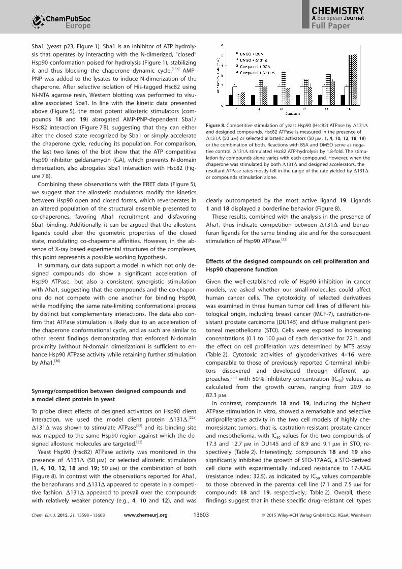
presence of D131D (50 mm) or selected allosteric stimulators(1, 4, 10, 12, 18 and 19 ; 50 mm) or the combination of both
(Figure 8). In contrast with the observations reported for Aha1,the benzofurans and D131D appeared to operate in a competi-
tive fashion. D131D appeared to prevail over the compoundswith relatively weaker potency (e.g. , 4, 10 and 12), and was
clearly outcompeted by the most active ligand 19. Ligands
1 and 18 displayed a borderline behavior (Figure 8).These results, combined with the analysis in the presence of
Aha1, thus indicate competition between D131D and benzo-
furan ligands for the same binding site and for the consequentstimulation of Hsp90 ATPase.[32]
Effects of the designed compounds on cell proliferation andHsp90 chaperone function
Given the well-established role of Hsp90 inhibition in cancer
models, we asked whether our small-molecules could affecthuman cancer cells. The cytotoxicity of selected derivatives
was examined in three human tumor cell lines of different his-tological origin, including breast cancer (MCF-7), castration-re-
sistant prostate carcinoma (DU145) and diffuse malignant peri-
toneal mesothelioma (STO). Cells were exposed to increasingconcentrations (0.1 to 100 mm) of each derivative for 72 h, and
the effect on cell proliferation was determined by MTS assay(Table 2). Cytotoxic activities of glycoderivatives 4–16 were
comparable to those of previously reported C-terminal inhibi-tors discovered and developed through different ap-proaches,[33] with 50 % inhibitory concentration (IC50) values, ascalculated from the growth curves, ranging from 29.9 to
82.3 mm.In contrast, compounds 18 and 19, inducing the highest
ATPase stimulation in vitro, showed a remarkable and selectiveantiproliferative activity in the two cell models of highly che-moresistant tumors, that is, castration-resistant prostate cancer
and mesothelioma, with IC50 values for the two compounds of17.3 and 12.7 mm in DU145 and of 8.9 and 9.1 mm in STO, re-
spectively (Table 2). Interestingly, compounds 18 and 19 also
significantly inhibited the growth of STO-17AAG, a STO-derivedcell clone with experimentally induced resistance to 17-AAG
(resistance index: 32.5), as indicated by IC50 values comparableto those observed in the parental cell line (7.1 and 7.5 mm for
compounds 18 and 19, respectively ; Table 2). Overall, thesefindings suggest that in these specific drug-resistant cell types
Figure 8. Competitive stimulation of yeast Hsp90 (Hsc82) ATPase by D131D
and designed compounds. Hsc82 ATPase is measured in the presence ofD131D (50 mm) or selected allosteric activators (50 mm, 1, 4, 10, 12, 18, 19)or the combination of both. Reactions with BSA and DMSO serve as nega-tive control. D131D stimulated Hsc82 ATP-hydrolysis by 1.8-fold. The stimu-lation by compounds alone varies with each compound. However, when thechaperone was stimulated by both D131D and designed accelerators, theresultant ATPase rates mostly fell in the range of the rate yielded by D131D
or compounds stimulation alone.
Chem. Eur. J. 2015, 21, 13598 – 13608 www.chemeurj.org Ó 2015 Wiley-VCH Verlag GmbH & Co. KGaA, Weinheim13603
Full Paper
the Hsp90 machinery could pop-ulate an ensemble of conforma-
tions more sensitive to the newderivatives.
Moreover, the activating com-
pounds did not disrupt theHsp90/HSF1 complex (Figure S4
in the Supporting Information)and did not promote heat shock
factor 1 (HSF1) dissociation, asclassical N-terminal inhibitors are
known to do.[19a] HSF1 dissocia-
tion is indeed the first step trig-gering the heat shock responsethat limits the application of NTD inhibitors in anticancer ther-apy. The inability of the benzofurans to activate this response
is a promising feature for further development of these com-pounds towards therapeutic applications.
To verify whether the observed cytotoxic activity was due to
the breakdown of multiple cell survival pathways as a conse-quence of the interaction of our compounds with Hsp90, we
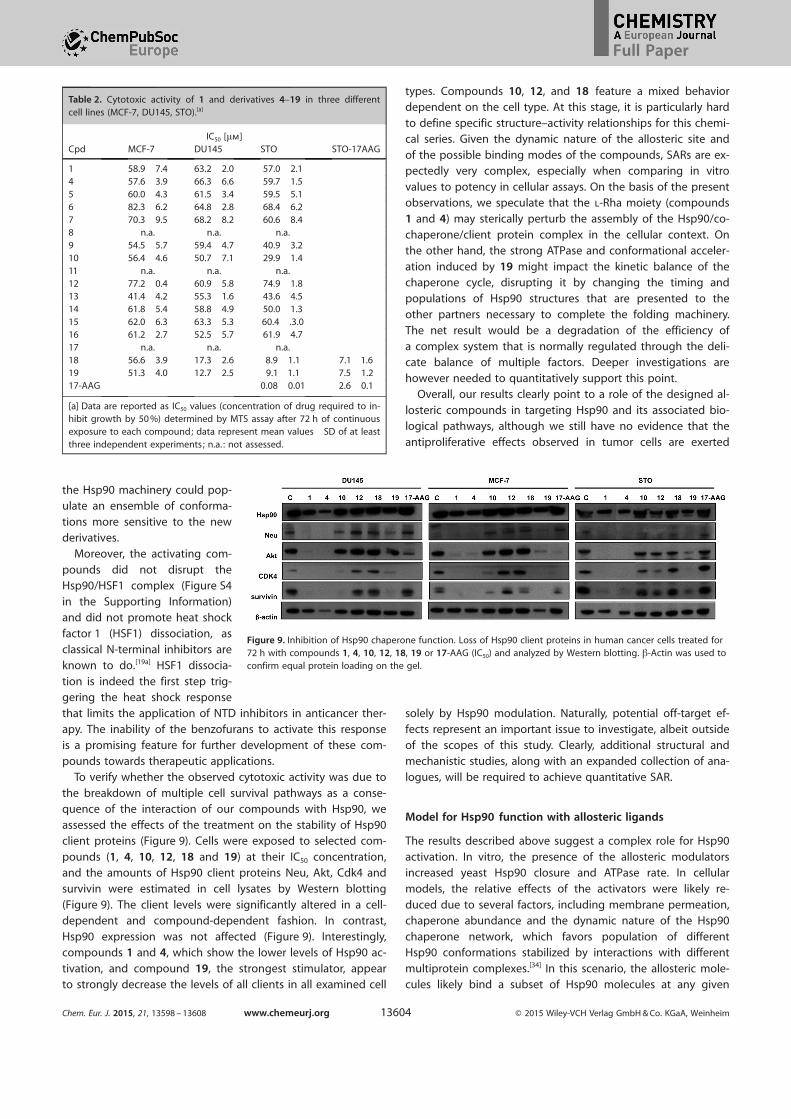
assessed the effects of the treatment on the stability of Hsp90client proteins (Figure 9). Cells were exposed to selected com-
pounds (1, 4, 10, 12, 18 and 19) at their IC50 concentration,and the amounts of Hsp90 client proteins Neu, Akt, Cdk4 and
survivin were estimated in cell lysates by Western blotting
(Figure 9). The client levels were significantly altered in a cell-dependent and compound-dependent fashion. In contrast,
Hsp90 expression was not affected (Figure 9). Interestingly,compounds 1 and 4, which show the lower levels of Hsp90 ac-
tivation, and compound 19, the strongest stimulator, appearto strongly decrease the levels of all clients in all examined cell
types. Compounds 10, 12, and 18 feature a mixed behaviordependent on the cell type. At this stage, it is particularly hard
to define specific structure–activity relationships for this chemi-cal series. Given the dynamic nature of the allosteric site and
of the possible binding modes of the compounds, SARs are ex-pectedly very complex, especially when comparing in vitro
values to potency in cellular assays. On the basis of the presentobservations, we speculate that the l-Rha moiety (compounds1 and 4) may sterically perturb the assembly of the Hsp90/co-
chaperone/client protein complex in the cellular context. Onthe other hand, the strong ATPase and conformational acceler-
ation induced by 19 might impact the kinetic balance of thechaperone cycle, disrupting it by changing the timing and
populations of Hsp90 structures that are presented to theother partners necessary to complete the folding machinery.
The net result would be a degradation of the efficiency ofa complex system that is normally regulated through the deli-cate balance of multiple factors. Deeper investigations arehowever needed to quantitatively support this point.
Overall, our results clearly point to a role of the designed al-
losteric compounds in targeting Hsp90 and its associated bio-logical pathways, although we still have no evidence that the
antiproliferative effects observed in tumor cells are exerted
solely by Hsp90 modulation. Naturally, potential off-target ef-fects represent an important issue to investigate, albeit outside
of the scopes of this study. Clearly, additional structural andmechanistic studies, along with an expanded collection of ana-
logues, will be required to achieve quantitative SAR.
Model for Hsp90 function with allosteric ligands
The results described above suggest a complex role for Hsp90
activation. In vitro, the presence of the allosteric modulatorsincreased yeast Hsp90 closure and ATPase rate. In cellular
models, the relative effects of the activators were likely re-
duced due to several factors, including membrane permeation,chaperone abundance and the dynamic nature of the Hsp90
chaperone network, which favors population of differentHsp90 conformations stabilized by interactions with different
multiprotein complexes.[34] In this scenario, the allosteric mole-cules likely bind a subset of Hsp90 molecules at any given
Table 2. Cytotoxic activity of 1 and derivatives 4–19 in three differentcell lines (MCF-7, DU145, STO).[a]
IC50 [mm]Cpd MCF-7 DU145 STO STO-17AAG
1 58.9�7.4 63.2�2.0 57.0�2.14 57.6�3.9 66.3�6.6 59.7�1.55 60.0�4.3 61.5�3.4 59.5�5.16 82.3�6.2 64.8�2.8 68.4�6.27 70.3�9.5 68.2�8.2 60.6�8.48 n.a. n.a. n.a.9 54.5�5.7 59.4�4.7 40.9�3.210 56.4�4.6 50.7�7.1 29.9�1.411 n.a. n.a. n.a.12 77.2�0.4 60.9�5.8 74.9�1.813 41.4�4.2 55.3�1.6 43.6�4.514 61.8�5.4 58.8�4.9 50.0�1.315 62.0�6.3 63.3�5.3 60.4� .3.016 61.2�2.7 52.5�5.7 61.9�4.717 n.a. n.a. n.a.18 56.6�3.9 17.3�2.6 8.9�1.1 7.1�1.619 51.3�4.0 12.7�2.5 9.1�1.1 7.5�1.217-AAG 0.08�0.01 2.6�0.1
[a] Data are reported as IC50 values (concentration of drug required to in-hibit growth by 50 %) determined by MTS assay after 72 h of continuousexposure to each compound; data represent mean values�SD of at leastthree independent experiments; n.a. : not assessed.
Figure 9. Inhibition of Hsp90 chaperone function. Loss of Hsp90 client proteins in human cancer cells treated for72 h with compounds 1, 4, 10, 12, 18, 19 or 17-AAG (IC50) and analyzed by Western blotting. b-Actin was used toconfirm equal protein loading on the gel.
Chem. Eur. J. 2015, 21, 13598 – 13608 www.chemeurj.org Ó 2015 Wiley-VCH Verlag GmbH & Co. KGaA, Weinheim13604
Full Paper

time, leaving the rest available for normal client and co-chaper-one interactions.
To generate a structural model of the mechanism, we carriedout MD simulations of the closed ATP-bound Hsp90 in com-
plex with 1, 4, 10, 12, 18, and 19 at the C-terminal site andcompared the results with those obtained in the absence of
compounds (ATP-only). MD simulations were aimed to shedlight on the microscopic perturbations of Hsp90 internal dy-
namics induced by allosteric ligands that could be linked to
the activation of functional states.We first characterized the overall rigidity/flexibility patterns
in the complexes using the coordination propensity (CP) analy-sis[20a] (Figure S5 in the Supporting Information). In the pres-
ence of the compound, the NTDs from one protomer were de-coupled from the MD and CTD of the other. By contrast, the
high degree of internal coordination between the NTD and
MD within each protomer was maintained upon compoundaddition. In this picture, high intraprotomer coordination of
the two domains favors the proper positioning of the residuesnecessary for catalysis, while interprotomer flexibility can be
aptly exploited to speed up the search for the closed activestate, consistent with the observed increases in ATPase and
closure rates.
The analysis of the effects of compounds on the internal dy-namics of Hsp90 highlighted interesting differences. For exam-
ple, compounds 1 and 19, representing the starting lead andthe strongest activator, respectively, appear to determine dif-
ferent coordination patterns (Figure S4 in the Supporting Infor-mation). In particular, the coordination patterns in the different
subdomains of the two protomers indicate that the dynamics
of Hsp90 in the presence of 1 is more similar to the ATP-onlycase than in the presence of 19. In the latter, the two proto-
mers are moreover characterized by a highly asymmetric dy-namic organization: in protomer A, in fact, high coordination
entails all the NTD and M-large regions, which emerge as a co-herent dynamic domain as defined by Morra et al.[20] The sub-
domains of protomer B, on the other hand, show the same
patterns observed for the ATP-only and Hsp90-1 cases.Consistent with the above observations, representative
structures for the complex with 19 showed an evident distor-tion of one of the two protomers, suggesting a role for the al-
losteric ligands in shifting the population to a closed asymmet-ric state reminiscent of the one observed in the crystal struc-
ture of the mitochondrial isoform of Hsp90 (named TRAP1).[14d]
This state was also identified in solution with SAXS measure-ments as a general conformation of all Hsp90 chaperones, as
a high-energy state that facilitates ATP hydrolysis[14d, 35]
(Figure 10).
Rearrangement to an asymmetric state could help explainthe observed synergistic activation by Aha1, through the selec-
tion of Hsp90 conformations more favorable for co-chaperone
binding, a model supported by our data in yeast lysates. Con-sistent with the general model of asymmetric Hsp90 activa-
tion,[14d] ligand-induced structural asymmetry in Hsp90 coulddetermine an expansion of conformations that are primed for
ATP processing and activated for Aha1 recognition. Aha1 acti-vation was in fact shown to be asymmetric,[36] one Aha1 mole-
cule per Hsp90 dimer is sufficient to bridge the two protomers,
stabilizing the N-dimerized catalytic state. In our model, the al-losteric ligands could preorganize an asymmetric Hsp90 con-
formation with which Aha1 preferentially interacts, stimulatingATP hydrolysis.
In the light of in vitro and cell results, we thus suggest thatallosteric ligands act as conformational catalysts bringing
Hsp90 into an asymmetric state primed for sequential ATP hy-
drolysis steps as proposed by Lavery et al.[14d] As an importantcaveat, it must be underlined that the data obtained from MD
simulations and coordination analysis must be consideredmostly qualitative: they indicate possible mechanistic differen-
ces among different protein complexes that can be linked tothe observed small-molecule modulations of yeast Hsp90
ATPase. However, they do not indicate quantitative trends be-
tween dynamic variables and experimental ATPase stimulationdata.
In summary, our findings indicate that the designed alloste-ric accelerators represent novel chemical tools to investigatesalient aspects of the relationships between Hsp90 structuraldynamics and functional regulation. These gain-of-function
probes offer the possibility to address the role of enzymaticand conformational dynamics in the protein endogenous envi-ronment. This would complement biochemical and molecularbiology approaches in shedding light on the roles of Hsp90mechanisms at different stages of the chaperone cycle.[30, 37]
The observed cytotoxic activities in geldanamycin-resistantcancer cells and the lack of heat shock response induction indi-
cate possible therapeutic perspectives for this class of com-
pounds.
Conclusion
Proteins participate in biochemical interaction networks byswitching among structural substates, which favor adaptation
Figure 10. The structural changes induced on Hsp90 by 19 compared to thesymmetric 2CG9 structure. The superposition exemplifies the distortion in-duced by the small molecules in protomer B (in red) compared to the origi-nal X-ray structure (PDB ID: 2CG9, in blue). Protomer A from both structuresare highly superimposable and are shown in light gray for simplicity.
Chem. Eur. J. 2015, 21, 13598 – 13608 www.chemeurj.org Ó 2015 Wiley-VCH Verlag GmbH & Co. KGaA, Weinheim13605
Full Paper

to different partners and fine-tuning of functions. Such confor-mational changes are induced by several factors, including
ligand binding.Herein, we rationally designed new allosteric ligands of the
molecular chaperone Hsp90, with the goal of exploring theirbiochemical and cellular effects. The initial set of molecules, 4–
16, was developed on the basis of compound 1, which waspreviously proven to bind Hsp90-CTD and to have promising
anticancer effects.[16] Interestingly, when probed in ATPase and
FRET kinetic experiments, these compounds turned out to beactivators of the enzymatic activity and conformational dynam-ics of the chaperone. Starting from this intriguing observation,we combined computational biology, synthetic chemistry, bio-chemical, biophysical and cell biology approaches, to designand partially optimize compounds 17–19 to further increase
ATPase activities. These latter molecules may be considered as
new molecular probes able to act as chemical switches tuningthe properties of the molecular chaperone Hsp90.
The ligands were directed towards an allosteric pocket re-cently identified in Hsp90 by coordination propensity analysis
of extended dynamic simulations of the protein. This pocket islocated at the interface between the CTD and the M-domain
and is dynamically coordinated to the ATP-binding site in the
NTD, so that modification of the allosteric pocket are translat-ed into variation of the ATPase activity of Hsp90. Given the in-
trinsically flexible nature of allosteric pockets, computationalstrategies for the discovery of allosteric ligands need to be de-
veloped ad hoc. Flexibility has so far hampered the resolutionof crystal structures with Hsp90 complexed to C-terminal tar-
geted ligands. Here we turned to extensive molecular dynam-
ics simulation of the protein in the presence of a lead com-pound (1), previously identified by virtual screening in a phar-
macophore model and validated as a CTD binder and an allo-steric modulator by a number of experimental techniques.[16]
The identification of the ligand–protein interactions most rele-vant in determining the dynamic cross-talk between the bind-
ing partners guided the evolution of initial activators towards
molecules with higher activities. Computational results pointedto specific charged and hydrophobic residues in the putative
Hsp90 allosteric pocket that could be targeted by ligands de-signed to contain specific and complementary chemical func-
tionalities.In particular, these results showed that the sugar moiety of
1 interacts extensively with the protein and suggested toexpand the SAR by glycodiversification of the aglycon of 1 (2)and of its synthetic precursor 3. Analysis of the interaction ofcompounds 4–16 with Hsp90 revealed that most of them actas stimulators, rather than inhibitors, of the ATPase activity,
a feature that was totally unprecedented, until very recently[27]
activators were found by large scale screening. We would like
to underline that the case we present here is the first one
where these results are obtained using a rational design ap-proach. The validity of our protocol was further corroborated
by the development of second generation activators : addition-al modeling reinforced the starting pharmacophoric hypothesis
and suggested a second set of modifications, finally leading to19, which accelerates Hsp90 ATPase by a factor of six, similar
to the most active known endogenous activator, the co-chap-erone Aha1.
Using FRET, we showed that ATPase activation by the benzo-furan probes is connected to acceleration of Hsp90 conforma-
tional dynamics. We characterized the activity of the mostpotent probes in the presence of the endogenous activator
Aha1, which is known to bind the N-M domains of Hsp90 andthus in principle should not compete with the ligands, and ofthe model client protein D131D, which binds in the proximity
of the proposed allosteric site. Importantly, the allosteric li-gands showed a synergistic effect with Aha1 and a competition
with D131D in regulating Hsp90 ATPase. Consistent with thesynergistic effect with Aha1, co-immunoprecipitation experi-ments also showed that association of the Hsp90 ATPase inhib-itory co-chaperone Sba1 was reduced in the presence of ben-
zofuran stimulators. Finally, the stimulatory activity observedon the isolated protein was found to translate to cytotoxicityin specific cell lines, which are known to be sensitive to Hsp90deregulation. The molecules tested were shown to affect theviability of cancer cells, in particular those resistant to the
Hsp90-targeted drug 17AAG, providing new opportunities togenerate anticancer interventions based on novel mechanisms
of action. Remarkably, the benzofurans described here were
shown not to activate the heat shock response in treated cells,a known drawback of Hsp90 NTD inhibitors.
Whether activation of the chaperone is the only mechanismoperating in cellular studies, it still needs to be fully demon-
strated. Nonetheless, these designed allosteric activators mayrepresent innovative gain-of-function probes to directly ad-
dress the roles of Hsp90 ATPase and conformational dynamics
in determining its cell functions. Moreover, our approach mayultimately generate effective anticancer drugs with novel
mechanisms of action, based on the perturbation of the Hsp90machinery, whereby the acceleration of conformational dynam-
ics can eventually translate into impaired chaperone functionsand cell death.
Experimental Section
Design and synthesis
Docking calculations and MD simulations : Docking was carriedout with the Maestro Suite (Release 2013-1-9.4, Schrçdinger, LLC,New York, NY, 2013). The shape and chemical properties of theCTD binding site were mapped onto a grid with dimensions of36 æ (enclosing box) and 14 æ (ligand diameter midpoint box).Docking calculations were performed using Glide[38] (version 5.8)and carried out in XP-mode with the OPLS-AA force field.[39] The re-sulting consensus poses were used for molecular dynamics simula-tion.
The structures of the three most populated clusters for complexeswith 1, 4, 10, 12, 18, 19 were used to start 100 ns explicit solventMD. Three simulations were run for each complex (total of1800 ns). Control simulations were run with Hsp90 in complex withATP only. All calculations were carried out with GROMACS.[40] De-tails are provided in the Supporting Information.
Synthesis of allosteric ligands : Synthesis of compounds 1–16 wasdescribed previously.[25] For 17–19, aglycon 3 was treated with dif-
Chem. Eur. J. 2015, 21, 13598 – 13608 www.chemeurj.org Ó 2015 Wiley-VCH Verlag GmbH & Co. KGaA, Weinheim13606
Full Paper

ferent alkylating agents under phase-transfer catalysis conditionsanalogous to those previously reported.[25] Details of the synthesisand full compound characterization are collected the in SupportingInformation.
Biochemical and biophysical assays
Hsp90 ATPase assay : ATPase activity of Hsp90 was measured bythe NADH-coupled ATPase assay. Briefly, Hsc82 (2 mm) or humanHsp90a (2 mm) was premixed with NADH (0.18 mm), l-lactate dehy-drogenase (50 U mL¢1), PEP (1 mm), pyruvate kinase (50 U mL¢1)and compounds (dissolved in DMSO to a final concentration of50 mm ; 5 mm for radicicol). The reaction was initiated by the addi-tion of ATP (1 mm). The reaction was carried out at 30 8C in buffercomposed of HEPES (20 mm, pH 7.5), KCl (100 mm) and MgCl2
(1 mm). Absorbance data were collected using a microplate spec-trophotometer (Spectra M5, Molecular Devices) at 360 nm.
FRET-based Hsp90 conformational change assay : The conforma-tional change rate of Hsp90 was measured by a FRET-basedassay.[41] Briefly, D61C or E329C mutation was introduced intoHsc82 for labeling with Alex Fluor 647 or Alex Fluor 555 (Life Tech-nologies), respectively. Labeled Hsp90 populations were mixed toproduce Hsp90 heterodimers. The conformational change ofHsp90 was initiated by adding AMP-PNP (1 mm) in the presence ofcompounds. The fluorescence from different dyes was monitoredon a microplate spectrophotometer with excitation/emission wave-length as follows: Ex525/Em568 (AF555), Ex525/Em668 (AF647).The assay was carried out at room temperature in the same bufferas Hsp90 ATPase assay.
Cytotoxicity and effects on Hsp90 interactions of designedcompounds
Cell lines : Human breast cancer (MCF-7) and castration-resistantprostate carcinoma (DU145) cell lines were obtained from theAmerican Type Culture Collection (Rockville, MD, USA). The humandiffuse malignant peritoneal mesothelioma cell line (STO) was es-tablished from a tumor specimen of a patient who underwent sur-gery at the Istituto Nazionale dei Tumori, Milan.[42] The resistantsubline STO-17AAG was derived by continuous exposure of theoriginal parental cell line (STO) to increasing concentrations of 17-AAG.
Cell proliferation assay : After harvesting in the logarithmicgrowth phase, 4500 cells per 50 mL were plated in 96-well flat-bot-tomed microtiter plates for 24 h and treated with increasing con-centrations of CTD ligands (1–100 mm) or 17-AAG (0.05–50 mm) for72 h. Control cells received vehicle alone (DMSO). At the end ofdrug exposure, cell growth inhibition was determined with theCellTiter 96Ò AQueous one solution cell proliferation assay (MTS;Promega). Optical density was read at 490 nm on a microplatereader (POLARstar OPTIMA) and the results were expressed asa percentage relative to DMSO-treated cells. Dose-response curveswere created and IC50 values were determined graphically from thecurve for each compound.
Analysis of Hsp90 client proteins : To monitor changes in Hsp90client proteins, cells were harvested, solubilized in lysis buffer(0.01 % NP40, 10 mm Tris pH 7.5, 50 mm KCl, 5 mm MgCl2, 2 mmDTT, 20 % glycerol plus protease inhibitors) and analyzed by West-ern blotting primary antibodies specific for survivin (AbCam),Hsp90, CD K4, Neu (Santa Cruz Biotechnology), and Akt (Cell Signal-ing Technology). Briefly, total cellular lysates were separated ona 4–12 % NuPAGE bis-tris gel (Life Technologies) and transferred tonitrocellulose using standard protocols. The filters were blocked in
PBS 1 Õ Tween-20 with 5 % skim milk or 5 % BSA and incubated,overnight, with primary antibodies. The filters were then incubatedwith the secondary peroxidase-linked whole antibodies (Life Tech-nologies). Bound antibodies were detected using the Novex ECL,HRP chemiluminescent substrate reagent kit (Life Technologies). Fil-ters were autoradiographed and images were acquired by Biospec-trum imaging system (Ultra-Violet Products Ltd.). b-Actin (AbCam)was used on each blot to ensure equal loading of proteins.
Hsp90 co-chaperone interaction analysis (Co-IP analysis): Yeastexpressing His-tagged Hsc82 (yeast Hsp90) as their sole Hsp90 pro-tein (yeast strain pp30 [hsc82hsp82]) were lysed as previously de-scribed.[43] Protein lysates were incubated either with buffer (¢negative control) or with 5 mm AMP-PNP as indicated, as well aswith 50 mm allosteric compounds, for 10 min at 308 prior to affinityprecipitation of His-tagged Hsp82 with Ni-NTA agarose. Hsp82-as-sociated Sba1 was detected by immunoblotting.
Acknowledgements
We acknowledge funding from Fondazione Cariplo throughgrant 2011.1800, “Premio Fondazione Cariplo per la Ricerca di
Frontiera”. G.C. acknowledges funding from the AIRC (Associa-
zione Italiana Ricerca sul Cancro) grant IG 15420; Universita’degli Studi di Milano is acknowledged for a grant to S.S. (As-
segno di ricerca tipo A).
Keywords: allostery · drug design · functional dynamics ·glycoconjugates · Hsp90
[1] R. Nussinov, C.-J. Tsai, Cell 2013, 153, 293 – 305.[2] a) V. J. Hilser, Science 2010, 327, 653 – 654; b) R. G. Smock, L. M. Gierasch,
Science 2009, 324, 198 – 203.[3] J. A. Zorn, J. A. Wells, Nat. Chem. Biol. 2010, 6, 179 – 188.[4] a) Z. Fang, C. Grìtter, D. Rauh, ACS Chem. Biol. 2013, 8, 58 – 70; b) C. J.
Wenthur, P. R. Gentry, T. P. Mathews, C. W. Lindsley, Rev. Pharm. Toxicol.2014, 54, 165 – 184; c) A. Rodina, P. D. Patel, Y. Kang, Y. Patel, I. Baaklini,M. J. Wong, T. Taldone, P. Yan, C. Yang, R. Maharaj, A. Gozman, M. R.Patel, H. J. Patel, W. Chirico, H. Erdjument-Bromage, T. T. Talele, J. C.Young, G. Chiosis, Chem. Biol. 2013, 20, 1469 – 1480.
[5] M. Taipale, D. F. Jarosz, S. Lindquist, Nat. Rev. Mol. Cell. Biol. 2010, 11,515 – 528.
[6] S. E. Jackson, Top. Curr. Chem. 2013, 328, 155 – 240.[7] L. Whitesell, S. Lindquist, Nat. Rev. Cancer 2005, 5, 761 – 772.[8] V. Shah, R. Wiest, G. Garcia-Cardena, G. Cadelina, R. J. Groszmann, W. C.
Sessa, Am. J. Physiol. 1999, 277, 463 – 468.[9] W. Luo, W. Sun, T. Taldone, A. Rodina, G. Chiosis, Mol. Neurodegener.
2010, 5, 24.[10] D. F. Jarosz, S. Lindquist, Science 2010, 330, 1820 – 1824.[11] M. Taipale, I. Krykbaeva, M. Koeva, C. Kayatekin, K. D. Westover, G. I.
Karras, S. Lindquist, Cell 2012, 150, 987 – 1001.[12] a) P. C. Echeverr�a, A. Bernthaler, P. Dupuis, B. Mayer, D. Picard, PLoS ONE
2011, 6, e26044; b) E. Kirschke, D. Goswami, D. Southworth, P. R. Griffin,D. A. Agard, Cell 2014, 157, 1685 – 1697.
[13] K. A. Krukenberg, T. O. Street, L. A. Lavery, D. A. Agard, Q. Rev. Biophys.2011, 44, 229 – 255.
[14] a) M. M. U. Ali, S. M. Roe, C. K. Vaughan, P. Meyer, B. Panaretou, P. W.Piper, C. Prodromou, L. H. Pearl, Nature 2006, 440, 1013 – 1017; b) A. K.Shiau, S. F. Harris, D. R. Southworth, D. A. Agard, Cell 2006, 127, 329 –340; c) D. E. Dollins, J. J. Warren, R. M. Immormino, D. T. Gewirth, Mol.Cell 2007, 28, 41 – 56; d) L. A. Lavery, J. R. Partridge, T. A. Ramelot, D. El-natan, M. A. Kennedy, D. A. Agard, Mol. Cell 2014, 53, 330 – 343.
[15] a) A. Zuehlke, J. L. Johnson, BIOPOLYMERS 2010, 93, 211 – 217; b) J. Li, J.Buchner, Biomed. J. 2013, 36, 106 – 117.
Chem. Eur. J. 2015, 21, 13598 – 13608 www.chemeurj.org Ó 2015 Wiley-VCH Verlag GmbH & Co. KGaA, Weinheim13607
Full Paper

[16] G. Morra, M. A. C. Neves, C. J. Plescia, S. Tsutsumi, L. Neckers, G. Ver-khivker, D. C. Altieri, G. Colombo, J. Chem. Theory Comput. 2010, 6,2978 – 2989.
[17] M. Taddei, S. Ferrini, L. Giannotti, M. Corsi, F. Manetti, G. Giannini, L.Vesci, F. M. Milazzo, D. Alloatti, M. B. Guglielmi, M. Castorina, M. Cervoni,M. Barbarino, R. Foder�, V. Carollo, C. Pisano, S. Armaroli, W. Cabri, J.Med. Chem. 2014, 57, 2258 – 2274.
[18] S. M. Roe, C. Prodromou, R. O’Brien, J. E. Ladbury, P. W. Piper, L. H. Pearl,J. Med. Chem. 1999, 42, 260 – 266.
[19] a) P. Workman, F. J. Burrows, L. Neckers, N. Rosen, Ann. N. Y. Acad. Sci.2007, 1113, 202 – 216; b) L. Whitesell, R. Bagatell, R. Falsey, Curr. CancerDrug Targets 2003, 3, 349 – 358.
[20] a) G. Morra, R. Potestio, C. Micheletti, G. Colombo, PLoS Comput. Biol.2012, 8, e1002433; b) G. Morra, G. M. Verkhivker, G. Colombo, PLoSComput. Biol. 2009, 5, e1000323.
[21] a) E. Moroni, H. Zhao, B. S. Blagg, G. Colombo, J. Chem. Inf. Model 2014,24397468; b) H. Zhao, E. Moroni, G. Colombo, B. S. Blagg, ACS Med.Chem. Lett 2014, 5, 84 – 88; c) H. Zhao, E. Moroni, B. Yan, G. Colombo,B. S. J. Blagg, ACS Med. Chem. Lett. 2013, 4, 57 – 62.
[22] a) T. O. Street, L. A. Lavery, D. A. Agard, Mol. Cell 2011, 42, 96 – 105; b) O.Genest, M. Reidy, T. O. Street, J. R. Hoskins, J. L. Camberg, D. A. Agard,D. C. Masison, S. Wickner, Mol. Cell 2013, 49, 464 – 473.
[23] E. Moroni, A. Paladino, G. Colombo, Curr. Top. Med. Chem. 2015, 15,2043 – 2055.
[24] R. D. Goff, J. S. Thorson, Med. Chem. Commun. 2014, 5, 1036 – 1047.[25] L. Morelli, A. Bernardi, S. Sattin, Carbohydr. Res. 2014, 390C, 33 – 41.[26] B. Panaretou, C. Prodromou, S. M. Roe, R. O’Brien, J. E. Ladbury, P. W.
Piper, L. H. Pearl, EMBO J. 1998, 17, 4829 – 4836.[27] B. K. Zierer, M. Weiwad, M. Rubbelke, L. Freiburger, G. Fischer, O. R.
Lorenz, M. Sattler, K. Richter, J. Buchner, Angew. Chem. Int. Ed. 2014, 53,12257 – 12262; Angew. Chem. 2014, 126, 12454 – 12459.
[28] A. Kranjc, S. Bongarzone, G. Rossetti, X. Biarnes, A. Cavalli, M. L. Bolog-nesi, M. Roberti, G. Legname, P. Carloni, J. Chem. Theory Comput. 2009,5, 2565 – 2573.
[29] a) M. Mickler, M. Hessling, C. Ratzke, J. Buchner, T. Hugel, Nat. Struct.Mol. Biol. 2009, 16, 281 – 286; b) D. R. Southworth, D. A. Agard, Mol. Cell2008, 32, 631 – 640.
[30] L. Pullen, D. N. Bolon, J. Biol. Chem. 2011, 286, 11091 – 11098.
[31] J. Li, K. Richter, J. Reinstein, J. Buchner, Nat. Struct. Mol. Biol. 2013, 20,326 – 331.
[32] S. H. McLaughlin, H. W. Smith, S. E. Jackson, J. Mol. Biol. 2002, 315, 787 –798.
[33] H. P. Zhao, A. C. Donnelly, B. R. Kusuma, G. E. L. Brandt, D. Brown, R. A.Rajewski, G. Vielhauer, J. Holzbeierlein, M. S. Cohen, B. S. J. Blagg, J.Med. Chem. 2011, 54, 3839 – 3853.
[34] I. Fierro-Monti, P. Echeverria, J. Racle, C. Hernandez, D. Picard, M. Quad-roni, PLoS ONE 2013, 8, e80425.
[35] J. R. Partridge, L. A. Lavery, D. Elnatan, N. Naber, R. Cooke, D. A. Agard,eLife 2014, DOI : 10.7554/eLife.03487.
[36] M. Retzlaff, F. Hagn, L. Mitschke, M. Hessling, F. Gugel, H. Kessler, K.Richter, J. Buchner, Mol. Cell 2010, 37, 344 – 354.
[37] K. Beebe, M. Mollapour, B. Scroggins, C. Prodromou, W. Xu, M. Tokita, T.Taldone, L. Pullen, B. K. Zierer, M. J. Lee, J. Trepel, J. Buchner, D. N.Bolon, G. Chiosis, L. Neckers, Oncotarget 2013, 4, 1065 – 1074.
[38] R. A. Friesner, R. B. Murphy, M. P. Repasky, L. L. Frye, J. R. Greenwood,T. A. Halgren, P. C. Sanschagrin, D. T. Mainz, J. Med. Chem. 2006, 49,6177 – 6196.
[39] W. L. Jorgensen, D. S. Maxwell, J. Tirado-Rives, J. Am. Chem. Soc. 1996,118, 11225 – 11236.
[40] B. Hess, C. Kutzner, D. van der Spoel, E. Lindahl, J. Chem. Theory Comput.2008, 4, 435 – 447.
[41] M. Hessling, K. Richter, J. Buchner, Nat. Struct. Mol. Biol. 2009, 16, 287 –293.
[42] N. Zaffaroni, A. Costa, M. Pennati, C. De Marco, E. Affini, M. Madeo, R.Erdas, A. Cabras, S. Kusamura, D. Baratti, M. Deraco, M. G. Daidone, CellOncol. 2007, 29, 453 – 466.
[43] M. Mollapour, D. Bourboulia, K. Beebe, M. R. Woodford, S. Polier, A.Hoang, R. Chelluri, Y. Li, A. Guo, M. J. Lee, E. Fotooh-abadi, S. Khan, T.Prince, N. Miyajima, S. Yoshida, S. Tusutsumi, W. P. Xu, B. Panaretou,W. G. Stetler-Stevenson, G. Bratslavsky, J. B. Trepel, C. Prodromou, L.Neckers, Mol. Cell 2014, 53, 317 – 329.
Received: May 7, 2015
Published online on August 18, 2015
Chem. Eur. J. 2015, 21, 13598 – 13608 www.chemeurj.org Ó 2015 Wiley-VCH Verlag GmbH & Co. KGaA, Weinheim13608
Full Paper


![Hsp90-Targeted Library - Chemdiv · HtpG (high-temperature protein G), whereas Archaebacteria lack a Hsp90 representative [24]. All eukaryotes possess cytosolic members, called Hsp90](https://static.fdocuments.net/doc/165x107/5c687a8609d3f2f5638b9b2b/hsp90-targeted-library-htpg-high-temperature-protein-g-whereas-archaebacteria.jpg)


