Actinomycosis and Sialolithiasis in Submandibular Gland · 41 Jin Seok Kang et al . Actinomycosis...
4
Archives of Craniofacial Surgery Copyright © 2015 The Korean Cleft Palate-Craniofacial Association This is an Open Access article distributed under the terms of the Creative Commons Attribution Non-Commercial License (http://creativecommons.org/ licenses/by-nc/3.0/) which permits unrestricted non-commercial use, distribution, and reproduction in any medium, provided the original work is properly cited. www.e-acfs.org pISSN 2287-1152 eISSN 2287-5603 39 Arch Craniofac Surg Vol.16 No.1, 39-42 http://dx.doi.org/10.7181/acfs.2015.16.1.39 Correspondence: Hwan Jun Choi Department of Plastic and Reconstructive Surgery, Soonchunhyang University Cheonan Hospital, Soonchunhyang University College of Medicine, 31 Suncheonhyang 6-gil, Dongnam-gu, Cheonan 330-721, Korea E-mail: [email protected] *This work was supported by Soonchunhyang University Research Fund. Received January 16, 2015 / Revised February 27, 2015 / Accepted April 2, 2015 INTRODUCTION Actinomycosis is a subacute or chronic suppurative infection caused by Actinomyces species, which are anaerobic Gram-posi- tive bacteria that normally colonize the human mouth and diges- tive and urogenital tracts [1]. To date, multiple clinical features of actinomycosis have been described, and the clinical types of ac- tinomycosis are cervicofacial, thoracic, abdominal, and female genitalia. Cervicofacial actinomycosis is the most common form and comprises about 50% of all reported cases [2]. e most fre- quently affected cervicofacial sites are the parotids and the man- dible [3]. Cervicofacial actinomycosis is associated with an abscess Actinomycosis and Sialolithiasis in Submandibular Gland Actinomycosis is a subacute or chronic suppurative infection caused by Actinomyces species, which are anaerobic Gram-positive bacteria that normally colonize the hu- man mouth and digestive and urogenital tracts. Cervicofacial actinomycosis is the most frequent clinical form of actinomycosis, and is associated with odontogenic infection. Characterized by an abscess and mandibular involvement with or without fistula, but the cervicofacial form of actinomycosis is often misdiagnosed because the presentation is not specific and because it can mimic numerous infectious and non-infectious diseases, including malignant tumors. We report a rare case of actinomycosis infection with co- existing submandibular sialolithiasis. The patient presented with a 1×1 cm abscess-like lesion below the lower lip. Punch biopsy of the lesion revealed atypical squamous cell proliferation with infiltrative growth, suggestive of squamous cell carcinoma. The patient underwent wide excision of this lesion, where the lesion was found to be an abscess for- mation with multiple submandibular sialolithiases. The surgical specimen was found to contain Actinomyces without any evidence of a malignant process. We assumed that as- sociated predisposing factors such as poor oral hygiene may have caused a dehydrated condition of the oral cavity, leading to coexistence of actinomycosis and sialolithiasis. Keywords: Actinomycosis / Sialolithiasis Jin Seok Kang 1 , Hwan Jun Choi 2 , Min Sung Tak 1 1 Department of Plastic and Reconstructive Surgery, Soonchunhyang University Seoul Hospital, Seoul; 2 Department of Plastic and Reconstructive Surgery, Soonchunhyang University Cheonan Hospital, Soonchunhyang University College of Medicine, Cheonan, Korea No potential conflict of interest relevant to this article was reported. and mandibular involvement with or without fistula. Oſten mis- diagnosed, clinical presentation is not specific, and the lesions can mimic numerous infectious and non-infectious diseases, includ- ing malignant tumors [1]. We report a case of actinomycosis of the lower lip with multiple submandibular gland calcifications that mimicked squamous cell carcinoma. CASE REPORT An 87-year-old woman presented to the outpatient dermatology clinic with a complaint of a progressive abscess-like lesion on the lower lip which was painful and discharging pus (Fig. 1). e le- sion had developed about 1 year ago and initially it was non-ten- der but later it became tender. Physical examination revealed an abscess-like lesion measuring about 1×1 cm below the lower lip and multiple palpable hard masses on the lower lip. Purulent yel- low exudate was discharged upon expression. No cervical lymph- adenopathy was noted. ere was no history of trauma, but the Case Report
-
Upload
duongkhuong -
Category
Documents
-
view
217 -
download
0
Transcript of Actinomycosis and Sialolithiasis in Submandibular Gland · 41 Jin Seok Kang et al . Actinomycosis...
Archives of Craniofacial Surgery
Copyright © 2015 The Korean Cleft Palate-Craniofacial Association This is an Open Access article distributed under the terms of the Creative Commons Attribution Non-Commercial License (http://creativecommons.org/
licenses/by-nc/3.0/) which permits unrestricted non-commercial use, distribution, and reproduction in any medium, provided the original work is properly cited.
www.e-acfs.orgpISSN 2287-1152eISSN 2287-5603
39
Arch Craniofac Surg Vol.16 No.1, 39-42http://dx.doi.org/10.7181/acfs.2015.16.1.39
Correspondence: Hwan Jun ChoiDepartment of Plastic and Reconstructive Surgery, Soonchunhyang University Cheonan Hospital, Soonchunhyang University College of Medicine, 31 Suncheonhyang 6-gil, Dongnam-gu, Cheonan 330-721, KoreaE-mail: [email protected]
*This work was supported by Soonchunhyang University Research Fund.
Received January 16, 2015 / Revised February 27, 2015 / Accepted April 2, 2015
INTRODUCTION
Actinomycosis is a subacute or chronic suppurative infection
caused by Actinomyces species, which are anaerobic Gram-posi-
tive bacteria that normally colonize the human mouth and diges-
tive and urogenital tracts [1]. To date, multiple clinical features of
actinomycosis have been described, and the clinical types of ac-
tinomycosis are cervicofacial, thoracic, abdominal, and female
genitalia. Cervicofacial actinomycosis is the most common form
and comprises about 50% of all reported cases [2]. The most fre-
quently affected cervicofacial sites are the parotids and the man-
dible [3]. Cervicofacial actinomycosis is associated with an abscess
Actinomycosis and Sialolithiasis in Submandibular Gland
Actinomycosis is a subacute or chronic suppurative infection caused by Actinomyces species, which are anaerobic Gram-positive bacteria that normally colonize the hu-man mouth and digestive and urogenital tracts. Cervicofacial actinomycosis is the most frequent clinical form of actinomycosis, and is associated with odontogenic infection. Characterized by an abscess and mandibular involvement with or without fistula, but the cervicofacial form of actinomycosis is often misdiagnosed because the presentation is not specific and because it can mimic numerous infectious and non-infectious diseases, including malignant tumors. We report a rare case of actinomycosis infection with co-existing submandibular sialolithiasis. The patient presented with a 1×1 cm abscess-like lesion below the lower lip. Punch biopsy of the lesion revealed atypical squamous cell proliferation with infiltrative growth, suggestive of squamous cell carcinoma. The patient underwent wide excision of this lesion, where the lesion was found to be an abscess for-mation with multiple submandibular sialolithiases. The surgical specimen was found to contain Actinomyces without any evidence of a malignant process. We assumed that as-sociated predisposing factors such as poor oral hygiene may have caused a dehydrated condition of the oral cavity, leading to coexistence of actinomycosis and sialolithiasis.
Keywords: Actinomycosis / Sialolithiasis
Jin Seok Kang1,Hwan Jun Choi2,Min Sung Tak1
1Department of Plastic and Reconstructive Surgery, Soonchunhyang University Seoul Hospital, Seoul; 2Department of Plastic and Reconstructive Surgery, Soonchunhyang University Cheonan Hospital, Soonchunhyang University College of Medicine, Cheonan, Korea
No potential conflict of interest relevant to this article was reported.
and mandibular involvement with or without fistula. Often mis-
diagnosed, clinical presentation is not specific, and the lesions can
mimic numerous infectious and non-infectious diseases, includ-
ing malignant tumors [1]. We report a case of actinomycosis of the
lower lip with multiple submandibular gland calcifications that
mimicked squamous cell carcinoma.
CASE REPORT
An 87-year-old woman presented to the outpatient dermatology
clinic with a complaint of a progressive abscess-like lesion on the
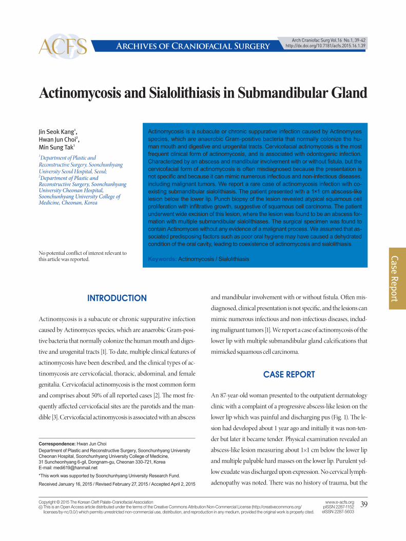
lower lip which was painful and discharging pus (Fig. 1). The le-
sion had developed about 1 year ago and initially it was non-ten-
der but later it became tender. Physical examination revealed an
abscess-like lesion measuring about 1×1 cm below the lower lip
and multiple palpable hard masses on the lower lip. Purulent yel-
low exudate was discharged upon expression. No cervical lymph-
adenopathy was noted. There was no history of trauma, but the
Case Report
Archives of Craniofacial Surgery Vol. 16, No. 1, 2015
www.e-acfs.org40
patient had poor oral hygiene with a relatively dehydrated cavity.
Punch biopsy of the lesion revealed atypical squamous cell prolif-
eration with infiltrative growth, suggestive of squamous cell carci-
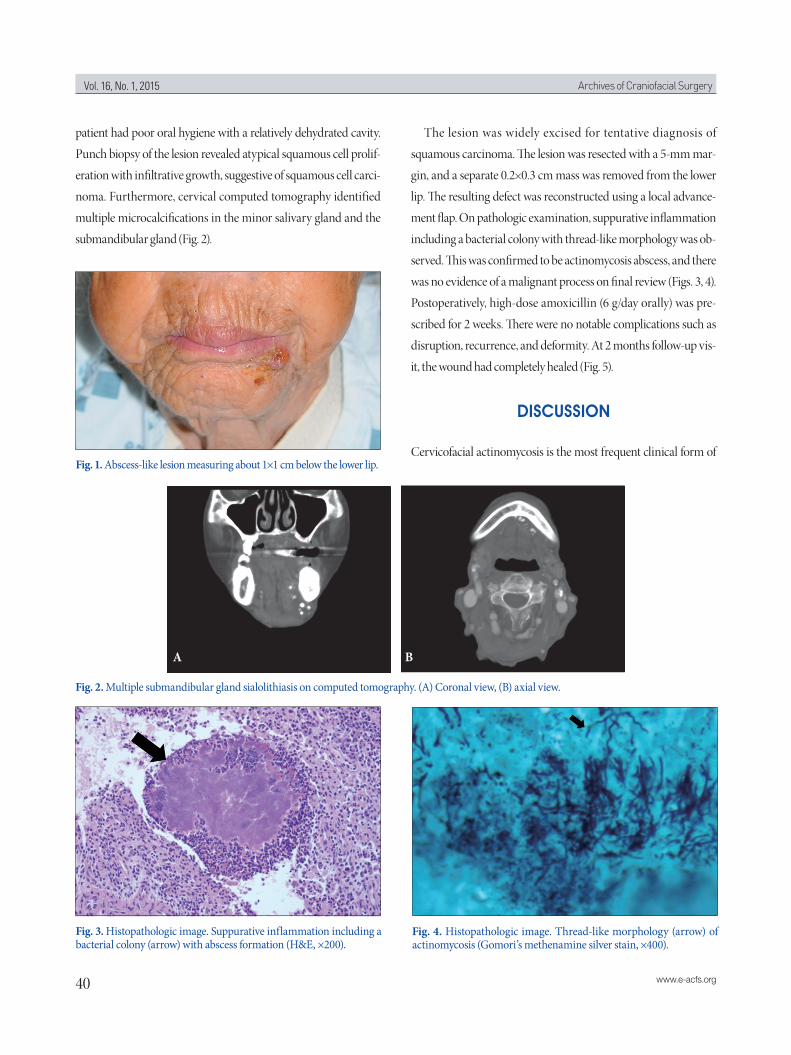
noma. Furthermore, cervical computed tomography identified
multiple microcalcifications in the minor salivary gland and the
submandibular gland (Fig. 2).
The lesion was widely excised for tentative diagnosis of
squamous carcinoma. The lesion was resected with a 5-mm mar-
gin, and a separate 0.2×0.3 cm mass was removed from the lower
lip. The resulting defect was reconstructed using a local advance-
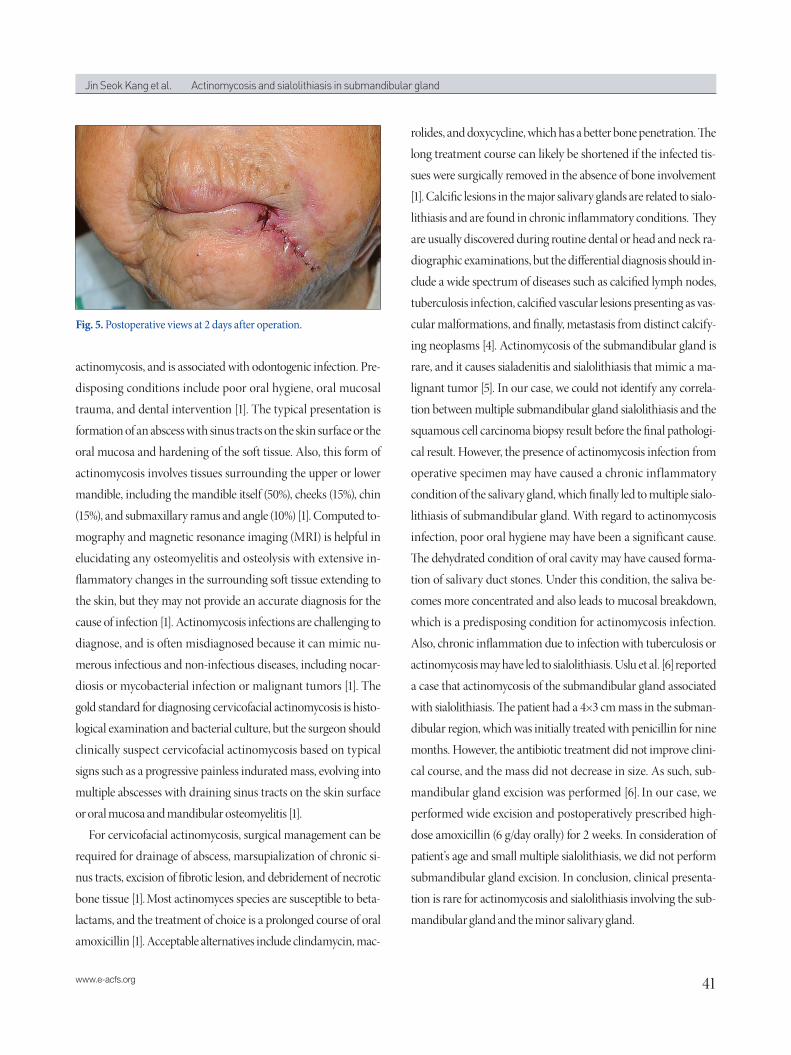
ment flap. On pathologic examination, suppurative inflammation
including a bacterial colony with thread-like morphology was ob-
served. This was confirmed to be actinomycosis abscess, and there
was no evidence of a malignant process on final review (Figs. 3, 4).
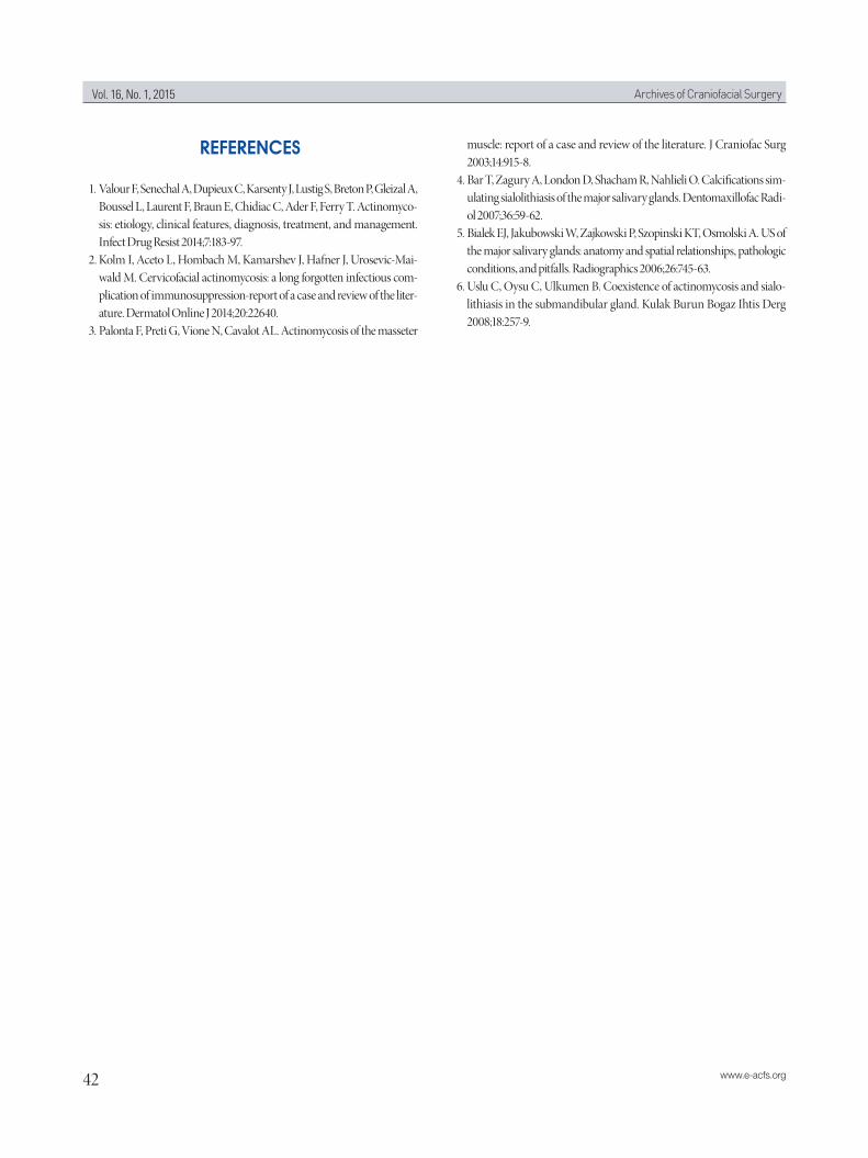
Postoperatively, high-dose amoxicillin (6 g/day orally) was pre-
scribed for 2 weeks. There were no notable complications such as
disruption, recurrence, and deformity. At 2 months follow-up vis-
it, the wound had completely healed (Fig. 5).
DISCUSSION
Cervicofacial actinomycosis is the most frequent clinical form of Fig. 1. Abscess-like lesion measuring about 1×1 cm below the lower lip.
Fig. 3. Histopathologic image. Suppurative inflammation including a bacterial colony (arrow) with abscess formation (H&E, ×200).
Fig. 4. Histopathologic image. Thread-like morphology (arrow) of actinomycosis (Gomori’s methenamine silver stain, ×400).
Fig. 2. Multiple submandibular gland sialolithiasis on computed tomography. (A) Coronal view, (B) axial view.
A B
41www.e-acfs.org
Jin Seok Kang et al. Actinomycosis and sialolithiasis in submandibular gland
actinomycosis, and is associated with odontogenic infection. Pre-
disposing conditions include poor oral hygiene, oral mucosal
trauma, and dental intervention [1]. The typical presentation is
formation of an abscess with sinus tracts on the skin surface or the
oral mucosa and hardening of the soft tissue. Also, this form of
actinomycosis involves tissues surrounding the upper or lower
mandible, including the mandible itself (50%), cheeks (15%), chin
(15%), and submaxillary ramus and angle (10%) [1]. Computed to-
mography and magnetic resonance imaging (MRI) is helpful in
elucidating any osteomyelitis and osteolysis with extensive in-
flammatory changes in the surrounding soft tissue extending to
the skin, but they may not provide an accurate diagnosis for the
cause of infection [1]. Actinomycosis infections are challenging to
diagnose, and is often misdiagnosed because it can mimic nu-
merous infectious and non-infectious diseases, including nocar-
diosis or mycobacterial infection or malignant tumors [1]. The
gold standard for diagnosing cervicofacial actinomycosis is histo-
logical examination and bacterial culture, but the surgeon should
clinically suspect cervicofacial actinomycosis based on typical
signs such as a progressive painless indurated mass, evolving into
multiple abscesses with draining sinus tracts on the skin surface
or oral mucosa and mandibular osteomyelitis [1].
For cervicofacial actinomycosis, surgical management can be
required for drainage of abscess, marsupialization of chronic si-
nus tracts, excision of fibrotic lesion, and debridement of necrotic
bone tissue [1]. Most actinomyces species are susceptible to beta-
lactams, and the treatment of choice is a prolonged course of oral
amoxicillin [1]. Acceptable alternatives include clindamycin, mac-
rolides, and doxycycline, which has a better bone penetration. The
long treatment course can likely be shortened if the infected tis-
sues were surgically removed in the absence of bone involvement
[1]. Calcific lesions in the major salivary glands are related to sialo-
lithiasis and are found in chronic inflammatory conditions. They
are usually discovered during routine dental or head and neck ra-
diographic examinations, but the differential diagnosis should in-
clude a wide spectrum of diseases such as calcified lymph nodes,
tuberculosis infection, calcified vascular lesions presenting as vas-
cular malformations, and finally, metastasis from distinct calcify-
ing neoplasms [4]. Actinomycosis of the submandibular gland is
rare, and it causes sialadenitis and sialolithiasis that mimic a ma-
lignant tumor [5]. In our case, we could not identify any correla-
tion between multiple submandibular gland sialolithiasis and the
squamous cell carcinoma biopsy result before the final pathologi-
cal result. However, the presence of actinomycosis infection from
operative specimen may have caused a chronic inflammatory
condition of the salivary gland, which finally led to multiple sialo-
lithiasis of submandibular gland. With regard to actinomycosis
infection, poor oral hygiene may have been a significant cause.
The dehydrated condition of oral cavity may have caused forma-
tion of salivary duct stones. Under this condition, the saliva be-
comes more concentrated and also leads to mucosal breakdown,
which is a predisposing condition for actinomycosis infection.
Also, chronic inflammation due to infection with tuberculosis or
actinomycosis may have led to sialolithiasis. Uslu et al. [6] reported
a case that actinomycosis of the submandibular gland associated
with sialolithiasis. The patient had a 4×3 cm mass in the subman-
dibular region, which was initially treated with penicillin for nine
months. However, the antibiotic treatment did not improve clini-
cal course, and the mass did not decrease in size. As such, sub-
mandibular gland excision was performed [6]. In our case, we
performed wide excision and postoperatively prescribed high-
dose amoxicillin (6 g/day orally) for 2 weeks. In consideration of
patient’s age and small multiple sialolithiasis, we did not perform
submandibular gland excision. In conclusion, clinical presenta-
tion is rare for actinomycosis and sialolithiasis involving the sub-
mandibular gland and the minor salivary gland.
Fig. 5. Postoperative views at 2 days after operation.
Archives of Craniofacial Surgery Vol. 16, No. 1, 2015
www.e-acfs.org42
REFERENCES
1. Valour F, Senechal A, Dupieux C, Karsenty J, Lustig S, Breton P, Gleizal A, Boussel L, Laurent F, Braun E, Chidiac C, Ader F, Ferry T. Actinomyco-sis: etiology, clinical features, diagnosis, treatment, and management. Infect Drug Resist 2014;7:183-97.
2. Kolm I, Aceto L, Hombach M, Kamarshev J, Hafner J, Urosevic-Mai-wald M. Cervicofacial actinomycosis: a long forgotten infectious com-plication of immunosuppression-report of a case and review of the liter-ature. Dermatol Online J 2014;20:22640.
3. Palonta F, Preti G, Vione N, Cavalot AL. Actinomycosis of the masseter
muscle: report of a case and review of the literature. J Craniofac Surg 2003;14:915-8.
4. Bar T, Zagury A, London D, Shacham R, Nahlieli O. Calcifications sim-ulating sialolithiasis of the major salivary glands. Dentomaxillofac Radi-ol 2007;36:59-62.
5. Bialek EJ, Jakubowski W, Zajkowski P, Szopinski KT, Osmolski A. US of the major salivary glands: anatomy and spatial relationships, pathologic conditions, and pitfalls. Radiographics 2006;26:745-63.
6. Uslu C, Oysu C, Ulkumen B. Coexistence of actinomycosis and sialo-lithiasis in the submandibular gland. Kulak Burun Bogaz Ihtis Derg 2008;18:257-9.