Tashi the Foolish Fox - by Jyotsna Patwal and Aaditya Singh Kathait
Upload
dr-harshavardhan-patwalCategory
view
580download
5
Actinobacillus Actinomycetemcomitans
Dr Harshavardhan G Patwal
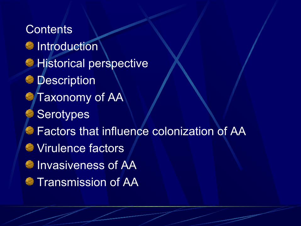
Contents
Introduction
Historical perspective
Description
Taxonomy of AA
Serotypes
Factors that influence colonization of AA
Virulence factors
Invasiveness of AA
Transmission of AA
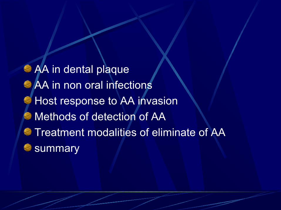
AA in dental plaque
AA in non oral infections
Host response to AA invasion
Methods of detection of AA
Treatment modalities of eliminate of AA
summary
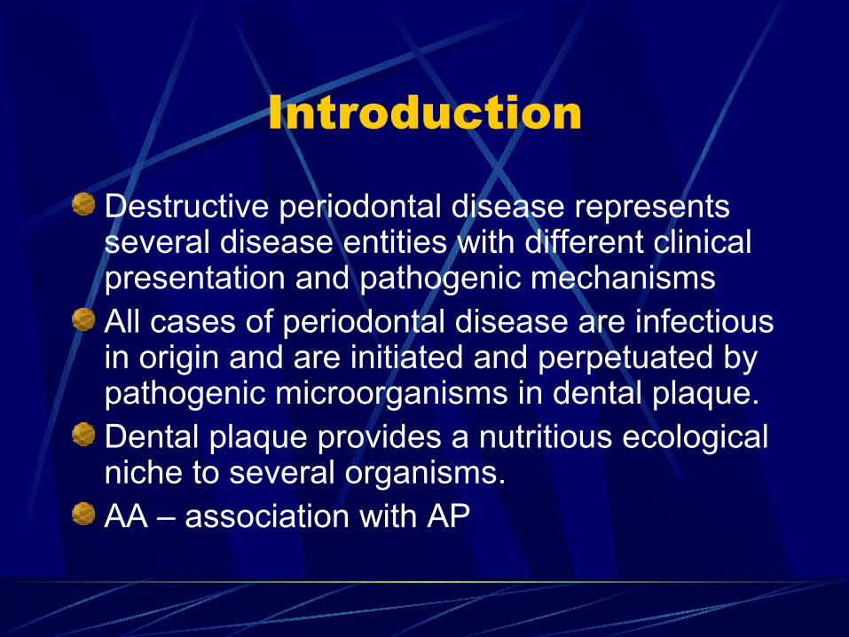
Introduction
Destructive periodontal disease represents several disease entities with different clinical presentation and pathogenic mechanismsAll cases of periodontal disease are infectious in origin and are initiated and perpetuated by pathogenic microorganisms in dental plaque.Dental plaque provides a nutritious ecological niche to several organisms. AA – association with AP
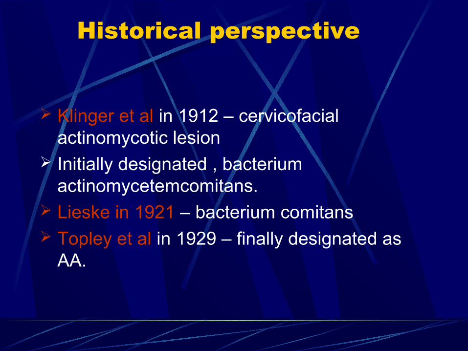
Historical perspective
Klinger et al in 1912 – cervicofacial actinomycotic lesion
Initially designated , bacterium actinomycetemcomitans.
Lieske in 1921 – bacterium comitans Topley et al in 1929 – finally designated as
AA.
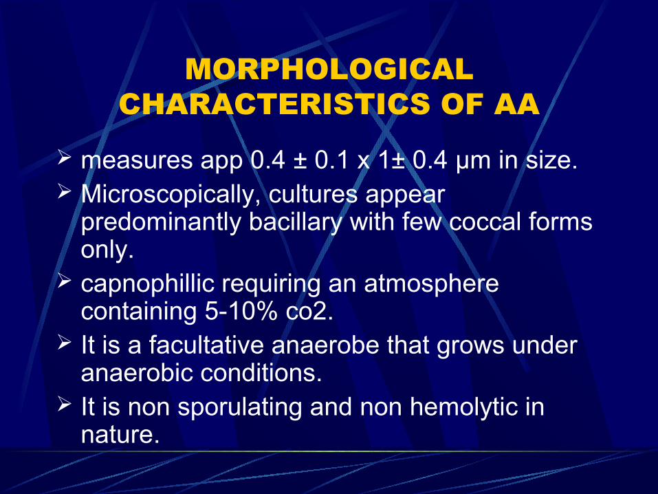
MORPHOLOGICAL CHARACTERISTICS OF AA
measures app 0.4 ± 0.1 x 1± 0.4 μm in size. Microscopically, cultures appear
predominantly bacillary with few coccal forms only.
capnophillic requiring an atmosphere containing 5-10% co2.
It is a facultative anaerobe that grows under anaerobic conditions.
It is non sporulating and non hemolytic in nature.
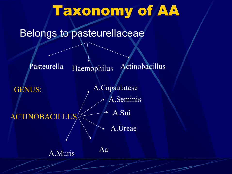
Taxonomy of AABelongs to pasteurellaceae
Pasteurella Haemophilus Actinobacillus
GENUS:
ACTINOBACILLUS
A.Capsulatese
A.Seminis
A.Sui
A.Ureae
AaA.Muris
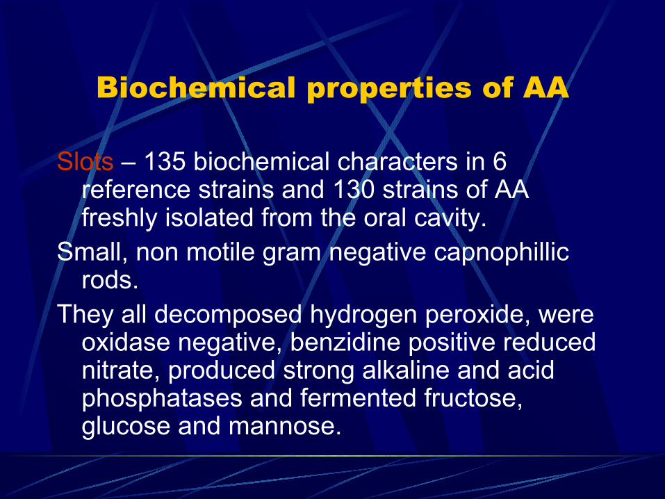
Biochemical properties of AA
Slots – 135 biochemical characters in 6 reference strains and 130 strains of AA freshly isolated from the oral cavity.
Small, non motile gram negative capnophillic rods.
They all decomposed hydrogen peroxide, were oxidase negative, benzidine positive reduced nitrate, produced strong alkaline and acid phosphatases and fermented fructose, glucose and mannose.
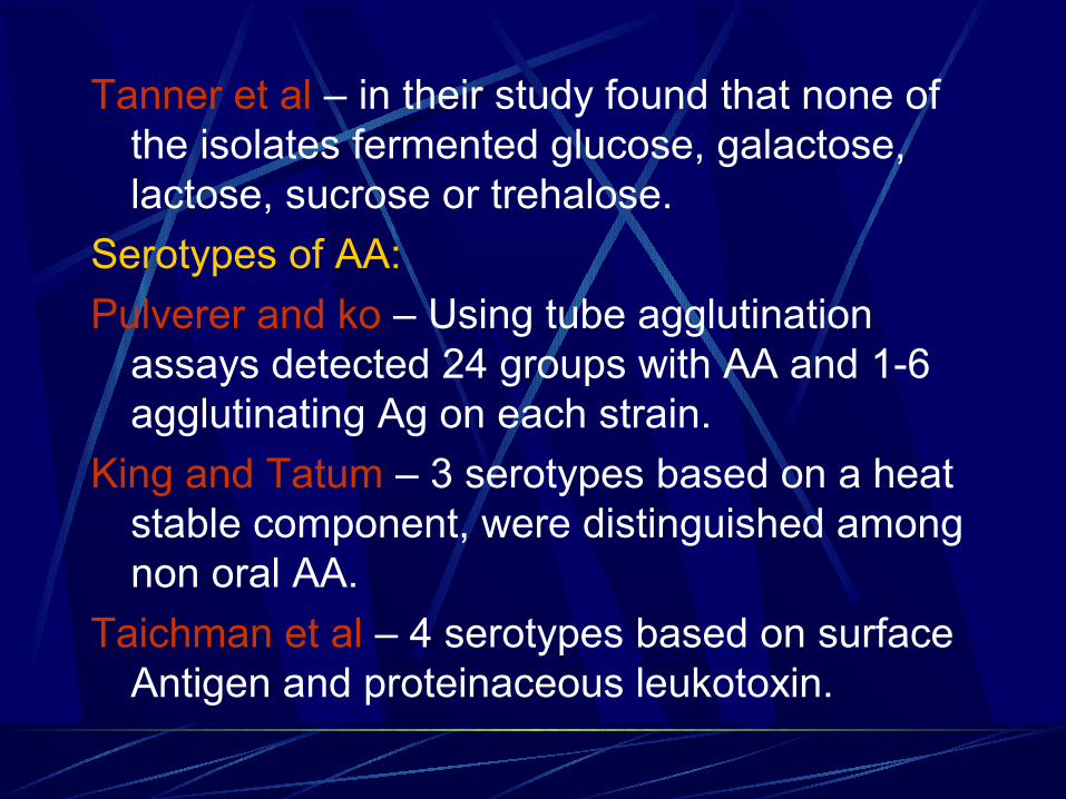
Tanner et al – in their study found that none of the isolates fermented glucose, galactose, lactose, sucrose or trehalose.
Serotypes of AA:
Pulverer and ko – Using tube agglutination assays detected 24 groups with AA and 1-6 agglutinating Ag on each strain.
King and Tatum – 3 serotypes based on a heat stable component, were distinguished among non oral AA.
Taichman et al – 4 serotypes based on surface Antigen and proteinaceous leukotoxin.
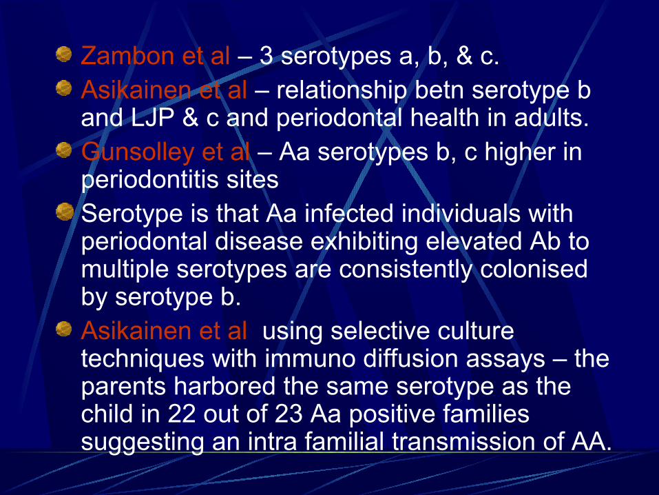
Zambon et al – 3 serotypes a, b, & c.Asikainen et al – relationship betn serotype b and LJP & c and periodontal health in adults.Gunsolley et al – Aa serotypes b, c higher in periodontitis sitesSerotype is that Aa infected individuals with periodontal disease exhibiting elevated Ab to multiple serotypes are consistently colonised by serotype b.Asikainen et al using selective culture techniques with immuno diffusion assays – the parents harbored the same serotype as the child in 22 out of 23 Aa positive families suggesting an intra familial transmission of AA.
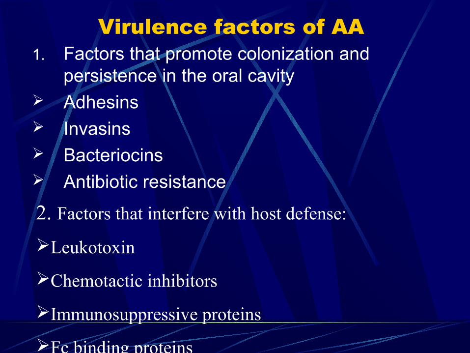
Virulence factors of AA1. Factors that promote colonization and
persistence in the oral cavity Adhesins Invasins Bacteriocins Antibiotic resistance
2. Factors that interfere with host defense:
Leukotoxin
Chemotactic inhibitors
Immunosuppressive proteins
Fc binding proteins
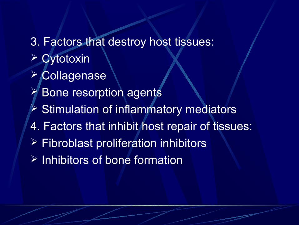
3. Factors that destroy host tissues: Cytotoxin Collagenase Bone resorption agents Stimulation of inflammatory mediators
4. Factors that inhibit host repair of tissues: Fibroblast proliferation inhibitors Inhibitors of bone formation
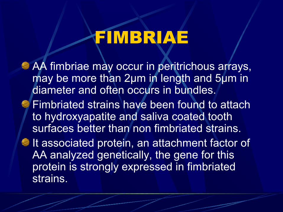
FIMBRIAEAA fimbriae may occur in peritrichous arrays, may be more than 2µm in length and 5µm in diameter and often occurs in bundles.Fimbriated strains have been found to attach to hydroxyapatite and saliva coated tooth surfaces better than non fimbriated strains.It associated protein, an attachment factor of AA analyzed genetically, the gene for this protein is strongly expressed in fimbriated strains.

vesicles
These structures which are LPS in nature, originate from and continuous with the outer membrane.
The surface of highly leukotoxic AA strians an abundance of extracellular membranous vesicles.
AA vesicles also exhibit adhesive properties.
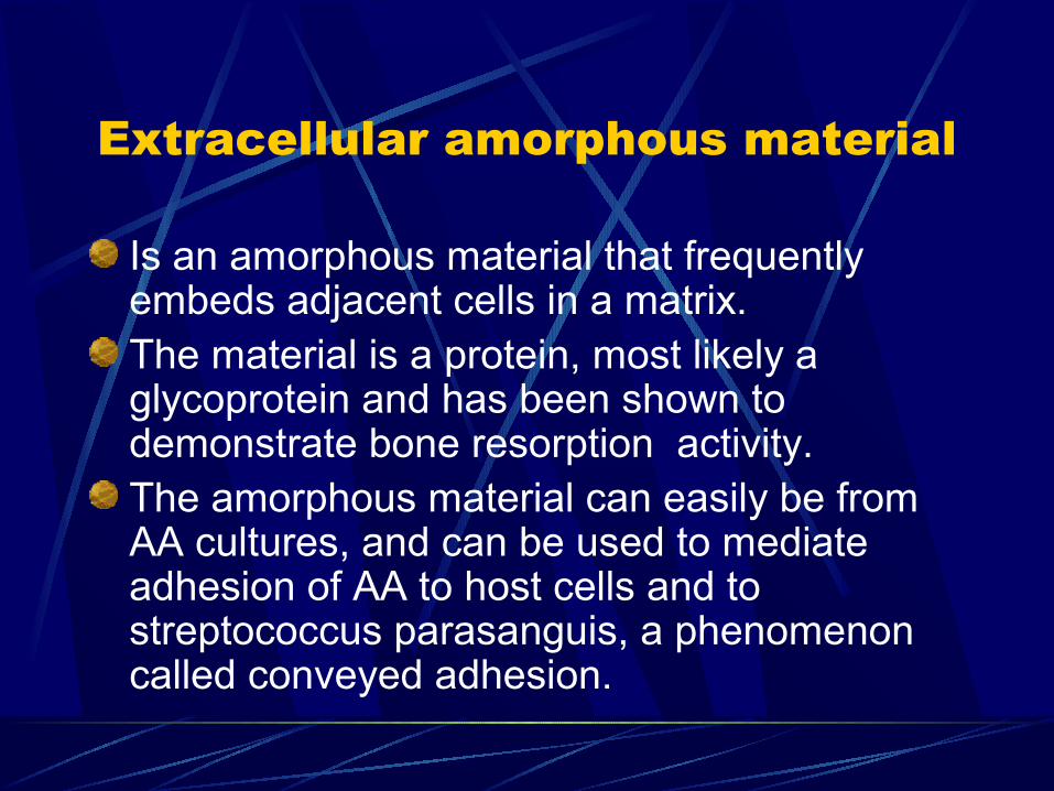
Extracellular amorphous material
Is an amorphous material that frequently embeds adjacent cells in a matrix. The material is a protein, most likely a glycoprotein and has been shown to demonstrate bone resorption activity. The amorphous material can easily be from AA cultures, and can be used to mediate adhesion of AA to host cells and to streptococcus parasanguis, a phenomenon called conveyed adhesion.
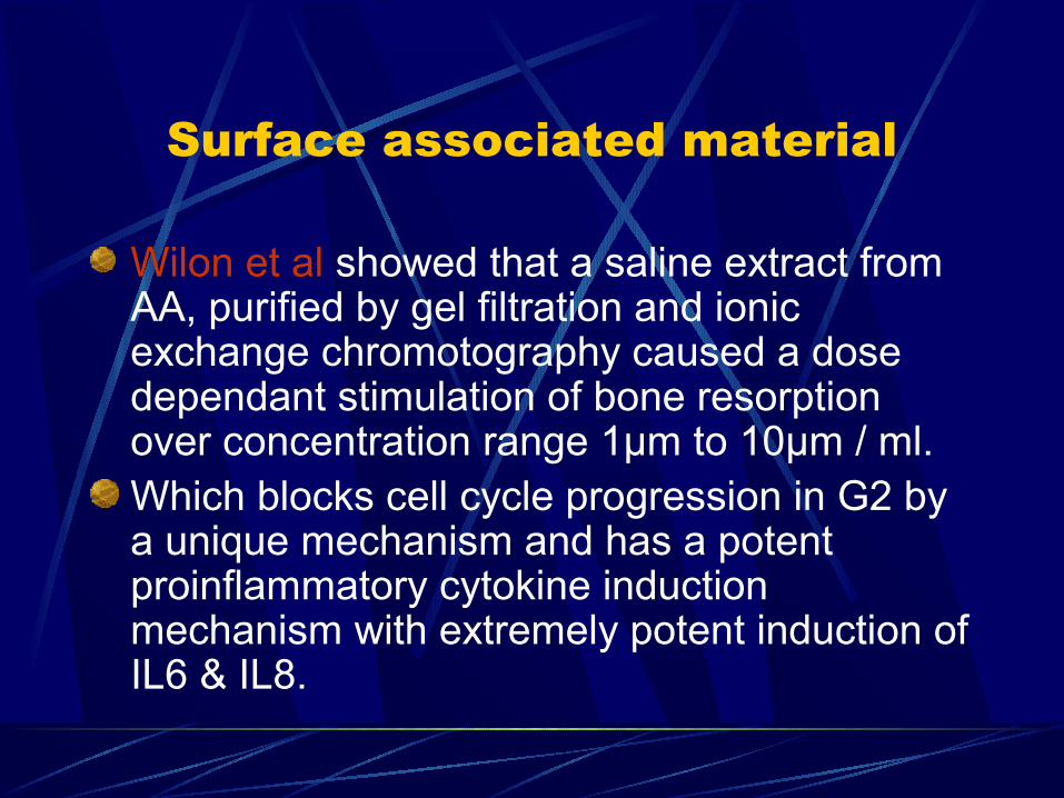
Surface associated material
Wilon et al showed that a saline extract from AA, purified by gel filtration and ionic exchange chromotography caused a dose dependant stimulation of bone resorption over concentration range 1µm to 10µm / ml.Which blocks cell cycle progression in G2 by a unique mechanism and has a potent proinflammatory cytokine induction mechanism with extremely potent induction of IL6 & IL8.
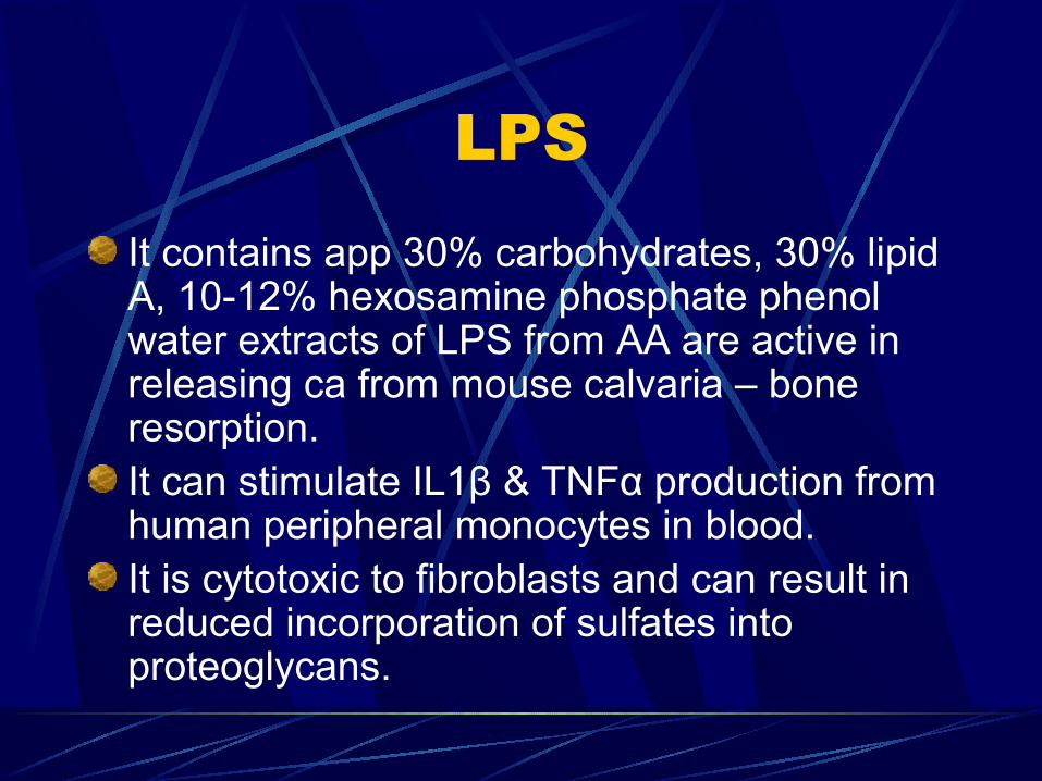
LPSIt contains app 30% carbohydrates, 30% lipid A, 10-12% hexosamine phosphate phenol water extracts of LPS from AA are active in releasing ca from mouse calvaria – bone resorption.It can stimulate IL1β & TNFα production from human peripheral monocytes in blood.It is cytotoxic to fibroblasts and can result in reduced incorporation of sulfates into proteoglycans.
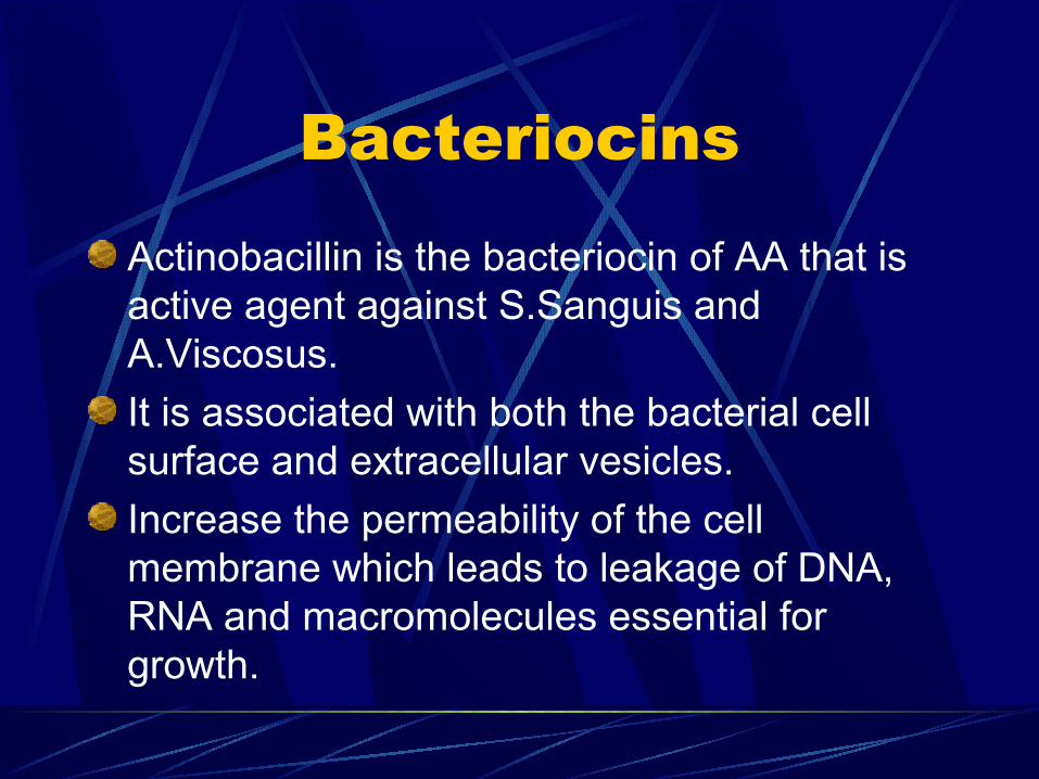
Bacteriocins
Actinobacillin is the bacteriocin of AA that is active agent against S.Sanguis and A.Viscosus.
It is associated with both the bacterial cell surface and extracellular vesicles.
Increase the permeability of the cell membrane which leads to leakage of DNA, RNA and macromolecules essential for growth.
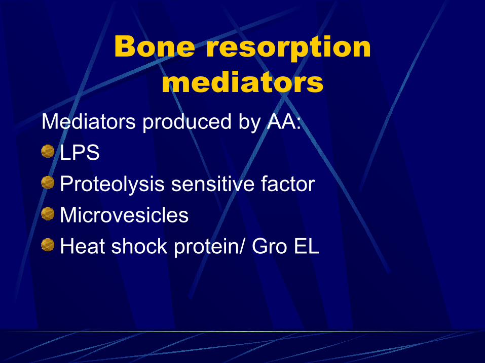
Bone resorption mediators
Mediators produced by AA:
LPS
Proteolysis sensitive factor
Microvesicles
Heat shock protein/ Gro EL
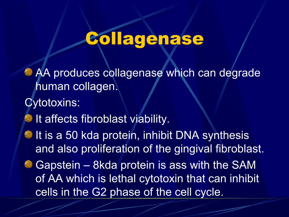
Collagenase
AA produces collagenase which can degrade human collagen.
Cytotoxins:
It affects fibroblast viability.
It is a 50 kda protein, inhibit DNA synthesis and also proliferation of the gingival fibroblast.
Gapstein – 8kda protein is ass with the SAM of AA which is lethal cytotoxin that can inhibit cells in the G2 phase of the cell cycle.
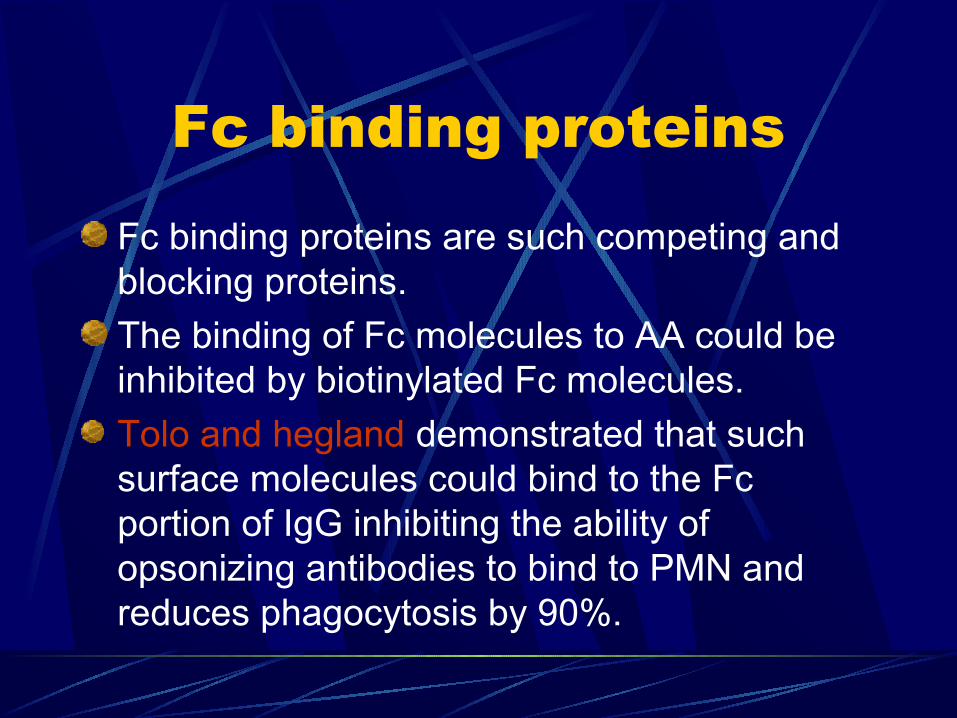
Fc binding proteins
Fc binding proteins are such competing and blocking proteins.
The binding of Fc molecules to AA could be inhibited by biotinylated Fc molecules.
Tolo and hegland demonstrated that such surface molecules could bind to the Fc portion of IgG inhibiting the ability of opsonizing antibodies to bind to PMN and reduces phagocytosis by 90%.
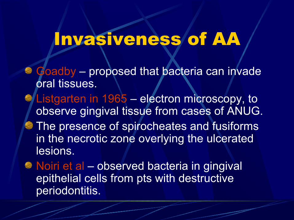
Invasiveness of AAGoadby – proposed that bacteria can invade oral tissues.Listgarten in 1965 – electron microscopy, to observe gingival tissue from cases of ANUG. The presence of spirocheates and fusiforms in the necrotic zone overlying the ulcerated lesions. Noiri et al – observed bacteria in gingival epithelial cells from pts with destructive periodontitis.
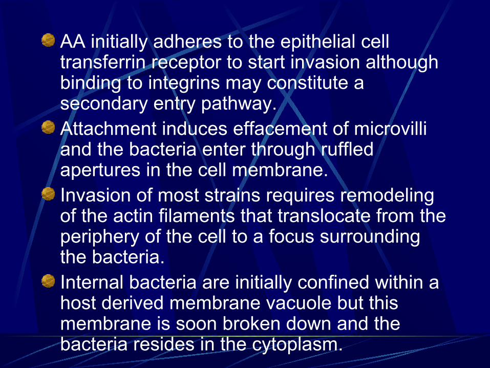
AA initially adheres to the epithelial cell transferrin receptor to start invasion although binding to integrins may constitute a secondary entry pathway.Attachment induces effacement of microvilli and the bacteria enter through ruffled apertures in the cell membrane.Invasion of most strains requires remodeling of the actin filaments that translocate from the periphery of the cell to a focus surrounding the bacteria.Internal bacteria are initially confined within a host derived membrane vacuole but this membrane is soon broken down and the bacteria resides in the cytoplasm.
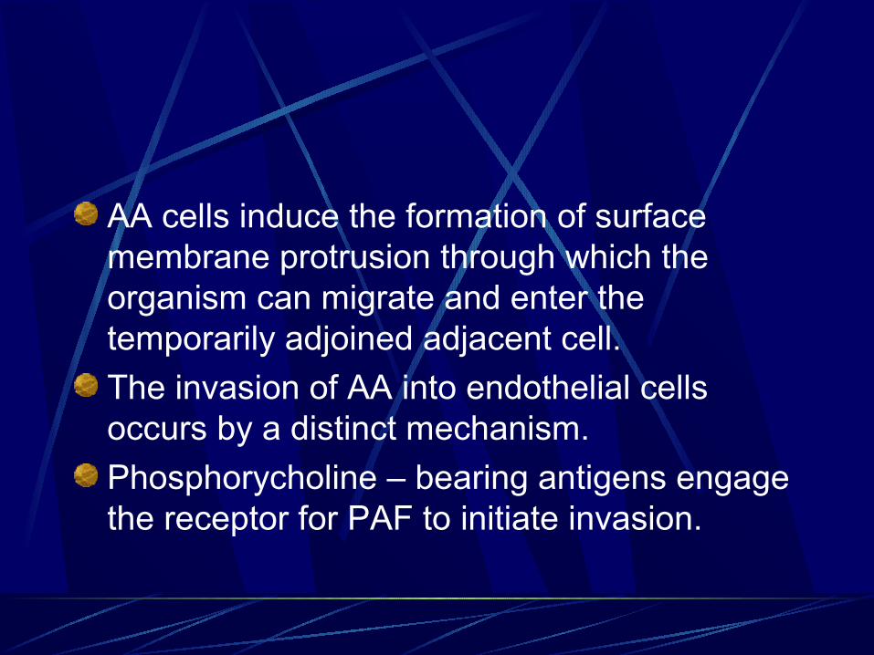
AA cells induce the formation of surface membrane protrusion through which the organism can migrate and enter the temporarily adjoined adjacent cell.
The invasion of AA into endothelial cells occurs by a distinct mechanism.
Phosphorycholine – bearing antigens engage the receptor for PAF to initiate invasion.
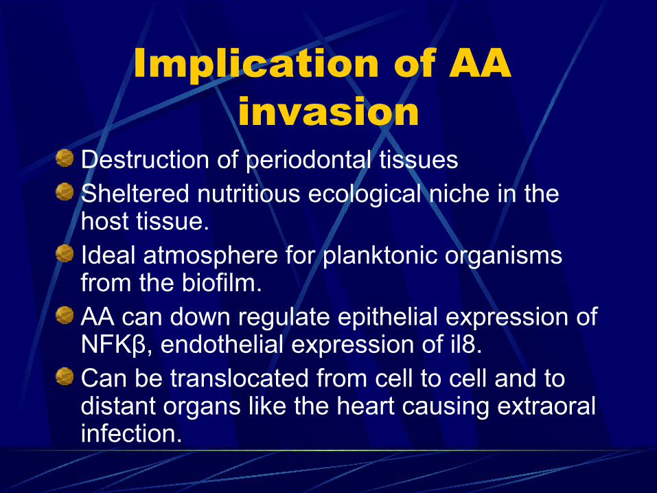
Implication of AA invasion
Destruction of periodontal tissuesSheltered nutritious ecological niche in the host tissue.Ideal atmosphere for planktonic organisms from the biofilm.AA can down regulate epithelial expression of NFKβ, endothelial expression of il8.Can be translocated from cell to cell and to distant organs like the heart causing extraoral infection.
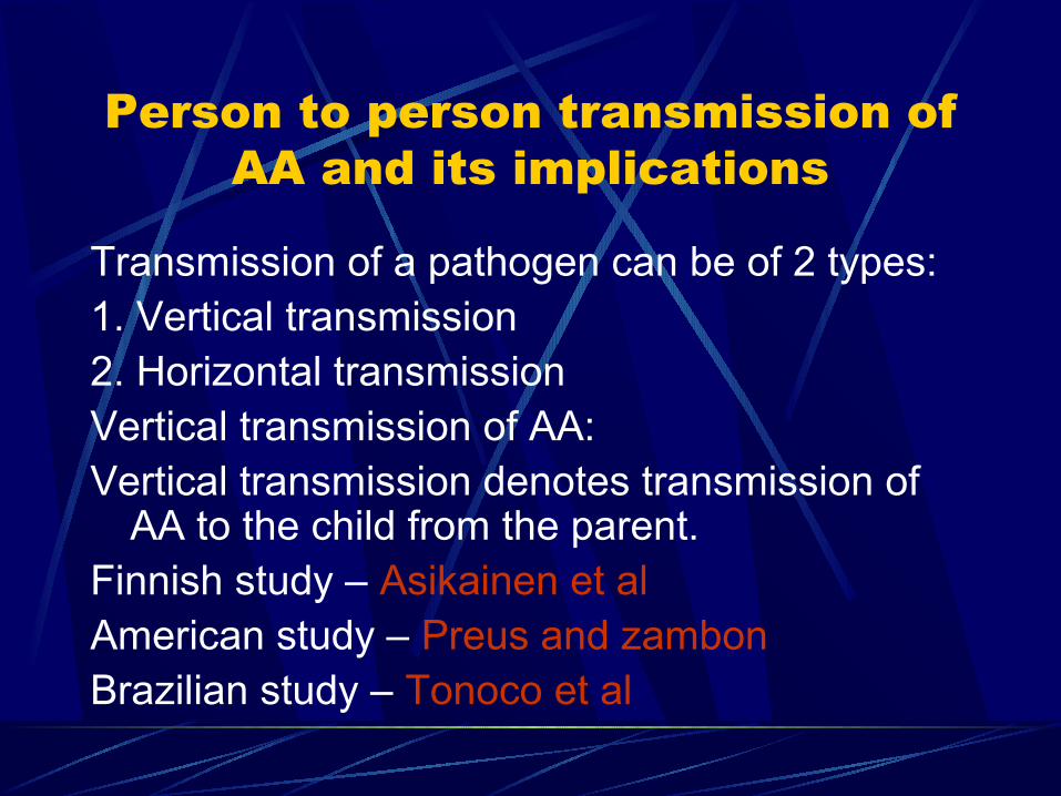
Person to person transmission of AA and its implications
Transmission of a pathogen can be of 2 types:1. Vertical transmission2. Horizontal transmissionVertical transmission of AA:Vertical transmission denotes transmission of
AA to the child from the parent.Finnish study – Asikainen et alAmerican study – Preus and zambonBrazilian study – Tonoco et al

Horizontal transmission
May occur betn siblings or between spouses.
Di rienzo et al – 89%
Tinoco et al – 7%
Asikainen et al – evidenced by transmission between married couples.
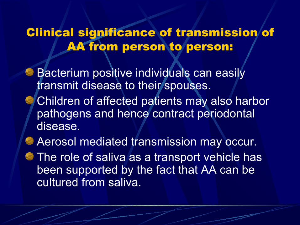
Clinical significance of transmission of AA from person to person:
Bacterium positive individuals can easily transmit disease to their spouses.Children of affected patients may also harbor pathogens and hence contract periodontal disease.Aerosol mediated transmission may occur.The role of saliva as a transport vehicle has been supported by the fact that AA can be cultured from saliva.
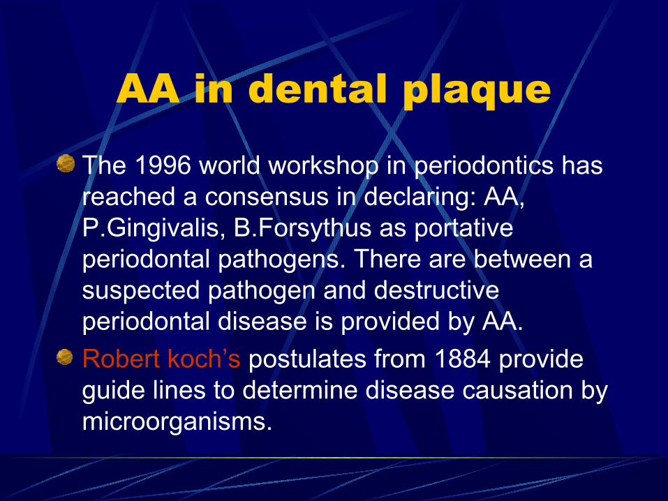
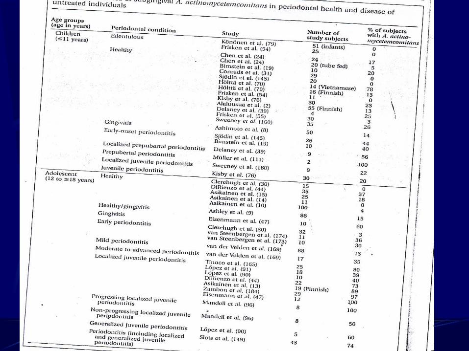
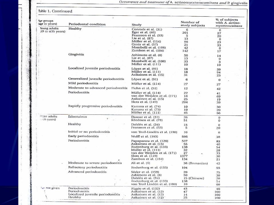
AA in dental plaque
The 1996 world workshop in periodontics has reached a consensus in declaring: AA, P.Gingivalis, B.Forsythus as portative periodontal pathogens. There are between a suspected pathogen and destructive periodontal disease is provided by AA.
Robert koch’s postulates from 1884 provide guide lines to determine disease causation by microorganisms.
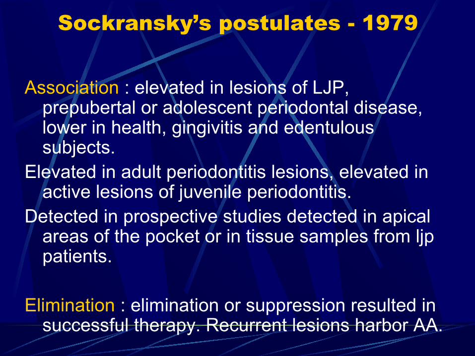
Sockransky’s postulates - 1979
Association : elevated in lesions of LJP, prepubertal or adolescent periodontal disease, lower in health, gingivitis and edentulous subjects.
Elevated in adult periodontitis lesions, elevated in active lesions of juvenile periodontitis.
Detected in prospective studies detected in apical areas of the pocket or in tissue samples from ljp patients.
Elimination : elimination or suppression resulted in successful therapy. Recurrent lesions harbor AA.
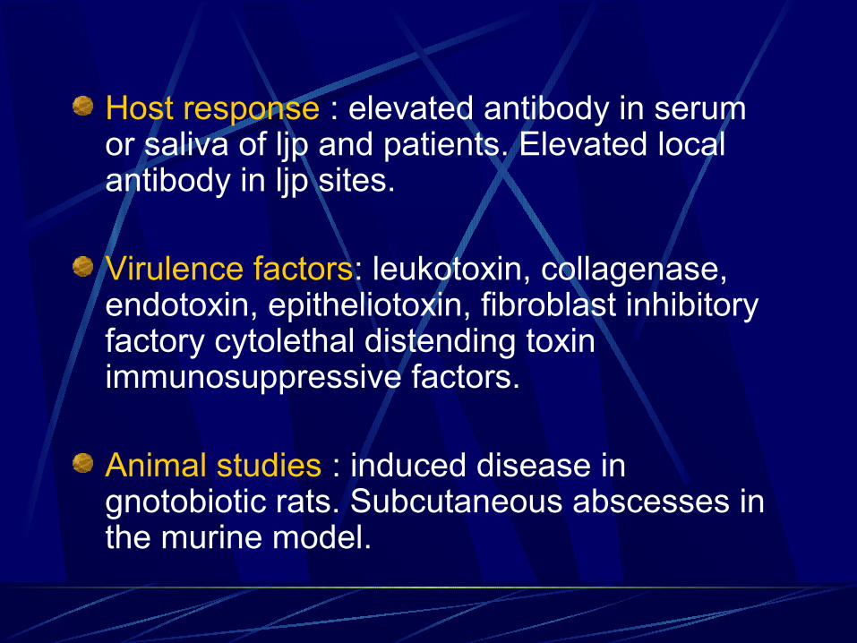
Host response : elevated antibody in serum or saliva of ljp and patients. Elevated local antibody in ljp sites.
Virulence factors: leukotoxin, collagenase, endotoxin, epitheliotoxin, fibroblast inhibitory factory cytolethal distending toxin immunosuppressive factors.
Animal studies : induced disease in gnotobiotic rats. Subcutaneous abscesses in the murine model.
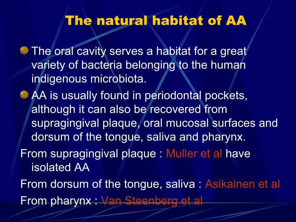
The natural habitat of AA
The oral cavity serves a habitat for a great variety of bacteria belonging to the human indigenous microbiota.
AA is usually found in periodontal pockets, although it can also be recovered from supragingival plaque, oral mucosal surfaces and dorsum of the tongue, saliva and pharynx.
From supragingival plaque : Muller et al have isolated AA
From dorsum of the tongue, saliva : Asikainen et al
From pharynx : Van Steenberg et al
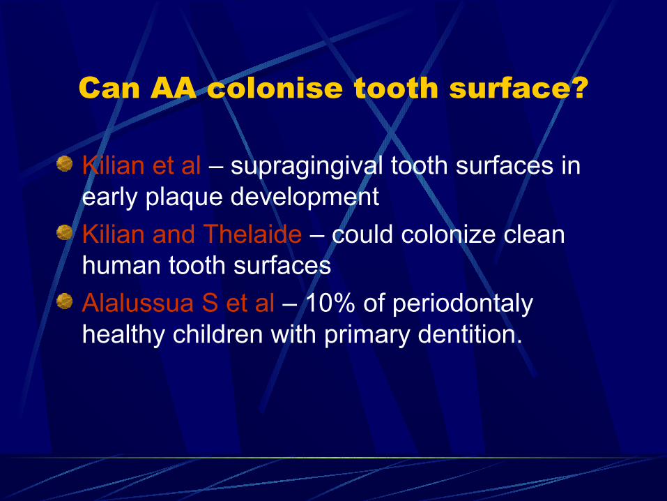
Can AA colonise tooth surface?
Kilian et al – supragingival tooth surfaces in early plaque development
Kilian and Thelaide – could colonize clean human tooth surfaces
Alalussua S et al – 10% of periodontaly healthy children with primary dentition.
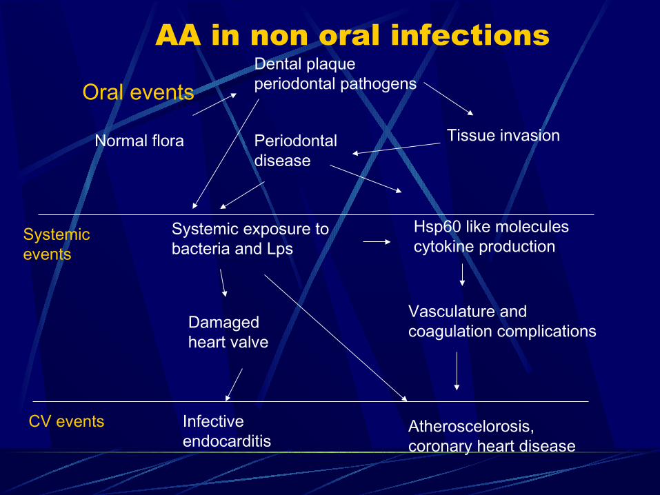
AA in non oral infections
Oral eventsDental plaque periodontal pathogens
Normal flora Tissue invasionPeriodontal disease
Systemic exposure to bacteria and Lps
Hsp60 like molecules cytokine production
Systemic events
Damaged heart valve
Vasculature and coagulation complications
Infective endocarditis
Atheroscelorosis, coronary heart disease
CV events
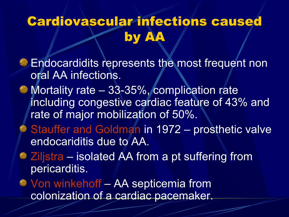
Cardiovascular infections caused by AA
Endocardidits represents the most frequent non oral AA infections.Mortality rate – 33-35%, complication rate including congestive cardiac feature of 43% and rate of major mobilization of 50%.Stauffer and Goldman in 1972 – prosthetic valve endocariditis due to AA.Ziljstra – isolated AA from a pt suffering from pericarditis.Von winkehoff – AA septicemia from colonization of a cardiac pacemaker.
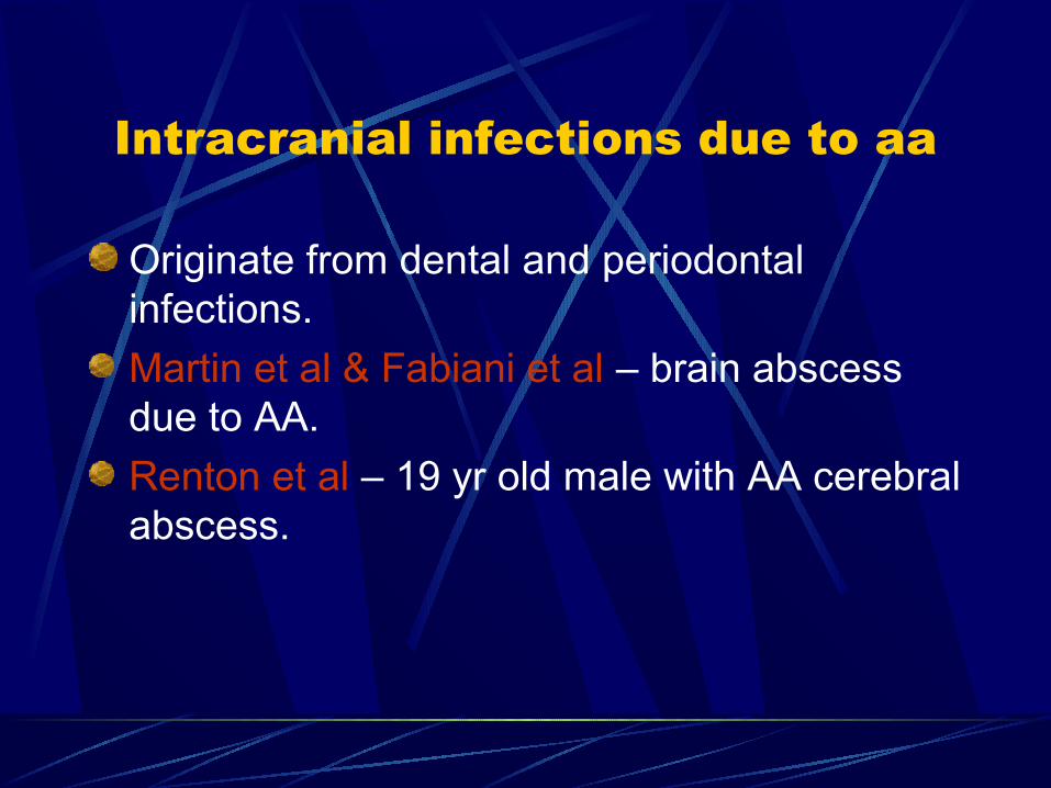
Intracranial infections due to aa
Originate from dental and periodontal infections.
Martin et al & Fabiani et al – brain abscess due to AA.
Renton et al – 19 yr old male with AA cerebral abscess.
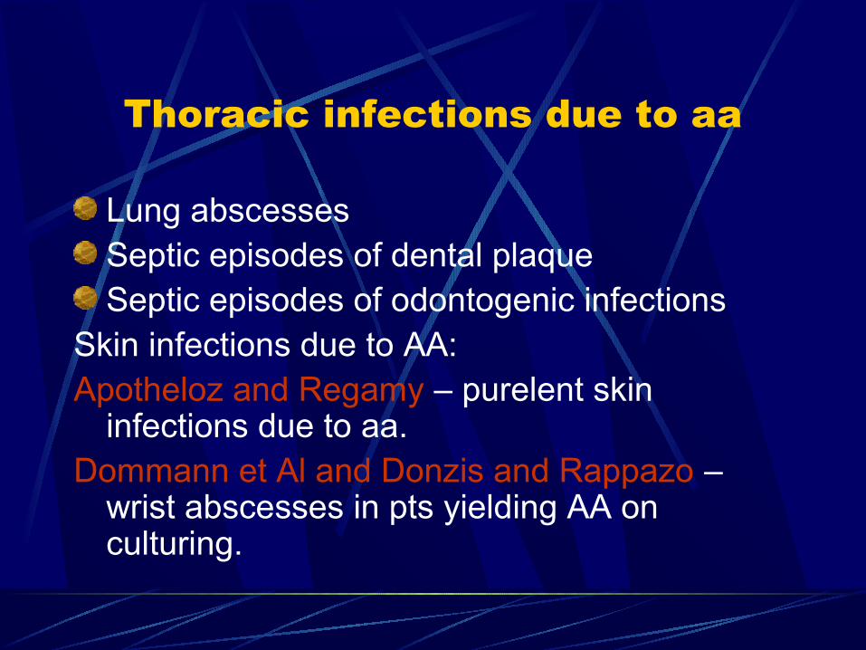
Thoracic infections due to aa
Lung abscessesSeptic episodes of dental plaqueSeptic episodes of odontogenic infections
Skin infections due to AA:Apotheloz and Regamy – purelent skin
infections due to aa.Dommann et Al and Donzis and Rappazo –
wrist abscesses in pts yielding AA on culturing.
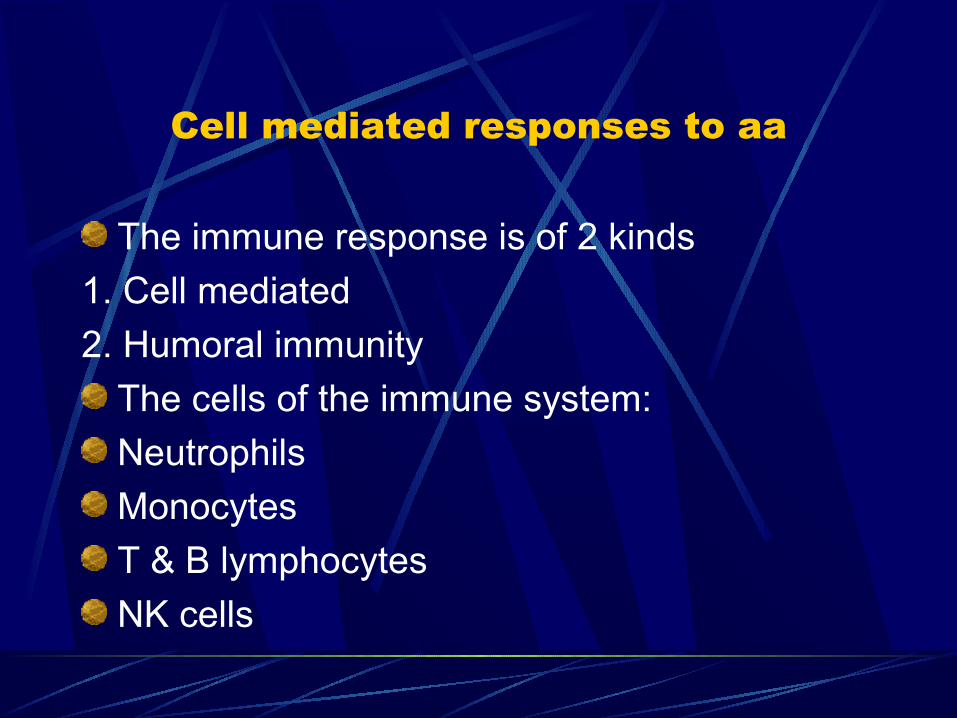
Cell mediated responses to aa
The immune response is of 2 kinds
1. Cell mediated
2. Humoral immunity
The cells of the immune system:
Neutrophils
Monocytes
T & B lymphocytes
NK cells
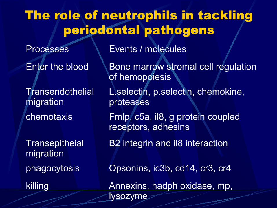
The role of neutrophils in tackling periodontal pathogens
Processes Events / molecules
Enter the blood Bone marrow stromal cell regulation of hemopoiesis
Transendothelial migration
L.selectin, p.selectin, chemokine, proteases
chemotaxis Fmlp, c5a, il8, g protein coupled receptors, adhesins
Transepitheial migration
Β2 integrin and il8 interaction
phagocytosis Opsonins, ic3b, cd14, cr3, cr4
killing Annexins, nadph oxidase, mp, lysozyme
AA evade pmn leukocyte defenses
Impairment of chemotaxis:Increase epithelial and endothelial expression
of IL8 genes and hence jeopardizes the chemotactic gradients.
The RTX leukotoxin:Which binds to LFA-1 on the neutrophil by
interaction with 1 domain of the cd11a subunit.
Lfa1 is expressed in both myeloid an lympoid cells and binding to leukotoxin causes death of the cells.
Immuno suppression:AA produces immuno suppressive factors that
impede the functioning of the immune system.
Humoral responses to AA:Individuals colonized by AA generally respond
with humoral response by production of Ig. There is a rise in local as well as systemic
levels of Ig in response to bacterial antigens such as fimbriae and adhesins.
Ebersole et al – association betn increased levels of and increased frequency of occurrence of antibody to AA in LJP.
Later study – showed increased levels of IgG to AA serotype b in 90% of LJP and 25% of adult periodontitis.
North American population – association between antibody to AA and LJP.
A study from brazil – elevated serotype b antibodies in down’s syndrome patients suffering from AP.
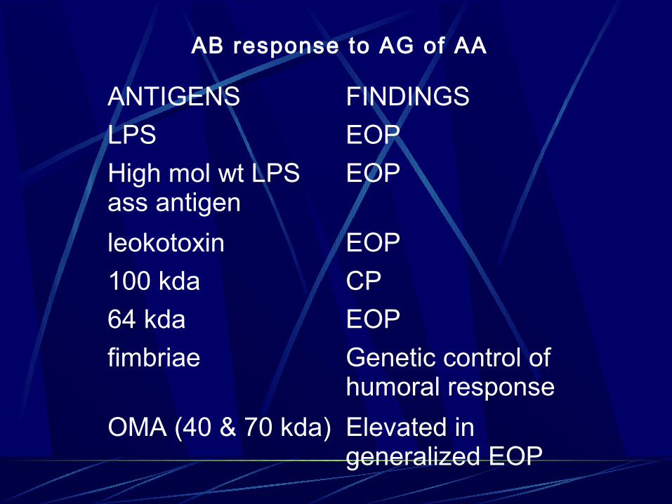
ANTIGENS FINDINGS
LPS EOP
High mol wt LPS ass antigen
EOP
leokotoxin EOP
100 kda CP
64 kda EOP
fimbriae Genetic control of humoral response
OMA (40 & 70 kda) Elevated in generalized EOP
AB response to AG of AA
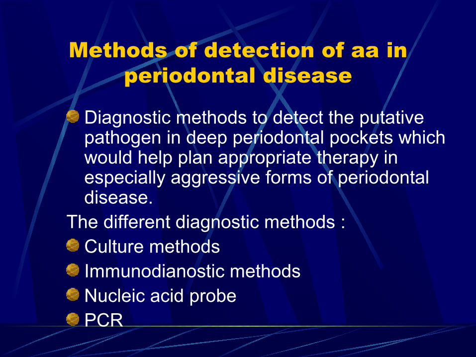
Methods of detection of aa in periodontal disease
Diagnostic methods to detect the putative pathogen in deep periodontal pockets which would help plan appropriate therapy in especially aggressive forms of periodontal disease.
The different diagnostic methods :Culture methodsImmunodianostic methodsNucleic acid probePCR
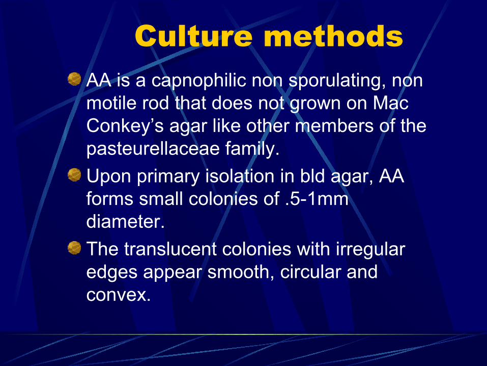
Culture methodsAA is a capnophilic non sporulating, non motile rod that does not grown on Mac Conkey’s agar like other members of the pasteurellaceae family.
Upon primary isolation in bld agar, AA forms small colonies of .5-1mm diameter.
The translucent colonies with irregular edges appear smooth, circular and convex.
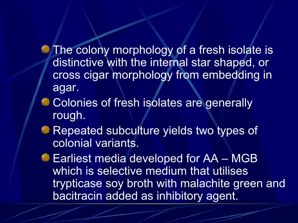
The colony morphology of a fresh isolate is distinctive with the internal star shaped, or cross cigar morphology from embedding in agar.Colonies of fresh isolates are generally rough.Repeated subculture yields two types of colonial variants.Earliest media developed for AA – MGB which is selective medium that utilises trypticase soy broth with malachite green and bacitracin added as inhibitory agent.
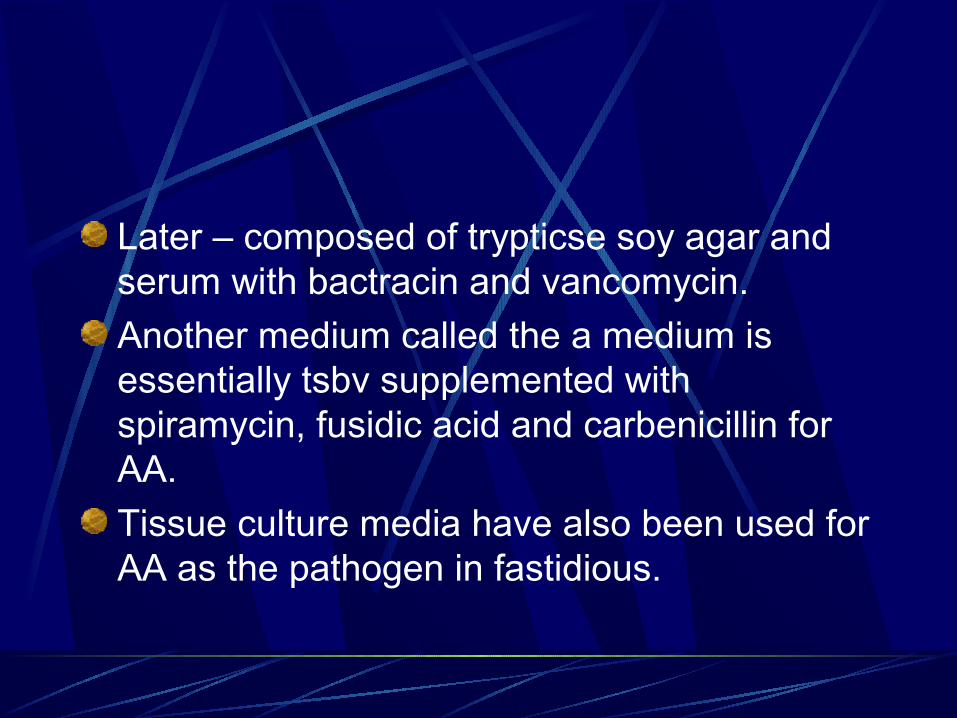
Later – composed of trypticse soy agar and serum with bactracin and vancomycin.
Another medium called the a medium is essentially tsbv supplemented with spiramycin, fusidic acid and carbenicillin for AA.
Tissue culture media have also been used for AA as the pathogen in fastidious.
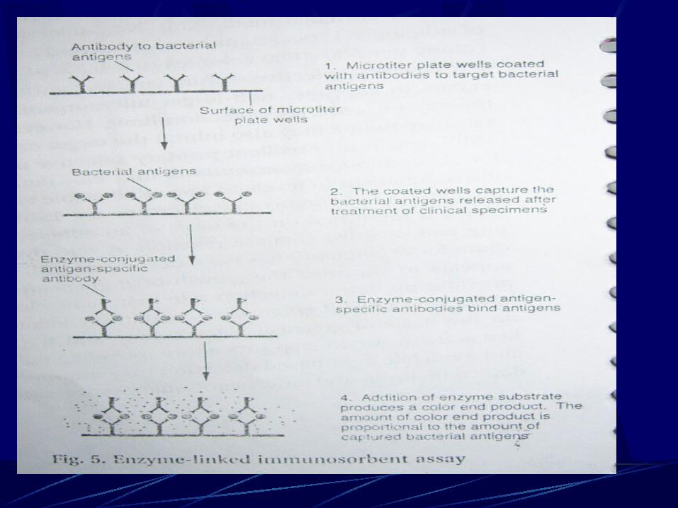
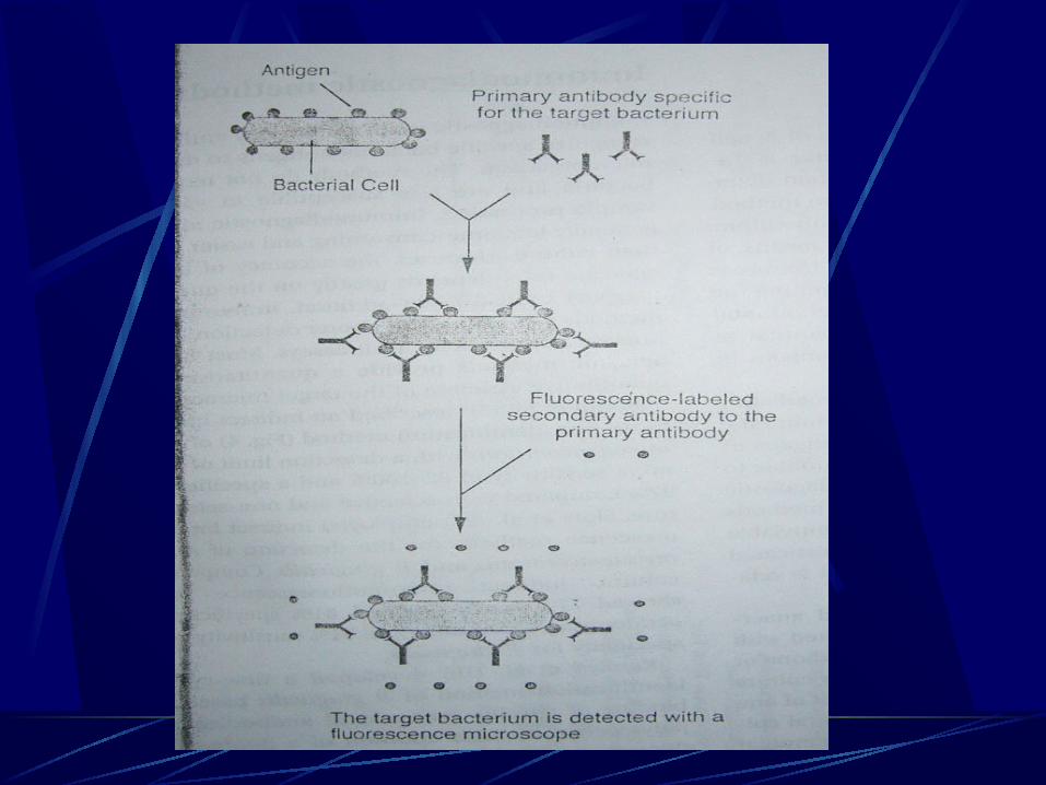
Immunodiagnostic methods to detect aa
IDM employ Ab that recognize specific bacterial Ag to detect that target microorganisms.S.Bonta et al – detection of AA with a detection limit of 500 cells/ml with a sensitivity of 82-100% and specificity of 92%.Slots et al – evalusite test is a commercially developed Ab based ELISA for the detection of AA. The sample wells are coated first with Ab against Ag specific for the bacterial specific targeted.Ag-Ab reactions are then detected by enzyme linked Ag specific antibodies to the sample wells.Detection limits are 105 AA cells/sample.
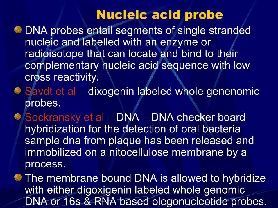
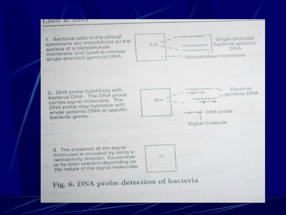
Nucleic acid probeDNA probes entail segments of single stranded nucleic and labelled with an enzyme or radioisotope that can locate and bind to their complementary nucleic acid sequence with low cross reactivity.Savdt et al – dixogenin labeled whole genenomic probes.Sockransky et al – DNA – DNA checker board hybridization for the detection of oral bacteria sample dna from plaque has been released and immobilized on a nitocellulose membrane by a process.The membrane bound DNA is allowed to hybridize with either digoxigenin labeled whole genomic DNA or 16s & RNA based olegonucleotide probes.
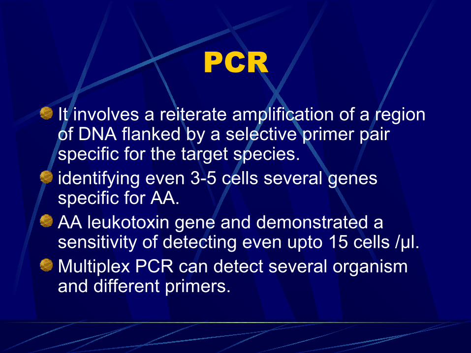
PCRIt involves a reiterate amplification of a region of DNA flanked by a selective primer pair specific for the target species.identifying even 3-5 cells several genes specific for AA.AA leukotoxin gene and demonstrated a sensitivity of detecting even upto 15 cells /μl.Multiplex PCR can detect several organism and different primers.
Treatment modalities to eliminate AA
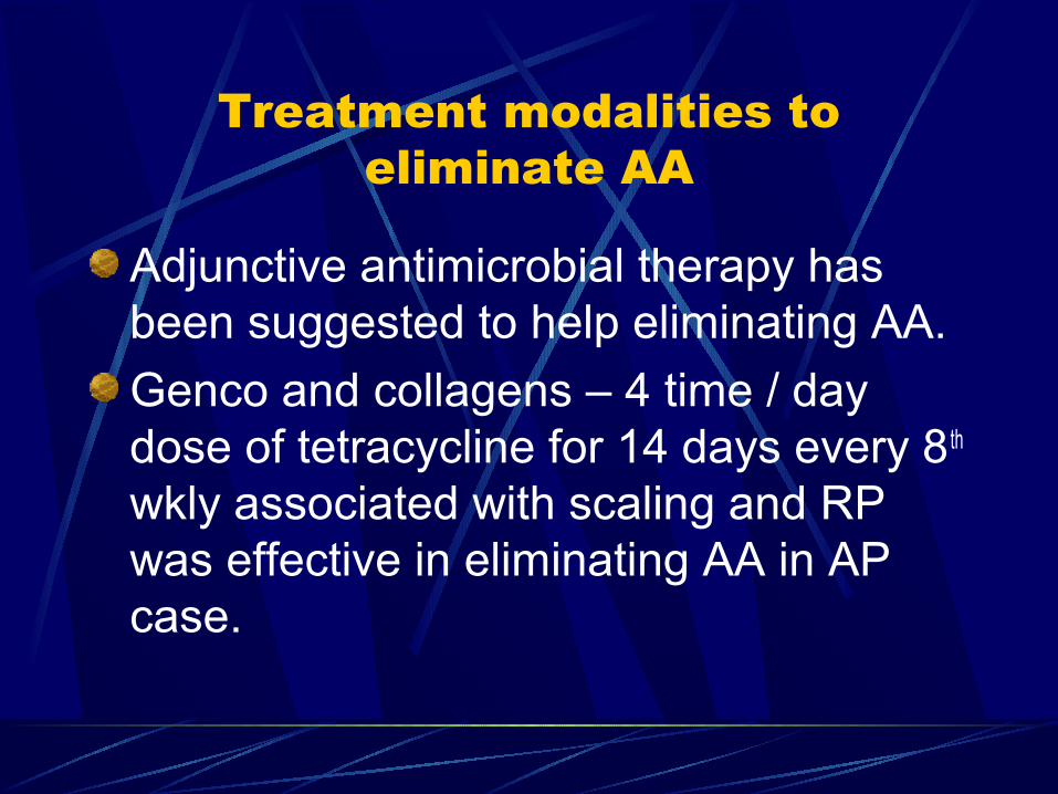
Adjunctive antimicrobial therapy has been suggested to help eliminating AA.
Genco and collagens – 4 time / day dose of tetracycline for 14 days every 8 th wkly associated with scaling and RP was effective in eliminating AA in AP case.
Principles of intervention in AA elimination
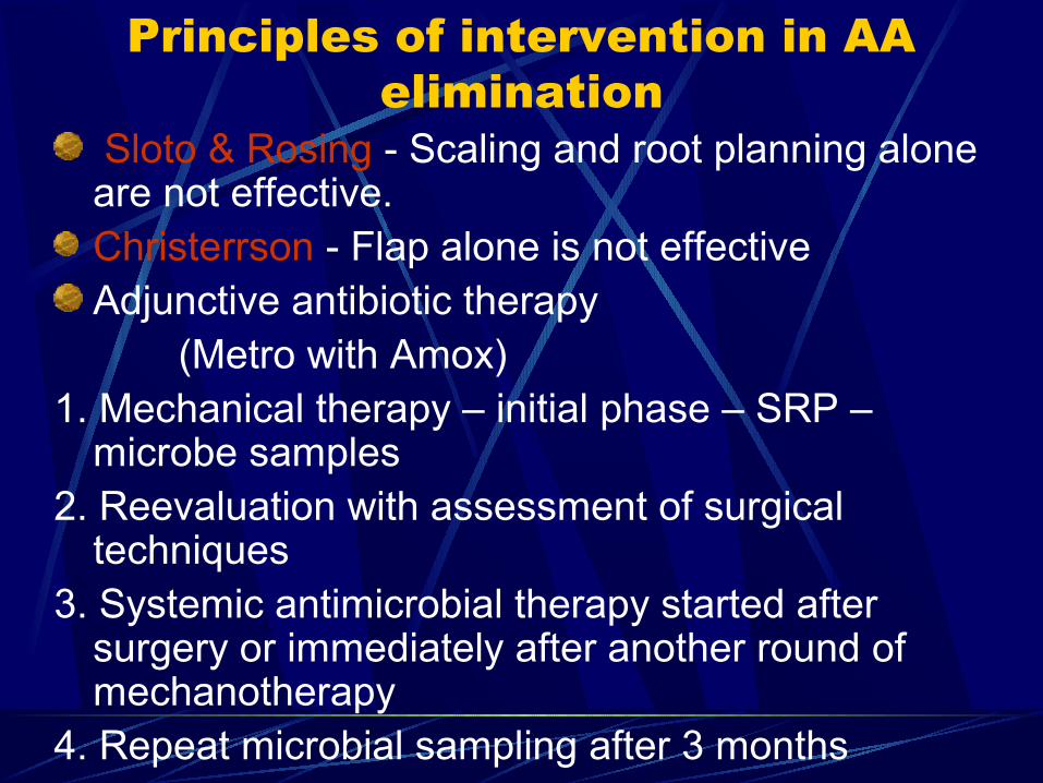
Sloto & Rosing - Scaling and root planning alone are not effective.Christerrson - Flap alone is not effectiveAdjunctive antibiotic therapy
(Metro with Amox)1. Mechanical therapy – initial phase – SRP –
microbe samples2. Reevaluation with assessment of surgical
techniques3. Systemic antimicrobial therapy started after
surgery or immediately after another round of mechanotherapy
4. Repeat microbial sampling after 3 months
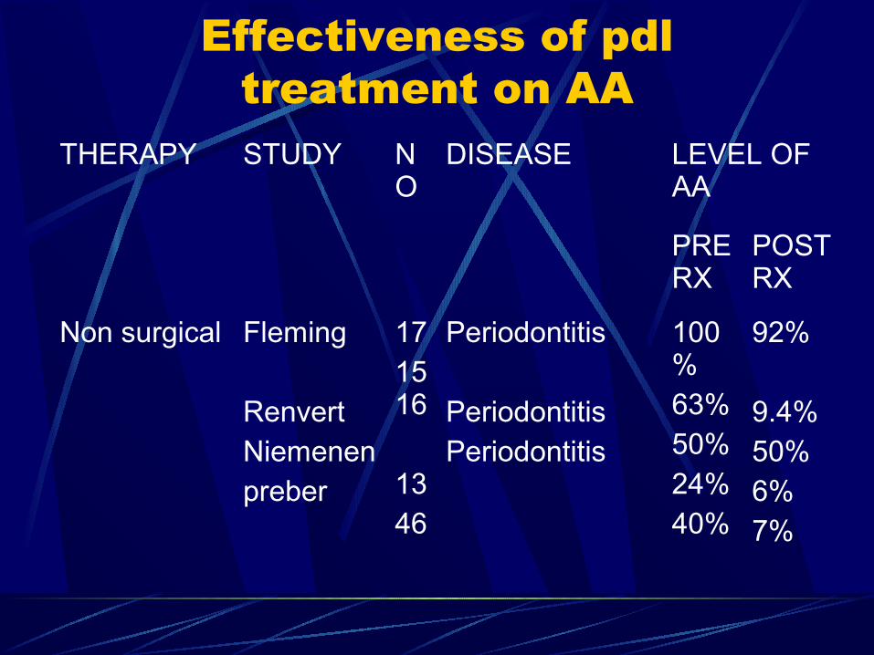
Effectiveness of pdl treatment on AA
THERAPY STUDY NO
DISEASE LEVEL OF AA
PRE RX
POST RX
Non surgical Fleming
RenvertNiemenenpreber
171516
1346
Periodontitis
PeriodontitisPeriodontitis
100%63%50%24%40%
92%
9.4%50%6%7%
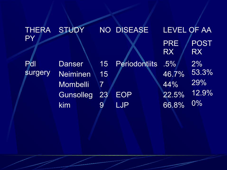
THERAPY
STUDY NO DISEASE LEVEL OF AA
PRE RX
POST RX
Pdl surgery
DanserNeiminenMombelliGunsollegkim
15157239
Periodontiits
EOPLJP
.5%46.7%44%22.5%66.8%
2% 53.3%29%12.9%0%
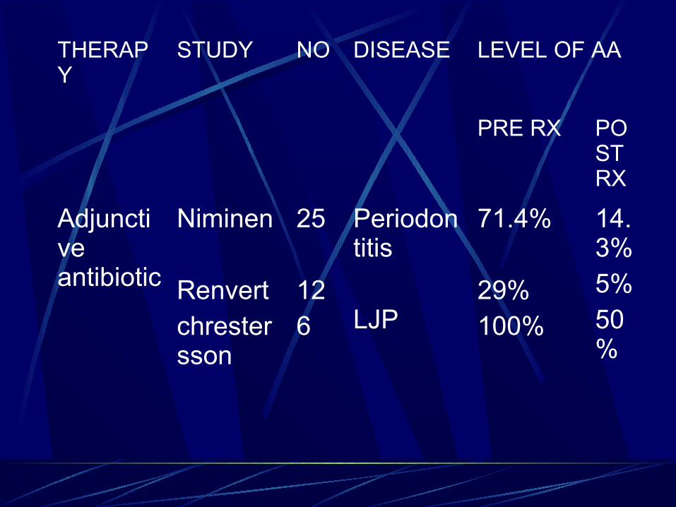
THERAPY
STUDY NO DISEASE LEVEL OF AA
PRE RX POST RX
Adjunctive antibiotic
Niminen
Renvertchrestersson
25
126
Periodontitis
LJP
71.4%
29%100%
14.3%5%50%