Actinobacillosis
16
Actinobacillosis Hafeez ullah khan 11-arid-983 Ayesha Azad 11-arid-985
-
Upload
ayesha-azad -
Category
Health & Medicine
-
view
15 -
download
2
Transcript of Actinobacillosis

Actinobacillosis
Hafeez ullah khan 11-arid-983Ayesha Azad
11-arid-985
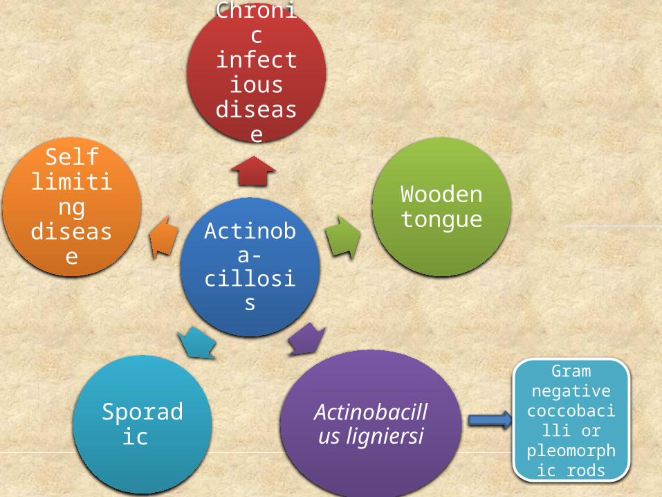
Actinoba-cillosis
Chronic infectious
disease
Wooden tongue
Actinobacillus ligniersiSporadic
Self limiting disease
Gram negative
coccobacilli or
pleomorphic rods

Epidemiology• Distribution: The disease in cattle is worldwide in
distribution and usually of sporadic occurrence on individual farms and reported in Egypt.
• Areas with copper deficiency• Animal susceptibility: Cattle, buffaloes (mature
and of dairy breed are more susceptible), sheep and goats.
• Predisposing factors : Oral mucosa injuries by fibrous feed materials or by foreign bodies and during oral manipulation by hand of owner or veterinarian

Mode of infectionSource of infection: Pus or infected discharges are the
main source of infection.
Mode of transmission: The disease is transmitted by ingestion of contaminated food and water with the presence of oral mucosa injury (wounds or abrasions).
A. lignieresii normal rumen inhabitant of sheep and cattle.
It survives 4 to 5 days in forage
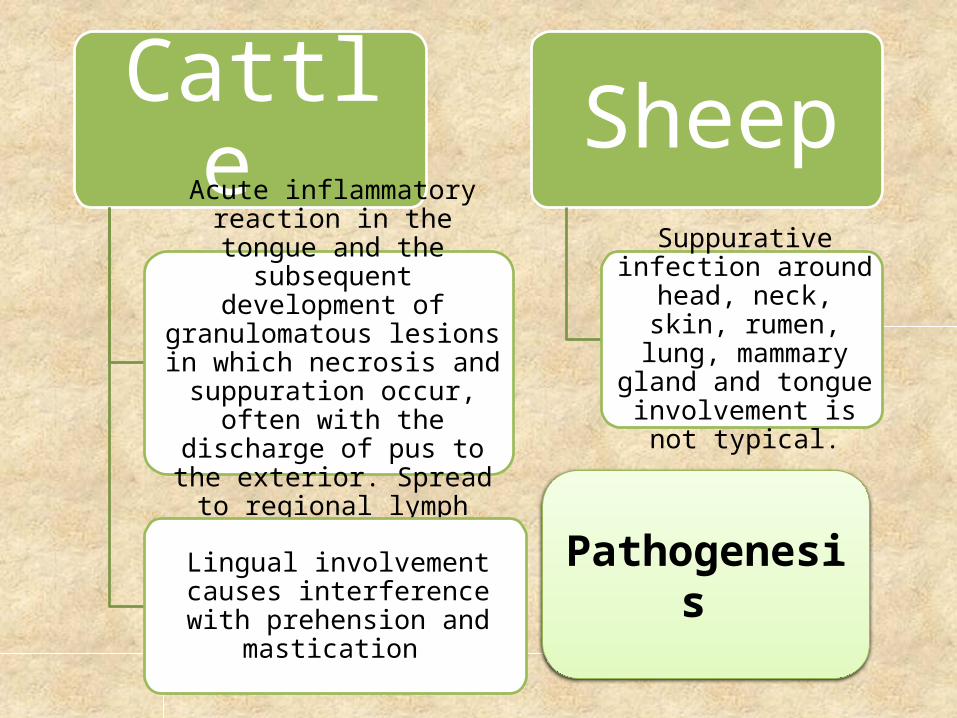
Cattle Acute inflammatory reaction in the tongue and the subsequent development of granulomatous lesions in which necrosis and suppuration occur, often with
the discharge of pus to the exterior. Spread to regional
lymph nodes is usual.
Lingual involvement causes interference with prehension and
mastication
Sheep Suppurative infection
around head, neck, skin, rumen, lung, mammary
gland and tongue involvement is not
typical.
Pathogenesis
Clinical signs
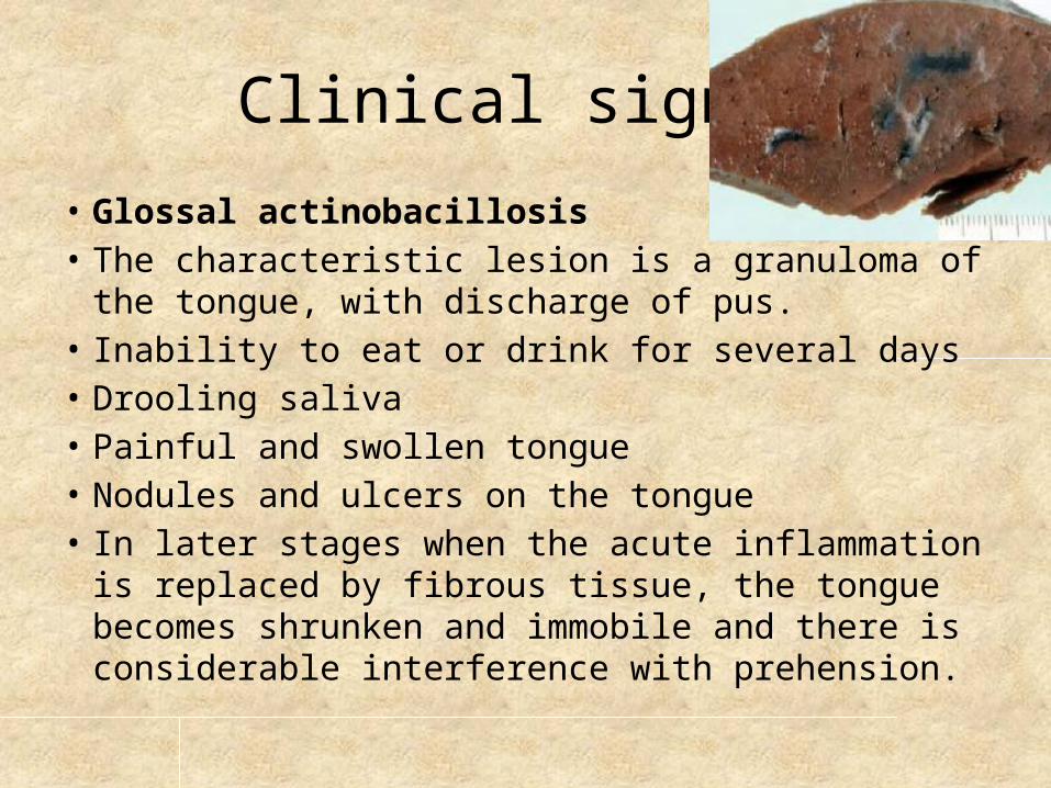
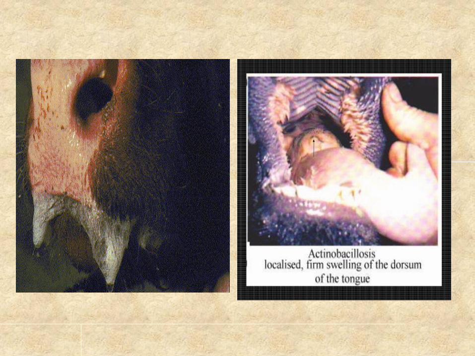
• Glossal actinobacillosis• The characteristic lesion is a granuloma of the tongue, with
discharge of pus.• Inability to eat or drink for several days• Drooling saliva• Painful and swollen tongue• Nodules and ulcers on the tongue• In later stages when the acute inflammation is replaced by
fibrous tissue, the tongue becomes shrunken and immobile and there is considerable interference with prehension.

Conti….
• Cutaneous actinobacillosis is also recorded with actinobacillosis granulomas occurring on atypical but visible areas such as the external nares, cheeks, skin or eyelid, and hind limbs.
• In sheep, Tongue is not usually involved. lesion up to 8 cm in diameter present on lower jaw, face, nose, in the skin folds from lower jaw to sternum, these lesions are superficial or deep, usually extended to cranial or cervical lymph nodes, it discharge viscid yellow green pus containing granules through number of openings.

Neck
Oral cavity
Leg
Udder
Chest
Tongue
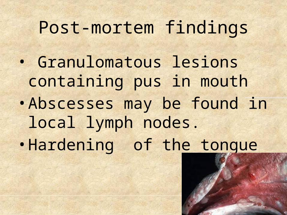
Post-mortem findings
• Granulomatous lesions containing pus in mouth• Abscesses may be found in local
lymph nodes.• Hardening of the tongue

Diagnosis • Field diagnosis: It depends on clinical signs of disease as
fever, tongue protrusion, salivation and history of feeding on hard food objects beside the epidemiology of the disease.
• Laboratory diagnosis:• Samples: Pus, smear or biopsy from the lesion, parts of
lesion on ice or formalin, blood and serum.• Laboratory procedures:
– Direct examination of stained smears after staining with Gram stain.
– Culture of the suspected material on blood agar.– Histopathological findings.– Serotests.

Differential diagnosis
• The disease may be confused with:Actinomycosis: It involves hard tissue and rarely
soft one.TB, especially with atypical form, differentiates on
basis of tuberculin test.Abscess of throat region, contain single cavity and
discharge thin pus and readily heal after drainage
Treatment
Flushing with iodine.Administration of potassium iodide orally (6 to
10 g a day for 10 days)Intravenous injection of sodium iodide at 10 %
(8 g for 100kg)Streptomycin (5 g/day for 3 days) treatment of
choice, Tetracylcines and tilmicosin also effective.

Control
• Restriction of the spread of disease is best implemented by quick treatment of affected animals and the prevention of contamination of pasture and feed troughs.
• Isolation or disposal of animals with discharging lesions is essential, although the disease does not spread readily unless predisposing environmental factors cause a high incidence of oral or skin lacerations.

QUESTIONS….?????

THANKS…….