Acral melanoma with hyperkeratosis mimicking a pigmented wart file38 Observation | Dermatol Pract...
3
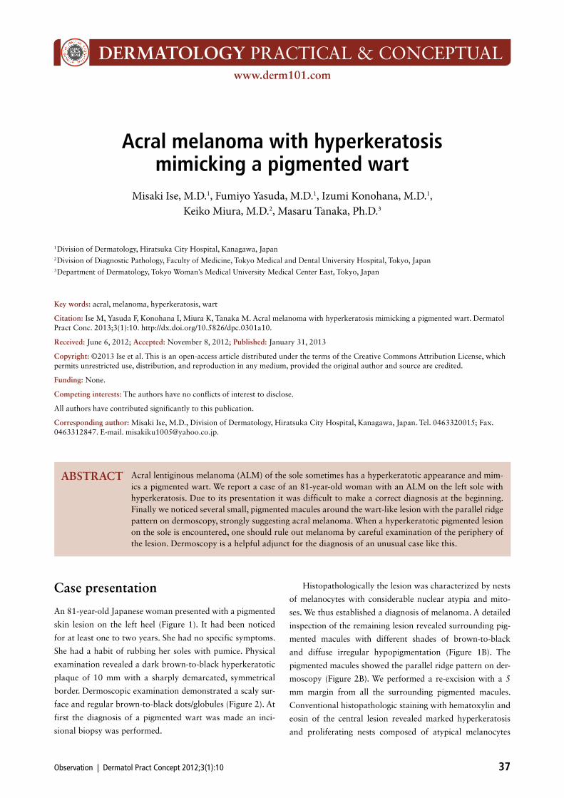
DERMATOLOGY PRACTICAL & CONCEPTUAL www.derm101.com Observation | Dermatol Pract Concept 2012;3(1):10 37 Case presentation An 81-year-old Japanese woman presented with a pigmented skin lesion on the left heel (Figure 1). It had been noticed for at least one to two years. She had no specific symptoms. She had a habit of rubbing her soles with pumice. Physical examination revealed a dark brown-to-black hyperkeratotic plaque of 10 mm with a sharply demarcated, symmetrical border. Dermoscopic examination demonstrated a scaly sur- face and regular brown-to-black dots/globules (Figure 2). At first the diagnosis of a pigmented wart was made an inci- sional biopsy was performed. Histopathologically the lesion was characterized by nests of melanocytes with considerable nuclear atypia and mito- ses. We thus established a diagnosis of melanoma. A detailed inspection of the remaining lesion revealed surrounding pig- mented macules with different shades of brown-to-black and diffuse irregular hypopigmentation (Figure 1B). The pigmented macules showed the parallel ridge pattern on der- moscopy (Figure 2B). We performed a re-excision with a 5 mm margin from all the surrounding pigmented macules. Conventional histopathologic staining with hematoxylin and eosin of the central lesion revealed marked hyperkeratosis and proliferating nests composed of atypical melanocytes Acral melanoma with hyperkeratosis mimicking a pigmented wart Misaki Ise, M.D. 1 , Fumiyo Yasuda, M.D. 1 , Izumi Konohana, M.D. 1 , Keiko Miura, M.D. 2 , Masaru Tanaka, Ph.D. 3 1 Division of Dermatology, Hiratsuka City Hospital, Kanagawa, Japan 2 Division of Diagnostic Pathology, Faculty of Medicine, Tokyo Medical and Dental University Hospital, Tokyo, Japan 3 Department of Dermatology, Tokyo Woman’s Medical University Medical Center East, Tokyo, Japan Key words: acral, melanoma, hyperkeratosis, wart Citation: Ise M, Yasuda F, Konohana I, Miura K, Tanaka M. Acral melanoma with hyperkeratosis mimicking a pigmented wart. Dermatol Pract Conc. 2013;3(1):10. http://dx.doi.org/10.5826/dpc.0301a10. Received: June 6, 2012; Accepted: November 8, 2012; Published: January 31, 2013 Copyright: ©2013 Ise et al. This is an open-access article distributed under the terms of the Creative Commons Attribution License, which permits unrestricted use, distribution, and reproduction in any medium, provided the original author and source are credited. Funding: None. Competing interests: The authors have no conflicts of interest to disclose. All authors have contributed significantly to this publication. Corresponding author: Misaki Ise, M.D., Division of Dermatology, Hiratsuka City Hospital, Kanagawa, Japan. Tel. 0463320015; Fax. 0463312847. E-mail. [email protected]. Acral lentiginous melanoma (ALM) of the sole sometimes has a hyperkeratotic appearance and mim- ics a pigmented wart. We report a case of an 81-year-old woman with an ALM on the left sole with hyperkeratosis. Due to its presentation it was difficult to make a correct diagnosis at the beginning. Finally we noticed several small, pigmented macules around the wart-like lesion with the parallel ridge pattern on dermoscopy, strongly suggesting acral melanoma. When a hyperkeratotic pigmented lesion on the sole is encountered, one should rule out melanoma by careful examination of the periphery of the lesion. Dermoscopy is a helpful adjunct for the diagnosis of an unusual case like this. ABSTRACT
Transcript of Acral melanoma with hyperkeratosis mimicking a pigmented wart file38 Observation | Dermatol Pract...
DERMATOLOGY PRACTICAL & CONCEPTUALwww.derm101.com
Observation | Dermatol Pract Concept 2012;3(1):10 37
Case presentation
An 81-year-old Japanese woman presented with a pigmented
skin lesion on the left heel (Figure 1). It had been noticed
for at least one to two years. She had no specific symptoms.
She had a habit of rubbing her soles with pumice. Physical
examination revealed a dark brown-to-black hyperkeratotic
plaque of 10 mm with a sharply demarcated, symmetrical
border. Dermoscopic examination demonstrated a scaly sur-
face and regular brown-to-black dots/globules (Figure 2). At
first the diagnosis of a pigmented wart was made an inci-
sional biopsy was performed.
Histopathologically the lesion was characterized by nests
of melanocytes with considerable nuclear atypia and mito-
ses. We thus established a diagnosis of melanoma. A detailed
inspection of the remaining lesion revealed surrounding pig-
mented macules with different shades of brown-to-black
and diffuse irregular hypopigmentation (Figure 1B). The
pigmented macules showed the parallel ridge pattern on der-
moscopy (Figure 2B). We performed a re-excision with a 5
mm margin from all the surrounding pigmented macules.
Conventional histopathologic staining with hematoxylin and
eosin of the central lesion revealed marked hyperkeratosis
and proliferating nests composed of atypical melanocytes
Acral melanoma with hyperkeratosis mimicking a pigmented wart
Misaki Ise, M.D.1, Fumiyo Yasuda, M.D.1, Izumi Konohana, M.D.1, Keiko Miura, M.D.2, Masaru Tanaka, Ph.D.3
1 Division of Dermatology, Hiratsuka City Hospital, Kanagawa, Japan2 Division of Diagnostic Pathology, Faculty of Medicine, Tokyo Medical and Dental University Hospital, Tokyo, Japan3 Department of Dermatology, Tokyo Woman’s Medical University Medical Center East, Tokyo, Japan
Key words: acral, melanoma, hyperkeratosis, wart
Citation: Ise M, Yasuda F, Konohana I, Miura K, Tanaka M. Acral melanoma with hyperkeratosis mimicking a pigmented wart. Dermatol Pract Conc. 2013;3(1):10. http://dx.doi.org/10.5826/dpc.0301a10.
Received: June 6, 2012; Accepted: November 8, 2012; Published: January 31, 2013
Copyright: ©2013 Ise et al. This is an open-access article distributed under the terms of the Creative Commons Attribution License, which permits unrestricted use, distribution, and reproduction in any medium, provided the original author and source are credited.
Funding: None.
Competing interests: The authors have no conflicts of interest to disclose.
All authors have contributed significantly to this publication.
Corresponding author: Misaki Ise, M.D., Division of Dermatology, Hiratsuka City Hospital, Kanagawa, Japan. Tel. 0463320015; Fax. 0463312847. E-mail. [email protected].
Acral lentiginous melanoma (ALM) of the sole sometimes has a hyperkeratotic appearance and mim-ics a pigmented wart. We report a case of an 81-year-old woman with an ALM on the left sole with hyperkeratosis. Due to its presentation it was difficult to make a correct diagnosis at the beginning. Finally we noticed several small, pigmented macules around the wart-like lesion with the parallel ridge pattern on dermoscopy, strongly suggesting acral melanoma. When a hyperkeratotic pigmented lesion on the sole is encountered, one should rule out melanoma by careful examination of the periphery of the lesion. Dermoscopy is a helpful adjunct for the diagnosis of an unusual case like this.
ABSTRACT

38 Observation | Dermatol Pract Concept 2012;3(1):10
son for hyperkeratosis might be that our patient had tinea
pedis. Coexistence of tylosis, clavus, human papilloma virus
(HPV) infection, or other hyperkeratotic disorders may have
an influence on keratinization of melanoma. We performed
anti-HPV staining of the specimen but with a negative result.
Furthermore, similar to previous cases [2,4], hyperkeratosis
was conspicuous since melanoma cells mainly proliferated in
the epidermis rather than the dermis. Melanoma cells may
directly affect overlying epidermis and induce keratinization.
Hyperkeratotic cases of melanoma have often been mis-
diagnosed. For example, two cases [2,4] in Japan showed
amelanotic melanoma, one of which was diagnosed as a
hematoma at first. An additional two cases [3] were diag-
nosed as warts at first and treated by curettage or cryother-
apy. Since misdiagnosis and inadequate treatment may lead
to dissemination of the disease, it is essential to diagnose
melanoma correctly without any delay.
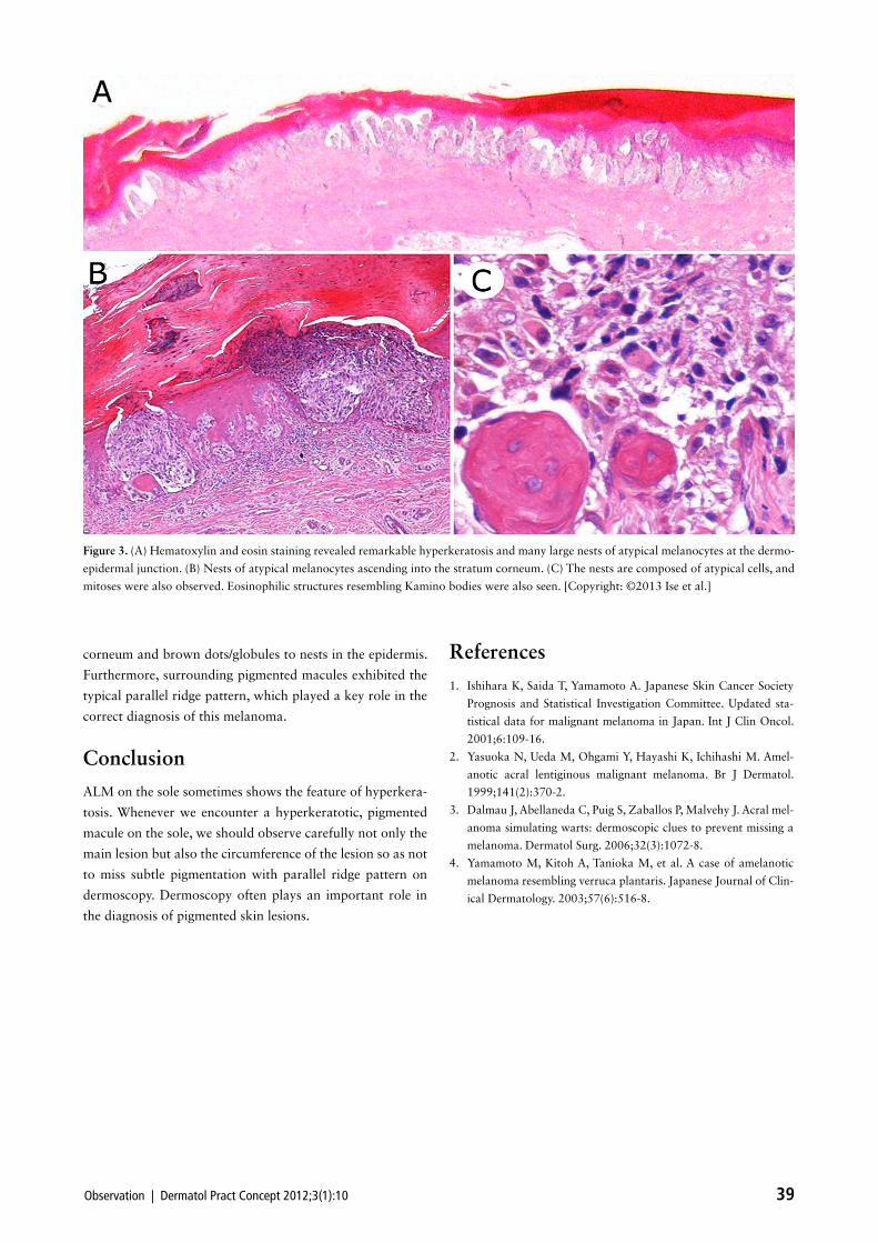
Dermoscopic findings were very helpful for a correct
diagnosis in this case. Aggregated dots/globules strongly indi-
cate melanocytic lesion. Their color depends on the amount
and depth of melanin. Therefore we estimated that black
dots/globules correspond to aggregated nests in the stratum
within the epidermis (Figure 3A). Nests ascending into the
stratum corneum were also seen (Figure 3B). Atypical melano-
cytes forming nests demonstrated substantial nuclear atypia
and mitoses (Figure 3C). Eosinophilic structures resembling
Kamino bodies were also seen. The surrounding pigmented
macules revealed atypical melanocytes proliferating singly or
forming small nests. These atypical melanocytes were hardly
observed in the hypopigmented areas that were characterized
by lymphocytic infiltration, aggregated melanophages and
sparse papillary dermal fibrosis. A hemorrhage in the stratum
corneum or epidermis that could induce black or brown dots/
globules in dermoscopic observation was not seen histologi-
cally. The tumor cells stained positively for S-100 protein and
HMB-45 antigen. We confirmed by melan-A staining that
melanoma cells were confined to the epidermis both at the
central and surrounding lesions. A diagnosis of acral lentigi-
nous melanoma (ALM) in situ was eventually made.
Computed tomography scan of the abdomen and tho-
rax revealed no obvious metastasis. Serum 5-S-cysteinyldopa
level was normal. A full thickness skin graft from her abdo-
men was placed on the operative wound. The patient has
been free of disease for eight months since the operation.
Discussion
ALM is the most common type of melanoma in the Japa-
nese population. In Japan about one-half of cases of cuta-
neous melanoma affect acral skin and approximately 30%
of them occur on the sole [1]. ALM on the sole sometimes
can be hyperkeratotic [2,3,4]. In previous reports, hyper-
keratotic lesions were seen at sites on which acute pressure
was exerted. Our case was not necessarily at a site of acute
pressure, but the patient had been habitually rubbing it. This
might have caused hyperkeratosis in our case. Another rea-
Figure 1. (A) A dark brown-to-black hyperkeratotic plaque measur-
ing 10 mm in diameter on the left sole. The border was well cir-
cumscribed and symmetrical. (B) A clinical examination after biopsy
revealed several pigmented macules of different shades varying from
brown to black around the central hyperkeratotic lesion. [Copy-
right: ©2013 Ise et al.]
Figure 2. Dermoscopic examinations of: (A) A hyperkeratotic le-
sion—scaly surface and regular brown-to-black dots/globules. (B)
Surrounding macules—many brown-to-black macules. Most of
them showed parallel ridge pattern. [Copyright: ©2013 Ise et al.]
Observation | Dermatol Pract Concept 2012;3(1):10 39
References
1. Ishihara K, Saida T, Yamamoto A. Japanese Skin Cancer Society
Prognosis and Statistical Investigation Committee. Updated sta-
tistical data for malignant melanoma in Japan. Int J Clin Oncol.
2001;6:109-16.
2. Yasuoka N, Ueda M, Ohgami Y, Hayashi K, Ichihashi M. Amel-
anotic acral lentiginous malignant melanoma. Br J Dermatol.
1999;141(2):370-2.
3. Dalmau J, Abellaneda C, Puig S, Zaballos P, Malvehy J. Acral mel-
anoma simulating warts: dermoscopic clues to prevent missing a
melanoma. Dermatol Surg. 2006;32(3):1072-8.
4. Yamamoto M, Kitoh A, Tanioka M, et al. A case of amelanotic
melanoma resembling verruca plantaris. Japanese Journal of Clin-
ical Dermatology. 2003;57(6):516-8.
corneum and brown dots/globules to nests in the epidermis.
Furthermore, surrounding pigmented macules exhibited the
typical parallel ridge pattern, which played a key role in the
correct diagnosis of this melanoma.
Conclusion
ALM on the sole sometimes shows the feature of hyperkera-
tosis. Whenever we encounter a hyperkeratotic, pigmented
macule on the sole, we should observe carefully not only the
main lesion but also the circumference of the lesion so as not
to miss subtle pigmentation with parallel ridge pattern on
dermoscopy. Dermoscopy often plays an important role in
the diagnosis of pigmented skin lesions.
Figure 3. (A) Hematoxylin and eosin staining revealed remarkable hyperkeratosis and many large nests of atypical melanocytes at the dermo-
epidermal junction. (B) Nests of atypical melanocytes ascending into the stratum corneum. (C) The nests are composed of atypical cells, and
mitoses were also observed. Eosinophilic structures resembling Kamino bodies were also seen. [Copyright: ©2013 Ise et al.]