
Acoustic Property of Cvd-Diamond Film and Acoustic … · 2012-09-24 · erated ultrasonics. We...
11
J. Acoustic Emission, 21 (2003) 131 © 2003 Acoustic Emission Group ACOUSTIC PROPERTY OF CVD-DIAMOND FILM AND ACOUSTIC EMISSION ANALYSIS FOR INTEGRITY EVALUATION R. IKEDA 1 , Y. HAYASHI 2 and M. TAKEMOTO 2 1 R and D Div., Asahi Diamond Co., Ltd, 787, Tabi, Ichihara, Chiba, 290-0515, JAPAN, 2 Faculty of Science and Engineering, Aoyama Gakuin University Fuchinobe 5-10-1, Sagamihara, Kanagawa, 229-8558, JAPAN Abstract The authors determined the acoustic properties of CVD-diamond film using a laser ultrasonic system. Three elastic stiffness coefficients, i.e, C 11 , C 12 , C 44 for cubic structure, were estimated by inverse processing of Lamb waves detected for a free-standing diamond film. Two stiffness coefficients; C 11 (1026 GPa) and C 12 (250 GPa), estimated from sheet velocity and velocity dis- persion of Ao-Lamb wave, agreed well with those reported for natural diamond, but C 44 (388 GPa) was estimated to be 80 % that of natural diamond. Residual stress of diamond film depos- ited on WC-Co substrate was measured as 2.2 to 2.4 GPa in compression by the Raman spectros- copy. We analyzed the Lamb-wave AE signals from two kinds of diamond film deposited in differ- ent area on a 1-mm thick WC-Co substrate during four-point bending. No AE was detected for the specimen with small area diamond film (Type-A) under tensile and compressive bending. We also detected no AE for the specimen with large area diamond (Type-B) when it is subjected to tensile bending. Diamond film suffered damages (fragmentation and decohesion) when the sub- strate suffered macroscopic unstable fractures at +1.56 GPa. In contrast, we detected frequent AE signals from compressive surface stress (–0.58 GPa) when Type-B specimen was subjected to compressive bending. AE events rapidly increased above the surface stress of –0.58 GPa. Most AE signals with small peak amplitudes were located outside the sensors and their fracture types were not classified. A few AE signals with low amplitude and fast rise time of So-mode, detected at around –0.60 GPa, were classified to the Mode-I cleavage fracture of the film. Though frag- mentary diamond film exfoliated from the substrate, we could not monitor the AE events due to the film spalling. Fracture mechanism of diamond film under tensile and compressive loading was discussed in relation to the compressive residual stress. Key Words: CVD-diamond, Acoustic property, Residual stress, Four-point bending, Decohe- sion, Cleavage fracture 1. Introduction Diamond film was first synthesized by chemical vapor deposition (CVD) method almost 20 years ago. Since then diamond film has been expected to become new functional material in tools and machinery as hard coating, SAW devices and so on. Diamond is the hardest material and shows high thermal conductance but good electrical insulation. It is brittle and shows low fracture toughness. Utilization of the hardest diamond as cutting tools has been the most suc- ceeding industrial practice, but it is still limited on the market due to poor reliability against fracture and decohesion of the film during machining. Interpretation of decohesion mechanism and effective countermeasure against it are indispensable for further expansion of the diamond as
Transcript of Acoustic Property of Cvd-Diamond Film and Acoustic … · 2012-09-24 · erated ultrasonics. We...

J. Acoustic Emission, 21 (2003) 131 © 2003 Acoustic Emission Group
ACOUSTIC PROPERTY OF CVD-DIAMOND FILM AND ACOUSTICEMISSION ANALYSIS FOR INTEGRITY EVALUATION
R. IKEDA1, Y. HAYASHI2 and M. TAKEMOTO2
1 R and D Div., Asahi Diamond Co., Ltd, 787, Tabi, Ichihara, Chiba, 290-0515, JAPAN,2 Faculty of Science and Engineering, Aoyama Gakuin University
Fuchinobe 5-10-1, Sagamihara, Kanagawa, 229-8558, JAPAN
Abstract
The authors determined the acoustic properties of CVD-diamond film using a laser ultrasonicsystem. Three elastic stiffness coefficients, i.e, C11, C12, C44 for cubic structure, were estimatedby inverse processing of Lamb waves detected for a free-standing diamond film. Two stiffnesscoefficients; C11 (1026 GPa) and C12 (250 GPa), estimated from sheet velocity and velocity dis-persion of Ao-Lamb wave, agreed well with those reported for natural diamond, but C44 (388GPa) was estimated to be 80 % that of natural diamond. Residual stress of diamond film depos-ited on WC-Co substrate was measured as 2.2 to 2.4 GPa in compression by the Raman spectros-copy.
We analyzed the Lamb-wave AE signals from two kinds of diamond film deposited in differ-ent area on a 1-mm thick WC-Co substrate during four-point bending. No AE was detected forthe specimen with small area diamond film (Type-A) under tensile and compressive bending. Wealso detected no AE for the specimen with large area diamond (Type-B) when it is subjected totensile bending. Diamond film suffered damages (fragmentation and decohesion) when the sub-strate suffered macroscopic unstable fractures at +1.56 GPa. In contrast, we detected frequent AEsignals from compressive surface stress (–0.58 GPa) when Type-B specimen was subjected tocompressive bending. AE events rapidly increased above the surface stress of –0.58 GPa. MostAE signals with small peak amplitudes were located outside the sensors and their fracture typeswere not classified. A few AE signals with low amplitude and fast rise time of So-mode, detectedat around –0.60 GPa, were classified to the Mode-I cleavage fracture of the film. Though frag-mentary diamond film exfoliated from the substrate, we could not monitor the AE events due tothe film spalling. Fracture mechanism of diamond film under tensile and compressive loadingwas discussed in relation to the compressive residual stress.
Key Words: CVD-diamond, Acoustic property, Residual stress, Four-point bending, Decohe-sion, Cleavage fracture
1. Introduction
Diamond film was first synthesized by chemical vapor deposition (CVD) method almost 20years ago. Since then diamond film has been expected to become new functional material intools and machinery as hard coating, SAW devices and so on. Diamond is the hardest materialand shows high thermal conductance but good electrical insulation. It is brittle and shows lowfracture toughness. Utilization of the hardest diamond as cutting tools has been the most suc-ceeding industrial practice, but it is still limited on the market due to poor reliability againstfracture and decohesion of the film during machining. Interpretation of decohesion mechanismand effective countermeasure against it are indispensable for further expansion of the diamond as

132
a cutting-tool material. Driving force of decohesion damages are the residual, thermal and ap-plied (cutting) stresses. Cutting stress is often dynamic or impact. As the diamond film is depos-ited on hard substrate material such as WC-Co at temperatures higher than 1073K, extremelyhigh residual stresses exist in the film. Interfacial quality or bond strength of the diamond filmdepends on the area, thickness, deposition condition and so on. Estimation of the interfacialquality is particularly important for establishing countermeasures against decohesion damages,but has not been studied much.
We first estimated the elastic properties of polycrystalline CVD-diamond utilizing laser gen-erated ultrasonics. We estimated elastic stiffness coefficients for isotropic cubic structure andcompared with those reported for natural diamond. Both the zero-th order symmetric (So) andanti-symmetric (Ao) mode Lamb waves detected for free-standing diamond film were analyzed.Residual stress was estimated by Raman spectroscopy.
We studied effects of residual stress and film area on the film damage by monitoring AEduring four-point bending. Diamond films with different deposition area on WC-Co plates weresubjected to compressive and tensile bending. Threshold stress to cause film or substrate fractureunder tension and compression were determined by AE monitoring. Next, we studied fracturetypes of the film and progression of fractures by waveform analysis of S0 component of LambAE signals.
2. Acoustic Properties And Residual Stress Of Cvd-Diamond Film
Two kinds of polycrystalline diamond film were prepared by hot-filament CVD method ofmethane and hydrogen gas mixture. One is a free standing film with average grain size of ~50µm, mechanically exfoliated after being deposited on 5.4 mm thick sintered SiC substrate (de-noted hereafter as 50FSD). Film surface and transverse structure of 50FSD film are shown inFig. 1. Surface of the diamond film was polished and coated with gold, depending on the pur-pose. Another specimen is a film deposited on 1 mm thick WC-Co substrate with average grainsize of about 3 µm (denoted hereafter as 3MD). This one is submitted to the residual stressmeasurement by Raman spectroscopy.
Fig. 1 SEM images of as-grown surface (a) and cross-section (b) of CVD-diamond with averagegrain size of 50 µm (50SFD).

133
Fig. 2 Effects of density (ρ) and Lame’s constants (λ) and (µ) on the group-velocity dispersionof Ao-mode Lamb waves for free-standing diamond film.
We estimated elastic stiffness coefficients of the diamond film from the sheet velocity of So-mode and the group velocity dispersion of Ao-mode. Natural diamond is known to be acousti-cally cubic (m3m). Table 1 shows the stiffness coefficients reported for natural and CVD dia-mond [1-5]. It is noted that data are grouped into two. Two groups reported 238 and 250 GPa forC12, and two others 125 GPa. Jiang et al. [5] measured the coefficient C12 of CVD diamond as122 GPa by using Brillouin scattering.
Table 1 Reported data for elastic stiffness (C11, C12, C44), anisotropy (η) and density (ρ) of dia-mond.
We first attempted to utilize the velocity dispersion of Rayleigh wave. However, Rayleighwave can only exist in a limited velocity range from 6900 (Rayleigh wave velocity of the sub-strate) to 8200 m/s (shear wave velocity of the diamond). This suggests that the estimation ofstiffness coefficients from the Rayleigh wave was very difficult. Thus, we utilized the Lambwaves measured on the free-standing diamond film.
Figure 2 shows the effects of density (ρ) and Lame’s constants (λ = C12, µ = C44) on thegroup velocity dispersion of the Ao-mode Lamb waves of the diamond film. These dispersioncurves are computed using the C12 and C44 reported by Auld [1]. It is noted that λ gives smalleffect on the dispersion and is difficult to estimate from the Ao-mode. Therefore, we measuredthe sheet velocity of So-mode Lamb.
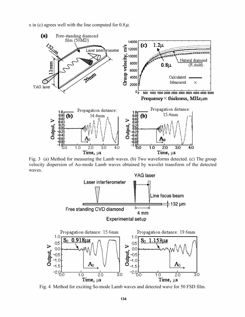
Figure 3 shows the method for measuring the Lamb waves. Here, a pulse YAG laser with du-ration of 5 ns was irradiated on the Au-coated distal plane of the diamond film and monitored bylaser interferometer at propagation distances of 14.1 and 15.4 mm. Two of the waveforms de-tected are shown in (b), from which the group velocity dispersion of Ao-mode Lamb was ob-tained by wavelet transform (see (c)). The bold solid line in (c) was computed using the proper-ties by Auld (1) for natural diamond. Fine lines are the dispersion curves computed by changingonly the value of µ from 0.8 to 1.2 times that obtained by Auld. Data points shown by
134
x in (c) agrees well with the line computed for 0.8 µ.
Fig. 3 (a) Method for measuring the Lamb waves. (b) Two waveforms detected. (c) The groupvelocity dispersion of Ao-mode Lamb waves obtained by wavelet transform of the detectedwaves.
Fig. 4 Method for exciting So-mode Lamb waves and detected wave for 50 FSD film.

135
Using the Simplex-assisted inverse scheme, we estimated the stiffness coefficients fromgroup velocity dispersion of Ao-Lamb waves. Assuming the density as 3515 kg/m3, C11 and C44
were estimated for the case of C12= 125 GPa and 250 GPa. For C12= 125 GPa, C11= 941 GPa andC44= 407 GPa were obtained, and C11= 1026 GPa, C44=388 GPa for C12= 250 GPa. The sheetvelocity is computed as 16,200 m/s for the former case (C12=125 GPa) and 16,600 m/s for thelater case (C12= 250 GPa).
Next we measured the So-mode Lamb wave using the method shown in the upper part ofFig. 4. A line-focused beam of a high-energy YAG laser was irradiated onto the un-coated dia-mond film normal to the surface. So-mode Lamb wave was excited by the thermal expansion ofthe diamond film. Beam position was moved and So-packets detected at the limit distances of15.6 and 19.6 mm are shown. The sheet velocity was measured as 17,000 m/s and close to thecomputed one for C12= 250 GPa. The Rayleigh wave velocity calculated from C12= 250GPa,C11= 1026 m/s and C44= 388 GPa is 9550 m/s and agrees well with 10,000 m/s measured usingan ultrasonic microscope (at 200 MHz). Finally, C44 of CVD diamond was estimated to be 80%of that reported by Auld (1) for natural diamond. Accurate determination of the three stiffnesscoefficients from limited acoustic data is difficult, but we have demonstrated that the elasticproperties of CVD-diamond is very close to those of natural diamond and almost isotropic.
Raman spectrpscopy is occasionally utilized for the estimation of residual stress. Sharma etal. [6] measured Raman spectra of diamond under static high pressure, and found a linear rela-tion between drift amount (Δn) of the peak from the position under no stress and applied stress(σ), by Eq. (1).
σ (GPa) = -0.422 x Δn (cm –1) (1)
We measured residual stress using Raman spectroscopy on the CVD-diamond film (3MD)deposited on WC-Co substrate. Residual stress was measured as –2.2 to –2.4 GPa (in compres-sion).
3. Analyses of AE during Four-Point Bending of Coated Diamond
Experimental MethodWe monitored AE signals from diamond films subjected to four-point bending using the
equipment as shown in the left of Fig. 5. Distance of the inner span is 73 mm. It is noted the bothsides of specimen were rigidly clamped to steel blocks. Fine-structure diamond films (3MD) of10-µm thickness were deposited on the WC-Co plate of 1 mm thick, 100 mm long and 30 mmwide. Two types of specimen were tested. One is Type-A specimen with the diamond film de-posited within 16 mm square at the central portion of the plate. Type-B is the specimen withdiamond film deposited over central 60 mm x 30 mm area. Diamond film of Type-B extended tothe areas under the steel blocks. The diamond film was loaded so as to apply both compressionand tension. Four AE sensors (PAC Type PICO) were mounted on the corner of 30 mm x 16 mmrectangle at specimen center as shown in Fig. 5 (right). In case of Type-B, the sensors are on thediamond film. Sensor outputs were amplified 40 dB using NF9913 (NF Circuit Co.), digitized bya fast A/D converter H310 (Autonics Co.) and fed to a personal computer.
Both fracture-type classification and source location were performed using the So-packet ofLamb AE signals. Sheet velocity of WC-Co is as fast as 6600 m/s, and fast sampling interval of50 ns were used for accurate determination of the arrival time. Amplification of the system wasadjusted so as to detect only the first portion of the So-packet.

136
Fig. 5 Experimental setup for AE monitoring during four-point bending of diamond film depos-ited on WC-Co plate.
Fig. 6 (a) Surface photograph of Type-A specimen after bending to the surface strain of 0.16%.(b) At 0.21% strain, substrate fractured and diamond film exfoliated in parts.
Results of Type-A specimen with small area diamond filmDiamond films deposited within 16-mm square area were submitted to tensile or compressive
bending. No AE was detected for any diamond film below the surface strain of 0.16% and stressof 1 GPa. Only one AE event was detected when the substrate WC-Co suffered unstable fractureat strain of 0.21% (1.56 GPa). This finding was the same when the diamond film is subjected totension or compression. Figure 6 shows the surface photographs of the diamond film subjected tothe surface strain of 0.16% and 0.21%. At 0.16%, no macroscopic or microscopic damages wereobserved. At the strain of 0.21%, the WC-Co plate sustained several unstable fractures andcaused partial exfoliation of diamond film, shown in (b). This demonstrates that the CVD-diamond film deposited in a limited area possesses very high adhesion to the substrate WC-Co.
Results of Type-B Specimen with Large Area Diamond FilmWe observed no AE during the tensile bending up to the surface stress of 1 GPa (0.16%
strain), which is close to the tensile fracture strength (1.5 GPa) of the substrate. AE sensors wereremoved from the specimen above the stress of 1 GPa to avoid the sensor damage due to unsta-ble macroscopic fracture of brittle WC-Co. This finding demonstrates that the CVD-diamondfilm, even if it is coated over wide area, has strong adhesion to the WC-Co substrate under ten-sion. Diamond film was found to be free of any damages after being unloaded as shown in Fig.7.

137
Fig. 7 Surface photograph of the CVD-diamond subjected to bending to surface tensile stress of1 GPa.
Fig. 8 Compressive stress vs. strain diagram with cumulative AE counts.
Contrary to the high performance of the diamond film under tension, we detected frequentAE signals when the diamond films are subjected to compressive stresses in bending. Figure 8shows compressive stress vs. strain with cumulative AE counts. AE counts rapidly increased atcompressive strains larger than 0.078 % (corresponding to surface stress of 0.58 GPa). Most AEevents (event number 1 to 12) detected below 0.078% strain are, as indicated by symbol ▼, lo-cated at the edges of steel block (rigid fastening) as shown in Fig. 9. These events are likely to becontact noise. Above 0.078% strain, we heard frequent crack-sounds and simultaneously de-tected many AE signals. AE events denoted by symbol ▼ with number 17, 18, 22, 23, 25 and 27were located near Sensors #1, 2 and 3. Further we detected five other AE events (event 13, 14,16, 26 and 28) designated by symbol ◇. Three of them (13, 14 and 16) at around the surfacestrain of 0.08% were located along the upper edge of the specimen, but other two (26, 28) de

138
tected at higher stresses were located inside the monitoring area. It is noted that no AE was lo-cated near the specimen center.
Fig. 9 Source location of AE events detected during compressive bending of WC-Co with largearea CVD-diamond film (Type-B specimen).
Fig. 10 First portion of the So-packet of Lamb AE signals (◇, No. 14) indicating the Mode-Icleavage fracture of the diamond film under compressive bending.
Figure 10 shows Lamb waveform of event No. 14 (◇) located near the sensor #1. Polarity ofthe first So-peak of four sensors is positive, and indicates the Mode-I fracture with crack openingvector parallel to the surface. This radiation characteristic agrees well with those of So-Lambwaves produced by adiabatic expansion of line focused pulse laser (Mode-I fracture with anopening vector parallel to the surface) reported by Sato et al. [7]. Fast rise time of the first So-mode, 0.8 to 0.9 µs, indicates the fast crack opening of diamond film, while the weak amplitudeless than 1 mV shows a small crack volume.
We primarily produced Lamb wave utilizing a 2-mm wide and 5-mm long compression-typePZT element with an opening (expansion) of 8 x 10⁻⁶ mm at 1.5 µs rise time. This simulates theMode-I decohesion of the film with opening vector in the surface normal direction. We detectedstrong So-peak of 20 mV with negative polarity. Polarity and peak amplitude of this simulatedSo-Lamb wave are much different from these observed in Fig. 10. As shown in Fig. 11, exfoli-ated diamond film are fragmented with sizes from less than 1 mm to a few mm. Thus, the signal◇ shown in Figs. 8 and 9 are not produced by the large-size Mode-I decohesion (normal to sur-face) but from small-size Mode-I fracture (in-plane) due to buckling of the diamond film.

139
Fig. 11 Fragments of diamond film from WC-Co substrate after being subjected to bending incompression up to 0.75 GPa.
Fig. 12 (a) Method for Mode-II shear fracture simulation. (b) Resultant waveforms. (c) The ra-diation pattern of the first peak of So-packet of the Lamb waves.
4. Discussion of Interfacial Quality of CVD-Diamond
Some important findings were revealed by this study. These are:1) Large compressive residual stress of –2.3 GPa exists in CVD-diamond films.2) Diamond film deposited in a limited area shows strong adhesion to the substrate under both
tensile and compressive bending.3) Diamond film deposited in a large area shows strong adhesion under tension but lower
bending strength under compression.
140
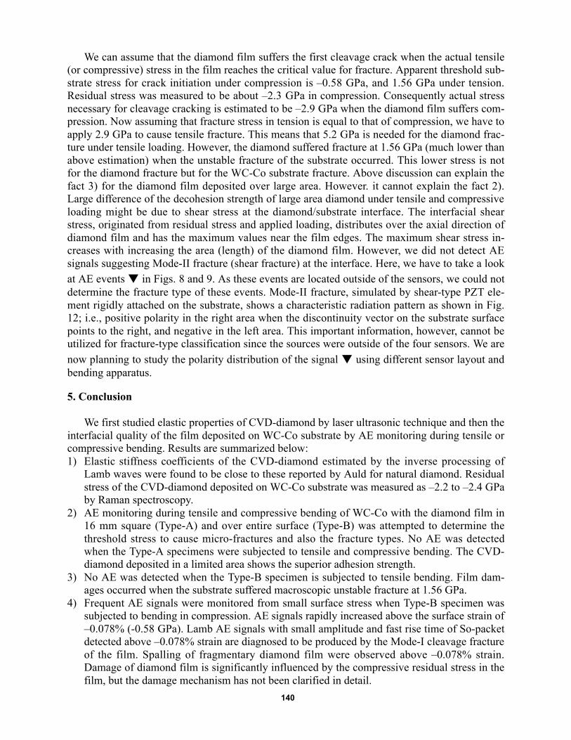
We can assume that the diamond film suffers the first cleavage crack when the actual tensile(or compressive) stress in the film reaches the critical value for fracture. Apparent threshold sub-strate stress for crack initiation under compression is –0.58 GPa, and 1.56 GPa under tension.Residual stress was measured to be about –2.3 GPa in compression. Consequently actual stressnecessary for cleavage cracking is estimated to be –2.9 GPa when the diamond film suffers com-pression. Now assuming that fracture stress in tension is equal to that of compression, we have toapply 2.9 GPa to cause tensile fracture. This means that 5.2 GPa is needed for the diamond frac-ture under tensile loading. However, the diamond suffered fracture at 1.56 GPa (much lower thanabove estimation) when the unstable fracture of the substrate occurred. This lower stress is notfor the diamond fracture but for the WC-Co substrate fracture. Above discussion can explain thefact 3) for the diamond film deposited over large area. However. it cannot explain the fact 2).Large difference of the decohesion strength of large area diamond under tensile and compressiveloading might be due to shear stress at the diamond/substrate interface. The interfacial shearstress, originated from residual stress and applied loading, distributes over the axial direction ofdiamond film and has the maximum values near the film edges. The maximum shear stress in-creases with increasing the area (length) of the diamond film. However, we did not detect AEsignals suggesting Mode-II fracture (shear fracture) at the interface. Here, we have to take a lookat AE events ▼ in Figs. 8 and 9. As these events are located outside of the sensors, we could notdetermine the fracture type of these events. Mode-II fracture, simulated by shear-type PZT ele-ment rigidly attached on the substrate, shows a characteristic radiation pattern as shown in Fig.12; i.e., positive polarity in the right area when the discontinuity vector on the substrate surfacepoints to the right, and negative in the left area. This important information, however, cannot beutilized for fracture-type classification since the sources were outside of the four sensors. We arenow planning to study the polarity distribution of the signal ▼ using different sensor layout andbending apparatus.
5. Conclusion
We first studied elastic properties of CVD-diamond by laser ultrasonic technique and then theinterfacial quality of the film deposited on WC-Co substrate by AE monitoring during tensile orcompressive bending. Results are summarized below:1) Elastic stiffness coefficients of the CVD-diamond estimated by the inverse processing of
Lamb waves were found to be close to these reported by Auld for natural diamond. Residualstress of the CVD-diamond deposited on WC-Co substrate was measured as –2.2 to –2.4 GPaby Raman spectroscopy.
2) AE monitoring during tensile and compressive bending of WC-Co with the diamond film in16 mm square (Type-A) and over entire surface (Type-B) was attempted to determine thethreshold stress to cause micro-fractures and also the fracture types. No AE was detectedwhen the Type-A specimens were subjected to tensile and compressive bending. The CVD-diamond deposited in a limited area shows the superior adhesion strength.
3) No AE was detected when the Type-B specimen is subjected to tensile bending. Film dam-ages occurred when the substrate suffered macroscopic unstable fracture at 1.56 GPa.
4) Frequent AE signals were monitored from small surface stress when Type-B specimen wassubjected to bending in compression. AE signals rapidly increased above the surface strain of–0.078% (-0.58 GPa). Lamb AE signals with small amplitude and fast rise time of So-packetdetected above –0.078% strain are diagnosed to be produced by the Mode-I cleavage fractureof the film. Spalling of fragmentary diamond film were observed above –0.078% strain.Damage of diamond film is significantly influenced by the compressive residual stress in thefilm, but the damage mechanism has not been clarified in detail.
141
References
1) B. A. Auld, “Acoustic Field and Waves in Solids”, Wiley-Interscience Publ., (1973), 368.2) A. Tourlog, W. Li and D. Achenbach, Applied Physics Letters, 69 (1996), 3680.3) H. J. McSkimin and W. L. Bond, Physical Review, 105 (1956), 116.4) M. H. Grimsditch and A. K. Ramdas, Physical Review B, 11 (1975), 3139.5) X. Jiang, J. V. Harzer, B. Hillbrands, Ch. Wild and P. Koidl, Applied Physics Letters, 59(1991), 1055.6) S. K. Sharma, J. of Raman Spectroscopy, 16 (1985), 350.7) T. Sato, M. Takemoto and K. Ono, Japanese Journal of Applied Physics, 38 (1999), 3193.


![Nonlinear UT for NDT [Kompatibilitetsläge] · Nonlinear ultrasonics for NDTNonlinear ultrasonics for NDT Linear Ultrasonics: Detection of Flaws/Discontinuities • Detect geometric](https://static.fdocuments.net/doc/165x107/5eb54bd032d9642d8e2c4d0a/nonlinear-ut-for-ndt-kompatibilitetslge-nonlinear-ultrasonics-for-ndtnonlinear.jpg)


