Acoustic Break-up of Potential Cerebral Emboli · Cerebral emboli are associated with neurological...
3
18 th Australasian Fluid Mechanics Conference Launceston, Australia 3-7 December 2012 Acoustic Break-up of Potential Cerebral Emboli F. Secretain 1 , A. Pollard 1 and B. Milne 2 1 Department of Mechanical and Materials Engineering Queen’s University at Kingston, Ontario, K7L3N6, Canada 2 Department of Anesthesiology and Preoperative Medicine Kingston General Hospital, Kingston, Ontario, K7L2V7, Canada Abstract Risks associated with air emboli (bubbles) introduced during open-heart surgery have been highlighted by reports of postoperative neuropsychological disorders with increasing cerebral emboli. Cerebral emboli are associated with neurological dysfunction ranging from stroke or coma to cognitive deficits, which are among the most common complications following successful cardiac surgery. Presently, there are no standard, effective methods for detecting, quantifying or eliminating potential emboli. Typically, transcranial Doppler is used to detect emboli from the middle cerebral arteries even though the amount of air observed during cardiac procedures would overload common systems. Our multidisciplinary engineering-medical research team has developed software that can detect and quantify air emboli within the ascending aorta and/or cardiac chambers during cardiac surgery in real-time before emboli reach the brain. In this paper, we introduce acoustic fields to set up surface waves on bubbles. The theoretical background for this phenomena is briefly described and high speed video images are presented that demonstrate bubble break-up. Introduction Open cardiac surgical procedures and particularly those using cardiopulmonary bypass inevitably introduce large amounts of air into the circulation. Particulate emboli are also a factor during cardiac surgery, but a recent study demonstrated that reducing the amount of air emboli during cardiac surgery improved executive function and verbal short-term memory three months following surgery while patients fitted with particulate emboli filters showed no improvement over control cardiac surgery patients [4]. Even so, with limited intra-operative information from trans- esophageal echocardiography (TEE) and trans-cranial Doppler and no standard de-aeration methods or guidelines, the ability for health care providers to eliminate air introduced during cardiac surgery is limited. As a result, large quantities of air are often observed in the cardiac chambers following closure of the heart (figure 1) and following separation from bypass. Trapped air circulates through the body until it is either absorbed or lodged in small capillaries in the brain, heart or other organs to result in a myriad of adverse events including death [1]. Air introduced during cardiac surgery correlates highly with neurological dysfunction and this is associated with a reduced quality of life even at 5 years following cardiac bypass surgery [7]. The long-term objective of the current research is to improve methods of detection, quantification and removal of air introduced during open cardiac procedures. This would translate to a reduced incidence of postoperative complications including postoperative cognitive disorders, which can have a major impact upon recovery, autonomy and quality of life following cardiac surgery [2,7]. Given that at least a million open cardiac procedures are performed annually around the world [8], this translates to an enormous number of people being adversely affected by poor air detection, quantification and de-aeration methods during cardiac surgery. Figure 1: Left side of the heart viewed using TEE transducer (a) before and (b) after weaning from the cardiopulmonary bypass machine, where LA=Left Atrium and LV=Left Ventricle. Theory The Rayleigh-Plesset equation (1) describes the dynamics of a bubble that undergoes purely spherical motion. The linear harmonic solution to the Rayleigh-Plesset equation reveals the natural frequency of spherical motion, the 0 th or breathing mode of the bubble, equation 2. (1) (2)
Transcript of Acoustic Break-up of Potential Cerebral Emboli · Cerebral emboli are associated with neurological...

18th Australasian Fluid Mechanics Conference Launceston, Australia 3-7 December 2012
Acoustic Break-up of Potential Cerebral Emboli
F. Secretain1, A. Pollard1 and B. Milne2
1Department of Mechanical and Materials Engineering Queen’s University at Kingston, Ontario, K7L3N6, Canada 2Department of Anesthesiology and Preoperative Medicine
Kingston General Hospital, Kingston, Ontario, K7L2V7, Canada
Abstract
Risks associated with air emboli (bubbles) introduced during open-heart surgery have been highlighted by reports of postoperative neuropsychological disorders with increasing cerebral emboli. Cerebral emboli are associated with neurological dysfunction ranging from stroke or coma to cognitive deficits, which are among the most common complications following successful cardiac surgery. Presently, there are no standard, effective methods for detecting, quantifying or eliminating potential emboli. Typically, transcranial Doppler is used to detect emboli from the middle cerebral arteries even though the amount of air observed during cardiac procedures would overload common systems. Our multidisciplinary engineering-medical research team has developed software that can detect and quantify air emboli within the ascending aorta and/or cardiac chambers during cardiac surgery in real-time before emboli reach the brain. In this paper, we introduce acoustic fields to set up surface waves on bubbles. The theoretical background for this phenomena is briefly described and high speed video images are presented that demonstrate bubble break-up.
Introduction
Open cardiac surgical procedures and particularly those using cardiopulmonary bypass inevitably introduce large amounts of air into the circulation. Particulate emboli are also a factor during cardiac surgery, but a recent study demonstrated that reducing the amount of air emboli during cardiac surgery improved executive function and verbal short-term memory three months following surgery while patients fitted with particulate emboli filters showed no improvement over control cardiac surgery patients [4]. Even so, with limited intra-operative information from trans-esophageal echocardiography (TEE) and trans-cranial Doppler and no standard de-aeration methods or guidelines, the ability for health care providers to eliminate air introduced during cardiac surgery is limited. As a result, large quantities of air are often observed in the cardiac chambers following closure of the heart (figure 1) and following separation from bypass. Trapped air circulates through the body until it is either absorbed or lodged in small capillaries in the brain, heart or other organs to result in a myriad of adverse events including death [1]. Air introduced during cardiac surgery correlates highly with neurological dysfunction and this is associated with a reduced quality of life even at 5 years following cardiac bypass surgery [7]. The long-term objective of the current research is to improve methods of detection, quantification and removal of air introduced during open cardiac procedures. This would translate to a reduced incidence of postoperative complications including postoperative cognitive disorders, which can have a major impact
upon recovery, autonomy and quality of life following cardiac surgery [2,7]. Given that at least a million open cardiac procedures are performed annually around the world [8], this translates to an enormous number of people being adversely affected by poor air detection, quantification and de-aeration methods during cardiac surgery.
Figure 1: Left side of the heart viewed using TEE transducer (a) before and (b) after weaning from the cardiopulmonary bypass machine, where LA=Left Atrium and LV=Left Ventricle.
Theory
The Rayleigh-Plesset equation (1) describes the dynamics of a bubble that undergoes purely spherical motion. The linear harmonic solution to the Rayleigh-Plesset equation reveals the natural frequency of spherical motion, the 0th or breathing mode of the bubble, equation 2.
(1)
(2)
where R=R(t) is the bubble wall radius, the single and double dots represents first and second time derivatives, respectively, ρ is the liquid density, po is the equilibrium static pressure, σ is the gas-liquid surface tension, Ro is the equilibrium bubble radius, γ is the polytropic index of the gas, µ and υ is the liquid dynamic and kinematic viscosity, respectively, p∞(t) is the applied external pressure, and ωo is the natural frequency in Hz.
When spherical bubble motion oscillations become unstable and spherical symmetry is lost, the analysis of the bubble dynamics becomes exceedingly complex. The first issue is to describe or specify the bubble shape. A solution that has been extensively used [5,9] for non-spherical bubble dynamics is a spherical harmonic expansion of the bubble radius:
(3)
where r(θ,φ,t) is the instantaneous bubble, anm is the amplitude of the spherical harmonic component of order n and degree m, and Yn
m(θ,φ) is the normalized spherical harmonic shape distortion of the bubble surface.
Plesset [9] solved the equation of motion of an incompressible, inviscid, unbounded fluid with a free surface using a small amplitude approximation (i.e. anm/R<<1). In this approximation the equations for R(t) (the average bubble radius) and the anm(t)’s are uncoupled, the first just being Rayleigh’s equation. The solution as derived by Plesset [9] of the bubble surface distortions (anm(t)’s) is:
(4)
Note, that the coefficients of the surface distortion governing equation (4) is dependant on the dynamics of the average radius (R(t)) of the bubble surface. Second, the n=1 solution corresponds to a translation of the bubble centre, which is of little interest and shall be neglected [10]. Therefore, we shall restrict our considerations to the cases for n>1 solutions. The linear harmonic solution to equation 4 reveals the natural frequencies for surface oscillations for n>1, equation 5.
(5)
where ωn is the surface distortion frequency of order n in Hz.
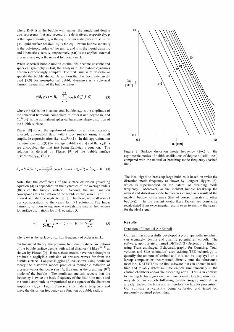
On linearized theory, the pressure field due to shape oscillations of the bubble surface decays with radial distance (r) like r-(n+1) as shown by Plesset [9]. Hence, these modes have been thought to produce a negligible emission of pressure waves far from the bubble surface. Longuet-Higgins [6] has shown using nonlinear theory the distortion modes produce a monopole radiation of pressure waves that decays as 1/r, the same as the breathing (0th) mode of the bubble. The nonlinear analysis reveals that the frequency is twice the basic frequency of the distortion mode and the sound amplitude is proportional to the square of the distortion amplitude (anm). Figure 2 presents the natural frequency and twice the distortion frequency as a function of bubble radius.
Figure 2: Surface distortion mode frequency (2ωn) of the asymmetric modes of bubble oscillations of degree n (solid lines) compared with the natural or breathing mode frequency (dashed line).
The ideal signal to break-up large bubbles is based on twice the distortion mode frequency as shown by Longuet-Higgins [6], which is superimposed on the natural or breathing mode frequency. Moreover, as the incident bubble breaks-up the natural and distortion mode frequencies change as a result of the incident bubble losing mass (that of course migrates to other bubbles). In the current work, these factors are constantly recalculated from experimental results so as to narrow the search for the ideal signal.
Results
Detection of Potential Air Emboli
Our team has successfully developed a prototype software which can accurately identify and quantify potential air emboli. The software, appropriately named DETECTS (Detection of Emboli using Trans-esophageal Echocardiography for Counting, Total volume, and Size estimation) uses existing TEE technology to quantify the amount of emboli and this can be displayed on a laptop computer or incorporated directly into the ultrasound machine. DETECTS is the first software that can operate in real-time and reliably detect multiple emboli simultaneously in the cardiac chambers and/or the ascending aorta. This is in contrast to existing technologies such as trans-cranial Doppler, which can only detect air emboli following cardiac surgery once it has already reached the brain and is therefore too late for prevention. Our software is currently being calibrated and tested on previously obtained patient data.

Acoustic Break-up of Potential Air Emboli
We are currently developing a multi-frequency acoustic procedure which breaks-up larger air bubbles (1-2 mm diameter) into smaller bubbles (figure 3). Evidence is lacking in terms of precise cut-off size at which point air emboli become clinically significant. It is, however, generally accepted that large emboli are more clinically significant than small ones [7]. Small potential emboli that remain following application of our break-up technology are less likely to be associated with adverse events than larger bubbles such as those introduced during cardiac surgery.
The objectives of the current series of experiments will be to further investigate the effect of a time dependent, large bandwidth frequency signals on the surface oscillations and instabilities of large bubble dynamics before, during and following break-up. The specific objective is to find the ideal signal to break-up bubbles with maximal effect with minimal time, power and cell damage.
Figure 3: 1.6 mm radius bubble break-up evolution sequence using a linear tone sweep from 1.5 to 1.6 kHz acoustic signal; images shown at 1000 frames per second.
Our experimental rig automatically produces a bubble of known size and then exposes it to a known acoustic signal. The dynamics and break-up are captured at 30,000 fps using a high-speed camera. The bubble fission rate is measured using an edge detection software and feedback to create a new signal. This process is repeated until the maximum bubble fission rate is found.
Our primary goal is to find the combined frequencies for optimal bubble break-up (i.e., maximal effect with minimal time, power and cell damage). The hypothesis is that if we break-up potential air emboli within the arterial system using acoustic methods to a size equal to or smaller than 40 µm, micro-bubbles should be more likely to travel harmlessly through the body until being diffused into the blood. Acoustic signals have been used previously for cavitation both within medicine and industry but
these used single frequency ultrasonic transducers [3] which are insufficient for breaking-up large air bubbles (i.e. > 100 µm) like those introduced during cardiac surgery.
Closing Remarks
A reliable software program that could quantify the potential air emboli would not only standardize de-aeration techniques for all open-heart cardiac surgeries, but also be the standard for quantification of potential air emboli. A record or history of the bubbles seen during the surgery could be kept and correlated to neurological dysfunction in future studies.
The non-intrusive break-up of air emboli would completely change the de-aeration procedures set in place today. This device would potentially become a standard for use during all cardiac surgical procedures. This method of non-invasive break-up can be applied to all potential air emboli sources within medicine and even industrial sources as well. Cavitation from propeller blades could potentially use this device to break-up bubbles before causing irreversible cavitation damage on fan blades. Further research could make industrial cavitation and neurological dysfunction from air bubbles be a thing of the past.
Acknowledgements
The authors acknowledge Dr. R. Tanzola for supporting the clinical aspects of this work. Funding was provided by the Natural Sciences and Engineering Research Council of Canada, The Garfield Kelly Cardiovascular Fund of Queen’s University and the Province of Ontario.
References
[1] Barak M, Yeshayahu K. Microbubbles: Pathophysiology and Clinical Implications. Chest 2005 128: 2918-2932
[2] Barbut D, Lo YW, Gold JP, Trifiletti RR, Yao FS, Hager DN, Hinton RB, Isom OW. Impact of embolization during coronary artery bypass grafting on outcomes and length of stay. Ann Thorac Surg 1997 63: 998-1002
[3] Bekeredjian R, Grayburn P, Shohet R. Use of Ultrasound contrast agents for gene or drug delivery in cardiovascular medicine. J Am Coll Cardiol 2005 45(3): 329-335
[4] Gerriets T, Schwarz N, Sammer G, Baehr J, Stolz E, Kaps M, Kloevekorn WP, Bachmann G, Schonburg M. Protecting the brain from gaseous and solid micro-emboli during coronary artery bypass grafting: A randomized controlled trial. Eur Heart J 2010 31(3): 278-80
[5] Lamb H., Hydrodynamics, Cambridge University Press, 1932 Art 275
[6] Longuet-Higgins M., Monopole emission of sound by asymmetric bubble oscillations. Part 1. Normal modes, J. Fluid Mech., 1989 201:525-541
[7] Newman MF, Grocott HP, Mathew JP, White WD, Landolfo K, Reves JG, Laskowitz DT, Mark DB, Blumenthal JA. Report of substudy assessing the impact or neurocognitive functioning on quality of life 5 years after cardiac surgery. Stroke 2001 32: 2874-2881
[8] Pezzela AT. International aspects of cardiac surgery. Ann Thorac Surg 1998 65: 903-904
[9] Plesset M., On the Stability of Fluid Flows with Spherical Symmetry, J. App. Phys., 1954 25:96-98.
[10] Plesset M., Prosperetti A., Bubble Dynamics and Cavitation, Ann. Rev. Fluid Mech., 1977 9:145-1