Achilles tendon healing in rats is improved by...
24
Achilles tendon healing in rats is improved by intermittent mechanical loading during the inflammatory phase Therese Andersson, Pernilla Eliasson and Per Aspenberg Linköping University Post Print N.B.: When citing this work, cite the original article. This is the pre-reviewed version of the following article: Therese Andersson, Pernilla Eliasson and Per Aspenberg, Achilles tendon healing in rats is improved by intermittent mechanical loading during the inflammatory phase, 2011, Journal of Orthopaedic Research, (30), 2, 274-279. which has been published in final form at: http://dx.doi.org/10.1002/jor.21511 Copyright: Wiley-Blackwell http://eu.wiley.com/WileyCDA/Brand/id-35.html Postprint available at: Linköping University Electronic Press http://urn.kb.se/resolve?urn=urn:nbn:se:liu:diva-70772
Transcript of Achilles tendon healing in rats is improved by...

Achilles tendon healing in rats is improved by
intermittent mechanical loading during the
inflammatory phase
Therese Andersson, Pernilla Eliasson and Per Aspenberg
Linköping University Post Print
N.B.: When citing this work, cite the original article.
This is the pre-reviewed version of the following article:
Therese Andersson, Pernilla Eliasson and Per Aspenberg, Achilles tendon healing in rats is
improved by intermittent mechanical loading during the inflammatory phase, 2011, Journal of
Orthopaedic Research, (30), 2, 274-279.
which has been published in final form at:
http://dx.doi.org/10.1002/jor.21511
Copyright: Wiley-Blackwell
http://eu.wiley.com/WileyCDA/Brand/id-35.html
Postprint available at: Linköping University Electronic Press
http://urn.kb.se/resolve?urn=urn:nbn:se:liu:diva-70772
For Peer Review
1
Achilles tendon healing in rats is improved by intermittent mechanical loading
during the inflammatory phase
Eliasson P 1a*, Andersson T1a, Aspenberg P1
a Equal contribution
1 Experimental Orthopaedics, Department of clinical and experimental medicine, Faculty of
health science, Linköping University
* Corresponding Author Pernilla Eliasson Experimental Orthopaedics KEF Faculty of health science Linköping University 58185 Linköping Sweden [email protected] +46-101034140
Running title: Tendon healing and early loading
Page 1 of 23
John Wiley & Sons, Inc.
Journal of Orthopaedic Research
123456789101112131415161718192021222324252627282930313233343536373839404142434445464748495051525354555657585960
For Peer Review
2
ABSTRACT
Tendons adapt to changes in mechanical loading, and numerous animal studies show that
immobilization of a healing tendon is detrimental to the healing process. The present study
addresses whether the effects of a few episodes of mechanical loading is different during
different phases of healing. Fifty female rats underwent Achilles tendon transection, and their
hind limbs were unloaded by tail suspension on the day after surgery. One group of 10 rats
was taken down from suspension to run on a treadmill for 30 minutes per day, on days 2-5
after transection. They were euthanized on day 8. Another group underwent similar treadmill
running on days 8-11 and was euthanized on day 14. Completely unloaded groups were
euthanized on day 8 and 14. Tendon specimens were then evaluated mechanically. The
results showed that just 4 loading episodes increased the strength of the healing tendon. This
was evident irrespective of the time-point when loading was applied (early or late). The
positive effect on early healing was unexpected, considering that the mechanical stimulation
was applied during the inflammatory phase, when the calluses were small and fragile. A
histological study of additional groups with early loading also showed some increased
bleeding in the loaded calluses. Our results indicate that a small amount of early loading may
improve the outcome of tendon healing. This could be of interest to clinical practice.
KEYWORDS
Early loading, tail-suspension, unloading, mechanical testing, cell differentiation
INTRODUCTION
The influence of early mobilization on the outcome of Achilles tendon healing has attracted
increasing attention. New rehabilitation programs recently tested include early weight bearing
(1, 2) and the use of orthoses, designed to permit certain ankle movement and some weight
Page 2 of 23
John Wiley & Sons, Inc.
Journal of Orthopaedic Research
123456789101112131415161718192021222324252627282930313233343536373839404142434445464748495051525354555657585960
For Peer Review
3
bearing (3, 4). The results from these studies indicate, to some extent, that early mobilization
and weight bearing have a positive effect on the healing process. However, clinical research
has so far not been able to distinguish between the effects of motion on the readaptation of
periarticular tissues and the effects of traction on tendon healing. It is unknown to what extent
weight bearing on the injured leg translates into traction forces in the healing Achilles tendon.
There is also probably a risk of complications, especially re-rupture, if loading is applied too
early. The improved clinical results, however, are in line with previous studies of the effects
of mechanical loading in animal models and on tendon cells in vitro. Cyclic loading regimes
up-regulate both the gene-expression and production of collagen by tendon fibroblasts in
vitro (5, 6). This increase in collagen expression can also be seen in intact animal tendons,
subjected to increased loading (7, 8). In fact there are many studies showing that intact
tendons adapt to changes in mechanical loading (9-14), and numerous animal studies show
that immobilization of a healing tendon is detrimental for the healing process (15-20).
To gain better understanding regarding the effect of mechanical loading on tendon healing,
we have previously studied the effects of short daily loading episodes on otherwise unloaded
healing Achilles tendons in rats (17). The results showed that 15 minutes of daily mechanical
loading, from the third day after tendon transection, was sufficient to increase callus
thickness and strength of the healing tendons at 14 days. There was only a weak correlation
between the duration of the loading episode (15, 30 or 60 minutes) and the effect on healing.
Moreover, an additional daily loading episode for 15 minutes, 8 hours after the first one, did
not further improve healing. This indicated that the response to a loading episode lasted for
several hours, and couldn’t be much boosted by more of the same type of loading the same
day. These results raised the question how many times a short loading episode has to be
repeated to improve tendon healing in otherwise unloaded tendons.
Page 3 of 23
John Wiley & Sons, Inc.
Journal of Orthopaedic Research
123456789101112131415161718192021222324252627282930313233343536373839404142434445464748495051525354555657585960

For Peer Review
4
In another model for unloaded tendon healing in rats, mechanical loading had different
effects on the gene expression pattern depending on the phase of tendon healing (21). At
three days of healing (during the inflammatory phase), gene expression of IL-1β, TNF-α and
TGF-β was decreased in loaded tendon calluses compared to calluses unloaded by Botox
injections. Also, procollagen I and III were less expressed in the loaded samples at that time-
point. However, at 14 days of healing (during the proliferative phase), procollagen I and
several tendon specific genes were all more expressed in the loaded samples compared to the
unloaded. These results suggested strong effects of loading also during the early
inflammatory phase of healing, while it is often thought that loading mainly influences matrix
formation and remodelling which occur later. The results raised the question if mechanical
loading has different effects on tissue strength when applied during different phases of
healing.
Having previously studied daily loading episodes for 11 days (days 3-13), we now
investigated if even as little as 4 loading episodes could improve the outcome of healing. We
also compared the effects of loading during the early, inflammatory phase of healing with
loading during the somewhat later proliferative phase. We had two primary hypotheses. First,
that 4 loading episodes are sufficient to stimulate healing. Second, that loading has less
positive effect during the early inflammatory phase compared to a later proliferative phase of
healing. We used tail suspended rats, to allow short and controlled episodes of loading of the
otherwise load-protected Achilles tendon. Tail suspension reduces maximal tension forces in
the Achilles tendon while maintaining full motion. Because no ground force is applied, the
only resistance to plantar flexion is the moment produced by the weaker dorsal flexors, with
their shorter lever arm. Even with maximal contraction of these muscles, the calf muscles will
be able to balance this moment with minimal effort. The suspended rats were either
completely unloaded, 24 hours a day, or allowed daily running periods of 30 min during a 4
Page 4 of 23
John Wiley & Sons, Inc.
Journal of Orthopaedic Research
123456789101112131415161718192021222324252627282930313233343536373839404142434445464748495051525354555657585960
For Peer Review
5
days period. We then compared tendon mechanical properties 3 days after the final loading
episode.
MATERIALS AND METHODS
The experiment was approved by the regional animal ethics committee for animal
experiments and institutional guidelines for care and treatment of laboratory animals were
adhered to.
Surgery: Fifty female Sprague-Dawley rats, 207 (SD 14) g, were used in this experiment,
40 rats for mechanical, and 10 for histological evaluation. All rats were habituated to a
treadmill apparatus (12 m/min), before the experiment started. Since rats are active during the
night, the 12 hour light and dark cycle was switched, so that handling of the animals was
done during their active period. On the day of surgery, the rats were anesthetized with
isoflurane gas (Forene, Abbot Scandinavia, Solna, Sweden) and given antibiotics (25 mg/kg,
Oxytetracycline, Engemycin; Intervet, Boxmeer, Holland), and analgesics (0.045 mg/kg,
Buprenorphine, Temgesic; Schering-Plough, Brussels, Belgium) preoperatively. The skin on
the right hind limb was shaved and washed with chlorhexidine ethanol, and the surgery was
performed under aseptic conditions. A transverse incision was made in the skin lateral to the
right Achilles tendon and the Achilles-plantaris tendon complex was exposed. The Achilles
tendon was transected transversely approximately 3 mm from the calcaneal bone, and a 3 mm
segment was removed. Thereafter the tendon was left to heal unsutured. The plantaris tendon
was completely removed to avoid interference during healing and mechanical testing.
Unloading: The day after surgery, all 50 rats were subjected to hindlimb unloading by tail
suspension. The tail suspension was carried out in special cages with an overhead system,
allowing the rats to rotate and move in all directions. The rats were kept one per cage and had
been acclimatized to the suspension cages before surgery. For suspension, an adhesive tape
was attached to the rat’s tail. The tape was connected to the overhead system by a fish-line
Page 5 of 23
John Wiley & Sons, Inc.
Journal of Orthopaedic Research
123456789101112131415161718192021222324252627282930313233343536373839404142434445464748495051525354555657585960
For Peer Review
6
swivel and a fish line. The hind limbs were lifted just above the cage floor during the entire
experiment and the rats moved around on their fore limbs, as described more in detail
elsewhere (22). The rats were housed in a room with temperature maintained at 24◦C and
were given food and water ad libitium.
Experimental set-up: The study consisted of two parts, one with biomechanical and one
with histological evaluation.
Biomechanical experiment: After unloading, 40 rats were randomized into four groups by
lottery. Two groups were completely unloaded for 8 or 14 days (early and late control group)
and two groups were subjected to exercise. The exercise groups were let down from the
suspension to run on the treadmill for 30 minutes per day (9 m/min, slightly uphill) on day 2-
5 (early exercise group) or on day 8-11 (late exercise group, fig 1) (17). The animals were
monitored during the entire exercise to ensure that loading was applied to the injured limb.
The animals in the two exercise groups were completely unloaded for 72 hours following the
final exercise episode (day 5 or 11), before they were sacrificed together with the
corresponding unloaded control animals on day 8 or 14.
Histological experiment: The histological experiment was performed to evaluate if early
weight bearing during the inflammatory phase caused bleeding or other signs of injury due to
the loading applied on the immature callus tissue. Ten rats were included in the histological
experiment. After unloading, the rats were randomized into two groups, one group was
completely unloaded for 5 days and one group was subjected to exercise by letting the rats
down from the suspension to run on the treadmill for 30 minutes per day (9 m/min, slightly
uphill) on day 2-5. The amount of loading corresponded to the loading applied in the early
exercise group in the biomechanical experiment. The animals in the exercise group were
euthanized one hour after finishing the final loading episode together with the corresponding
unloaded control group.
Page 6 of 23
John Wiley & Sons, Inc.
Journal of Orthopaedic Research
123456789101112131415161718192021222324252627282930313233343536373839404142434445464748495051525354555657585960

For Peer Review
7
Mechanical evaluation: On the day of sacrifice (after 8 or 14 days) the animals were
anaesthetized with a subcutaneous injection of dexmedetomidine (0.5 mg/kg, Dexdomitor;
Orion Pharma, Esbo, Finland) and ketamine (75 mg/kg, Ketaminol; Intervet, Boxmeer,
Holland), while still suspended, and then killed by an overdose of pentobarbital sodium
(APL, Stockholm, Sweden). The right Achilles tendon together with the calcaneal bone and
parts of the gastrocnemius and soleus muscle complex were harvested. Sagittal and transverse
diameters of the midpart of the callus tissue were measured with a slide caliper and cross-
sectional area was calculated by assuming an elliptical geometry. The old tendon stumps
were visualized by transillumination of the entire callus, and the distance between them was
measured and referred to as the gap distance. For mechanical testing, the muscles were
carefully scraped of the tendon fibers and fine sand paper was used to fix the fibers in a metal
clamp. The distance between the metal clamp and the calcaneal bone was used as an
approximation of the tendon length. The calcaneal bone was fixated in a custom-made clamp
in 30◦ dorsiflexion relative to the direction of traction. The tendon was mounted vertically via
the metal clamp in the materials testing machine (100R, DDL Inc., Eden Praire, USA) and
pulled at constant speed (0.1 mm/s) until failure. Force at failure (i.e. peak force) and
stiffness were calculated by the software of the testing machine. The investigator marked a
linear portion of the elastic phase of the curve for stiffness calculation. Peak stress and an
estimate for elastic modulus were calculated afterwards. The estimate for elastic modulus was
based on assumptions of tissue homogeneity and that the entire specimen was shaped as an
elliptic cylinder. It is likely to differ considerably from the modulus of the callus tissue in the
gap, but still allow group comparisons. All measurements and calculations were carried out
by an investigator blinded for treatment and group belonging of the specimens.
Histological evaluation: The rats were euthanized one hour after completing the final
exercise on day 5. The animals from the corresponding unloaded control group were
Page 7 of 23
John Wiley & Sons, Inc.
Journal of Orthopaedic Research
123456789101112131415161718192021222324252627282930313233343536373839404142434445464748495051525354555657585960

For Peer Review
8
sacrificed at the same time point. Before euthanasia, the rats were anaesthetized with a
subcutaneous injection of dexmedetomidine (0.5 mg/kg) and ketamine (75 mg/kg), while still
suspended, and then killed by an overdose of pentobarbital sodium. The hind limbs were kept
unloaded throughout the whole procedure. The right Achilles tendon callus was harvested
together with a small piece of the calf muscle (for orientation of the tissue) and fixed in 4%
phosphate buffered formaldehyde. After dehydration, the specimens were embedded in
paraffin and sectioned parallel to the longitudinal axis of the tendon. One slide per specimen,
comprising the full length of the tendon callus, was stained with Ehrlich Hematoxylin and
Eosin. The slides were analyzed in a light microscope to estimate the extent of bleeding in
each specimen, as a sign of iterated trauma. Each specimen was examined by two
independent investigators without any fixed criteria other than to estimate the occurrence of
bleeding. Bleeding was defined as large accumulations of erythrocytes in the tissue. Both
investigators independently graded the samples as either 0 or 1, where 1 corresponded to
extensive bleeding. One of the investigators classified one specimen as in-between (0.5). The
investigators were blinded for treatment during the evaluation.
Statistics: The mechanical results were analyzed by a two-way Anova with time and
loading status as independent variables. Peak force was the primary outcome variable.
According to plan, we first tested the hypothesis that 4 loading episodes had an influence on
healing, thereafter that there was an interaction between time and loading status. Having
found no such interaction, we formulated the secondary hypothesis that early loading would
have an effect on strength, and tested this by Student’s t-test, and finally did the same for
later loading. The rats were randomized into groups by lottery after surgery and all
measurements and calculations were performed by an investigator unaware of previous
treatment.
Page 8 of 23
John Wiley & Sons, Inc.
Journal of Orthopaedic Research
123456789101112131415161718192021222324252627282930313233343536373839404142434445464748495051525354555657585960
For Peer Review
9
RESULTS
Mechanical evaluation: A two-way Anova of all groups revealed that the 4 loading
episodes improved the mechanical properties of the tendons, with an increased peak force and
stiffness (p<0.0001 for both). The peak stress in the healing tendons was also improved
(p=0.006) as well as the energy uptake (p=0.002). All these four variables were also
increased with time (p≤0.002 for all). Elastic modulus was increased with time (p=0.04), but
was not significantly affected by loading.
After 8 days of healing, the peak force was 60% higher in the exercise group compared to
the corresponding controls (p=0.01, figure 2A, table 1). In order to confirm the statistical
robustness of this finding, we also made a Mann-Whitney test (p = 0.007).
The stiffness was 30% higher in the exercise group (p=0.05, table 1). Peak stress, a material
property, was 40% higher in the early exercise group compared to the controls (p=0.03,
figure 2b). Elastic modulus was not significantly affected.
After 14 days of healing, the pattern of the results was similar to the early time point but
with higher absolute values. The peak force was improved by 50% after late exercise
compared to the corresponding controls (p=0.006, figure 2, table 1) and the stiffness was 30%
higher (p=0.01, table 1). Peak stress increased by 40% (p=0.051) compared to the control
group.
The cross-sectional area did not differ between the unloaded controls and exercise groups at
any time point (table 1). There was also no difference in cross-sectional area between 8 and
14 days. The gap distance was also unaffected by loading at both time-points.
One rat in the early control group, one rat in the late exercise group, and two rats in the early
exercise group were excluded from the experiment because the suspension device on one
occasion loosened from the tail, resulting in unwanted loading (table 1).
Page 9 of 23
John Wiley & Sons, Inc.
Journal of Orthopaedic Research
123456789101112131415161718192021222324252627282930313233343536373839404142434445464748495051525354555657585960

For Peer Review
10
Histological evaluation: Signs of bleeding were found in all samples irrespective of
loading (fig. 3a, b). However, the regions with bleeding were more extensive in the exercise
group compared to the completely unloaded group (table 2).
The specimens were also divided into two groups (suspected exercise or suspected control)
based on the amount of bleeding. The two investigators, blinded for treatment, separately
came to the same results, which agreed completely with actual treatment. Larger amounts of
erythrocytes in the tissue were characteristic for the exercise group, whereas less bleeding
was rare but never absent in the unloaded control samples.
DISCUSSION
A few, short episodes of loading could improve the strength of the tendon callus, both when
applied during the early inflammatory phase or the later, proliferative phase of healing. A 50-
60% increase in peak force was found in both exercise groups compared to their unloaded
controls. The exercise applied during the early phase gave the tissue the same strength at 8
days as the unloaded controls at 14 days. This positive effect on early healing was
unexpected, considering that the mechanical stimulation was applied during the inflammatory
phase. At this time there is only a fragile callus at the healing site, and loading might cause
damage. Indeed, the tissue showed histological signs of repeated damage in the form of
increased bleeding. The fact that this added damage was not detrimental could be associated
with the rest periods between the short loading episodes, allowing trauma-boosted repair
mechanisms to work. The results might also be related to our previous finding that the
expression of some inflammation-related genes, for example IL-1 and TNF-α, are reduced by
mechanical loading in the early phase of healing (21). There are, however, a number of
differences between these two studies. The animals in the previous study were either
unloaded or loaded the whole time, whereas in the present study loading was only applied for
30 minutes per day.
Page 10 of 23
John Wiley & Sons, Inc.
Journal of Orthopaedic Research
123456789101112131415161718192021222324252627282930313233343536373839404142434445464748495051525354555657585960

For Peer Review
11
Although it might be unclear if reduction of inflammatory mediators should be expected to
be good or bad for healing, it has been shown that inhibition of IL-1 receptor signalling
reduces the deterioration caused by stress-shielding in intact patellar tendons (23). These
results and our previous gene expression data (21) together suggest that IL-1 might play an
important role for the detrimental effect of unloading in tendons.
In clinical practice, loading during the inflammatory phase is considered to be detrimental.
It has also previously been shown that tenotomized and sutured Achilles tendons in rats
become stronger at day 8 if immobilized during days 1-5, compared to tendons immobilized
days 1-2 and 5-8, suggesting that loading is detrimental specifically during days 1-5 in this
model (16). However, the main difference between that study and ours is that we allowed
loading only for short periods of time each day. The positive effect of short isolated loading
episodes might depend on sufficient time for the tissue to recover and respond before an
additional episode is applied. Also, despite loading, there was no elongation of the callus in
our experiment. In previous experiments with this model (17), we have even seen a
significant shortening of the tendon gap after daily 30 minutes loading episodes for 11 days.
This suggests that early loading might be more beneficial than previously thought, if only
sufficient periods for unloaded recovery are allowed.
The positive effect of loading on mechanical properties during the later phase of healing
was more expected. These findings agree with the generally accepted positive effects of
mobilization and weight bearing during the later phase of healing. Proliferating and
remodeling tissue is thought to contain a larger proportion of cells able to respond to
mechanical stimuli, compared to the granulation tissue of early healing. Still, it is interesting
that as little as 4 short episodes of exercise were enough to improve strength.
In this study, tissue quality (i. e. peak stress) was improved by mechanical stimulation. We
have not seen this before; previous experiments using this model have mainly shown an
Page 11 of 23
John Wiley & Sons, Inc.
Journal of Orthopaedic Research
123456789101112131415161718192021222324252627282930313233343536373839404142434445464748495051525354555657585960

For Peer Review
12
increased cross-sectional area, leading to increased force at failure and stiffness (17). We
have generally noted, also in other rat models (21), that loading increased the amount of
tissue and thereby the ultimate strength, but not the tissue quality. The explanation for the
discrepancy with previous findings might be that we now, contrary to previous studies,
included a 3 days recovery period before evaluation. Thus, the healing tissue seems to
respond to loading first by proliferating and producing more extra cellular matrix of the same
kind, whereas improvement of tissue quality comes as a later response, which perhaps
requires that loading is discontinued. This interpretation is built only on biomechanical
results. A gene expression analysis might show a switch from a proliferative to a remodeling
response after a certain time.
The results indicate that short episodes of loading have a positive effect on tendon healing,
suggesting that patients may be allowed short loading episodes following e.g. an Achilles
tendon rupture at an early stage. However, this study has several limitations. Animal models
are different from the clinical situation. Rats are quadrupeds and can better choose how much
loading they put on an injured leg. The rats also had healthy tendons cut transversely,
whereas in humans the ruptured tendons often have frayed ends and a history of
degeneration. The distance between the tendon stumps in this model is a few millimeters,
which probably corresponds to the situation in a human Achilles tendon, at least with non-
operative treatment. Our model could perhaps be seen as a model of one of many fringes in a
ruptured human Achilles tendon. Another weakness is that we used only female rats. Tendon
response to mechanical loading in humans has recently been shown to depend on sex (24).
Unfortunately, the tail suspension model is not suitable for male rats because of their rapid
continuous growth. A final weakness of our study is that the positive effect on early healing
was not specifically hypothesized in advance. Still, we find it a notable observation.
Page 12 of 23
John Wiley & Sons, Inc.
Journal of Orthopaedic Research
123456789101112131415161718192021222324252627282930313233343536373839404142434445464748495051525354555657585960

For Peer Review
13
In conclusion, we found that 4 short episodes of loading improved the strength and the
material properties of an otherwise unloaded healing Achilles tendon in the rat. There was a
positive effect of loading also during the early inflammatory phase.
ACKNOWLEDGEMENTS
We thank Bibbi Mårdh for assistance with the histology preparations. We also thank Mats
Christensson for manufacturing the tail-suspension cages. The study was supported by
unrestricted grants from the Swedish National Centre for Research in Sports, the Swedish
Research Council (VR 2009-6725) and the Östergötland County Council.
REFERENCES
1. Suchak AA, Bostick GP, Beaupre LA, et al. 2008. The influence of early weight-bearing
compared with non-weight-bearing after surgical repair of the Achilles tendon. J Bone Joint
Surg Am 90:1876-83.
2. Yotsumoto T, Miyamoto W, Uchio Y. Novel approach to repair of acute achilles tendon
rupture: early recovery without postoperative fixation or orthosis. Am J Sports Med 38:287-
92.
3. Mortensen HM, Skov O, Jensen PE. 1999. Early motion of the ankle after operative
treatment of a rupture of the Achilles tendon. A prospective, randomized clinical and
radiographic study. J Bone Joint Surg Am 81:983-90.
4. Majewski M, Schaeren S, Kohlhaas U, Ochsner PE. 2008. Postoperative rehabilitation
after percutaneous Achilles tendon repair: early functional therapy versus cast
immobilization. Disabil Rehabil 30:1726-32.
Page 13 of 23
John Wiley & Sons, Inc.
Journal of Orthopaedic Research
123456789101112131415161718192021222324252627282930313233343536373839404142434445464748495051525354555657585960
For Peer Review
14
5. Maeda E, Shelton JC, Bader DL, Lee DA. 2009. Differential regulation of gene expression
in isolated tendon fascicles exposed to cyclic tensile strain in vitro. J Appl Physiol 106:506-
12.
6. Yang G, Crawford RC, Wang JH. 2004. Proliferation and collagen production of human
patellar tendon fibroblasts in response to cyclic uniaxial stretching in serum-free conditions. J
Biomech 37:1543-50.
7. Heinemeier KM, Olesen JL, Haddad F, et al. 2007. Expression of collagen and related
growth factors in rat tendon and skeletal muscle in response to specific contraction types. J
Physiol 582:1303-16.
8. Olesen JL, Heinemeier KM, Haddad F, et al. 2006. Expression of insulin-like growth
factor I, insulin-like growth factor binding proteins, and collagen mRNA in mechanically
loaded plantaris tendon. J Appl Physiol 101:183-8.
9. Olesen JL, Heinemeier KM, Gemmer C, et al. 2007. Exercise-dependent IGF-I, IGFBPs,
and type I collagen changes in human peritendinous connective tissue determined by
microdialysis. J Appl Physiol 102:214-20.
10. Almeida-Silveira MI, Lambertz D, Perot C, Goubel F. 2000. Changes in stiffness induced
by hindlimb suspension in rat Achilles tendon. Eur J Appl Physiol 81:252-7.
11. Eliasson P, Fahlgren A, Pasternak B, Aspenberg P. 2007. Unloaded rat Achilles tendons
continue to grow, but lose viscoelasticity. J Appl Physiol 103:459-63.
12. Matsumoto F, Trudel G, Uhthoff HK, Backman DS. 2003. Mechanical effects of
immobilization on the Achilles' tendon. Arch Phys Med Rehabil 84:662-7.
13. Reeves ND, Maganaris CN, Ferretti G, Narici MV. 2005. Influence of 90-day simulated
microgravity on human tendon mechanical properties and the effect of resistive
countermeasures. J Appl Physiol 98:2278-86.
Page 14 of 23
John Wiley & Sons, Inc.
Journal of Orthopaedic Research
123456789101112131415161718192021222324252627282930313233343536373839404142434445464748495051525354555657585960

For Peer Review
15
14. Shin D, Finni T, Ahn S, et al. 2008. Effect of chronic unloading and rehabilitation on
human Achilles tendon properties: a velocity-encoded phase-contrast MRI study. J Appl
Physiol 105:1179-86.
15. Enwemeka CS. 1992. Functional loading augments the initial tensile strength and energy
absorption capacity of regenerating rabbit Achilles tendons. Am J Phys Med Rehabil 71:31-8.
16. Enwemeka CS, Spielholz NI, Nelson AJ. 1988. The effect of early functional activities on
experimentally tenotomized Achilles tendons in rats. Am J Phys Med Rehabil 67:264-9.
17. Andersson T, Eliasson P, Aspenberg P. 2009. Tissue memory in healing tendons: short
loading episodes stimulate healing. J Appl Physiol 107:417-21.
18. Palmes D, Spiegel HU, Schneider TO, et al. 2002. Achilles tendon healing: long-term
biomechanical effects of postoperative mobilization and immobilization in a new mouse
model. J Orthop Res 20:939-46.
19. Murrell GA, Lilly EG, 3rd, Goldner RD, et al. 1994. Effects of immobilization on
Achilles tendon healing in a rat model. J Orthop Res 12:582-91.
20. Virchenko O, Aspenberg P. 2006. How can one platelet injection after tendon injury lead
to a stronger tendon after 4 weeks? Interplay between early regeneration and mechanical
stimulation. Acta Orthop 77:806-12.
21. Eliasson P, Andersson T, Aspenberg P. 2009. Rat Achilles tendon healing: mechanical
loading and gene expression. J Appl Physiol 107:399-407.
22. Morey-Holton ER, Globus RK. 2002. Hindlimb unloading rodent model: technical
aspects. J Appl Physiol 92:1367-77.
23. Miyatake S, Tohyama H, Kondo E, et al. 2008. Local administration of interleukin-1
receptor antagonist inhibits deterioration of mechanical properties of the stress-shielded
patellar tendon. J Biomech 41:884-9.
Page 15 of 23
John Wiley & Sons, Inc.
Journal of Orthopaedic Research
123456789101112131415161718192021222324252627282930313233343536373839404142434445464748495051525354555657585960

For Peer Review
16
24. Magnusson SP, Hansen M, Langberg H, et al. 2007. The adaptability of tendon to loading
differs in men and women. Int J Exp Pathol 88:237-40.
Page 16 of 23
John Wiley & Sons, Inc.
Journal of Orthopaedic Research
123456789101112131415161718192021222324252627282930313233343536373839404142434445464748495051525354555657585960

For Peer Review
17
TABLE/FIGURE LEGENDS
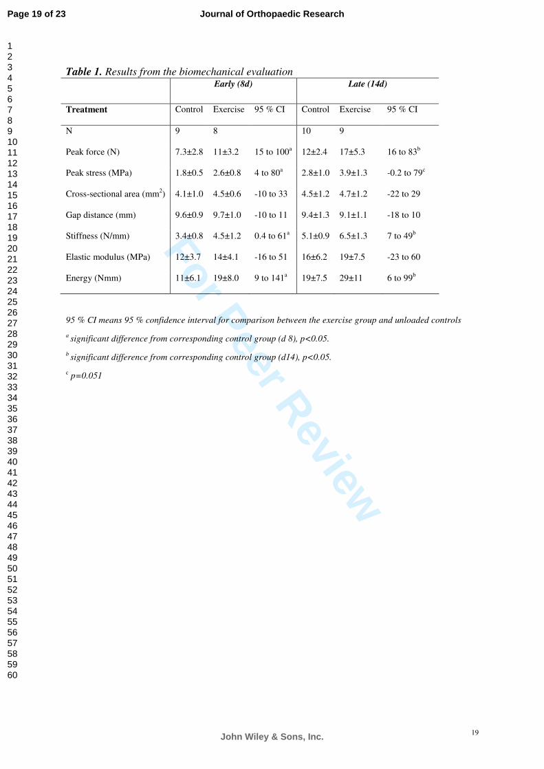
Table 1. Animal numbers in each group (N) and biomechanical findings in tendon calluses at
8 and 14 days after tendon transection. Controls were completely unloaded and exercise rats
were subjected to unloading in combination with 30 minutes of daily exercise for 4 days.
Values are expressed as mean±SD. Confidence intervals refer to the difference between
group mean values. This difference is expressed as percent of control mean.
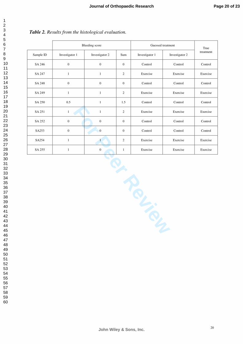
Table 2. Scores for each sample (SA246-SA255) from the histological evaluation. 1 means
extensive bleeding. 0 means less bleeding. Each investigator also guessed if a sample was
derived from exercise or control group.
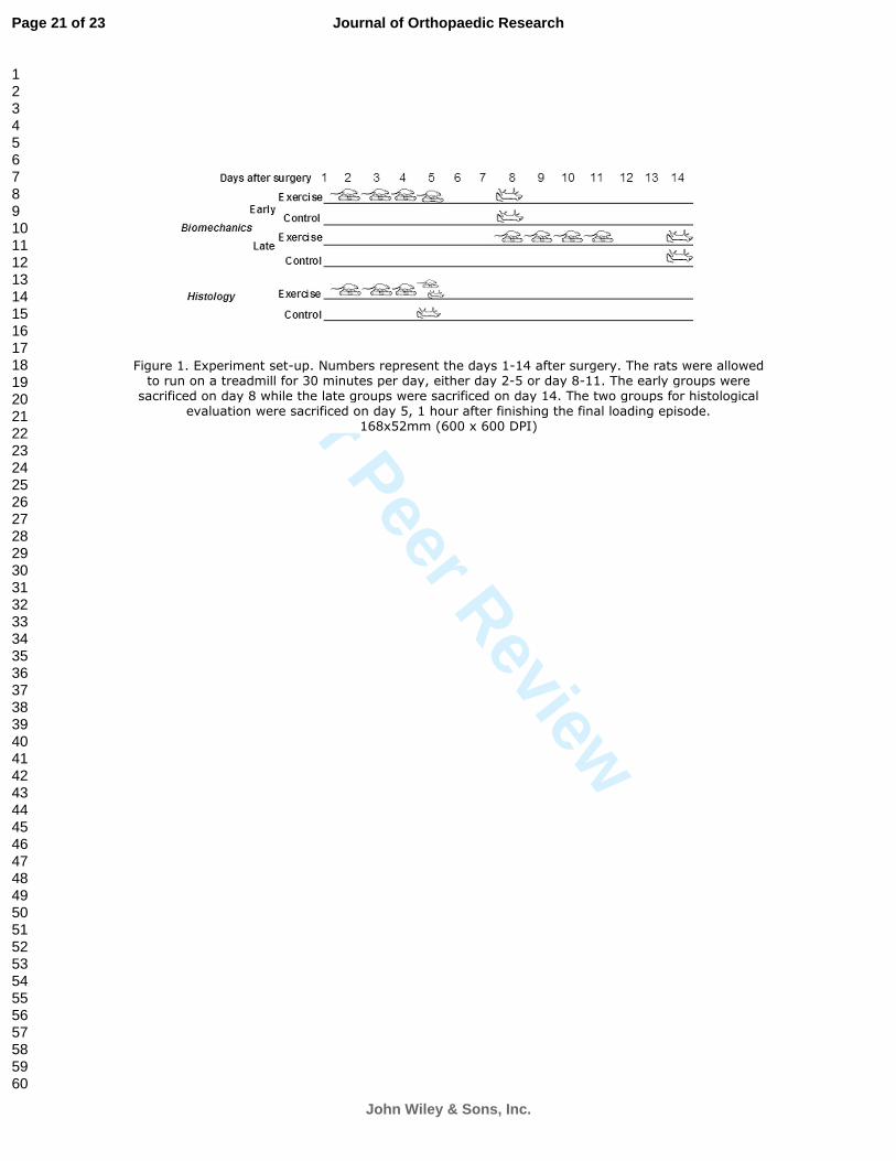
Figure 1. Experimental set-up. Numbers represent the days 1-14 after surgery. The rats were
allowed to run on a treadmill for 30 minutes per day, either day 2-5 or day 8-11. The early
group was euthanized on day 8, while the late group was euthanized on day 14. The two
groups for histological evaluation were sacrificed on day 5, 1 hour after the last loading
episode.
Figure 2. Peak force (A) and Peak stress (B) at 8 and 14 days after tendon transection with
or without loading. Controls were completely unloaded tendons harvested at the same time as
the exercise groups. Exercise consisted of 30 minutes of running on a treadmill, once a day
for 4 days. Peak force for the exercise groups differed from corresponding control groups at
each time point (p<0.01). Peak stress for the exercise group differed from the corresponding
control group at the early time point (p<0.05).
Page 17 of 23
John Wiley & Sons, Inc.
Journal of Orthopaedic Research
123456789101112131415161718192021222324252627282930313233343536373839404142434445464748495051525354555657585960

For Peer Review
18
Figure 3. Bleeding in the tendon callus. a) Overview of healing tendon tissue, black
broken line indicates the interface between callus tissue and transected tendon stump
(magnification x 2) and b) Area of bleeding, extravasated erythrocytes in the tissue
(magnification x 40).
Page 18 of 23
John Wiley & Sons, Inc.
Journal of Orthopaedic Research
123456789101112131415161718192021222324252627282930313233343536373839404142434445464748495051525354555657585960
For Peer Review
19
Table 1. Results from the biomechanical evaluation
Early (8d) Late (14d)
Treatment Control Exercise 95 % CI Control Exercise 95 % CI
N 9 8 10 9
Peak force (N) 7.3±2.8 11±3.2 15 to 100a 12±2.4 17±5.3 16 to 83b
Peak stress (MPa) 1.8±0.5 2.6±0.8 4 to 80a 2.8±1.0 3.9±1.3 -0.2 to 79c
Cross-sectional area (mm2) 4.1±1.0 4.5±0.6 -10 to 33 4.5±1.2 4.7±1.2 -22 to 29
Gap distance (mm) 9.6±0.9 9.7±1.0 -10 to 11 9.4±1.3 9.1±1.1 -18 to 10
Stiffness (N/mm) 3.4±0.8 4.5±1.2 0.4 to 61a 5.1±0.9 6.5±1.3 7 to 49b
Elastic modulus (MPa) 12±3.7 14±4.1 -16 to 51 16±6.2 19±7.5 -23 to 60
Energy (Nmm) 11±6.1 19±8.0 9 to 141a 19±7.5 29±11 6 to 99b
95 % CI means 95 % confidence interval for comparison between the exercise group and unloaded controls
a significant difference from corresponding control group (d 8), p<0.05.
b significant difference from corresponding control group (d14), p<0.05.
c p=0.051
Page 19 of 23
John Wiley & Sons, Inc.
Journal of Orthopaedic Research
123456789101112131415161718192021222324252627282930313233343536373839404142434445464748495051525354555657585960
For Peer Review
20
Table 2. Results from the histological evaluation.
Bleeding score Guessed treatment
Sample ID Investigator 1 Investigator 2 Sum Investigator 1 Investigator 2
True treatment
SA 246 0 0 0 Control Control Control
SA 247 1 1 2 Exercise Exercise Exercise
SA 248 0 0 0 Control Control Control
SA 249 1 1 2 Exercise Exercise Exercise
SA 250 0.5 1 1.5 Control Control Control
SA 251 1 1 2 Exercise Exercise Exercise
SA 252 0 0 0 Control Control Control
SA253 0 0 0 Control Control Control
SA254 1 1 2 Exercise Exercise Exercise
SA 255 1 0 1 Exercise Exercise Exercise
Page 20 of 23
John Wiley & Sons, Inc.
Journal of Orthopaedic Research
123456789101112131415161718192021222324252627282930313233343536373839404142434445464748495051525354555657585960
For Peer Review
Figure 1. Experiment set-up. Numbers represent the days 1-14 after surgery. The rats were allowed to run on a treadmill for 30 minutes per day, either day 2-5 or day 8-11. The early groups were
sacrificed on day 8 while the late groups were sacrificed on day 14. The two groups for histological evaluation were sacrificed on day 5, 1 hour after finishing the final loading episode.
168x52mm (600 x 600 DPI)
Page 21 of 23
John Wiley & Sons, Inc.
Journal of Orthopaedic Research
123456789101112131415161718192021222324252627282930313233343536373839404142434445464748495051525354555657585960

For Peer Review
Figure 2. Peak force (A) and Peak stress (B) at 8 and 14 days after tendon transection with or without loading. Controls were completely unloaded tendons harvested at the same time as the exercise groups. Exercise consisted of 30 minutes of walking on a treadmill, once a day for 4 days.
82x45mm (600 x 600 DPI)
Page 22 of 23
John Wiley & Sons, Inc.
Journal of Orthopaedic Research
123456789101112131415161718192021222324252627282930313233343536373839404142434445464748495051525354555657585960

For Peer Review
Figure 3. Bleeding in the tendon callus. a) Overview of healing tendon tissue, black broken line indicates the interface between callus tissue and transected tendon stump (magnification x 2) and
b) Area of bleeding, extravasated erythrocytes in the tissue (magnification x 40).
105x56mm (600 x 600 DPI)
Page 23 of 23
John Wiley & Sons, Inc.
Journal of Orthopaedic Research
123456789101112131415161718192021222324252627282930313233343536373839404142434445464748495051525354555657585960