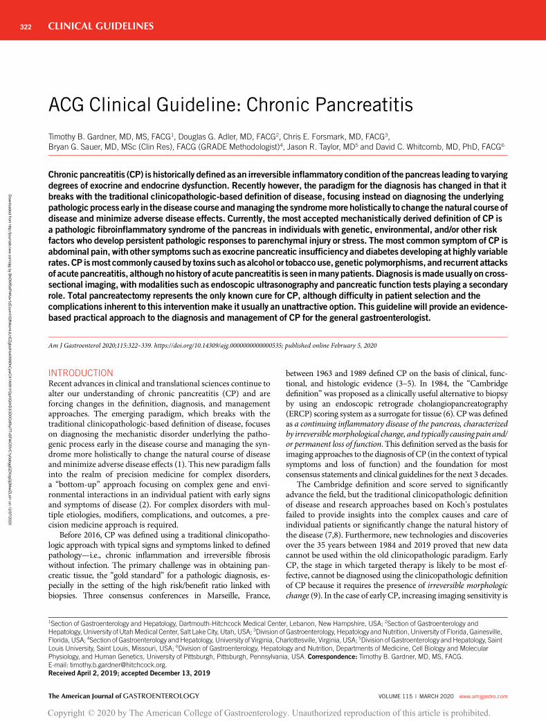
ACG Clinical Guideline: Chronic Pancreatitis
18
Downloaded from http://journals.lww.com/ajg by BhDMf5ePHKav1zEoum1tQfN4a+kJLhEZgbsIHo4XMi0hCywCX1AWnYQp/IlQrHD3i3D0OdRyi7TvSFl4Cf3VC1y0abggQZXdgGj2MwlZLeI= on 12/07/2020 ACG Clinical Guideline: Chronic Pancreatitis Timothy B. Gardner, MD, MS, FACG 1 , Douglas G. Adler, MD, FACG 2 , Chris E. Forsmark, MD, FACG 3 , Bryan G. Sauer, MD, MSc (Clin Res), FACG (GRADE Methodologist) 4 , Jason R. Taylor, MD 5 and David C. Whitcomb, MD, PhD, FACG 6 Chronic pancreatitis (CP) is historically defined as an irreversible inflammatory condition of the pancreas leading to varying degrees of exocrine and endocrine dysfunction. Recently however, the paradigm for the diagnosis has changed in that it breaks with the traditional clinicopathologic-based definition of disease, focusing instead on diagnosing the underlying pathologic process early in the disease course and managing the syndrome more holistically to change the natural course of disease and minimize adverse disease effects. Currently, the most accepted mechanistically derived definition of CP is a pathologic fibroinflammatory syndrome of the pancreas in individuals with genetic, environmental, and/or other risk factors who develop persistent pathologic responses to parenchymal injury or stress. The most common symptom of CP is abdominal pain, with other symptoms such as exocrine pancreatic insufficiency and diabetes developing at highly variable rates. CP is most commonly caused by toxins such as alcohol or tobacco use, genetic polymorphisms, and recurrent attacks of acute pancreatitis, although no history of acute pancreatitis is seen in many patients. Diagnosis is made usually on cross- sectional imaging, with modalities such as endoscopic ultrasonography and pancreatic function tests playing a secondary role. Total pancreatectomy represents the only known cure for CP, although difficulty in patient selection and the complications inherent to this intervention make it usually an unattractive option. This guideline will provide an evidence- based practical approach to the diagnosis and management of CP for the general gastroenterologist. Am J Gastroenterol 2020;115:322–339. https://doi.org/10.14309/ajg.0000000000000535; published online February 5, 2020 INTRODUCTION Recent advances in clinical and translational sciences continue to alter our understanding of chronic pancreatitis (CP) and are forcing changes in the definition, diagnosis, and management approaches. The emerging paradigm, which breaks with the traditional clinicopathologic-based definition of disease, focuses on diagnosing the mechanistic disorder underlying the patho- genic process early in the disease course and managing the syn- drome more holistically to change the natural course of disease and minimize adverse disease effects (1). This new paradigm falls into the realm of precision medicine for complex disorders, a “bottom-up” approach focusing on complex gene and envi- ronmental interactions in an individual patient with early signs and symptoms of disease (2). For complex disorders with mul- tiple etiologies, modifiers, complications, and outcomes, a pre- cision medicine approach is required. Before 2016, CP was defined using a traditional clinicopatho- logic approach with typical signs and symptoms linked to defined pathology—i.e., chronic inflammation and irreversible fibrosis without infection. The primary challenge was in obtaining pan- creatic tissue, the “gold standard” for a pathologic diagnosis, es- pecially in the setting of the high risk/benefit ratio linked with biopsies. Three consensus conferences in Marseille, France, between 1963 and 1989 defined CP on the basis of clinical, func- tional, and histologic evidence (3–5). In 1984, the “Cambridge definition” was proposed as a clinically useful alternative to biopsy by using an endoscopic retrograde cholangiopancreatography (ERCP) scoring system as a surrogate for tissue (6). CP was defined as a continuing inflammatory disease of the pancreas, characterized by irreversible morphological change, and typically causing pain and/ or permanent loss of function. This definition served as the basis for imaging approaches to the diagnosis of CP (in the context of typical symptoms and loss of function) and the foundation for most consensus statements and clinical guidelines for the next 3 decades. The Cambridge definition and score served to significantly advance the field, but the traditional clinicopathologic definition of disease and research approaches based on Koch’s postulates failed to provide insights into the complex causes and care of individual patients or significantly change the natural history of the disease (7,8). Furthermore, new technologies and discoveries over the 35 years between 1984 and 2019 proved that new data cannot be used within the old clinicopathologic paradigm. Early CP, the stage in which targeted therapy is likely to be most ef- fective, cannot be diagnosed using the clinicopathologic definition of CP because it requires the presence of irreversible morphologic change (9). In the case of early CP, increasing imaging sensitivity is 1 Section of Gastroenterology and Hepatology, Dartmouth-Hitchcock Medical Center, Lebanon, New Hampshire, USA; 2 Section of Gastroenterology and Hepatology, University of Utah Medical Center, Salt Lake City, Utah, USA; 3 Division of Gastroenterology, Hepatology and Nutrition, University of Florida, Gainesville, Florida, USA; 4 Section of Gastroenterology and Hepatology, University of Virginia, Charlottesville, Virginia, USA; 5 Division of Gastroenterology and Hepatology, Saint Louis University, Saint Louis, Missouri, USA; 6 Division of Gastroenterology, Hepatology and Nutrition, Departments of Medicine, Cell Biology and Molecular Physiology, and Human Genetics, University of Pittsburgh, Pittsburgh, Pennsylvania, USA. Correspondence: Timothy B. Gardner, MD, MS, FACG. E-mail: [email protected]. Received April 2, 2019; accepted December 13, 2019 The American Journal of GASTROENTEROLOGY VOLUME 115 | MARCH 2020 www.amjgastro.com CLINICAL GUIDELINES 322 Copyright © 2020 by The American College of Gastroenterology. Unauthorized reproduction of this article is prohibited.
Transcript of ACG Clinical Guideline: Chronic Pancreatitis
Dow
nloadedfrom
http://journals.lww.com
/ajgby
BhDMf5ePH
Kav1zEoum1tQ
fN4a+kJLhEZgbsIH
o4XMi0hC
ywCX1AW
nYQp/IlQ
rHD3i3D
0OdR
yi7TvSFl4Cf3VC
1y0abggQZXdgG
j2MwlZLeI=
on12/07/2020
Downloadedfromhttp://journals.lww.com/ajgbyBhDMf5ePHKav1zEoum1tQfN4a+kJLhEZgbsIHo4XMi0hCywCX1AWnYQp/IlQrHD3i3D0OdRyi7TvSFl4Cf3VC1y0abggQZXdgGj2MwlZLeI=on12/07/2020
ACG Clinical Guideline: Chronic PancreatitisTimothy B. Gardner, MD, MS, FACG1, Douglas G. Adler, MD, FACG2, Chris E. Forsmark, MD, FACG3,Bryan G. Sauer, MD, MSc (Clin Res), FACG (GRADE Methodologist)4, Jason R. Taylor, MD5 and David C. Whitcomb, MD, PhD, FACG6
Chronic pancreatitis (CP) is historically defined as an irreversible inflammatory condition of the pancreas leading to varying
degrees of exocrine and endocrine dysfunction. Recently however, the paradigm for the diagnosis has changed in that it
breaks with the traditional clinicopathologic-based definition of disease, focusing instead on diagnosing the underlying
pathologic process early in the disease course andmanaging the syndromemoreholistically to change the natural course of
disease and minimize adverse disease effects. Currently, the most accepted mechanistically derived definition of CP is
a pathologic fibroinflammatory syndrome of the pancreas in individuals with genetic, environmental, and/or other risk
factors who develop persistent pathologic responses to parenchymal injury or stress. The most common symptom of CP is
abdominal pain, with other symptoms such as exocrine pancreatic insufficiency and diabetes developing at highly variable
rates. CP ismost commonly causedby toxins such as alcohol or tobaccouse, genetic polymorphisms, and recurrent attacks
of acutepancreatitis, althoughnohistory of acute pancreatitis is seen inmanypatients. Diagnosis ismadeusually oncross-
sectional imaging, with modalities such as endoscopic ultrasonography and pancreatic function tests playing a secondary
role. Total pancreatectomy represents the only known cure for CP, although difficulty in patient selection and the
complications inherent to this interventionmake it usually an unattractive option. This guideline will provide an evidence-
based practical approach to the diagnosis and management of CP for the general gastroenterologist.
Am J Gastroenterol 2020;115:322–339. https://doi.org/10.14309/ajg.0000000000000535; published online February 5, 2020
INTRODUCTIONRecent advances in clinical and translational sciences continue toalter our understanding of chronic pancreatitis (CP) and areforcing changes in the definition, diagnosis, and managementapproaches. The emerging paradigm, which breaks with thetraditional clinicopathologic-based definition of disease, focuseson diagnosing the mechanistic disorder underlying the patho-genic process early in the disease course and managing the syn-drome more holistically to change the natural course of diseaseand minimize adverse disease effects (1). This new paradigm fallsinto the realm of precision medicine for complex disorders,a “bottom-up” approach focusing on complex gene and envi-ronmental interactions in an individual patient with early signsand symptoms of disease (2). For complex disorders with mul-tiple etiologies, modifiers, complications, and outcomes, a pre-cision medicine approach is required.
Before 2016, CP was defined using a traditional clinicopatho-logic approach with typical signs and symptoms linked to definedpathology—i.e., chronic inflammation and irreversible fibrosiswithout infection. The primary challenge was in obtaining pan-creatic tissue, the “gold standard” for a pathologic diagnosis, es-pecially in the setting of the high risk/benefit ratio linked withbiopsies. Three consensus conferences in Marseille, France,
between 1963 and 1989 defined CP on the basis of clinical, func-tional, and histologic evidence (3–5). In 1984, the “Cambridgedefinition” was proposed as a clinically useful alternative to biopsyby using an endoscopic retrograde cholangiopancreatography(ERCP) scoring system as a surrogate for tissue (6). CP was definedas a continuing inflammatory disease of the pancreas, characterizedby irreversiblemorphological change, and typically causingpainand/or permanent loss of function. This definition served as the basis forimaging approaches to the diagnosis of CP (in the context of typicalsymptoms and loss of function) and the foundation for mostconsensus statements and clinical guidelines for the next 3 decades.
The Cambridge definition and score served to significantlyadvance the field, but the traditional clinicopathologic definitionof disease and research approaches based on Koch’s postulatesfailed to provide insights into the complex causes and care ofindividual patients or significantly change the natural history ofthe disease (7,8). Furthermore, new technologies and discoveriesover the 35 years between 1984 and 2019 proved that new datacannot be used within the old clinicopathologic paradigm. EarlyCP, the stage in which targeted therapy is likely to be most ef-fective, cannot be diagnosed using the clinicopathologic definitionof CP because it requires the presence of irreversible morphologicchange (9). In the case of early CP, increasing imaging sensitivity is
1Section of Gastroenterology and Hepatology, Dartmouth-Hitchcock Medical Center, Lebanon, New Hampshire, USA; 2Section of Gastroenterology andHepatology, University of UtahMedical Center, Salt Lake City, Utah, USA; 3Division of Gastroenterology, Hepatology andNutrition, University of Florida, Gainesville,Florida, USA; 4Section of Gastroenterology andHepatology, University of Virginia, Charlottesville, Virginia, USA; 5Division of Gastroenterology andHepatology, SaintLouis University, Saint Louis, Missouri, USA; 6Division of Gastroenterology, Hepatology and Nutrition, Departments of Medicine, Cell Biology and MolecularPhysiology, and Human Genetics, University of Pittsburgh, Pittsburgh, Pennsylvania, USA. Correspondence: Timothy B. Gardner, MD, MS, FACG.E-mail: [email protected] April 2, 2019; accepted December 13, 2019
The American Journal of GASTROENTEROLOGY VOLUME 115 | MARCH 2020 www.amjgastro.com
CLINICAL GUIDELINES322
Copyright © 2020 by The American College of Gastroenterology. Unauthorized reproduction of this article is prohibited.
associated with decreasing specificity. Furthermore, examinationof the pathology, or images as a surrogate, offers little insight intoany of the dozens of potential underlying simple or complex eti-ologies.Genetics clearlyplay amajor role inpancreatic diseases, butbecause germline variants rarely link to specific symptoms or tissuepathologic features, they cannot be understood within the clini-copathologic framework. It follows that clinical genetic reports thatare framed by classic Mendelian geneticists or anatomic patholo-gists within the clinicopathologic framework provide little clinicalguidance, especially in the later stages of disease. In addition, thepractice of diagnosing and tracking disease progression based onfibrosis may be flawed because the degree of fibrosis correlatespoorly with pain, exocrine pancreatic insufficiency (EPI), diabetesmellitus (DM), progressive disease, or cancer risk—the primaryconcerns of clinical care (10–14).
In summary, the traditional clinicopathologic framework thatdefines theCP syndrome by irreversible damage results in years ofdelay between symptom onset and diagnosis and usually fails toidentify or address the underlying etiology, cannot predict theclinical course, cannot direct preventative treatments that changedisease trajectory, and remains limited to symptomatic or sup-portive care and replacement of lost gland function.
In 2016, a new Mechanistic Definition of CP was published,and later adopted, by themajor pancreas societies as the preferreddefinition worldwide (15,16). TheMechanistic Definition affirmsthe characteristics of end-stage disease as pancreatic atrophy, fi-brosis, pain syndromes, duct distortion and strictures, calcifica-tions, pancreatic exocrine dysfunction, pancreatic endocrinedysfunction, and dysplasia, but also addresses the disease mech-anism as a pathologic fibroinflammatory syndrome of the pancreasin individuals with genetic, environmental, and/or other risk fac-tors who develop persistent pathologic responses to parenchymalinjury or stress. The definition is linked to a progressive model toorganize risk factors, clinical scenarios, disease biomarkers, se-quential and progressive features, and individual variables withina lifetime. It was also designed to assess the differential diagnosisof disorders with pathologic features that overlap with early CP,such as fibrosis, atrophy, maldigestion, and diabetes.
Within the framework of the Mechanistic Definition, it isimportant to recognize the difference between pancreatic dys-function, pancreatitis-related disorders, and pancreatic disease.The term dysfunction is a dynamic term that describes a variationin the action of an entity that deviates from normal in a negativeway. A medical disorder indicates disruption of the normalfunctions of specialized cells or systems resulting in abnormalsigns, symptoms, biomarkers, and/or responses. A disease is anabnormal condition in a living animal that is defined by con-sensus criteria consisting of abnormal signs and symptoms, ab-normal biomarkers, and typical pathologic features.
An example of pancreatic dysfunction is the presence of a ge-netic mutation in the CFTR gene locus that causing variations inRNA expression or splicing, or changes in amino acid sequencecausing defective processing, trafficking, or channel opening. Thedysfunctionmay be tolerated by adaptivemechanisms and limitedcell stress so that the cells using CFTR do not fail under mostconditions. An example of a pancreatitis-related disorderwould beprotein dysfunction that causes the pancreatic duct cell to fail torespond normally when the cell is strongly stimulated or stressed,resulting in a duct cell dysfunction. When the compensatorythreshold is exceeded, then internal and external stress and injurysignals are generated thatmay cause clinical signs and symptomsof
diseases, such as an episode of acute pancreatitis (17). Pancreaticdiseases are conditions that are typically associated with pathologysuch as acute pancreatitis, recurrent acute pancreatitis (RAP), CP,and complex syndromes that affect the pancreas such as cysticfibrosis.When the clinical evaluation of the patient determines thepresenceof clinical, functional, andmorphologic features thatmeetconsensus criteria, then the patient may be diagnosed as havinga disease, such as CP. If multiple organs are involved, then thepatient is diagnosed with a genetic disease, such as cystic fibrosiswith pancreatic sufficiency or insufficiency. Precision medicinetherefore focuses on determining the dysfunction and the disorderand seeks to use targeted therapy to prevent the condition fromprogressing to a clinicopathologic-defined disease. However, newstudies are needed to evaluate the effectiveness of targeted therapiesin individual patients (2,18). At this time, a paucity of studies existsspecifically using the new Mechanistic Definition of CP.
In this context, a group of experts within the ACGwere taskedto complete a systematic review of the literature concerning CPand develop guidelines for the membership. Based on theframework of the traditional definition and approaches to CP,much of the older literature provides limited insights and contextfor strong recommendations. The authors expect, moving for-ward, that future guidelines will be more reliant on studies exe-cuted under the framework of the Mechanistic Definition of CP.Where possible, specific clinical questions are poised, followed bya review and recommendations based on the older literature andcomments on future directions.
METHODOLOGYWith the assistance of a health science librarian, a literaturesearch was completed through MEDLINE (1946-current),Embase (1974-current), Web of Science (1900-current), and theCochrane Library. All databases were searched up to February2018. The search strategy MeSH terms included chronic pan-creatitis or pancreatitis (chronic or minimal change). Searcheswere limited to the English language. Two authors (T.B.G. andJ.R.T.) independently reviewed all unique articles and includedthose articles that met consensus criteria. The authors also in-corporated articles from review of references in retrievedmanuscripts as well as relevant studies known to the authors. Thesearch results were primarily randomized trials. If these were notavailable, then meta-analyses and systematic reviews were used.
The guideline is structured in sections, each with recom-mendations or key concepts, and summaries of the evidencebased on the PICO question format that is a consistent “formula”for developing answerable, researchable questions. PICO is anacronym that includes the following 4 aspects important for re-search questions: 1. population/problem, 2. intervention, 3.comparison, and 4. outcome. The PICO questions were de-veloped by the consensus of the authors and served as the basis foreach recommendation and key concept (Table 1). Each recom-mendation statement has an associated assessment of the qualityof evidence and strength of recommendation based on theGrading of Recommendations Assessment, Development, andEvaluation (GRADE) process. The GRADE system was used toevaluate the quality of supporting evidence. A “Strong” recom-mendation is made when the benefits clearly outweigh the neg-atives and/or the result of no action. “Conditional” is used whensome uncertainty remains about the balance of benefits and po-tential harms. Statements with a “strong” recommendation arestated with “We recommend,” whereas statements with
© 2020 by The American College of Gastroenterology The American Journal of GASTROENTEROLOGY
Chronic Pancreatitis 323
Copyright © 2020 by The American College of Gastroenterology. Unauthorized reproduction of this article is prohibited.
a conditional recommendation are stated with “We suggest.” Thequality of evidence is graded from high to low. “High” qualityevidence indicates that further research is unlikely to change theauthors’ confidence in the estimate of the effect, and that we arevery confident that the true effect lies close to that of the estimateof the effect. “Moderate” quality evidence is associated withmoderate confidence in the effect estimate, although further re-search would be likely to have an impact on the confidence of theestimate, whereas “Low” quality evidence indicates that furtherstudy would likely have an important impact on the confidence inthe estimate of the effect and would likely change the estimate.“Very low” quality evidence indicates very little confidence in theeffect estimate, and that the true effect is likely to be substantiallydifferent from the estimate of the effect.
Key concepts are statements that are not amenable to theGRADE process, either because of the structure of the statement or
because of the available evidence. In some instances, key conceptsare based on the extrapolation of evidence and/or expert opinion.The GRADE recommendations and key concept statements fromthis guideline are found in Tables 2 and 3, respectively.
DIAGNOSIS OF CPRecommendation
1. We recommend computed tomography (CT) or MRI for the first-linediagnosis of CP. Either test should be the first choice for thediagnosis of CP. Endoscopic ultrasonography (EUS), because of itsinvasiveness and lack of specificity, should be used only if thediagnosis is in question after cross-sectional imaging is performed(strong recommendation, low quality of evidence).
Summary of evidence. The diagnosis of CP has been difficultbecause there is a debate about the gold-standard test that
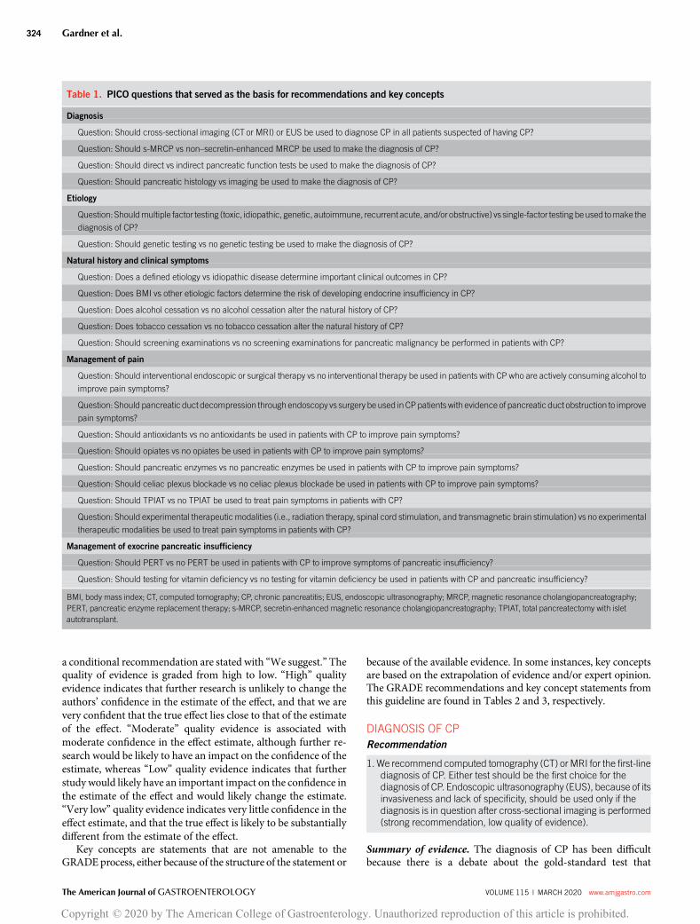
Table 1. PICO questions that served as the basis for recommendations and key concepts
Diagnosis
Question: Should cross-sectional imaging (CT or MRI) or EUS be used to diagnose CP in all patients suspected of having CP?
Question: Should s-MRCP vs non–secretin-enhanced MRCP be used to make the diagnosis of CP?
Question: Should direct vs indirect pancreatic function tests be used to make the diagnosis of CP?
Question: Should pancreatic histology vs imaging be used to make the diagnosis of CP?
Etiology
Question: Shouldmultiple factor testing (toxic, idiopathic, genetic, autoimmune, recurrent acute, and/or obstructive) vs single-factor testing beused tomake the
diagnosis of CP?
Question: Should genetic testing vs no genetic testing be used to make the diagnosis of CP?
Natural history and clinical symptoms
Question: Does a defined etiology vs idiopathic disease determine important clinical outcomes in CP?
Question: Does BMI vs other etiologic factors determine the risk of developing endocrine insufficiency in CP?
Question: Does alcohol cessation vs no alcohol cessation alter the natural history of CP?
Question: Does tobacco cessation vs no tobacco cessation alter the natural history of CP?
Question: Should screening examinations vs no screening examinations for pancreatic malignancy be performed in patients with CP?
Management of pain
Question: Should interventional endoscopic or surgical therapy vs no interventional therapy be used in patients with CP who are actively consuming alcohol to
improve pain symptoms?
Question: Should pancreatic duct decompression through endoscopy vs surgery be used in CPpatients with evidence of pancreatic duct obstruction to improve
pain symptoms?
Question: Should antioxidants vs no antioxidants be used in patients with CP to improve pain symptoms?
Question: Should opiates vs no opiates be used in patients with CP to improve pain symptoms?
Question: Should pancreatic enzymes vs no pancreatic enzymes be used in patients with CP to improve pain symptoms?
Question: Should celiac plexus blockade vs no celiac plexus blockade be used in patients with CP to improve pain symptoms?
Question: Should TPIAT vs no TPIAT be used to treat pain symptoms in patients with CP?
Question: Should experimental therapeutic modalities (i.e., radiation therapy, spinal cord stimulation, and transmagnetic brain stimulation) vs no experimental
therapeutic modalities be used to treat pain symptoms in patients with CP?
Management of exocrine pancreatic insufficiency
Question: Should PERT vs no PERT be used in patients with CP to improve symptoms of pancreatic insufficiency?
Question: Should testing for vitamin deficiency vs no testing for vitamin deficiency be used in patients with CP and pancreatic insufficiency?
BMI, body mass index; CT, computed tomography; CP, chronic pancreatitis; EUS, endoscopic ultrasonography; MRCP, magnetic resonance cholangiopancreatography;PERT, pancreatic enzyme replacement therapy; s-MRCP, secretin-enhanced magnetic resonance cholangiopancreatography; TPIAT, total pancreatectomy with isletautotransplant.
The American Journal of GASTROENTEROLOGY VOLUME 115 | MARCH 2020 www.amjgastro.com
Gardner et al.324
Copyright © 2020 by The American College of Gastroenterology. Unauthorized reproduction of this article is prohibited.
establishes the diagnosis. Furthermore, it represents a later stageof progressive disorders resulting in irreversible morphologicdamage with variable clinical consequences. The diagnosis ismade often using a combination of modalities, including expo-sure risk, underlying predisposition, cross-sectional imaging, anddirect and/or indirect pancreatic function tests. In fact, likely themost useful diagnostic test for CP is a careful history and physicalexamination, as the pretest probability and clinical suspicion areintegral for diagnosis—i.e., if the patient is in a high-risk group,the morphologic changes are a more accurate biomarker of CPrather than another disorder with a similar differential diagnosis.It is critical to assess the patient’s risk factors for CP, includingfamily and exposure history, the nature and character of thepatient’s pain, whether or not they have had previous episodes ofacute pancreatitis, and whether they have related conditions suchas steatorrhea and/or symptoms of vitamin deficiency.
However, in patients with clinical symptoms of an in-flammatory disorder of the pancreas (e.g., previous episode ofacute pancreatitis, characteristic pain, and/or maldigestion) and/or a suggestive gene–environment risk assessment, then cross-sectional imaging should be the first test used to establish thediagnosis of CP because it is universally available, reproducible,and valid when compared with other modalities.
No randomized controlled trials (RCTs) have been performedspecifically comparing cross-sectional imaging with EUS for thediagnosis of CP with the caveat that the test characteristics ofdiagnostic modalities are generally not amenable to RCTs. The
best evidence comparing modalities is from a systematic reviewand meta-analysis of 43 studies and 3460 patients with suspectedCP in which the sensitivity estimates of EUS, MRI, and CT were81% (95% confidence interval [CI]: 70%–89%), 78% (95% CI:69%–85%), and 75% (95% CI: 66%–83%), respectively, and didnot differ significantly from each other (19). Estimates of speci-ficity were comparable for EUS (90%; 95% CI: 82%–95%), ERCP(94%; 95% CI: 87%–98%), CT (91%; 95% CI: 81%–96%), MRI(96%; 95% CI: 90%–98%), and ultrasound (US) (98%; 95% CI:89%–100%). A limitation of thismeta-analysis, however, was thatnot all the studies included a histologic gold standard to establishthe type of inflammation for comparison.
Given the vast discrepancy in cost, availability, invasiveness,and objectivity, we believe that cross-sectional imaging should bethe first-line test for the diagnosis of CP.Owing to its invasivenessand issues surrounding availability, intrarate reproducibility, anddiscrepancy over the definition and importance of specific di-agnostic criteria, EUS should be used to diagnoseCP alone if thereis uncertainty following cross-sectional imaging (20).
Multiple other imaging modalities and scoring systems havebeen used to establish the diagnosis of CP, including contrast-enhanced EUS, ERCP, transcutaneous ultrasonography, andpancreatic elastography (6,21–24). However, high-quality RCTevidence is not available to warrant their inclusion as first-linediagnostic tests for CP in place of cross-sectional imaging or EUS.Practical clinical approach.Demonstrating typical morphologicchanges in the pancreas is a critical component of the definition of
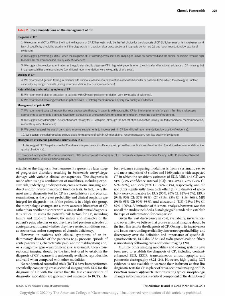
Table 2. Recommendations on the management of CP
Diagnosis of CP
1. We recommend CTor MRI for the first-line diagnosis of CP. Either test should be the first choice for the diagnosis of CP. EUS, because of its invasiveness and
lack of specificity, should be used only if the diagnosis is in question after cross-sectional imaging is performed (strong recommendation, low quality of
evidence).
2. We suggest performing s-MRCP when the diagnosis of CP following cross-sectional imaging or EUS is not confirmed and the clinical suspicion remains high
(conditional recommendation, low quality of evidence).
3. We suggest histological examination as the gold standard to diagnose CP in high-risk patients when the clinical and functional evidence of CP is strong, but
imaging modalities are inconclusive (conditional recommendation, very low quality of evidence).
Etiology of CP
4. We recommend genetic testing in patients with clinical evidence of a pancreatitis-associated disorder or possible CP in which the etiology is unclear,
especially in younger patients (strong recommendation, low quality of evidence).
Natural history and clinical symptoms of CP
5. We recommend alcohol cessation in patients with CP (strong recommendation, very low quality of evidence).
6. We recommend smoking cessation in patients with CP (strong recommendation, very low quality of evidence).
Management of pain in CP
7. We recommend surgical intervention over endoscopic therapy in patients with obstructive CP for the long-term relief of pain if first-line endoscopic
approaches to pancreatic drainage have been exhausted or unsuccessful (strong recommendation, moderate quality of evidence).
8. We suggest considering the use of antioxidant therapy for CP with pain, although the benefit of pain reduction is likely limited (conditional recommendation,
moderate quality of evidence).
9. We do not suggest the use of pancreatic enzyme supplements to improve pain in CP (conditional recommendation, low quality of evidence).
10. We suggest considering celiac plexus block for treatment of pain in CP (conditional recommendation, very low quality of evidence).
Management of exocrine pancreatic insufficiency in CP
11. We suggest PERTin patients with CP and exocrine pancreatic insufficiency to improve the complications of malnutrition (conditional recommendation, low
quality of evidence).
CT, computed tomography; CP, chronic pancreatitis; EUS, endoscopic ultrasonography; PERT, pancreatic enzyme replacement therapy; s-MRCP, secretin-enhancedmagnetic resonance cholangiopancreatography.
© 2020 by The American College of Gastroenterology The American Journal of GASTROENTEROLOGY
Chronic Pancreatitis 325
Copyright © 2020 by The American College of Gastroenterology. Unauthorized reproduction of this article is prohibited.
CP, as imaging is a surrogate for histology. In the absence ofa universally agreed-on gold standard for the diagnosis of CP andthe challenges in obtaining high-quality and representative his-tology, cross-sectional imaging is a familiar test for most clini-cians and should be used as the initial test for diagnosis.Endoscopic US should be used if the diagnosis is still in doubtafter cross-sectional imaging or if there is a concern about“minimal change disease” (CP without evidence of fibrosis) thatcannot be visualized on cross-sectional imaging. There is notenough quality evidence to recommend a specific type of EUSscoring system, nor the number or types of criteria that should beused to definitively diagnose CP using this modality. Therefore,CP remains a clinical diagnosis, integrating and balancing thestrength of evidence of the clinical scenario, the presence of riskfactors, and the exclusion of other diseases in the differentialdiagnosis.
Recommendation
2. We suggest performing secretin-enhanced magnetic resonancecholangiopancreatography (s-MRCP) when the diagnosis of CPfollowing cross-sectional imaging or EUS is not confirmed and theclinical suspicion remains high (conditional recommendation, lowquality of evidence).
Summary of evidence.When the diagnosis of CP cannot bemadefollowing standard cross-sectional imaging or EUS, s-MRCP issuggested. s-MRCP allows for better visualization of themain- andside-branch ducts by stimulating the release of bicarbonate fromthe pancreatic duct cells. It also allows for quantification of thedegree of filling into the duodenum, which may correlate with theseverity of CP and help quantify the degree of pancreatic exocrine
function (25–27). Given its expense, s-MRCP should be used onlywhen the diagnosis is not confirmed with first-line testing.
There are no RCTs specifically evaluating the role of s-MRCPin the diagnosis of CP, although one systematic review has beenperformed (28). Evaluating 69 original articles and 15 reviews, theauthors found that dynamic thick-slab 2-dimensionalMRCPwasthemost used imaging sequence (86%). The diameter of themainpancreatic duct (75%) and pancreatic exocrine function based onvisual grading of duodenal filling (67%) were the most evaluatedpancreatic features. Smaller studies suggest the diagnostic value ofsecretin-enhanced studies in childrenwith idiopathic CP and alsoin the identification of minimal change disease (29,30).Practical clinical approach. If cross-sectional imaging and EUSare not diagnostic of CP, s-MRCP can be used to identify subtleductal abnormalities such as dilated branches or an ectatic duct,which may indicate morphologic changes consistent with imag-ing criteria for CP. Structural imaging is a biomarker of thepancreas morphology and is most sensitive to changes caused byfibrosis with distortion of the ducts and calcifications. However,the diagnosis of CP should not be made solely on s-MRCPfindings or other imaging modalities, as noted previously.
Key concept
1. Pancreatic function testing is an important means of diagnosingEPI; however, its role in establishing the diagnosis of CP iscomplementary.
Summary of evidence.There is controversy surrounding theuse ofpancreatic function tests to make the diagnosis of CP. Pancreaticfunction tests are used to make the diagnosis of EPI, and as mostpatients with CP do not have clinically significant EPI, the
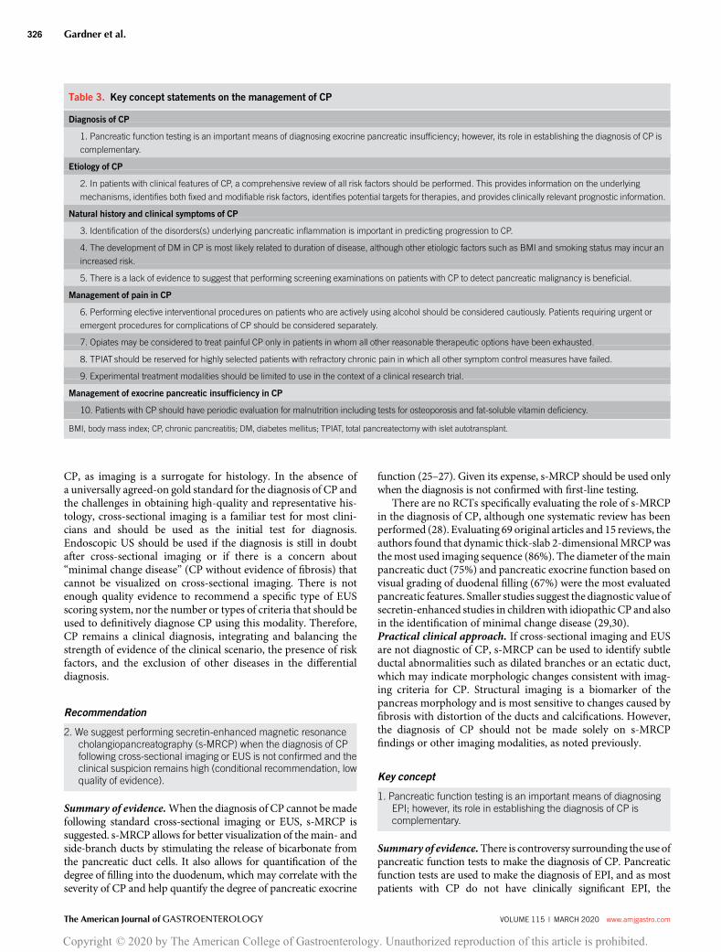
Table 3. Key concept statements on the management of CP
Diagnosis of CP
1. Pancreatic function testing is an important means of diagnosing exocrine pancreatic insufficiency; however, its role in establishing the diagnosis of CP is
complementary.
Etiology of CP
2. In patients with clinical features of CP, a comprehensive review of all risk factors should be performed. This provides information on the underlying
mechanisms, identifies both fixed and modifiable risk factors, identifies potential targets for therapies, and provides clinically relevant prognostic information.
Natural history and clinical symptoms of CP
3. Identification of the disorders(s) underlying pancreatic inflammation is important in predicting progression to CP.
4. The development of DM in CP is most likely related to duration of disease, although other etiologic factors such as BMI and smoking status may incur an
increased risk.
5. There is a lack of evidence to suggest that performing screening examinations on patients with CP to detect pancreatic malignancy is beneficial.
Management of pain in CP
6. Performing elective interventional procedures on patients who are actively using alcohol should be considered cautiously. Patients requiring urgent or
emergent procedures for complications of CP should be considered separately.
7. Opiates may be considered to treat painful CP only in patients in whom all other reasonable therapeutic options have been exhausted.
8. TPIAT should be reserved for highly selected patients with refractory chronic pain in which all other symptom control measures have failed.
9. Experimental treatment modalities should be limited to use in the context of a clinical research trial.
Management of exocrine pancreatic insufficiency in CP
10. Patients with CP should have periodic evaluation for malnutrition including tests for osteoporosis and fat-soluble vitamin deficiency.
BMI, body mass index; CP, chronic pancreatitis; DM, diabetes mellitus; TPIAT, total pancreatectomy with islet autotransplant.
The American Journal of GASTROENTEROLOGY VOLUME 115 | MARCH 2020 www.amjgastro.com
Gardner et al.326
Copyright © 2020 by The American College of Gastroenterology. Unauthorized reproduction of this article is prohibited.
sensitivity of pancreatic function testing to make the diagnosis ofCP is low.This is due in part to the large reservewithin the pancreasin which only significant loss of function (usually.90%) results inthe clinically apparent symptoms of steatorrhea, azotorrhea, andresultant vitamin deficiency (31). However, there are patientswhose only clinical manifestation of pancreatic exocrine celldamage may be EPI, and certainly, patients can have progressiveEPI over their disease course. In fact, EPI represents an imbalancein at least 4 domains; nutritional intake, pancreatic digestive en-zyme delivery to the small intestine, intestinal adaptation to dis-ease, and nutritional needs of each type of essential nutrient. Thus,failure of the pancreas to deliver sufficient enzymes to meeta patient’s nutritional needs is relative to the other 3 domains.
There are no RCTs, systematic reviews, or meta-analyses,which specifically detail the use of pancreatic function tests todiagnose CP. Based on the available evidence, the use of pan-creatic function testing to diagnose CP therefore should only beused as an ancillary test in making the diagnosis (22,32–34).
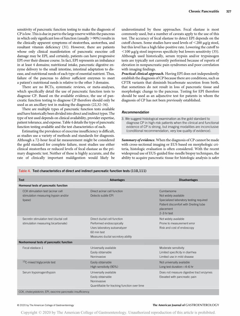
There are multiple types of pancreatic function tests availableandhavehistoricallybeendivided intodirect and indirect types.Thetype of test used depends on clinical availability, provider expertise,patient tolerance, and expense. Table 4 details the type of pancreaticfunction testing available and the test characteristics of each.
Estimating the prevalence of exocrine insufficiency is difficult,as studies use a variety of methods and standards for diagnosis.Although a 72-hour fecal fat measurement might be consideredthe gold standard for complete failure, most studies use eitherclinical steatorrhea or reduced levels of fecal elastase as the pri-mary diagnostic test. Neither of these is highly accurate, and therate of clinically important maldigestion would likely be
underestimated by these approaches. Fecal elastase is mostcommonly used, but a number of caveats apply to the use of thistest. The accuracy of fecal elastase to detect EPI depends on thecutoff chosen. Some studies have used levels of,200 mg/g stool,but this level has a high false-positive rate. Lowering the cutoff to,100 mg/g stool improves specificity but lowers sensitivity (35).Although used historically, serum trypsin and/or trypsinogentests are typically not currently performed because of reports ofelevation in nonpancreatic pain syndromes and poor correlationwith imaging findings.Practical clinical approach.Having EPI does not independentlyestablish the diagnosis of CP because there are conditions, such asCFTR variants that diminish bicarbonate secretion from birth,that sometimes do not result in loss of pancreatic tissue andmorphologic change to the pancreas. Testing for EPI thereforeshould be used as an adjunctive test for patients in whom thediagnosis of CP has not been previously established.
Recommendation
3. We suggest histological examination as the gold standard todiagnose CP in high-risk patients when the clinical and functionalevidence of CP is strong, but imaging modalities are inconclusive(conditional recommendation, very low quality of evidence).
Summary of evidence.When the diagnosis of CP cannot bemadewith cross-sectional imaging or EUS based on morphologic cri-teria, histologic evaluation is often considered. With the recentwidespread use of EUS-guided fine-needle biopsy techniques, theability to acquire pancreatic tissue for histologic analysis is safer
Table 4. Test characteristics of direct and indirect pancreatic function tests (110,111)
Test Advantages Disadvantages
Hormonal tests of pancreatic function
CCK stimulation test (acinar cell
stimulation measuring trypsin and/or
lipase)
Direct acinar cell function
Detects subtle EPI
Cumbersome
Not widely available
Specialized laboratory testing required
Patient discomfort with Dreiling tube
placement
2–3 hr test
Secretin stimulation test (ductal cell
stimulation measuring bicarbonate)
Direct ductal cell function
Performed endoscopically
Uses laboratory autoanalyzer
60 min test
Measures ductal secretory ability
Not widely available
Prone to measurement error
Risk and cost of endoscopy
Nonhormonal tests of pancreatic function
Fecal elastace-1 Universally available
Easily obtainable
Noninvasive
Moderate sensitivity
Limited specificity in diarrhea
Limited use in mild disease
13C-mixed triglyceride test Easily obtainable
High sensitivity (90%)
Not universally available
Long test duration—4–6 hr
Serum trypsinogen/trypsin Universally available
Easily obtainable
Noninvasive
Quantifiable for tracking function over time
Does not measure digestive tract enzymes
Elevated with pancreatic pain
CCK, cholecystokinin; EPI, exocrine pancreatic insufficiency.
© 2020 by The American College of Gastroenterology The American Journal of GASTROENTEROLOGY
Chronic Pancreatitis 327
Copyright © 2020 by The American College of Gastroenterology. Unauthorized reproduction of this article is prohibited.
and technically easier (36,37). However, the sensitivity of histo-logic evaluation for CP when tissue is available, compared withmorphologic evaluation, is often no better than chance (38,39).Histologic evaluation can be limited because of sampling error,complications inherent in obtaining the biopsy sample, the pat-chy nature of pancreatic inflammatory changes, and histologicinterpretation that is prone to subjectivity.
There are no RCTs, systematic reviews, or meta-analyses,which treat histologic evaluation as the diagnostic gold standardfor CP. Nonetheless, histologic confirmation can serve as thediagnostic gold standard, its value most important for ruling outCP when the diagnosis is under consideration. Histologic eval-uation should only be considered in high-risk patients afterclinical, functional, and imaging tests have not established theclinicopathologic diagnosis and a thoughtful informed consentprocess has been had with the patient.Practical clinic approach. Under the current clinicopathologicapproach to disease, histology is the gold standard test to diagnoseCP, and it is often used to “rule out” CP in patients in whom thediagnosis is being considered. However, both imaging and his-tology are biomarkers of an underlying disorder that may or maynot be true CP, and thus, the sensitivity of histology to make thediagnosis is low. As the mechanistic model of disease is in-vestigated and formalized further, histology will likely be lessimportant in making the diagnosis of CP.
ETIOLOGY OF CP
Key concept
2. In patients with clinical features of CP, a comprehensive review of allrisk factors should be performed. This provides information on theunderlying mechanisms, identifies both fixed and modifiable riskfactors, identifies potential targets for therapies, and providesclinically relevant prognostic information.
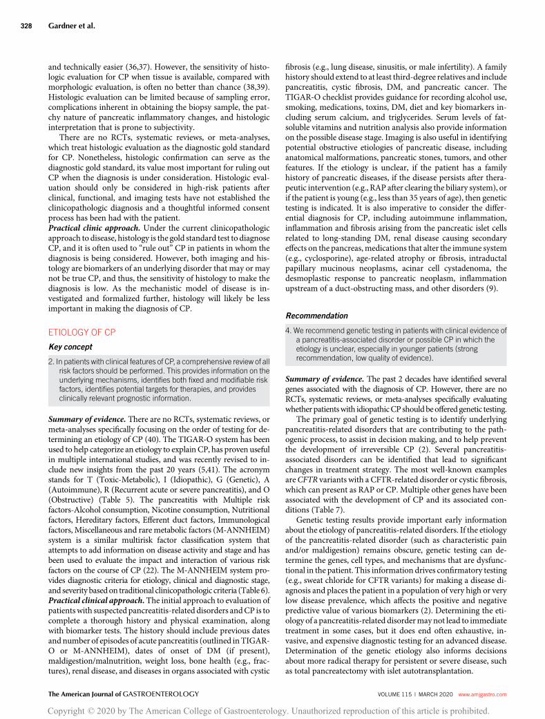
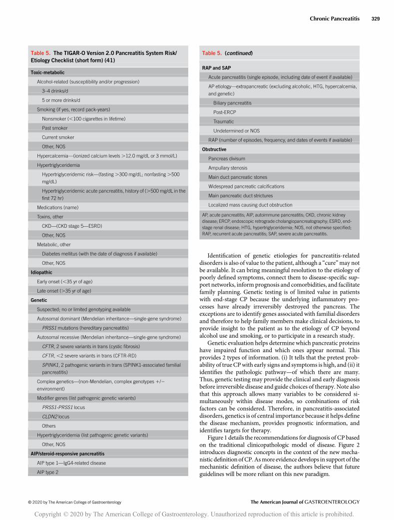
Summary of evidence. There are no RCTs, systematic reviews, ormeta-analyses specifically focusing on the order of testing for de-termining an etiology of CP (40). The TIGAR-O system has beenused to help categorize an etiology to explain CP, has proven usefulin multiple international studies, and was recently revised to in-clude new insights from the past 20 years (5,41). The acronymstands for T (Toxic-Metabolic), I (Idiopathic), G (Genetic), A(Autoimmune), R (Recurrent acute or severe pancreatitis), and O(Obstructive) (Table 5). The pancreatitis with Multiple riskfactors-Alcohol consumption, Nicotine consumption, Nutritionalfactors, Hereditary factors, Efferent duct factors, Immunologicalfactors, Miscellaneous and rare metabolic factors (M-ANNHEIM)system is a similar multirisk factor classification system thatattempts to add information on disease activity and stage and hasbeen used to evaluate the impact and interaction of various riskfactors on the course of CP (22). The M-ANNHEIM system pro-vides diagnostic criteria for etiology, clinical and diagnostic stage,and severity basedon traditional clinicopathologic criteria (Table 6).Practical clinical approach.The initial approach to evaluation ofpatientswith suspected pancreatitis-related disorders andCP is tocomplete a thorough history and physical examination, alongwith biomarker tests. The history should include previous datesandnumber of episodes of acute pancreatitis (outlined inTIGAR-O or M-ANNHEIM), dates of onset of DM (if present),maldigestion/malnutrition, weight loss, bone health (e.g., frac-tures), renal disease, and diseases in organs associated with cystic
fibrosis (e.g., lung disease, sinusitis, or male infertility). A familyhistory should extend to at least third-degree relatives and includepancreatitis, cystic fibrosis, DM, and pancreatic cancer. TheTIGAR-O checklist provides guidance for recording alcohol use,smoking, medications, toxins, DM, diet and key biomarkers in-cluding serum calcium, and triglycerides. Serum levels of fat-soluble vitamins and nutrition analysis also provide informationon the possible disease stage. Imaging is also useful in identifyingpotential obstructive etiologies of pancreatic disease, includinganatomical malformations, pancreatic stones, tumors, and otherfeatures. If the etiology is unclear, if the patient has a familyhistory of pancreatic diseases, if the disease persists after thera-peutic intervention (e.g., RAP after clearing the biliary system), orif the patient is young (e.g., less than 35 years of age), then genetictesting is indicated. It is also imperative to consider the differ-ential diagnosis for CP, including autoimmune inflammation,inflammation and fibrosis arising from the pancreatic islet cellsrelated to long-standing DM, renal disease causing secondaryeffects on the pancreas, medications that alter the immune system(e.g., cyclosporine), age-related atrophy or fibrosis, intraductalpapillary mucinous neoplasms, acinar cell cystadenoma, thedesmoplastic response to pancreatic neoplasm, inflammationupstream of a duct-obstructing mass, and other disorders (9).
Recommendation
4. We recommend genetic testing in patients with clinical evidence ofa pancreatitis-associated disorder or possible CP in which theetiology is unclear, especially in younger patients (strongrecommendation, low quality of evidence).
Summary of evidence. The past 2 decades have identified severalgenes associated with the diagnosis of CP. However, there are noRCTs, systematic reviews, or meta-analyses specifically evaluatingwhetherpatientswith idiopathicCPshouldbeoffered genetic testing.
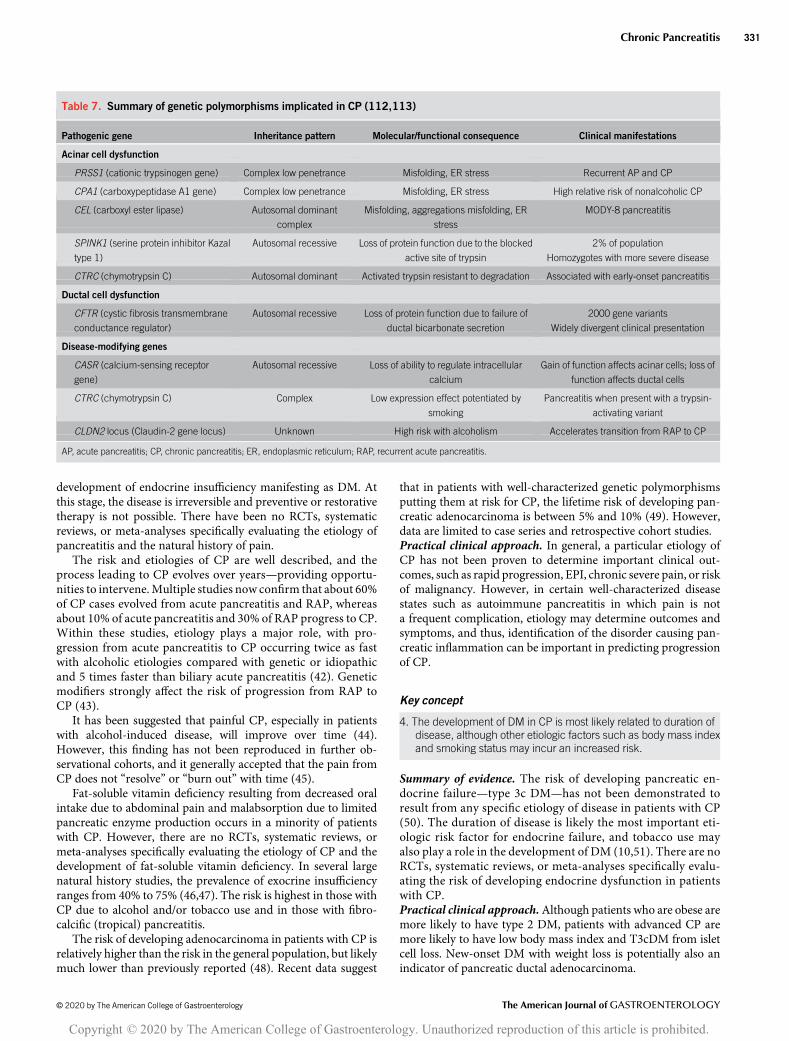
The primary goal of genetic testing is to identify underlyingpancreatitis-related disorders that are contributing to the path-ogenic process, to assist in decision making, and to help preventthe development of irreversible CP (2). Several pancreatitis-associated disorders can be identified that lead to significantchanges in treatment strategy. The most well-known examplesareCFTR variants with a CFTR-related disorder or cystic fibrosis,which can present as RAP or CP. Multiple other genes have beenassociated with the development of CP and its associated con-ditions (Table 7).
Genetic testing results provide important early informationabout the etiology of pancreatitis-related disorders. If the etiologyof the pancreatitis-related disorder (such as characteristic painand/or maldigestion) remains obscure, genetic testing can de-termine the genes, cell types, and mechanisms that are dysfunc-tional in the patient. This information drives confirmatory testing(e.g., sweat chloride for CFTR variants) for making a disease di-agnosis and places the patient in a population of very high or verylow disease prevalence, which affects the positive and negativepredictive value of various biomarkers (2). Determining the eti-ology of a pancreatitis-related disordermaynot lead to immediatetreatment in some cases, but it does end often exhaustive, in-vasive, and expensive diagnostic testing for an advanced disease.Determination of the genetic etiology also informs decisionsabout more radical therapy for persistent or severe disease, suchas total pancreatectomy with islet autotransplantation.
The American Journal of GASTROENTEROLOGY VOLUME 115 | MARCH 2020 www.amjgastro.com
Gardner et al.328
Copyright © 2020 by The American College of Gastroenterology. Unauthorized reproduction of this article is prohibited.
Identification of genetic etiologies for pancreatitis-relateddisorders is also of value to the patient, although a “cure”may notbe available. It can bring meaningful resolution to the etiology ofpoorly defined symptoms, connect them to disease-specific sup-port networks, inform prognosis and comorbidities, and facilitatefamily planning. Genetic testing is of limited value in patientswith end-stage CP because the underlying inflammatory pro-cesses have already irreversibly destroyed the pancreas. Theexceptions are to identify genes associated with familial disordersand therefore to help family members make clinical decisions, toprovide insight to the patient as to the etiology of CP beyondalcohol use and smoking, or to participate in a research study.
Genetic evaluation helps determine which pancreatic proteinshave impaired function and which ones appear normal. Thisprovides 2 types of information. (i) It tells that the pretest prob-ability of true CPwith early signs and symptoms is high, and (ii) itidentifies the pathologic pathway—of which there are many.Thus, genetic testing may provide the clinical and early diagnosisbefore irreversible disease and guide choices of therapy. Note alsothat this approach allows many variables to be considered si-multaneously within disease modes, so combinations of riskfactors can be considered. Therefore, in pancreatitis-associateddisorders, genetics is of central importance because it helps definethe disease mechanism, provides prognostic information, andidentifies targets for therapy.
Figure 1 details the recommendations for diagnosis of CP basedon the traditional clinicopathologic model of disease. Figure 2introduces diagnostic concepts in the context of the new mecha-nistic definition ofCP.Asmore evidence develops in support of themechanistic definition of disease, the authors believe that futureguidelines will be more reliant on this new paradigm.
Table 5. The TIGAR-O Version 2.0 Pancreatitis System Risk/
Etiology Checklist (short form) (41)
Toxic-metabolic
Alcohol-related (susceptibility and/or progression)
3–4 drinks/d
5 or more drinks/d
Smoking (if yes, record pack-years)
Nonsmoker (,100 cigarettes in lifetime)
Past smoker
Current smoker
Other, NOS
Hypercalcemia—(ionized calcium levels .12.0 mg/dL or 3 mmol/L)
Hypertriglyceridemia
Hypertriglyceridemic risk—(fasting .300 mg/dL; nonfasting .500
mg/dL)
Hypertriglyceridemic acute pancreatitis, history of (.500 mg/dL in the
first 72 hr)
Medications (name)
Toxins, other
CKD—(CKD stage 5—ESRD)
Other, NOS
Metabolic, other
Diabetes mellitus (with the date of diagnosis if available)
Other, NOS
Idiopathic
Early onset (,35 yr of age)
Late onset (.35 yr of age)
Genetic
Suspected; no or limited genotyping available
Autosomal dominant (Mendelian inheritance—single-gene syndrome)
PRSS1 mutations (hereditary pancreatitis)
Autosomal recessive (Mendelian inheritance—single-gene syndrome)
CFTR, 2 severe variants in trans (cystic fibrosis)
CFTR, ,2 severe variants in trans (CFTR-RD)
SPINK1, 2 pathogenic variants in trans (SPINK1-associated familial
pancreatitis)
Complex genetics—(non-Mendelian, complex genotypes 1/2
environment)
Modifier genes (list pathogenic genetic variants)
PRSS1-PRSS1 locus
CLDN2 locus
Others
Hypertriglyceridemia (list pathogenic genetic variants)
Other, NOS
AIP/steroid-responsive pancreatitis
AIP type 1—IgG4-related disease
AIP type 2
Table 5. (continued)
RAP and SAP
Acute pancreatitis (single episode, including date of event if available)
AP etiology—extrapancreatic (excluding alcoholic, HTG, hypercalcemia,
and genetic)
Biliary pancreatitis
Post-ERCP
Traumatic
Undetermined or NOS
RAP (number of episodes, frequency, and dates of events if available)
Obstructive
Pancreas divisum
Ampullary stenosis
Main duct pancreatic stones
Widespread pancreatic calcifications
Main pancreatic duct strictures
Localized mass causing duct obstruction
AP, acute pancreatitis; AIP, autoimmune pancreatitis; CKD, chronic kidneydisease; ERCP, endoscopic retrograde cholangiopancreatography; ESRD, end-stage renal disease; HTG, hypertriglyceridemia; NOS, not otherwise specified;RAP, recurrent acute pancreatitis; SAP, severe acute pancreatitis.
© 2020 by The American College of Gastroenterology The American Journal of GASTROENTEROLOGY
Chronic Pancreatitis 329
Copyright © 2020 by The American College of Gastroenterology. Unauthorized reproduction of this article is prohibited.
Practical clinical approach. Testing for germline mutations (asopposed to acquired somatic mutations in tumors for cancertherapy) is not diagnostic of CP, but rather (i) identifies a pop-ulation of patients with a high prevalence of pancreatitis-relateddisorders andCPso that it improves the accuracyof less sensitive orspecific biomarkers and (ii) identifies the dysfunctionalmechanismunderlying the pathogenic processes that cause biomarkers to beabnormal and lead to disease. This is important in patients of anyage because therapies (such as CFTR-modulating drugs) can targetmechanism, and knowing the mechanism allows the most ap-propriate drug and/or therapy to be selected. It also providesprognostic information for the management of complex syn-dromes without specific treatment (i.e., for total pancreatectomywith islet autotransplant [TPIAT] assessment) and can provideanswers to patients about the origin of their symptoms. In mostinstances, patients should be referred to a genetic counselor forevaluation; however, in centers inwhich experienced nongeneticistclinicians are comfortable ordering and evaluating the results, ge-netic referral is not necessary. At minimum, patients with idio-pathic CP should be evaluated for PRSS1, SPINK1, CFTR, andCTRCgenemutation analysis, althoughmore extendedpanelswithover a dozen susceptibility and modifier genes, hyper-triglyceridemia genes, and pharmacogenetics are available.
NATURAL HISTORY AND CLINICAL SYMPTOMS OF CP
Key concept
3. Identification of the disorders(s) underlying pancreaticinflammation is important in predicting progression to CP.
Summary of evidence. The primary clinical outcomes of patientswith CP are debilitating abdominal pain, fat-soluble vitamindeficiency leading to malnutrition and related conditions such asosteoporosis, the risk of pancreatic malignancy, and the
Table 6. The M-ANNHEIM scoring system for the grading of
chronic pancreatitis severity (22)
Clinical Features Points
Patient report of pain
No pain without therapy (patient reports
requiring no pain medication)
0
RAP (patient reports freedom from pain
between attacks of acute pancreatitis)
1
No pain with therapy (patient reports
freedom from pain with pain medication or
endoscopic intervention)
2
Intermittent pain (patient reports
intermittent pain-free episodes, either with
or without therapy; possibly additional
attacks of acute pancreatitis)
3
Continuous pain (patient reports absence
of pain-free episodes, either with or without
therapy; possibly additional attacks of
acute pancreatitis)
4
Pain control
No medication 0
Use of nonopioid drugs or use of mild
opioids (WHO step 1 or 2)
1
Use of potent opioids (WHO step 3) or
endoscopic intervention
2
Surgical intervention
Pancreatic surgical intervention for any
reason
4
Exocrine insufficiency
Absence of exocrine insufficiency 0
Presence of mild, moderate, or unproven
exocrine insufficiency not requiring
enzyme supplementation (including
patient reports of intermittent
diarrhea)
1
Presence of proven exocrine insufficiency
(according to exocrine function tests) or
presence of marked exocrine insufficiency
defined as steatorrhea (.7 g fat/24 hr),
normalized or markedly reduced by
enzyme supplementation
2
Endocrine insufficiency
Absence of DM 0
Presence of DM 4
Morphologic status on pancreatic imaging
(according to the Cambridge classification)
Normal 0
Equivocal 1
Mild 2
Moderate 3
Marked 4
Table 6. (continued)
Clinical Features Points
Severe organ complications
Absence of complications 0
Presence of possibly reversible
complications
2
Presence of irreversible complications 4
Severity index severity level point range
M-ANNHEIM A Minor 0–5 points
M-ANNHEIM B Increased 6–10 points
M-ANNHEIM C Advanced 11–15 points
M-ANNHEIM D Marked 16–20 points
M-ANNHEIM E Exacerbated.20 points
M-ANNHEIM scoring system points are added together, and the sum is used tocategorize a patient’s disease according to the M-ANNHEIM system.DM, diabetes mellitus; M-ANNHEIM, pancreatitis with Multiple risk factors-Alcohol consumption, Nicotine consumption, Nutritional factors, Hereditaryfactors, Efferent duct factors, Immunological factors, Miscellaneous and raremetabolic factors; RAP, recurrent acute pancreatitis; WHO, World HealthOrganization.
The American Journal of GASTROENTEROLOGY VOLUME 115 | MARCH 2020 www.amjgastro.com
Gardner et al.330
Copyright © 2020 by The American College of Gastroenterology. Unauthorized reproduction of this article is prohibited.
development of endocrine insufficiency manifesting as DM. Atthis stage, the disease is irreversible and preventive or restorativetherapy is not possible. There have been no RCTs, systematicreviews, or meta-analyses specifically evaluating the etiology ofpancreatitis and the natural history of pain.
The risk and etiologies of CP are well described, and theprocess leading to CP evolves over years—providing opportu-nities to intervene.Multiple studies now confirm that about 60%of CP cases evolved from acute pancreatitis and RAP, whereasabout 10% of acute pancreatitis and 30% of RAP progress to CP.Within these studies, etiology plays a major role, with pro-gression from acute pancreatitis to CP occurring twice as fastwith alcoholic etiologies compared with genetic or idiopathicand 5 times faster than biliary acute pancreatitis (42). Geneticmodifiers strongly affect the risk of progression from RAP toCP (43).
It has been suggested that painful CP, especially in patientswith alcohol-induced disease, will improve over time (44).However, this finding has not been reproduced in further ob-servational cohorts, and it generally accepted that the pain fromCP does not “resolve” or “burn out” with time (45).
Fat-soluble vitamin deficiency resulting from decreased oralintake due to abdominal pain and malabsorption due to limitedpancreatic enzyme production occurs in a minority of patientswith CP. However, there are no RCTs, systematic reviews, ormeta-analyses specifically evaluating the etiology of CP and thedevelopment of fat-soluble vitamin deficiency. In several largenatural history studies, the prevalence of exocrine insufficiencyranges from 40% to 75% (46,47). The risk is highest in those withCP due to alcohol and/or tobacco use and in those with fibro-calcific (tropical) pancreatitis.
The risk of developing adenocarcinoma in patients with CP isrelatively higher than the risk in the general population, but likelymuch lower than previously reported (48). Recent data suggest
that in patients with well-characterized genetic polymorphismsputting them at risk for CP, the lifetime risk of developing pan-creatic adenocarcinoma is between 5% and 10% (49). However,data are limited to case series and retrospective cohort studies.Practical clinical approach. In general, a particular etiology ofCP has not been proven to determine important clinical out-comes, such as rapid progression, EPI, chronic severe pain, or riskof malignancy. However, in certain well-characterized diseasestates such as autoimmune pancreatitis in which pain is nota frequent complication, etiology may determine outcomes andsymptoms, and thus, identification of the disorder causing pan-creatic inflammation can be important in predicting progressionof CP.
Key concept
4. The development of DM in CP is most likely related to duration ofdisease, although other etiologic factors such as body mass indexand smoking status may incur an increased risk.
Summary of evidence. The risk of developing pancreatic en-docrine failure—type 3c DM—has not been demonstrated toresult from any specific etiology of disease in patients with CP(50). The duration of disease is likely the most important eti-ologic risk factor for endocrine failure, and tobacco use mayalso play a role in the development of DM (10,51). There are noRCTs, systematic reviews, or meta-analyses specifically evalu-ating the risk of developing endocrine dysfunction in patientswith CP.Practical clinical approach.Although patients who are obese aremore likely to have type 2 DM, patients with advanced CP aremore likely to have low body mass index and T3cDM from isletcell loss. New-onset DM with weight loss is potentially also anindicator of pancreatic ductal adenocarcinoma.
Table 7. Summary of genetic polymorphisms implicated in CP (112,113)
Pathogenic gene Inheritance pattern Molecular/functional consequence Clinical manifestations
Acinar cell dysfunction
PRSS1 (cationic trypsinogen gene) Complex low penetrance Misfolding, ER stress Recurrent AP and CP
CPA1 (carboxypeptidase A1 gene) Complex low penetrance Misfolding, ER stress High relative risk of nonalcoholic CP
CEL (carboxyl ester lipase) Autosomal dominant
complex
Misfolding, aggregations misfolding, ER
stress
MODY-8 pancreatitis
SPINK1 (serine protein inhibitor Kazal
type 1)
Autosomal recessive Loss of protein function due to the blocked
active site of trypsin
2% of population
Homozygotes with more severe disease
CTRC (chymotrypsin C) Autosomal dominant Activated trypsin resistant to degradation Associated with early-onset pancreatitis
Ductal cell dysfunction
CFTR (cystic fibrosis transmembrane
conductance regulator)
Autosomal recessive Loss of protein function due to failure of
ductal bicarbonate secretion
2000 gene variants
Widely divergent clinical presentation
Disease-modifying genes
CASR (calcium-sensing receptor
gene)
Autosomal recessive Loss of ability to regulate intracellular
calcium
Gain of function affects acinar cells; loss of
function affects ductal cells
CTRC (chymotrypsin C) Complex Low expression effect potentiated by
smoking
Pancreatitis when present with a trypsin-
activating variant
CLDN2 locus (Claudin-2 gene locus) Unknown High risk with alcoholism Accelerates transition from RAP to CP
AP, acute pancreatitis; CP, chronic pancreatitis; ER, endoplasmic reticulum; RAP, recurrent acute pancreatitis.
© 2020 by The American College of Gastroenterology The American Journal of GASTROENTEROLOGY
Chronic Pancreatitis 331
Copyright © 2020 by The American College of Gastroenterology. Unauthorized reproduction of this article is prohibited.
Recommendation
5. We recommend alcohol cessation in patients with CP (strongrecommendation, very low quality of evidence).
Summary of evidence. There are no RCTs, systematic reviews, ormeta-analyses, which address the issue of whether alcohol ces-sation alters the natural history of CP pain. However, several caseseries have suggested that discontinuing alcohol use improves thepain in CP but does not necessarily alter the progression to en-docrine or exocrine dysfunction (52,53). There is 1 randomizedtrial, demonstrating that alcohol cessation counseling in patientsadmitted with an attack of acute alcoholic pancreatitis can limitfurther hospitalizations and pain attacks (54). Thus, alcoholcessation counseling is recommended for patients with CP, al-though the extent to which this intervention alters the naturalhistory of the disease is unknown (55).Practical clinical approach.Although the evidence is lowquality,strict alcohol avoidance should be a cornerstone of any treatmentprogram for patients with CP.
Recommendation
6. We recommend smoking cessation in patients with CP (strongrecommendation, very low quality of evidence).
Summary of evidence. Smoking cessation is very challenging forpatients, including those with CP (56). Smoking tobacco iswidely believed to be a risk factor for the development of CP, butonly single-center studies are available (57–59). There are no
RCTs, systematic reviews, or meta-analyses specifically evalu-ating whether smoking cessation is beneficial in improving thenatural history of CP. However, case series have reported a de-crease in the amount of pancreatic calcification progressionwhen smoking cessation occurs at the time of diagnosis ofCP (60).Practical clinical approach.Although the evidence is low quality,strict smoking avoidance should be a cornerstone of any treat-ment program for patients with CP, recognizing however that thelong-term success rate of smoking cessation is low.
Key concept
5. There is a lack of evidence to suggest that performing screeningexaminations on patients with CP to detect pancreatic malignancyis beneficial.
Summary of evidence. There is very little quality evidence sug-gesting performing screening examination for pancreatic malig-nancy in all patients and even in those at high risk for pancreaticmalignancy due to genetic or environmental risk factors. Al-though the overall prevalence of pancreatic malignancy is in-creased in patients with CP, there are no RCTs, systematicreviews, or meta-analyses to support screening this patient pop-ulation for pancreatic malignancy (48).Practical clinical approach. At this time, there is no definitivebenefit to screen patients with CP for pancreatic ductal adeno-carcinoma. This is based on the invasive and costly nature oftesting, the inherent difficulty in screening given the structuralchanges of CP, and the inability to alter in many cases the natural
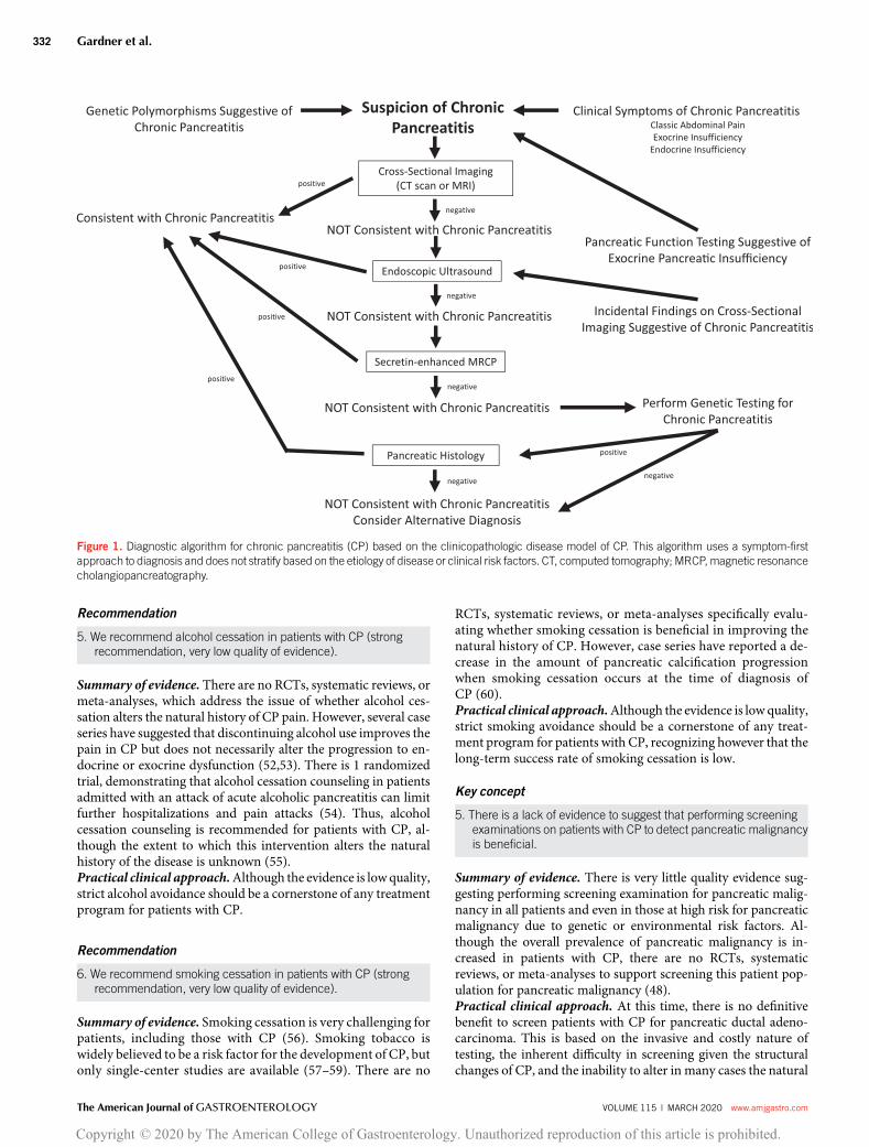
Figure 1. Diagnostic algorithm for chronic pancreatitis (CP) based on the clinicopathologic disease model of CP. This algorithm uses a symptom-firstapproach to diagnosis and does not stratify based on the etiology of disease or clinical risk factors. CT, computed tomography; MRCP, magnetic resonancecholangiopancreatography.
The American Journal of GASTROENTEROLOGY VOLUME 115 | MARCH 2020 www.amjgastro.com
Gardner et al.332
Copyright © 2020 by The American College of Gastroenterology. Unauthorized reproduction of this article is prohibited.
history of the disease even if malignancy is detected at an earlystage.
MANAGEMENT OF PAIN IN CP
Key concept
6. Performing elective interventional procedures on patients who areactively using alcohol should be considered cautiously. Patientsrequiring urgent or emergent procedures for complications of CPshould be considered separately.
Summary of evidence. The question of whether or not to un-dertake active interventions in patients with CP is complex andcan be evaluated from both medical and social points of view.There are no RCTs, systematic reviews, or meta-analyses, whichspecifically address this issue.
From a medical point of view, it can be argued that it is ill-advised to undertake aggressive endoscopic or surgical inter-ventions that may require ongoing or sequential procedures ina patient who is actively taking steps to harm themselves andexacerbate their underlying CP with ongoing and sustained al-cohol abuse. Alcohol abuse may be causing or worsening the verypain that these endoscopic and surgical procedures are meant totreat, and if alcohol cessation could be obtained, some (or all) ofthese interventions may not even be warranted.
From a social point of view, patients with CP who continue toconsume alcohol after appropriate patient education still warrantcare and should be encouraged to stop alcohol use by means ofcounseling, attendance at Alcoholics Anonymous, and/or otherprograms. Urgent interventions should be performed in patientswho continue to consume alcohol.
Practical clinical approach. In general, elective interventionalprocedures (such as celiac plexus interventions for pain palli-ation) should be performed with caution in patients who areactively consuming alcohol. Urgent interventions should beperformed in patients who continue to consume alcohol giventheir medical necessity. Patients making good faith efforts tostop or reduce alcohol consumption but who still occasionallyconsume alcohol can be evaluated for interventional proce-dures on a case-by-case basis, recognizing the difficulties in-volved in alcohol cessation and the need to treat clinicalsymptoms of CP.
Recommendation
7. We recommend surgical intervention over endoscopic therapy inpatients with obstructive CP for the long-term relief of pain if first-line endoscopic approaches to pancreatic drainage have beenexhausted or unsuccessful (strong recommendation, moderatequality of evidence).
Summary of evidence. Patients with CP often experience pain inthe setting of pancreatic duct obstruction. Duct obstruction canoccur because of pancreatic duct stones, pancreatic duct stric-tures, or a combination thereof. Endoscopic decompressiveprocedures include ERCP with pancreatic sphincterotomy,stone clearance, stricture dilation, and pancreatic duct stenting.Other endoscopic options include interventional EUS proce-dures that usually involve placement of a transluminal stent toallow for pancreatic duct decompression. Several surgicaldecompressive procedures exist (Puestow, Frey, and Begerprocedures) that may also include a component of partial
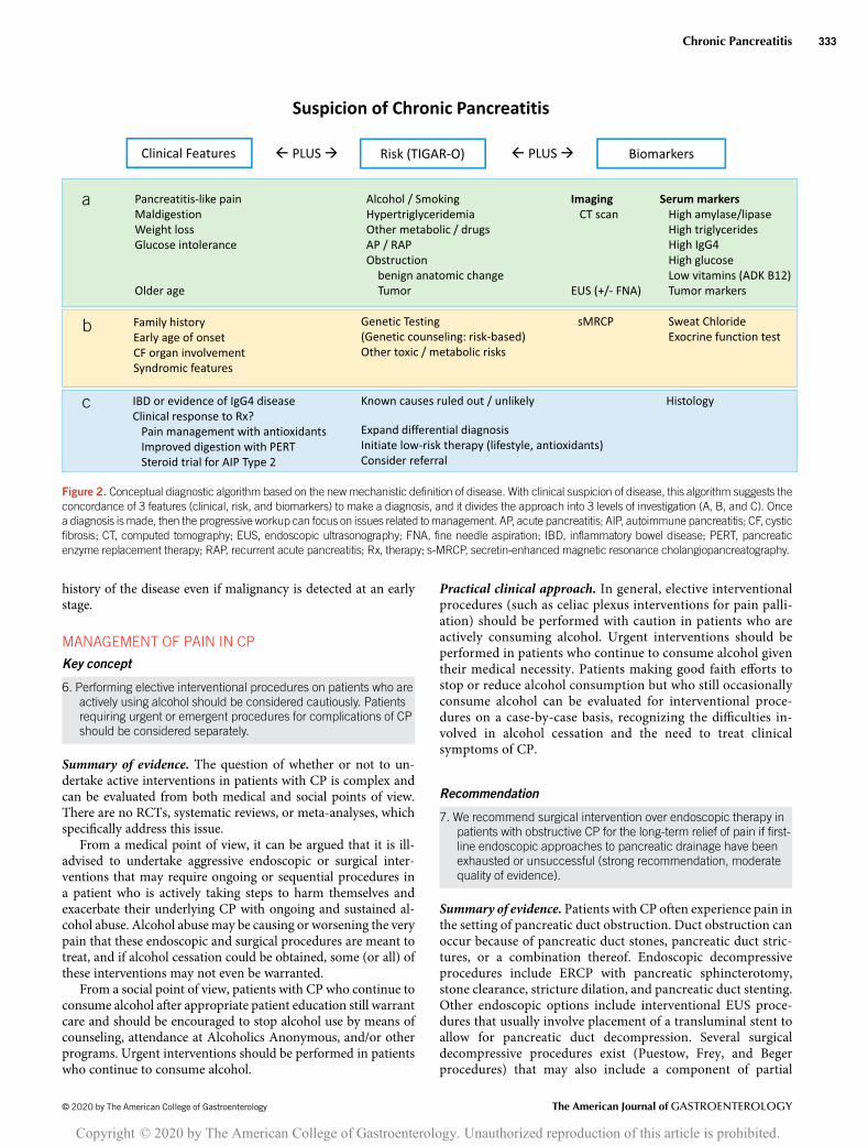
Figure 2. Conceptual diagnostic algorithm based on the newmechanistic definition of disease. With clinical suspicion of disease, this algorithm suggests theconcordance of 3 features (clinical, risk, and biomarkers) to make a diagnosis, and it divides the approach into 3 levels of investigation (A, B, and C). Oncea diagnosis ismade, then the progressive workup can focus on issues related tomanagement. AP, acute pancreatitis; AIP, autoimmune pancreatitis; CF, cysticfibrosis; CT, computed tomography; EUS, endoscopic ultrasonography; FNA, fine needle aspiration; IBD, inflammatory bowel disease; PERT, pancreaticenzyme replacement therapy; RAP, recurrent acute pancreatitis; Rx, therapy; s-MRCP, secretin-enhanced magnetic resonance cholangiopancreatography.
© 2020 by The American College of Gastroenterology The American Journal of GASTROENTEROLOGY
Chronic Pancreatitis 333
Copyright © 2020 by The American College of Gastroenterology. Unauthorized reproduction of this article is prohibited.
pancreatectomy. Unfortunately, there are no RCT or systematicreview data, which reliably report whether successful or un-successful endoscopic decompression predicts subsequentsuccessful drainage surgery.
Although widely performed, high-quality evidence regardingthese procedures is limited. Cahen et al. (61) randomized 39patients with CP to undergo either endoscopic or surgicaldrainage of the pancreatic duct. In this study, patients who un-derwent surgery had lower pain scores (P , 0.001) and betterphysical health summary scores (P 5 0.003) when validatedquestionnaires were used. At the end of 24 months of follow-up,complete or partial pain relief was achieved in 32% of patientswho underwent endoscopic drainage vs 75% of patients assignedto surgical drainage (P 5 0.007). Complication rates, length ofstay, and changes in pancreatic function were similar between thetreatment groups. As would be expected, patients receiving en-doscopic treatment required more interventions than patientswho underwent surgery (a median of 8 vs 3, P , 0.001).
This same group published a long-term follow-up study ofthese 39 patients lasting 79 months (62). Among patients in theoriginal endoscopic group, 68% underwent additional drainagecomparedwith 5%whounderwent surgery (P5 0.001). Length ofstay and costs were equivalent in the 2 groups. Forty-seven per-cent of patients treated through endoscopy ultimately underwentsurgery. Mean difference in validated pain scores was no longersignificant (39 vs 22; P5 0.12), although surgery was superior interms of pain relief (80% vs 38%; P 5 0.042). Overall, patients’quality of life and pancreatic function were not felt to be different.
An older study also compared endoscopic with surgicaltherapy in patients with CP (63). This study included 140patients, with a subgroup of 72 patients randomized to undergoeither surgical resective or drainage procedures vs endoscopictherapy that focused on pancreatic sphincterotomy and stoneremoval. Patients in both groups had similar initial success rates,but the complete absence of pain was low in both groups and wasmore common in patients undergoing surgery (37% vs 14%), withthe rate of partial relief being equivalent (49% vs 51%). Patientswho underwent surgery gained more weight, whereas patients inboth groups developed new-onset diabetes to an equal extent.
Various studies have compared outcomes between differenttypes of pancreatic drainage procedures in patients with CP, withno surgery being clearly identified as superior (64–66). Factorssuch as local expertise likely play a major role in the selection ofsurgical procedure. It should be stressed that many of thesestudies are older, and since their publication, interventional en-doscopic approaches are much more widely performed in thecurrent era. Newer studies comparing endoscopic and surgicalapproaches are warranted.Practical clinical approach. Although surgical approaches topancreatic duct decompression have been shown to providebetter long-term pain relief than endoscopic approaches, they arerarely first-line therapies and many surgeons only operate onceendoscopic approaches to pancreatic drainage have beenexhausted or unsuccessful. It is reasonable to perform endoscopicdrainage procedures through ERCP and/or EUS in patients witha symptomatic, obstructed pancreatic duct as first-line therapywith surgery reserved for treatment failures or those unwilling toundergomultiple endoscopic treatments if ductal decompressionis judged to be potentially successful. Means of ductal de-compression, including the use of lithotripsy, should be at thediscretion of the endoscopist based on local expertise.
Recommendation
8. We suggest considering the use of antioxidant therapy for CP withpain, although the benefit of pain reduction is likely limited(conditional recommendation, moderate quality of evidence).
Summary of evidence. Several studies have evaluated the ques-tion of whether or not antioxidant therapy has a benefit intreating pain in CP. The exact mechanism by which these agentscould reduce pain is not fully clear; most theories propose thatthese agents reduce oxidative stress and provide an anti-inflammatory effect. If these agents were helpful, they couldpotentially be used as an alternative to other medications in-cluding narcotics.
A randomized trial from 2009 evaluated patients receivinga mixture of 5 antioxidant agents (daily doses of 600 mg organicselenium, 0.54 g ascorbic acid, 9,000 IU b-carotene, 270 IUa-tocopherol, and 2 g methionine) vs placebo. Patients also re-ceived analgesics on demand and daily pancreatic enzyme sup-plementation. At the end of 6 months, patients receivingantioxidants had significantly fewer painful days per monthcompared with the placebo group. Analgesic use was significantlyless in those receiving antioxidants, and more patients receivingantioxidants became pain free (67).
A double-blind, randomized, controlled trial of antioxidanttherapy (38.5 mg selenium yeast, of which 50 mg was L-seleno-methionine, 113.4 mg D-a-tocopherol acetate, 126.3 mg ascorbicacid, and 480 mg L-methionine, together with the following sec-ondary ingredients: 285.6 mg microcrystalline cellulose, 14.0 mgcroscarmellose sodium, 7.0 mg colloidal anhydrous silica, and 3.0mg magnesium stearate. The coating included 4.2 mg b-caro-tene.) vs placebo in 70 patients with alcohol-induced CP was alsoperformed. Patients were followed up for 6months by a variety ofvalidated questionnaires. At the end of 6 months, there was nostatistically significant difference in pain scores between groups.Opiate use, hospital admissions, and outpatient visits were similarbetween groups. These authors concluded that antioxidants didnot improve pain or quality of life in patients with CP whosedisease was due to alcohol (68).
A multimodality treatment approach, which included anti-oxidants and interventional therapeutic modalities, did result inlong-term pain relief in a consecutive series of patients withpainful CP, although this was not a RCT (69).
Two meta-analyses that included 9 and 12 trials, respectively,both found that overall antioxidants did appear to reduce pain,but that the effect size was small (70,71).Practical clinical approach. Although the data for antioxidanttherapy in patients with CP are limited, their scientific plausibilityin CP is somewhat questionable and their use not Food and DrugAdministration (FDA)-regulated; these agents appear to be safeand may reduce pain and can be considered for clinical use, es-pecially early in the course of disease. The type of antioxidantsused has widely varied in clinical practice, but clinical trialsgenerally include at least selenium, ascorbic acid, b-carotene, andmethionine. Unfortunately, the optimal type of antioxidants anddosage is not clear from previous studies.
Key concept
7. Opiates may be considered to treat painful CP only in patients inwhom all other reasonable therapeutic options have beenexhausted.
The American Journal of GASTROENTEROLOGY VOLUME 115 | MARCH 2020 www.amjgastro.com
Gardner et al.334
Copyright © 2020 by The American College of Gastroenterology. Unauthorized reproduction of this article is prohibited.
Summary of evidence.The question ofwhether to treat pain inCPwith opiates has been debated for decades. Arguments in favor ofopioid treatment include their efficacy and low cost. Argumentsagainst opioid use include the risk of addiction, abuse, and toler-ance as well as concerns about providing patients whomay alreadyhave a history of substance abuse (i.e., alcoholism) with a narcoticagent. Side effects of narcotics (i.e., constipation) are also a concern.Physicians may also have valid concerns about prescribing nar-cotics in this setting. Despite its importance, few quality studieshave evaluated this question, and no RCTs, systematic reviews, ormeta-analyses on this subject exists to guide clinicians.
One trial evaluated the use of transdermal fentanyl vssustained-release morphine tablets in 18 patients with CP. Thestudy was a randomized cross-over trial. All patients were givenimmediate-release morphine tablets as a rescue medication. Theauthors concluded that transdermal fentanyl was not ideal forpatients with CP because the dosage had to be increased 50%above the manufacturer’s recommendation, and patients ontransdermal fentanyl required significantly more rescue mor-phine administration (72).Practical clinical approach.Overall, if possible, opiates should beavoided to treat pain in patients with CP given the risks of ad-diction, abuse, and tolerance. If a patient is unable to be palliatedwith other modalities, then opiates are justified for refractorypain. Patients who have been previously prescribed opiates for CPshould be encouraged to transition to other medications. Ideally,the prescribing clinician (gastroenterologist/primary careprovider/pain management center/and/or palliative care spe-cialist) should be knowledgeable about the natural history of thedisease and monitors the patient’s symptoms and response totreatment over time aswell as adheres to universal precautions forsafe prescribing (i.e., screening for opiate use disorder, patient–provider prescribing agreement, annual urine drug screen, annualcheck of automated prescription systems, counseling for potentialaddiction, and wrap-around services for chronic pain).
Recommendation
9. We do not suggest the use of pancreatic enzyme supplements toimprove pain in CP (conditional recommendation, low quality ofevidence).
Summary of evidence. Patients with CP often receive pancreaticenzyme supplementation, typically to treat symptomatic EPI.Although the beneficial effects of treating EPI with pancreaticenzyme supplementation are established, their role as a treatmentfor pain is less clear. Although it seems plausible that pain due tocramping, diarrhea, and other EPI symptoms could be treated byenzyme supplementation, it is unclear whether these agents treatprimary pancreatic pain due to inflammation, ductal stones, etc.This question has been addressed by several studies.
An older study of 26 patients with CP compared enteric-coated enzymes with placebo. Patients were treated for 4 monthsand then crossed over to the other medication. Overall, no dif-ference was found when placebo and enzymes were compared.The authors concluded that pancreatic enzymes were not bene-ficial to the treatment of pain in patients with CP (73).
Since that study, many other studies have been performed andseveral Cochrane database reviews and meta-analyses have beenperformed. A 2009 Cochrane database review evaluated 10 ran-domized trials that included 361 patients. Although the study
evaluated other outcomes besides analgesia, the analysis showedan equivocal result with regard to using enzymes to treat pain andfelt that the available studies were of less than ideal quality (74).
A different meta-analysis regarding the potential analgesiceffect of enzymes in patients with CP included 5 randomizedstudies. This study failed to show ameaningful effect in pain reliefin patients using pancreatic enzymes, although 1 study did showa benefit to non–enteric-coated enzymes (75).
Finally, a meta-analysis of 6 randomized, double-blind,placebo-controlled trials failed to show a significant benefitfrom pancreatic enzyme therapy on pain associated with CP (76).Practical clinical approach. Overall, pancreatic enzyme therapyshould not be used as a form of pain control in patients with CPgiven their expense and general lack of clinical efficacy. However,patientswithEPIusuallyderive somebenefits in termsof relief fromdiscomfort secondary to abdominal cramping, etc., frompancreaticenzyme replacement therapy (PERT). Nonetheless, if patients feelthat their pain is improved on pancreatic enzyme therapy, espe-cially non–enteric-coated formulations that have biologic plausi-bility, it is reasonable to continue these agents given their low risk ofuse and the lack of other low-risk analgesic alternatives.
Recommendation
10.We suggest considering celiac plexus block for treatment of pain inCP (conditional recommendation, very low quality of evidence).
Summary of evidence. Celiac plexus blockade refers to the in-jection of pharmaceuticals into and/or around the region of theceliac ganglia. The most common celiac plexus ingredients area combination of a local anesthetic and a steroid, i.e., bupivacaineand triamcinolone (77). Celiac plexus blockade can be performedthrough endoscopy, interventional radiology, or surgicalapproaches. Advantages of celiac plexus blockade include the factthat a single treatment can potentially provide pain reduction orrelief for 3–6 months, may reduce or eliminate the need for oralanalgesia, and can be performed quickly and repeated as needed.Disadvantages of celiac plexus blockade include the risks of theprocedure itself (bleeding, allergic reaction, etc.) and the risks of,and need for, sedation.
Although some studies have found that one approach is su-perior to another with endoscopic approaches often being fa-vored, in general celiac plexus blockade is felt to be similarlyeffective whether it is performed by an endoscopist, a radiologist,or a surgeon (78,79). The decision to choose one type of physicianover another can be made based on experience and availability.
A meta-analysis on the efficacy of celiac plexus blockade inpatients with painful CP favored EUS-guided injections overpercutaneous injections but acknowledged that the data re-garding percutaneous injections were limited. This same analysisfound a wide range of reported pain relief among patients(25%–96%) with a median response rate of EUS-guided proce-dures and percutaneous procedures of 68% and 61%, respectively.Overall, the authors felt that celiac plexus blockadewas effective atrelieving pain in CP (80).
The rationale behind the guideline recommendation is thatthere have been numerous studies as cited but most comparedceliac plexus blockade with EUS vs a percutaneous approach. Inaddition, the clinical success (pain relief) varied significantly in thestudies. “Selected patients” is added to highlight that numerousstudies have not shown significant benefit in patients with CP.
© 2020 by The American College of Gastroenterology The American Journal of GASTROENTEROLOGY
Chronic Pancreatitis 335
Copyright © 2020 by The American College of Gastroenterology. Unauthorized reproduction of this article is prohibited.
Practical clinical approach. Celiac plexus blockade representsa relatively low-risk, opioid-freemethod to reduce refractory painin certain patients with CP. Some patients can have a meaningfulreduction in their symptoms, although it is not clear whichpatients will derive the most benefit. Celiac plexus blockade canbe repeated on an “as-needed” basis, generally separating pro-cedures by 3months ormore if the patient has had clinical benefitfrom the initial celiac intervention.
Key concept
8. TPIATshould be reserved for highly selected patients with refractorychronic pain in which all other symptom control measures havefailed.
Summary of evidence. TPIAT is increasingly being used asa means of treating pain in patients with refractory painful CP.Offered at only selected centers, TPIAT is a procedure whoseoutcomes or effectiveness have not been subjected to RCTs, sys-tematic reviews, ormeta-analyses. Data consist exclusively of caseseries and cohort studies (81). As such, statements of its efficacyare limited, and it is critical that comparative effectiveness studieson important clinical outcomes be conducted in the future.
It is recommended that patients considering TPIAT be eval-uated at an expert center in which multidisciplinary evaluation isavailable. Patients undergoing TPIAT for treatment of painful CPneed to be thoroughly vetted and appraised of the subsequentrisks of type 3c DM and potential lifelong intestinal dysmotilitydisorders.Practical clinical approach.TPIAT should only be considered inpatients in whom all medical treatment options have beenexhausted. Resection procedures to treat painful CP should in-clude discussion of islet cell replacement therapy.
Key concept
9. Experimental treatment modalities should be limited to use in thecontext of a clinical research trial.
Summary of evidence. In hopes of treating refractory CP pain,various experimental treatment modalities such as spinal cordnerve stimulation, transmagnetic brain stimulation, or directradiation therapy to the pancreatic bed have been performed.Data on the efficacy of these modalities are limited to small caseseries and retrospective cohort studies. Therefore, it is recom-mended that use of these modalities be limited to a clinical re-search trial.Practical clinical approach. All patients in whom medical andsurgical interventions have failed for debilitating CP should beconsidered for referral to an expert center in which experimentaltherapies are offered.
MANAGEMENT OF EPI IN CPRecommendation
11. We suggest PERT in patients with CP and EPI to improve thecomplications of malnutrition (conditional recommendation, lowlevel of evidence).
Summary of evidence. The benefits of using PERT could includereduction in symptoms, gain of weight, improvement in fat ab-sorption, improvement in fat-soluble vitamin and trace element
levels, reduction in consequences of maldigestion (e.g., risk ofbone fracture due to osteoporosis), improvement in quality of life,or reduction in mortality. For most randomized trials of PERT,the outcome of choice is the coefficient of fat absorption (CFA),measured over 72 hours. This is the outcome that the FDArequires for approval of these products. The FDA guidance toindustry suggested a decrease in stool fat of 30% or more wasclinically meaningful in those with a CFA of less than 40%, CFAmust increase by at least 10%, and that studies would include atleast 200 patients studied over 6 months (or 100 over 1 year). Thenormal CFA is.93% (only 7% or less of dietary fat is lost in thestool). Some studies also use measures of nitrogen (protein)absorption.
Seventeen randomized trials have been conducted on the useof PERT in patients with CP, 12 using a cross-over design, and 5using a parallel design (82–98). Studies used enteric-coated cap-sules, non–enteric-coated tablets, microtablets, and granules, aswell as used some agents that are not currently available. The dailydosage (in lipase USP/day) varied substantially across thesestudies from,50,000 to.700,000. Some studies compared onePERT product or one dosage against another, whereas somecompared PERT against placebo. The timing of PERT ingestionvaried, with some using before meals and some during meals.Some studies added an H2 inhibitor of proton-pump inhibitor inaddition to the PERT.
A Cochrane database review in 2009 assessed the efficacy ofPERT therapy in reducing pain, improving steatorrhea, and im-proving quality of life (74). The analysis of 10 RCTs, comprising361 patients, noted no identifiable improvement in pain or re-duction in analgesic use. The use of enzymes did significantlyreduce fecal fat and demonstrated a nonsignificant trend to im-provement in weight and improvement in quality of life. Thisreview concluded that the data were equivocal and urged addi-tional studies. A more recent meta-analysis analyzed 17 trials(99). This analysis noted that 14 trials included data on the CFAcomparing PERT vs baseline, PERT vs placebo, and PERT vsPERT. PERT improved the CFA compared with baseline signif-icantly (83.7% 6 6.0 on PERT, vs 63.1 6 15.0 baseline, P ,0.00001). Fecal nitrogen excretion and stool weight were alsoreduced (P , 0.001). In some studies, PERT also improved flat-ulence and fecal consistency. Seven studies compared PERT withplacebo. PERT increased theCFA comparedwith placebo (83.265.5 vs 67.46 7.0, P5 0.0001, high heterogeneity I25 86%). Fourstudies analyzed high-dose vs low-dose PERT and noted a slightlyhigher CFA with dosages of lipase .60,000 units daily (not sig-nificant). This analysis concluded that PERT improved fat andprotein absorption, compared with baseline or placebo.
All the original studies included in this meta-analysis hadamaximum follow-up of only 2months. A few long-term follow-up studies over longer periods have noted significant improve-ment in nutritional parameters, weight, symptoms, and quality oflife (100–102). There are no data on the effect of PERT in pre-venting consequences of maldigestion (bone fracture). Retro-spective studies (23) have suggested EPI may have a detrimentaleffect on mortality, but there are no data on whether PERT mayimprove this condition (103).Practical clinical approach. EPI should be suspected in thosewith long-standing CP or in those with CP and weight loss,malnutrition, diarrhea, steatorrhea, osteoporosis, or osteopenia.In fact, a clinical suspicion is often sufficient tomake the diagnosiswithout formal fecal fat measurement in the proper clinical
The American Journal of GASTROENTEROLOGY VOLUME 115 | MARCH 2020 www.amjgastro.com
Gardner et al.336
Copyright © 2020 by The American College of Gastroenterology. Unauthorized reproduction of this article is prohibited.
context, with titration of pancreatic enzymes to improve symp-toms. An abnormal fecal elastase is the most easily availablediagnostic test. Therapy should include an adequate dosage(at least 40,000–50,000 USP units of lipase with each meal)administered during the meal. The measurement of fat-solublevitamin levels and bone density at baseline and periodicallythereafter are appropriate.
Key concept
10. Patients with CP should have periodic evaluation for malnutrition,including tests for osteoporosis and fat-soluble vitamin deficiency.
Summary of evidence. Testing for consequences of malnutritionis frequently performed, although there are no RCTs, systematicreviews, or meta-analyses to provide evidence for this practice.Single-center studies have demonstrated an increased risk ofosteoporosis and subsequent fracture risk in patients with CP(104–106). One RCT did evaluate the use of intramuscular vs oralvitamin D to achieve vitamin D sufficiency in CP and found theintramuscular route to bemore efficacious (107). No studies havebeen performed to determine the most appropriate test for boneloss.
Patients with CP are at risk for fat-soluble vitamin deficiency,as well as zinc and magnesium deficiency, regardless of whetherthey have proven EPI (108,109). The degree of abnormality likelycorresponds to the degree of EPI. There are no RCTs, systematicreviews, ormeta-analyses, which have evaluated the role of testingfor these deficiencies on important patient outcomes in CP. Northere are RCTs, systematic reviews, or meta-analyses evaluatingthe role of dietary interventions specifically in CP.Practical clinical approach. Although there is little high-qualityevidence behind this practice, periodic evaluation of fat-solublevitamin and zinc deficiency should be considered in patients withCP given their higher fracture risk and overall increased incidenceof malnutrition. In the absence of high-quality evidence, it isgenerally recommend that small, frequent meals without fat re-striction should be advised in patients with CP.
ACKNOWLEDGEMENTS
This guideline was produced in collaboration with the PracticeParameters Committee of theAmericanCollege ofGastroenterology.The Committee gives special thanks to Katarina B. Greer, MD, whoassisted with the GRADE methodology process.
CONFLICTS OF INTEREST
Guarantor of the article: Timothy B. Gardner, MD, MS, FACG.Specific author contributions: All authors contributed to theplanning, data analysis, writing, and the final revision of themanuscript.Financial support: C.E.F. receives grant support from theConsortium for the Study of Chronic Pancreatitis, Diabetes, andPancreatic Cancer (UO1DK108320). D.C.W. receives grant supportfrom US Department of Defense Award Nos. W81XWH-14-1-0376and W81XWH-17-1-0502, National Institutes of Health DK061451and DK108306. The remaining authors report no funding support.Potential competing interests: T.B.G. receives research grants fromthe ChiRhoClin Research Foundation. D.G.A. is a consultant forBoston Scientific and AbbVie. C.E.F. has no conflicts of interest.B.G.S. has no conflicts of interest. J.R.T. is a consultant for AbbVie.
D.C.W. is a consultant for AbbVie, Ariel Precision Medicine, andRegeneron and may have equity in Ariel Precision Medicine.
REFERENCES1. Petrov MS, Yadav D. Global epidemiology and holistic prevention of
pancreatitis. Nat Rev Gastroenterol Hepatol 2019;16(3):175–84.2. Whitcomb DC. Primer on precision medicine for complex chronic
disorders. Clin Trans Gastroenterol 2019;10(7):e00067.3. Sarles H. Proposal adopted unanimously in the participants of the
symposiumMarseilles 1963. BibliothecaGastroenterologica 1965;7:7–8.4. SingerMV,GyrK, SarlesH. Revised classification of pancreatitis: Report
of the second international symposium on the classification ofpancreatitis in Marseille, France, March 28-30, 1984. Gastroenterology1985;89(3):683–5.
5. Sarles H, Adler G, Dani R, et al. The pancreatitis classification ofMarseilles-Rome 1988. Scand J Gastroenterol 1989;24:641–2.
6. Sarner M, Cotton PB. Classification of pancreatitis. Gut 1984;25(7):756–9.
7. Steer ML, Waxman I, Freedman S. Chronic pancreatitis. N Engl J Med1995;332(22):1482–90.
8. Whitcomb DC. What is personalized medicine and what should itreplace? Nat Rev Gastroenterol Hepatol 2012;9(7):418–24.
9. WhitcombDC, ShimosegawaT, Chari ST, et al. International consensusstatements on early chronic pancreatitis: Recommendations from theworking group for the international consensus guidelines for chronicpancreatitis in collaboration with the International Association ofPancreatology, American Pancreatic Association, Japan PancreasSociety, PancreasFest Working Group and European Pancreatic Club.Pancreatology 2018;S1424-3903(18)30113–3.
10. Bellin MD, Whitcomb DC, Abberbock J, et al. Patient and diseasecharacteristics associated with the presence of diabetesmellitus in adultswith chronic pancreatitis in the United States. Am J Gastroenterol 2017;112(9):1457–65.
11. Wilcox CM, Yadav D, Tian Y, et al. Chronic pancreatitis pain patternand severity are independent of abdominal imaging findings. ClinGastroenterol Hepatol 2014;13(3):552–60.
12. Beyer G, Mahajan UM, Budde C, et al. Development and validation ofa chronic pancreatitis prognosis score in 2 independent cohorts.Gastroenterology 2017;153(6):1544–54.
13. Rebours V, Boutron-Ruault MC, Jooste V, et al. Mortality rate and riskfactors in patients with hereditary pancreatitis: Uni- andmultidimensional analyses. Am J Gastroenterol 2009;104(9):2312–7.
14. Rebours V, Levy P, Mosnier JF, et al. Pathology analysis reveals thatdysplastic pancreatic ductal lesions are frequent in patients withhereditary pancreatitis. Clin Gastroenterol Hepatol 2009;8(2):206–12.
15. Whitcomb DC, Frulloni L, Garg P, et al. Chronic pancreatitis: Aninternational draft consensus proposal for a newmechanistic definition.Pancreatology 2016;16:218–24.
16. Whitcomb DC. Peering into the “Black Box” of the complex chronicpancreatitis syndrome. Pancreas 2016;45(10):1361–4.
17. Sahin-Toth M. Genetic risk in chronic pancreatitis: The misfolding-dependent pathway. Curr Opin Gastroenterol 2017;33:390–5.
18. Forsmark CE, Andersen DK, Farrar JT, et al. Accelerating the drugdelivery pipeline for acute and chronic pancreatitis: Summary of theworking group on drug development and trials in chronic pancreatitis atthe National Institute of Diabetes and Digestive and Kidney Diseasesworkshop. Pancreas 2018;47(10):1200–7.
19. Issa Y, Kempeneers MA, van Santvoort HC, et al. Diagnosticperformance of imagingmodalities in chronic pancreatitis: A systematicreview and meta-analysis. Eur Radiol 2017;27(9):3820–44.
20. Gardner TB, Levy MJ. EUS diagnosis of chronic pancreatitis.Gastrointest Endosc 2010;71(7):1280–9.
21. CatalanoMF, Sahai A, LevyM, et al. EUS-based criteria for the diagnosisof chronic pancreatitis: The Rosemont classification. GastrointestEndosc 2009;69:1251–61.
22. Schneider A, Lohr JM, Singer MV. The MANNHEIM classification ofchronic pancreatitis: Introduction of a unifying classification systembased on a review of previous classifications of the disease.J Gastroenterol 2007;42:101–19.
23. Alpern MB, Sandler MA, Kellman GM, et al. Chronic pancreatitis:Ultrasonic features. Radiology 1985;155:215–9.
© 2020 by The American College of Gastroenterology The American Journal of GASTROENTEROLOGY
Chronic Pancreatitis 337
Copyright © 2020 by The American College of Gastroenterology. Unauthorized reproduction of this article is prohibited.
24. Jones SN, Lees WR, Frost RA. Diagnosis and grading of chronicpancreatitis by morphological criteria derived by ultrasound andpancreatography. Clin Radiol 1988;39:43–8.
25. ManfrediR,CostamagnaG,BriziMG,et al. Severe chronicpancreatitis versussuspected pancreatic disease: Dynamic MR cholangiopancreatography aftersecretin stimulation. Radiology 2000;214:849–55.
26. Bali MA, Sztantics A, Metens T, et al. Quantification of pancreaticexocrine function with secretin-enhanced magnetic resonancecholangiopancreatography: Normal values and short-term effects ofpancreatic duct drainage procedures in chronic pancreatitis. Initialresults. Eur Radiol 2005;15:2110–21.
27. Balci NC, Smith A, Momtahen AJ, et al. MRI and S-MRCP findings inpatients with suspected chronic pancreatitis: Correlation with endoscopicpancreatic function testing (ePFT). JMagnReson Imaging 2010;31:601–6.
28. Madzak A, Olesen SS, Wathle GK, et al. Secretin-stimulated magneticresonance imaging assessment of the benign pancreatic disorders:Systematic review and proposal for a standardized protocol. Pancreas2016;45(8):1092–103.
29. Manfredi R, Lucidi V, Gui B, et al. Idiopathic chronic pancreatitis inchildren: MR cholangiopancreatography after secretin administration.Radiology 2002;224:675–82.
30. Cappeliez O, Delhaye M, Deviere J, et al. Chronic pancreatitis:Evaluation of pancreatic exocrine function with MR pancreatographyafter secretin stimulation. Radiology 2000;215:358–64.
31. Dimagno EP, Go VL. Exocrine pancreatic insufficiency: Currentconcepts of pathophysiology. Postgrad Med 1972;52(1):135–40.
32. Conwell DL, Lee LS, Yadav D, et al. American Pancreatic Associationpractice guidelines in chronic pancreatitis: Evidence-based report ondiagnostic guidelines. Pancreas 2014;43:1143–62.
33. Ramesh H. Proposal for a new grading system for chronic pancreatitis:The ABC system. J Clin Gastroenterol 2002;35:67–70.
34. Bagul A, Siriwardena AK. Evaluation of the Manchester ClassificationSystem for Chronic Pancreatitis. J Pancreas 2006;7:390–6.
35. Vanga RR, Tansel A, Sidiq S, et al. Diagnostic performance ofmeasurement of fecal elastase-1 in detection of exocrine pancreaticinsufficiency: Systematic review and meta-analysis. Clin GastroenterolHepatol 2018;16:1220–8.
36. Albashir S, Bronner MP, Parsi MA, et al. Endoscopic ultrasound,secretin endoscopic pancreatic function test, and histology: Correlationin chronic pancreatitis. Am J Gastroenterol 2010;105:2498–503.
37. Trikudanathan G, Vega-Peralta J, Malli A, et al. Diagnostic performance ofendoscopic ultrasound (EUS) for non-calcific chronic pancreatitis (NCCP)based on histopathology. Am J Gastroenterol 2016;111(4):568–74.
38. Young MC, Theis JR, Hodges JS, et al. Preoperative computerizedtomography and magnetic resonance imaging of the pancreas predictspancreaticmass and functional outcomes after total pancreatectomy andislet autotransplant. Pancreas 2016;45(7):961–6.
39. Fritscher-Ravens A, Brand L, Knoffel WT, et al. Comparison ofendoscopic ultrasound-guidedfine needle aspiration for focal pancreaticlesions in patients with normal parenchyma and chronic pancreatitis.Am J Gastroenterol 2002;97:2768–75.
40. Rosendahl J, Landt O, Bernadova J, et al. CFTR, SPINK1, CTRC andPRSS1 variants in chronic pancreatitis: Is the role of mutated CFTRoverestimated? Gut 2013;62:582–92.
41. Whitcomb DC; the North American Pancreas Study Group. TIGAR-Oversion 2 risk/etiology checklist with topic reviews, updates and useprimers. Clin Trans Gastroenterol 2019;10:e00027.
42. Yadav D, O’Connell M, Papachristou GI. Natural history following thefirst attack of acute pancreatitis. Am JGastroenterol 2012;107:1096–103.
43. Kumar S, Ooi CY, Werlin S, et al. Risk factors associated with pediatricacute recurrent and chronic pancreatitis: Lessons from INSPPIRE.JAMA Pediatr 2016;170(6):562–9.
44. Ammann RW, Buehler H, Muench R, et al. Differences in the naturalhistory of idiopathic (nonalcoholic) and alcoholic chronic pancreatitis:A comparative long-term study of 287 patients. Pancreas 1987;2:368–77.
45. Mullady DK, Yadav D, Amann ST, et al. Type of pain, pain-associatedcomplications, quality of life, disability and resource utilization inchronic pancreatitis: A prospective cohort study. Gut 2011;60:77–84.
46. AmmannRW,Akovbiantz A, Largiarder F, et al. Course and outcome ofchronic pancreatitis: Longitudinal study of a mixed medical-surgicalseries of 245 patients. Gastroenterology 1984;86:820–8.
47. Lankisch PG, Lohr-Happe A, Otto J, et al. Natural course in chronicpancreatitis: Pain, exocrine and endocrine pancreatic insufficiency andprognosis of the disease. Digestion 1993;54:148–55.
48. Lowenfels AB, Maisonneuve P, Cavallini G, et al. Pancreatitis and therisk of pancreatic cancer. International Pancreatitis StudyGroup.NEnglJ Med 1993;328(20):1433–7.
49. Shelton CA, Umapathy C, Stello K, et al. Hereditary pancreatitis in theUnited States: Survival and rates of pancreatic cancer. Am JGastroenterol 2018;113(9):1376–84.
50. Malka D, Hammel P, Sauvanet A, et al. Risk factors for diabetes mellitusin chronic pancreatitis. Gastroenterology 2000;119:1324–32.
51. Rebours V, Boutron-Ruault MC, Schnee M, et al. The natural history ofhereditary pancreatitis: A national series. Gut 2009;58:97–103.
52. StrumWB. Abstinence in alcoholic chronic pancreatitis: Effect on painand outcome. J Clin Gastroenterol 1995;20:37–41.
53. Yadav D, Whitcomb DC. The role of alcohol and smoking inpancreatitis. Nat Rev Gastroenterol Hepatol 2010;7(3):131–45.
54. Nordback I, Pelli H, Lappalainen-Lehto R, et al. The recurrence of acutealcohol-associated pancreatitis can be reduced: A randomizedcontrolled trial. Gastroenterology 2009;136(3):848–55.
55. Pfutzer RH, Schneider A. Treatment of alcoholic pancreatitis. Dig Dis2005;23(3–4):241–6.
56. Han S, Kheder J, Bocelli L, et al. Smoking cessation in a chronicpancreatitis population. Pancreas 2016;45(9):1303–8.
57. Sankaran SJ, Xiao AY, Wu LM, et al. Frequency of progression fromacute to chronic pancreatitis and risk factors: A meta-analysis.Gastroenterology 2015;149:1490–500.
58. Tolstrup JS, Kristiansen L, BeckerU, et al. Smoking and risk of acute andchronic pancreatitis among women and men: A population-basedcohort study. Arch Intern Med 2009;169:603–9.
59. Cote GA, YadavD, Slivka A, et al. Alcohol and smoking as risk factors inan epidemiology study of patients with chronic pancreatitis. ClinGastroenterol Hepatol 2011r;9(3):266–73.
60. Talamini G, Bassi C, Falconi M, et al. Smoking cessation at the clinicalonset of chronic pancreatitis and risk of pancreatic calcifications.Pancreas 2007;35(4):320–6.
61. Cahen DL, Gouma DJ, Nio Y, et al. Endoscopic versus surgical drainageof the pancreatic duct in chronic pancreatitis. N Engl JMed 2007;356(7):676–84.
62. Cahen DL, Gouma DJ, Laramee P, et al. Long-term outcomes ofendoscopic vs surgical drainage of the pancreatic duct in patients withchronic pancreatitis. Gastroenterology 2011;141(5):1690–5.
63. Díte P, Ruzicka M, Zboril V, et al. A prospective, randomized trialcomparing endoscopic and surgical therapy for chronic pancreatitis.Endoscopy 2003;35(7):553–8.
64. BuchlerMW, FriessH,MullerMW, et al. Randomized trial of duodenum-preserving pancreatic head resection versus pylorus-preserving Whipplein chronic pancreatitis. Am J Surg 1995;169(1):65–9.
65. Duffas JP, Suc B, Msika S, et al. A controlled randomized multicentertrial of pancreatogastrostomy or pancreatojejunostomy afterpancreatoduodenectomy. Am J Surg 2005;189(6):720–9.
66. Gurusamy KS, Lusuku C, Halkias C, et al. Duodenum-preservingpancreatic resection versus pancreaticoduodenectomy for chronicpancreatitis. Cochrane Database Syst Rev 2016;2:CD011521.
67. Bhardwaj P, Garg PK, Maulik SK, et al. A randomized controlled trial ofantioxidant supplementation for pain relief in patients with chronicpancreatitis. Gastroenterology 2009;136(1):149–59.
68. Siriwardena AK, Mason JM, Sheen AJ, et al. Antioxidant therapy doesnot reduce pain in patients with chronic pancreatitis: TheANTICIPATEstudy. Gastroenterology 2012;143(3):655–63.
69. Shalimar, Midha S, Hasan A, et al. Long-term pain relief with optimizedmedical treatment including antioxidants and step-up interventionaltherapy in patients with chronic pancreatitis. J Gastroenterol Hepatol2017;32(1):270–7.
70. Cai GH, Huang J, Zhao Y, et al. Antioxidant therapy for pain relief inpatients with chronic pancreatitis: Systematic review andmeta-analysis.Pain Physician 2013;16:521–32.
71. Ahmed Ali U, Jens S, Busch ORC, et al. Antioxidants for pain in chronicpancreatitis. Cochrane Database Syst Rev 2014;21(8):CD008945.
72. Niemann T, Madsen LG, Larsen S, et al. Opioid treatment of painfulchronic pancreatitis. Int J Pancreatol 2000;27:235–40.
73. Malesci A, Gaia E, FiorettaA, et al. No effect of long-term treatment withpancreatic extract on recurrent abdominal pain in patients with chronicpancreatitis. Scand J Gastroenterol 1995;30(4):392–8.
74. Shafiq N, Rana S, Bhasin D, et al. Pancreatic enzymes for chronicpancreatitis. Cochrane Database Syst Rev 2009(4):CD006302.
The American Journal of GASTROENTEROLOGY VOLUME 115 | MARCH 2020 www.amjgastro.com
Gardner et al.338
Copyright © 2020 by The American College of Gastroenterology. Unauthorized reproduction of this article is prohibited.
75. Yaghoobi M, McNabb-Baltar J, Bijarchi R, et al. Pancreatic enzymesupplements are not effective for relieving abdominal pain in patients withchronic pancreatitis: Meta-analysis and systematic review of randomizedcontrolled trials. Can J Gastroenterol Hepatol 2016;2016:8541839.
76. Brown A, Hughes M, Tenner S, et al. Does pancreatic enzymesupplementation reduce pain in patients with chronic pancreatitis: Ameta-analysis. Am J Gastroenterol 1997;92(11):2032–5.
77. Wyse JM, Battat R, Sun S, et al. Practice guidelines for endoscopicultrasound-guided celiac plexus neurolysis. Endosc Ultrasound 2017;6(6):369–75.
78. Santosh D, Lakhtakia S, Gupta R, et al. Clinical trial: A randomized trialcomparing fluoroscopy guided percutaneous technique vs. endoscopicultrasound guided technique of coeliac plexus block for treatment ofpain in chronic pancreatitis. Aliment Pharmacol Ther 2009;29(9):979–84.
79. Gress F, Schmitt C, Sherman S, et al. A prospective randomizedcomparison of endoscopic ultrasound- and computed tomography-guided celiac plexus block for managing chronic pancreatitis pain. Am JGastroenterol 1999;94(4):900–5.
80. ParkW,Manickavasagan H, Kumar A. Efficacy of celiac plexus block inchronic pancreatitis-a systematic review. Gastrointest Endo 2017:AB341–AB342.
81. Chinnakotla S, BeilmanGJ, DunnTB, et al. Factors predicting outcomesafter a total pancreatectomy and islet autotransplantation lessonslearned from over 500 cases. Ann Surg 2015;262:610–22.
82. Graham DY. An enteric-coated pancreatic enzyme preparation thatworks. Dig Dis Sci 1979;24:906–9.
83. Dutta SK, Rubin J, Harvey J. Comparative evaluation of the therapeuticefficacy of a pH-sensitive enteric coated pancreatic enzyme preparationwith conventional pancreatic enzyme therapy in the treatment ofexocrine pancreatic insufficiency. Gastroenterology 1983;84:476–82.
84. Lankisch PG, Lembcke B, Goke B, et al. Therapy of pancreatogenicsteatorrhoea: Does acid protection of pancreatic enzymes offer anyadvantage? Z Gastroenterol 1986;24:753–7.
85. HalgreenH, PedersenNT,WorningH. Symptomatic effect of pancreaticenzyme therapy in patients with chronic pancreatitis. Scand JGastroenterol 1986;21:104–8.
86. Gouerou H, Dain MP, Parrondo I, et al. Alipase versus nonenteric-coated enzymes in pancreatic insufficiency: A French multicentercrossover comparative study. Int J Pancreatol 1989;5(Suppl):45–50.
87. Jørgensen BB, Pedersen NT, Worning H. Monitoring the effect ofsubstitution therapy in patients with exocrine pancreatic insufficiency.Scand J Gastroenterol 1991;26:321–6.
88. Paris JC. A multicentre double-blind placebo-controlled study of theeffect of a pancreatic enzyme formulation (Panzytrat 25000) onimpaired lipid digestion in adults with chronic pancreatitis. Drug Invest1993;5:229–37.
89. Delhaye M, Meuris S, Gohimont AC, et al. Comparative evaluation ofa high lipase pancreatic enzyme preparation and a standard pancreaticsupplement for treating exocrine pancreatic insufficiency in chronicpancreatitis. Eur J Gastroenterol Hepatol 1996;8:699–703.
90. Opekun AR, Sutton FM, Graham DY. Lack of dose-response withPancrease MT for the treatment of exocrine pancreatic insufficiency inadults. Aliment Pharmacol Ther 1997;11:981–6.
91. Halm U, Loser C, Lohr M, et al. A double-blind, randomized,multicentre, crossover study to prove equivalence of pancreatinminimicrospheres versus microspheres in exocrine pancreaticinsufficiency. Aliment Pharmacol Ther 1999;13:951–7.
92. O’Keefe SJ, Cariem AK, Levy M. The exacerbation of pancreaticendocrine dysfunction by potent pancreatic exocrine supplements inpatients with chronic pancreatitis. J Clin Gastroenterol 2001;32:319–23.
93. Domínguez-Muñoz JE, Iglesias-García J, Iglesias-Rey M, et al. Effect ofthe administration schedule on the therapeutic efficacy of oral pancreaticenzyme supplements in patients with exocrine pancreatic insufficiency:A randomized, three-way crossover study. Aliment Pharmacol Ther2005;21:993–1000.
94. Vecht J, Symersky T, Lamers CB, et al. Efficacy of lower than standarddoses of pancreatic enzyme supplementation therapy during acid
inhibition in patients with pancreatic exocrine insufficiency. J ClinGastroenterol 2006;40:721–5.
95. Safdi M, Bekal PK,Martin S, et al. The effects of oral pancreatic enzymes(Creon 10 capsule) on steatorrhea: A multicenter, placebo-controlled,parallel group trial in subjects with chronic pancreatitis. Pancreas 2006;33:156–62.
96. Whitcomb DC, Lehman GA, Vasileva G, et al. Pancrelipase delayed-release capsules (CREON) for exocrine pancreatic insufficiency due tochronic pancreatitis or pancreatic surgery: A double-blind randomizedtrial. Am J Gastroenterol 2010;105:2276–86.
97. Toskes PP, Secci A, Thieroff-Ekerdt R, et al. Efficacy of a novel pancreaticenzyme product, EUR-1008 (Zenpep), in patients with exocrinepancreatic insufficiency due to chronic pancreatitis. Pancreas 2011;40:376–82.
98. Thorat V, ReddyN, Bhatia S, et al. Randomised clinical trial: The efficacyand safety of pancreatin enteric-coatedminimicrospheres (Creon 40000MMS) in patients with pancreatic exocrine insufficiency due to chronicpancreatitis—a double-blind, placebo-controlled study. AlimentPharmacol Ther 2012;36:426–36.
99. De la Iglesia-GarciaD,HuangW, SzatmaryP, et al. Efficacy of pancreaticenzyme replacement therapy in chronic pancreatitis: Systematic reviewand meta-analysis. Gut 2017;66:1354–5.
100. Gubergrits N, Malecka-Panas E, Lehman GA, et al. A 6-month, open-label clinical trial of pancrelipase delayed-release capsules (Creon) inpatients with exocrine pancreatic insufficiency due to chronicpancreatitis or pancreatic surgery. Aliment Pharmacol Ther 2011;33:1152–61.
101. RameshH,ReddyN, Bhatia S, et al. A 51-week, open-label clinical trial inIndia to assess the efficacy and safety of pancreatin 40000 enteric-coatedminimicrospheres in patients with pancreatic exocrine insufficiency dueto chronic pancreatitis. Pancreatology 2013;13:133–9.
102. D’Haese JG, Ceyhan GO, Demir IE, et al. Pancreatic enzymereplacement therapy in patients with exocrine pancreatic insufficiencydue to chronic pancreatitis: A 1-year disease management study onsymptom control and quality of life. Pancreas 2014;43:834–41.
103. De la Iglesia-Garcia D,Vallejo-SenraN, Iglesias-Garcia J, et al. Increasedrisk of mortality associated with pancreatic exocrine insufficiency inpatients with chronic pancreatitis. J Clin Gastroenterol 2018;52:e63–72.
104. Tignor AS, Wu BU, Whitlock TL, et al. High prevalence of low-traumafracture in chronic pancreatitis. Am J Gastroenterol 2010;105(12):2680–6.
105. Duggan SN, Purcell C, Kilbane M, et al. An association betweenabnormal bone turnover, systemic inflammation, and osteoporosis inpatients with chronic pancreatitis: A case-matched study. Am JGastroenterol 2015;110:336–45.
106. Sikkens EC, Cahen DL, Koch AD, et al. The prevalence of fat-solublevitamin deficiencies and a decreased bone mass in patients with chronicpancreatitis. Pancreatology 2013;13:238–42.
107. Reddy SV, Ramesh V, Bhatia E. Double blind randomized control studyof intramuscular vitamin D3 supplementation in tropical calcificpancreatitis. Calcif Tissue Int 2013;93:48–54.
108. Duggan SN, Smyth ND, O’Sullivan M, et al. The prevalence ofmalnutrition and fat-soluble vitamin deficiencies in chronic pancreatitis.Nutr Clin Pract 2014;29:348–54.
109. Girish BN, Rajesh G, Vaidyanathan K, et al. Zinc status in chronicpancreatitis and its relationship with exocrine and endocrineinsufficiency. J Pancreas 2009;10:651–6.
110. Dominguez Munoz JE, Iglesias-Garcia J, Vilarino-Insua M, et al. 13C-mixed triglyceride breath test to assess oral enzyme substitution therapyin patients with chronic pancreatitis. Clin Gastroenterol Hepatol 2007;5:484–8.
111. Stevens T, Parsi MA. Update on endoscopic pancreatic function testing.World J Gastroenterol 2011;17:3957–61.
112. LaRusch J, Solomon S, Whitcomb DC. Pancreatitis overview. In: AdamMP, Ardinger HH, Pagon RA, et al (eds). GeneReviews [Internet].University of Washington, Seattle: Seattle, WA, 1993–2018.
113. Hasan A, Moscoso DI, Kastrinos F. The role of genetics in pancreatitis.Gastrointest Endosc Clin N Am 2018;28:587–603.
© 2020 by The American College of Gastroenterology The American Journal of GASTROENTEROLOGY
Chronic Pancreatitis 339
Copyright © 2020 by The American College of Gastroenterology. Unauthorized reproduction of this article is prohibited.