Acellular Microbes
33
Acellular Microbes Acellular Microbes
description
Acellular Microbes. Infectious Agents. Viruses. Range from 10-300 nm. academic.pgcc.edu/.../Chapter%2013/size.html. All Organisms Have the Potential to be Infected by Some Type of Virus. Tobacco Mosaic Virus . www.ncbi.nlm.nih.gov/.../Milne /tobamo1.htm. Small pox virus. - PowerPoint PPT Presentation
Transcript of Acellular Microbes

Acellular MicrobesAcellular Microbes

Infectious AgentsInfectious Agents

VirusesViruses
Range from 10-300 nm.
academic.pgcc.edu/.../Chapter%2013/size.html
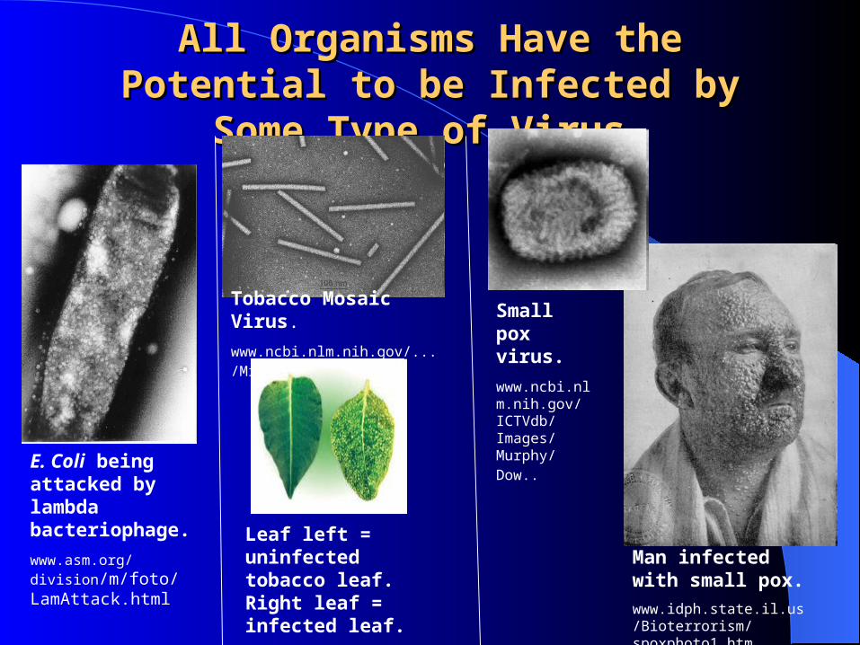
All Organisms Have the Potential to be All Organisms Have the Potential to be Infected by Some Type of Virus.Infected by Some Type of Virus.
E. Coli being attacked by lambda bacteriophage.
www.asm.org/division/m/foto/LamAttack.html
Tobacco Mosaic Virus.
www.ncbi.nlm.nih.gov/.../Milne/tobamo1.htm
Leaf left = uninfected tobacco leaf. Right leaf = infected leaf.
www.nature.com/.../v411/n6839/full/411848a0.html
Man infected with small pox.
www.idph.state.il.us/Bioterrorism/spoxphoto1.htm
Small pox virus.
www.ncbi.nlm.nih.gov/ICTVdb/Images/Murphy/Dow..
Sooooo……….Sooooo……….what is a virus?what is a virus?

Virus CharacteristicsVirus Characteristics
1. Have genetic material, either DNA or RNA. 2. Can’t replicate without a host cell. 3. Can’t divide by binary fission, mitosis, or
meiosis. 4. Can’t make their own energy (steal it from host
cell). 5. Can’t make their own protein or genetic
material (steal from host cell).
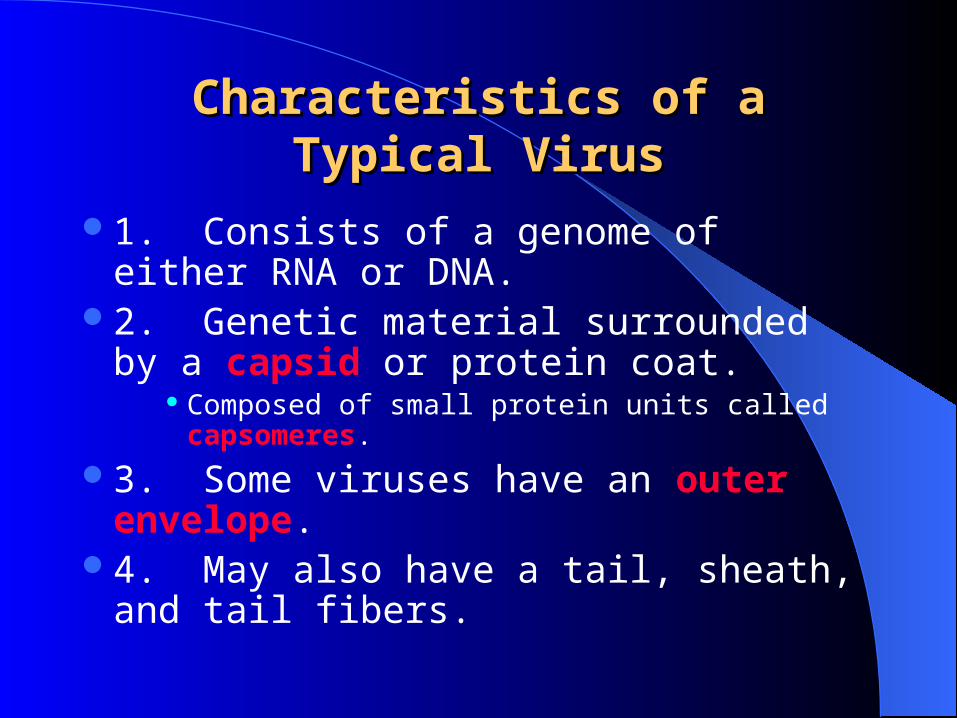
Characteristics of a Typical VirusCharacteristics of a Typical Virus
1. Consists of a genome of either RNA or DNA.
2. Genetic material surrounded by a capsid or protein coat.
Composed of small protein units called capsomeres.
3. Some viruses have an outer envelope.4. May also have a tail, sheath, and tail
fibers.

A Typical VirusA Typical Virus
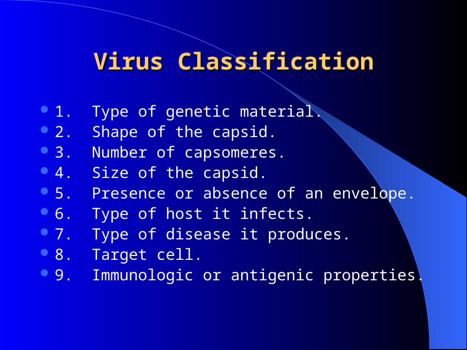
Virus ClassificationVirus Classification
1. Type of genetic material. 2. Shape of the capsid. 3. Number of capsomeres. 4. Size of the capsid. 5. Presence or absence of an envelope. 6. Type of host it infects. 7. Type of disease it produces. 8. Target cell. 9. Immunologic or antigenic properties.

Viral ClassificationViral Classification
www.biotech100.com www.antibac2k.com

Where did Where did viruses come from?viruses come from?

Virus OriginVirus Origin
3 Major Theories1. Viruses are remnants of past infections
(e.g. mitochondrion/chloroplasts).2. Cells came before viruses.
• Viruses are degenerate cells or cell fragments.
3. Viruses represent a separate evolutionary branch
What if. . . .What if. . . .
Scientists found a cell that was as large or larger than some bacteria and that cell had the capabilities to produce almost everything it needed to “live.”
It required a host cell only to make some ribosomes.
Is it a virus or a living cell?
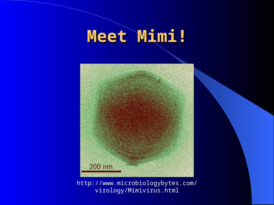
Meet Mimi!Meet Mimi!
http://www.microbiologybytes.com/virology/Mimivirus.html

RetrovirusRetrovirus Genetic information is
single-stranded RNA. Have a special enzyme
called reverse transcriptase.
This enzyme makes DNA from RNA.
Integrate their newly formed double-stranded DNA into the host cell.
Example = HIV
Human Immunodeficiency Virus (www.msu.edu)
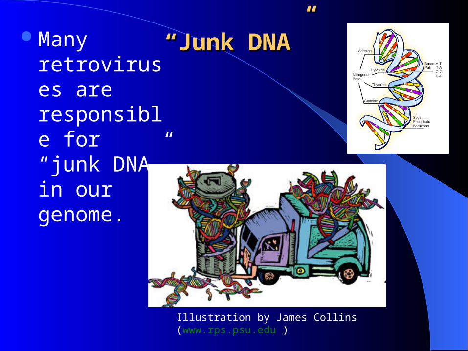
““Junk DNA”Junk DNA”Many retroviruses are responsible for “junk DNA” in our genome.
Illustration by James Collins (www.rps.psu.edu )

Sheep, Viruses, and Dr. SpencerSheep, Viruses, and Dr. Spencer
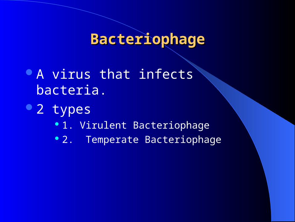
BacteriophageBacteriophage
A virus that infects bacteria.2 types
1. Virulent Bacteriophage 2. Temperate Bacteriophage

Virulent BacteriophageVirulent Bacteriophage
Causes Lytic Cycle (5 steps)
1. Attachment 2. Penetration 3. Biosynthesis 4. Assembly 5. Release
textbookofbacteriology.net oceanworld.tamu.edu

Temperate BacteriophageTemperate Bacteriophage
Do not immediately begin lytic cycle.Their DNA remains embedded in bacterial
cell chromosome.
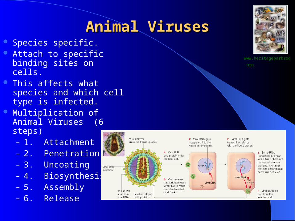
Animal VirusesAnimal Viruses Species specific. Attach to specific binding
sites on cells. This affects what species
and which cell type is infected.
Multiplication of Animal Viruses (6 steps)– 1. Attachment– 2. Penetration– 3. Uncoating– 4. Biosynthesis– 5. Assembly– 6. Release
www.heritageparkzoo.org
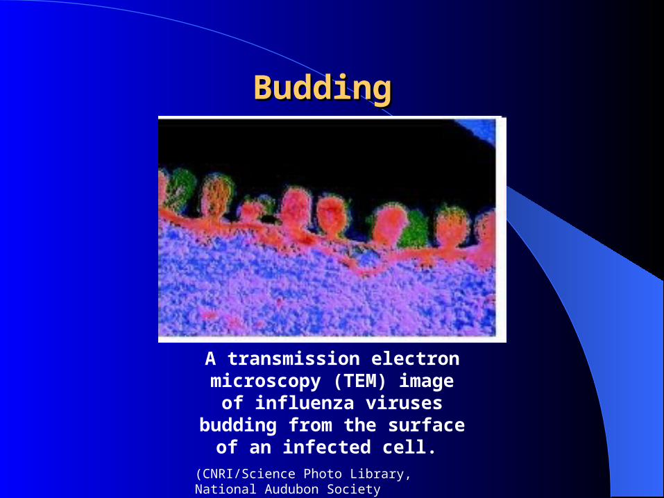
Budding Budding
A transmission electron microscopy (TEM) image of
influenza viruses budding from the surface of an infected cell.
(CNRI/Science Photo Library, National Audubon Society Collection/Photo Researchers, Inc.)

Antiviral AgentsAntiviral Agents Interfere with the
phases of viral multiplication.
May disrupt a binding site.
May disrupt an enzyme or protein.
May interfere with the synthesis of viral parts like DNA, RNA, or protein synthesis.

Oncogenic VirusesOncogenic Viruses
Viruses that cause cancer.
Ex. Human papillomaviruses (HPV – wart viruses) cause different types of cancers.– i.e. cervical cancer
and other types of cancers of the genital tract. Kaposi Sarcoma – caused by human
herpesvirus 8.
www.hyle.org
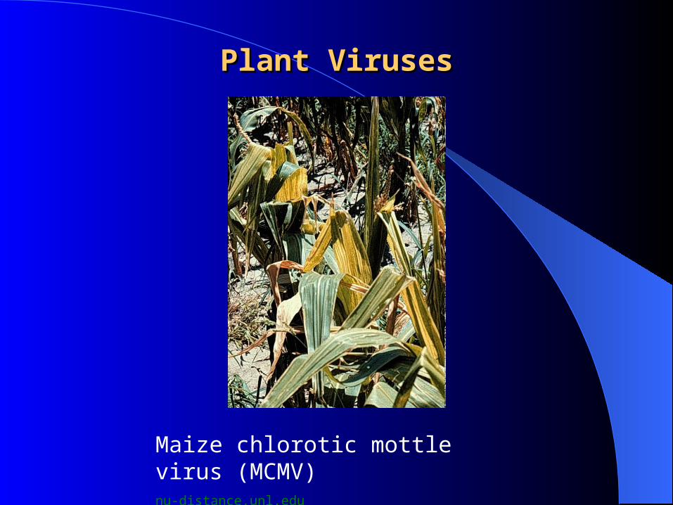
Plant VirusesPlant Viruses
Maize chlorotic mottle virus (MCMV) nu-distance.unl.edu

Viroids and PrionsViroids and Prions
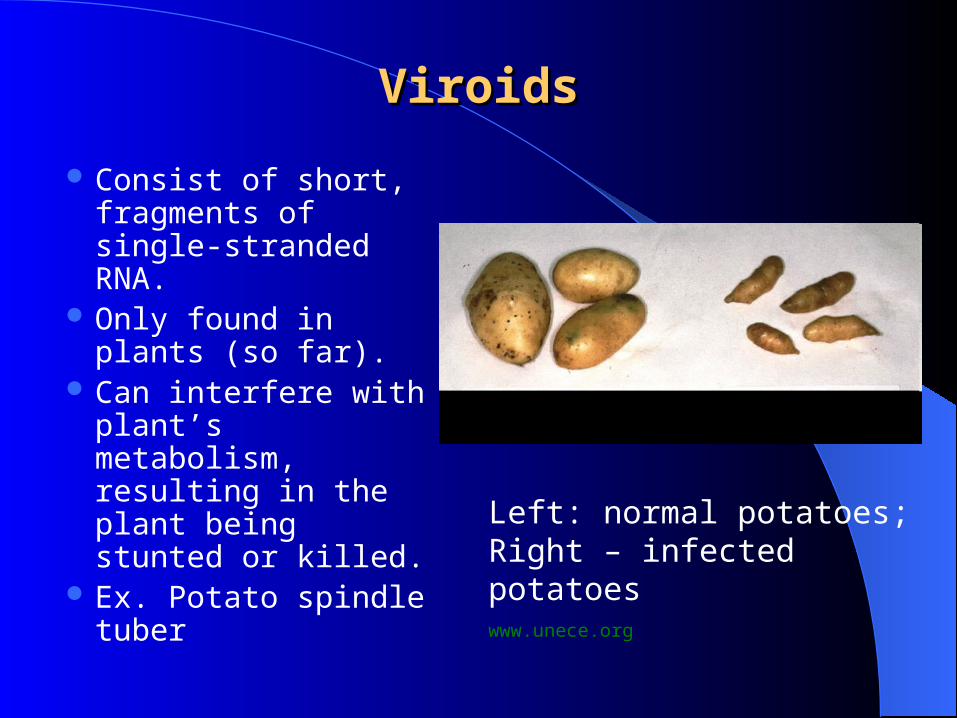
ViroidsViroids
Consist of short, fragments of single-stranded RNA.
Only found in plants (so far).
Can interfere with plant’s metabolism, resulting in the plant being stunted or killed.
Ex. Potato spindle tuber
Left: normal potatoes; Right – infected potatoeswww.unece.org

ProteinsProteins
Left = unfolded protein; Right = folded protein

PrionsPrions
Left = correct protein folding; Right = wrong protein foldingwww.cogs.susx.ac.uk

PrionsPrions
Small infectious proteins.Cause fatal neurologic diseases in animals.Cause fatal spongiform encephalopathies.
– Brain becomes riddled with holes.
Brain with spongiform encephalopathy
webs.wichita.edu

Prion Animal InfectionsPrion Animal Infections
Sheep infected with scrapie. (www.gov.mb.ca)
Deer infected with “chronic wasting disease.” (http://www.fw.delaware.gov)
Bovine spongiform encephalopathy, “mad cow disease.”
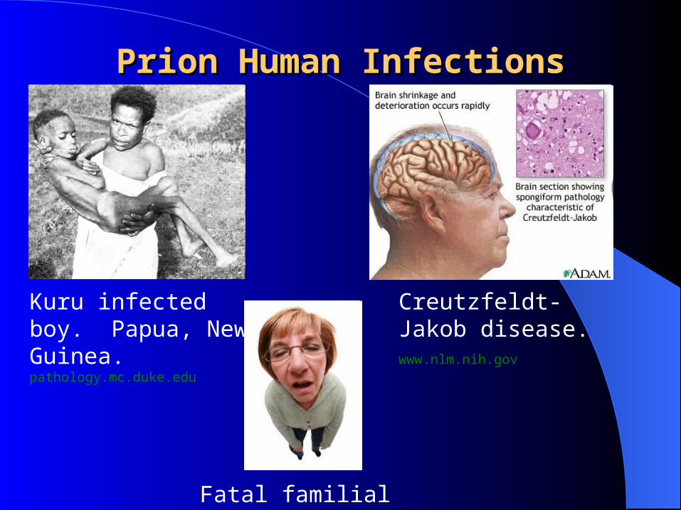
Prion Human InfectionsPrion Human Infections
Kuru infected boy. Papua, New Guinea. pathology.mc.duke.edu
Creutzfeldt-Jakob disease. www.nlm.nih.gov
Fatal familial insomnia

The EndThe End